Buffalo sperm surface proteome profiling reveals an intricate relationship between innate immunity and reproduction
←
→
Page content transcription
If your browser does not render page correctly, please read the page content below
Batra et al. BMC Genomics      (2021) 22:480
https://doi.org/10.1186/s12864-021-07640-z

 RESEARCH                                                                                                                                            Open Access

Buffalo sperm surface proteome profiling
reveals an intricate relationship between
innate immunity and reproduction
Vipul Batra1, Vanya Bhushan2†, Syed Azmal Ali2†, Parul Sarwalia1, Ankit Pal1, Seema Karanwal1, Subhash Solanki1,
Arumugam Kumaresan3, Rakesh Kumar1 and Tirtha Kumar Datta1*

  Abstract
  Background: Low conception rate (CR) despite insemination with morphologically normal spermatozoa is a
  common reproductive restraint that limits buffalo productivity. This accounts for a significant loss to the farmers
  and the dairy industry, especially in agriculture-based economies. The immune-related proteins on the sperm
  surface are known to regulate fertility by assisting the spermatozoa in their survival and performance in the female
  reproductive tract (FRT). Regardless of their importance, very few studies have specifically catalogued the buffalo
  sperm surface proteome. The study was designed to determine the identity of sperm surface proteins and to
  ascertain if the epididymal expressed beta-defensins (BDs), implicated in male fertility, are translated and applied
  onto buffalo sperm surface along with other immune-related proteins.
  Results: The raw mass spectra data searched against an in-house generated proteome database from UniProt using
  Comet search engine identified more than 300 proteins on the ejaculated buffalo sperm surface which were bound
  either by non-covalent (ionic) interactions or by a glycosylphosphatidylinositol (GPI) anchor. The singular
  enrichment analysis (SEA) revealed that most of these proteins were extracellular with varied binding activities and
  were involved in either immune or reproductive processes. Flow cytometry using six FITC-labelled lectins confirmed
  the prediction of glycosylation of these proteins. Several beta-defensins (BDs), the anti-microbial peptides including
  the BuBD-129 and 126 were also identified amongst other buffalo sperm surface proteins. The presence of these
  proteins was subsequently confirmed by RT-qPCR, immunofluorescence and in vitro fertilization (IVF) experiments.
  Conclusions: The surface of the buffalo spermatozoa is heavily glycosylated because of the epididymal secreted
  (glyco) proteins like BDs and the GPI-anchored proteins (GPI-APs). The glycosylation pattern of buffalo sperm-
  surface, however, could be perturbed in the presence of elevated salt concentration or incubation with PI-PLC. The
  identification of numerous BDs on the sperm surface strengthens our hypothesis that the buffalo BDs (BuBDs) assist
  the spermatozoa either in their survival or in performance in the FRT. Our results suggest that BuBD-129 is a sperm-
  surface BD that could have a role in buffalo sperm function. Further studies elucidating its exact physiological
  function are required to better understand its role in the regulation of male fertility.
  Keywords: Buffalo, Sperm, Beta-defensins, Epididymis, Glycosylation

* Correspondence: tirtha.datta@icar.gov.in
†
 Vanya Bhushan and Syed Azmal Ali contributed equally to this work.
1
 Animal Genomics Lab., Animal Biotechnology Centre, National Dairy
Research Institute, Karnal, India
Full list of author information is available at the end of the article

                                         © The Author(s). 2021 Open Access This article is licensed under a Creative Commons Attribution 4.0 International License,
                                         which permits use, sharing, adaptation, distribution and reproduction in any medium or format, as long as you give
                                         appropriate credit to the original author(s) and the source, provide a link to the Creative Commons licence, and indicate if
                                         changes were made. The images or other third party material in this article are included in the article's Creative Commons
                                         licence, unless indicated otherwise in a credit line to the material. If material is not included in the article's Creative Commons
                                         licence and your intended use is not permitted by statutory regulation or exceeds the permitted use, you will need to obtain
                                         permission directly from the copyright holder. To view a copy of this licence, visit http://creativecommons.org/licenses/by/4.0/.
                                         The Creative Commons Public Domain Dedication waiver (http://creativecommons.org/publicdomain/zero/1.0/) applies to the
                                         data made available in this article, unless otherwise stated in a credit line to the data.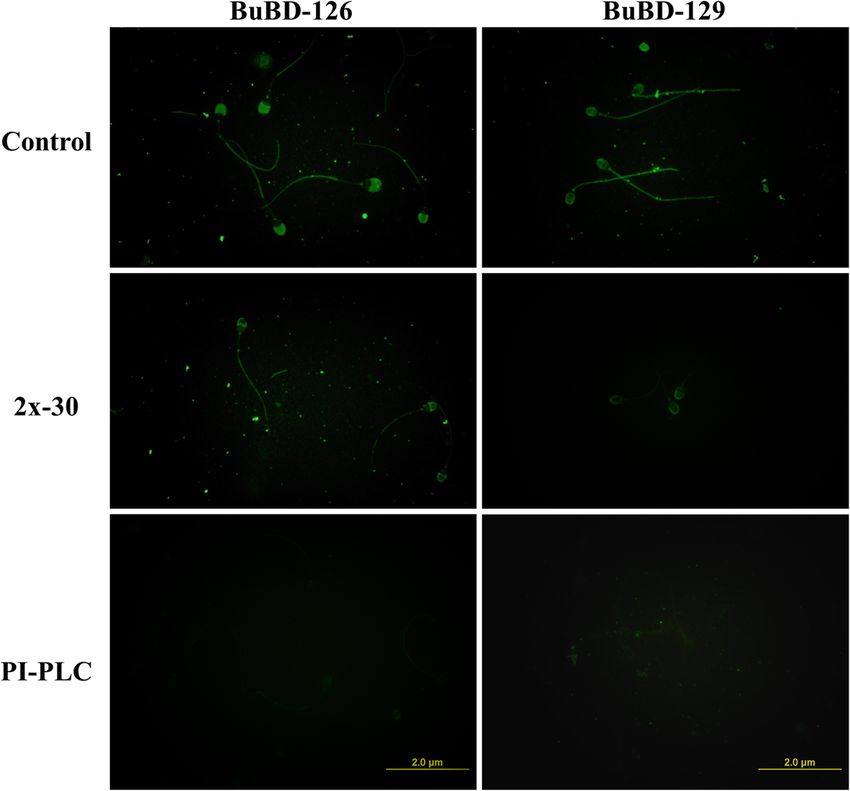
Batra et al. BMC Genomics   (2021) 22:480                                                                          Page 2 of 18

Introduction                                                    surface coat on the spermatozoa, not only acts as a barrier
The voyage of the spermatozoa in the female reproductive        between the spermatozoa and female immune system but
tract (FRT) entails surmounting of numerous impedi-             also assists spermatozoa in cervical mucus penetration
ments including the physical, thermal, chemical and im-         (CMP), oviductal epithelial cell (OEC) binding, identifica-
munological barriers. These include the vaginal acidic pH,      tion of the zona pellucida and oolemma, apposition of the
the mucus in the cervix, the leukocytes and anti-sperm          sperm and the oocyte plasma membrane [3, 19, 29–31].
antibodies of the immune system especially in the uterus,       The sperm-surface proteins and their associated glycans
and the narrow utero-tubal junction in the oviduct [1–5].       also play a key role in the acquisition of motility and fertil-
To overcome these obstructions the spermatozoa must             izing ability in the epididymis, their protection, selection
acquire surface properties primarily customized for this        and secondary maturation in the FRT [19, 32].
arduous journey. The process of sperm surface remodel-             The buffalo was considered as a model for this
ling (SSR), which occurs during the epididymal transit of       study due to its economic importance in agriculture-
the spermatozoa tailors the sperm surface which assists         based economies. It has been reported that more
them in survival and fertilization in the FRT [6–8]. The        people depend on buffalo than on any other domes-
bio-molecular constitution of the mammalian testicular          tic animal [33]. Although it is a premier dairy animal
spermatozoa changes continuously and progressively in           with superior milk-producing ability, idiopathic male
the luminal fluid of the various epididymal regions due to      infertility is a common reproductive limitation in
the secretory and re-absorptive actions of the epithelial       buffalo. A sizeable number of high genetic merit bull
cells that line this organ [9–12]. The remodelling events       calves originally selected for AI programs are dis-
include a) enzymatic cleavage of the membrane-associated        carded because their semen ends up yielding dismal
proteins b) variations in the composition of membrane-          conception rates (CRs) between 30 to 50%, reflecting
lipids c) re-organization of the glycoconjugates (GCs) as-      poor fertilizing ability [34–36]. The factors that con-
sociated with the sperm glycocalyx d) removal or addition       tribute to male fertility are relatively poorly under-
of (glyco) proteins [9, 13, 14]. A blend of distinct secreta-   stood, especially in bovine species [37]. The
gogues is known to be added onto the sperm-surface in           prediction of fertility assessment currently relies on
these three epididymal regions viz. caput, corpus and           analyses of sperm functional parameters apart from
cauda. A majority of these secretagogues include the            the physical examination of the bulls, nonetheless,
immune-related (glyco) proteins often implicated in sperm       the correlation between these parameters and the CR
survival and fertility. The epididymal secreted proteins        is often inconsistent [38]. Therefore, a better under-
which are involved in sperm maturation could be loosely         standing of the novel factors which regulate fertility
adhered to the sperm plasma membrane or could be                e.g. sperm-surface proteins is required to gain in-
transmembrane. Many of these proteins bind transiently,         sights into the factors behind idiopathic male
for example, the ones acquired in the distal epididymal re-     infertility.
gions [15, 16]. These loosely adhered proteins in the per-         The objective of this study was the identification and
ipheral sperm environment bind the sperm-surface either         in silico characterization of the post-testicular matur-
by electrostatic or hydrophobic interactions. These pro-        ation antigens and peripheral proteins that interact with
teins change the sperm-surface characteristics as they          the buffalo sperm plasma membrane either through
interact with the transiting spermatozoa. The epididymal        non-covalent (ionic) interactions or through a GPI-
secretome also involves the glycan-modifying enzymes            anchor. We also sought to determine the existence of
such as glycosidases and glycosyltransferases [17–19], pro-     the epididymal expressed BDs on buffalo spermatozoa
teases and protease inhibitors [10, 11, 20], proteins in-       and to predict their reproductive functional significance
volved in immunological protection [3, 4] and the ones          through RT-qPCR, immunofluorescence and in vitro
that protect the sperm from oxidative injuries [18, 21]. Be-    fertilization experiments.
sides, various membranous, extracellular vesicles (EVs)
rich in cholesterol, sphingolipids and Ca+ 2 known as epi-      Results
didymosomes have been reported to exist in the epididy-         The ejaculates that were milky or creamy in colour,
mal lumen [22, 23]. These EVs mainly carry the GPI-APs,         homogenous in consistency i.e. free from flakes/clumps
many of which are inserted in the sperm plasma mem-             with a minimum sperm concentration of 600 × 106/ml
brane [24]. As mentioned earlier, many of the added             were considered for swim-up and further downstream
(glyco) proteins on sperm-surface belong to defence fam-        experiments. The average motility and viability of seven
ily and their glycosylation patterns are critical for either    representative sample ejaculates after processing was
stabilizing the sperm-membrane during the immune at-            81.84 ± 1.20% and 85.85 ± 1.16%, respectively. The sam-
tack by immune cells or assisting in immune-evasion in          ples were diluted according to the experiments, as men-
the FRT [25–28]. The rendering of a highly glycosylated         tioned, wherever required.
Batra et al. BMC Genomics (2021) 22:480 Page 3 of 18
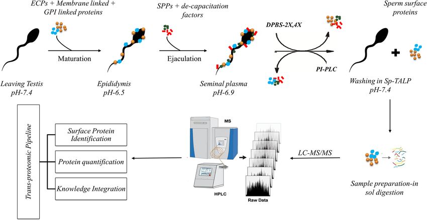
Hundreds of proteins could be extracted from the buffalo beta-defensins (CA-BDs) viz. BD-129 and 126 amongst
sperm-surface after elevated salt (DPBS) or PI-PLC the other sperm-surface proteins Notwithstanding, only
treatment 85 proteins were found to be common among the elevated
The extracted sperm-surface proteins identified after LC- salt and PI-PLC treatments which were predicted to be
MS/MS data processing indicated enough diversity among extracellular or present on the buffalo sperm surface (Sup-
the types of proteins removed using the two treatment plementary Fig. 2 and Supplementary sheet-Results). A re-
classes representing the a) the elevated salt extractions markable diversity in the range of MW and pI was
(2x-30, 2x-60, 4x-30, 4x-60) and b) the PI-PLC extractions observed among these proteins (Supplementary sheet-
(1 U/mL, 1.5 U/mL and 2 U/mL) (Supplementary Fig. 1 Results). Amongst the mapped entries, the BDs like BD-
and 2). The extracted sperm-surface proteins produced > 134, BD-126 and Spag-11D were found to be among the
20,000 PEP-XML spectra for each of the sub-groups of ei- proteins with the lowest molecular weight (Mr = 5.33, 7.44
ther treatment (elevated salt or PI-PLC). The iprophet tool and 11.95, respectively) while the angiotensin-converting
correctly identified more than 300 proteins in all of the enzyme (ACE) and the two uncharacterized proteins (Uni-
treatments at p > =0.99 where p indicates the probability Prot ID: F1MD73 and F1MQ37) were the on the other
that the spectra have been correctly matched to its analo- end of the scale (Mr = 141.24, 190.10 and 227.10 respect-
gous peptide (Table 1). A total of 317, 391, 394 and 432 ively) (Supplementary sheet-Results-Mapped Entries). The
proteins were identified in 2x-30, 4x-30, 2x-60 and 4x-60 BDs like Spag-11D, BD-129 and BD-126, however, had
(DPBS) treatments respectively. On the other hand, 385, high pI values (9.5, 9.49, and 9.48, respectively) whereas
353 and 364 proteins were identified in the 1 U, 1.5 U and the Acrosin inhibitor 1 had the lowest pI (4.25) among the
2 U/mL PI-PLC treatments, respectively. At p ≥ 0.99 (iPro- mapped entries. Only three proteins viz. Sperm acrosome
phet probability) zero proteins were found to be incor- membrane-associated protein1, Angiotensin-converting
rectly identified. Many proteins were found to be unique enzyme and an uncharacterized protein (F1MD73) were
to each sub-group of either treatment demonstrating that predicted to contain a transmembrane segment (Supple-
the individual combinations of incubation time and salt/ mentary sheet-Results-Mapped Entries). A high level of
enzyme concentration exerted disparate effects on dis- PTMs, especially glycosylation appears to modify the ana-
rupting the non-covalent/GPI mediated interactions of lyzed proteins because more than 80% of the analyzed
the buffalo sperm surface proteins. Moreover, nearly 30% proteins were predicted to possess at least either 1 N-
of the proteins were common between any two treatment glycosylation site or one O-glycosylation site. BD-126 was
subgroups (Supplementary Fig. 1 and 2). predicted to contain two O-glycosylation sites whereas the
Overall, we report a total of 352 buffalo sperm-surface BD-129 predicted to contain eight such sites (Supplemen-
proteins that were identified in the protein fractions ex- tary sheet-Results). The BD-126 and 134 were predicted
tracted by the two treatment classes. The LC-MS/MS data to contain 1 N-glycosylation site while the BD-129 was
analysis identified several BDs including the two Class-A predicted to contain three such sites.
Table 1 The seven treatment sub-groups for sperm-surface protein extraction from two treatment classes and the corresponding
TPP results indicating total spectra, correctly and incorrectly identified proteins at p ≥ 0.6 and 0.99
Sample PEP-XML Ipro.pep.xml Prot.xml Total proteins Incorrectly identified Total proteins Incorrectly identified
Total Total spectra Total with p ≥ 0.6 with iProphet p ≥ 0.6 with p ≥ 0.99 with iProphet p ≥ 0.99
spectra proteins
2x- 26,535 7252 1733 875 76 317 0
DPBS-
30 min
4x- 26,470 8399 1725 956 76 391 0
DPBS-
30 min
2x- 25,947 7742 1898 1013 90 395 0
DPBS-
60 min
4x- 25,951 8210 2162 1244 99 432 0
DPBS-
60 min
1 U/ml 24,829 6557 1788 992 99 385 0
1.5 U/ml 24,587 6343 1932 1005 76 353 0
2 U/ml 24,881 5978 1662 793 63 364 0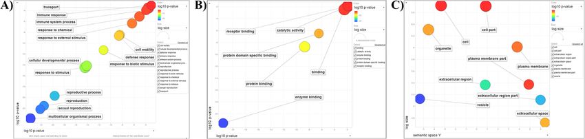
Batra et al. BMC Genomics      (2021) 22:480                                                                                           Page 4 of 18

Proteins involved in the immune response and                                sheet-Results). Overall, these results suggested that the
reproductive processes adorn the buffalo sperm surface                      buffalo sperm surface is adorned with extracellular/ves-
The gene ontology (GO) analysis was performed on the                        icular-origin proteins which are involved in reproduction
85 extracellular (EC) sperm-surface proteins which were                     specific activities, immune responses, responses to bi-
common between the two treatment classes (Supple-                           otic/abiotic stimuli usually performing catalytic or
mentary Fig. 2c) wherein the annotation terms for                           carbohydrate/protein binding functions.
Biological Process, Molecular Function and Cellular
Component were determined (Fig. 1 and Table 2). These                       Cytometry reveals removal of glycosylated proteins from
proteins were successfully mapped to 63 entries in the                      buffalo sperm surface after elevated salt/PI-PLC treatment
background dataset. The singular enrichment analysis                        The Flow cytometry analyses were performed on the control
(SEA) for Biological Process terms identified reproduct-                    spermatozoa sample (NCM), spermatozoa incubated in ele-
ive processes, sexual reproduction, immune response                         vated salt for 30 min (2x-DPBS) and spermatozoa incubated
and response to biotic/abiotic stimulus terms as the                        with PI-PLC (2 U/mL) to assess the corresponding changes
major GO annotations in the input list vis-à-vis the                        in sperm-surface glycosylation after either treatment. The
background reference dataset, the Bovine genome locus                       analyses revealed a reduction in both the O-linked and the
(Bovine Genome Database): GLEAN_03528 (Table 2).                            N-linked glycans after either treatment as assessed by the de-
The scatter plot analysis (SPA) for Biological Process                      cline in the MFI values which are produced upon binding of
similarly indicated higher semantic similarities between                    FITC-bound lectins on the buffalo sperm-surface (Fig. 2). A
reproductive process functions, immune response and                         panel of five O-linked glycans specific lectins viz. ABL, JAC,
response to biotic/abiotic stimulus terms as observed by                    MAL-II, LCA, PNA and 1 N-linked glycan specific lectin,
their closeness in the displayed two-dimensional space                      LEL was used. The Brown-Forsythe test for all the lectins
(Fig. 1a). The SEA for Molecular Function indicated that                    was found to produce a non-significant p-value (p > 0.05) in-
the majority of proteins were involved in catalytic and                     dicating no differences in standard deviations of the MFIs
binding (carbohydrate or protein) functions (Table 2).                      produced in these groups. The unstained spermatozoa were
The SPA for Molecular Function also identified protein                      excluded from the analysis by gating and the singlets were
binding and catalytic activity as the major GO terms                        chosen for analyses which were performed on single, stained
with the highest uniqueness index values and the least                      spermatozoa. The O-linked glycan-binding lectin, ABL pref-
dispensability scores (Fig. 1b and Supplementary sheet-                     erentially binds the Thomsen-Friedenreich antigen, galactosyl
Results). Most of the proteins were found to be extracel-                   (β-1, 3) N-acetylgalactosamine [39]. It produced a mean
lular, vesicular or part of the plasma membrane as indi-                    fluorescence intensity (MFI) of 1, 56,610.0 units in the con-
cated by the SEA and SPA for the Cellular Component                         trol sample which differed significantly (p < 0.001) from the
terms (Fig. 1c and Supplementary sheet-Results). The                        MFI produced in the spermatozoa incubated in 2x-DPBS (1,
low p-values from the Fisher’s test and the results of the                  25,032.0 units) or treated with PI-PLC (1, 29,399.0 units) as
Yekutieli test (low FDRs) are indicative of high confi-                     assessed by one-way ANOVA (Fig. 2 and Supplementary
dence in the determined annotation terms for the input                      Fig. 3). The lectin JAC which has a sugar specificity towards
list in the SEA (Table 2). Similarly, the lower log10 p-                    galactose of O-linked glycans preferring the structure galac-
values and dispensability score with high uniqueness                        tosyl (β-1, 3) N-acetylgalactosamine also produced higher
index indicate the reliability of the GO annotation terms                   MFI in the control sample (2, 47,848.0 units) in comparison
for the input list used for the SPA (Supplementary                          to either the 2x-30 sample (1, 71,757.0 units) or the PI-PLC

 Fig. 1 The semantic-similarity based scatter plots depicting the summarized lists of GO terms for a Biological Process, b Molecular Function and c
 Cellular Component domains for the buffalo sperm surface proteins
Batra et al. BMC Genomics           (2021) 22:480                                                                                       Page 5 of 18

Table 2 The major GO annotation terms, Fisher’s p-values and Yekutieli result (FDR under dependency) for the singular enrichment
analysis (SEA) of Biological Process, Molecular Function and Cellular Component annotation terms performed on the sperm-surface
proteins in the input list
Biological Process                                      Molecular Function                              Cellular Component
Term                                  p-       FDR      Term                          p-       FDR      Term                          p-       FDR
                                      value                                           value                                           value
Multicellular organismal process      2.00E-   1.40E-   Enzyme binding                2.90E-   2.90E-   Membrane-bounded vesicle      5.70E-   6.10E-
                                      27       24                                     24       22                                     85       83
Sexual reproduction                   1.90E-   6.60E-   Ubiquitin protein ligase      8.20E-   4.10E-   Vesicle                       5.70E-   6.10E-
                                      25       23       binding                       16       14                                     85       83
Reproduction                          1.30E-   3.00E-   Protein binding               6.00E-   2.00E-   Extracellular region part     3.80E-   2.70E-
                                      24       22                                     11       09                                     70       68
Reproductive process                  1.20E-   2.10E-   Unfolded protein binding      3.60E-   9.00E-   Extracellular region          3.60E-   1.90E-
                                      22       20                                     09       08                                     62       60
Positive regulation of biological     1.90E-   2.70E-   Protein domain specific       1.80E-   3.50E-   Cytoplasm                     3.90E-   1.60E-
process                               21       19       binding                       08       07                                     32       30
Anatomical structure                  4.70E-   5.50E-   Carbohydrate binding          8.40E-   0.00014 Membrane-bounded organelle     1.60E-   5.70E-
development                           21       19                                     06                                              29       28
Regulation of biological quality      4.20E-   4.20E-   Binding                       0.00037 0.0037    Extracellular space           9.30E-   2.20E-
                                      18       16                                                                                     25       23
Positive regulation of cellular       5.30E-   4.70E-   Nucleotide binding            0.00041 0.0037    Organelle                     1.60E-   3.50E-
process                               18       16                                                                                     23       22
System development                    1.10E-   8.90E-   Enzyme regulator activity     0.00041 0.0037    Intracellular part            8.60E-   1.70E-
                                      17       16                                                                                     20       18
Response to stimulus                  5.10E-   3.60E-   Catalytic activity            0.0022   0.012    Plasma membrane               7.80E-   1.30E-
                                      17       15                                                                                     18       16
Cellular developmental process        1.10E-   7.00E-   Receptor binding              0.0024   0.012    Cytoplasmic membrane-bounded 4.80E-    6.10E-
                                      16       15                                                       vesicle                      15        14
Response to biotic stimulus           1.90E-   5.60E-                                                   Plasma membrane part          5.00E-   3.90E-
                                      13       12                                                                                     09       08
                                                                                                        Cell part                     4.80E-   2.60E-
                                                                                                                                      06       05
                                                                                                        Cell                          4.80E-   2.60E-
                                                                                                                                      06       05

treated sample (1, 27,951.0 units). The post-hoc analysis indi-                     produced a significantly higher MFI in the control sample
cated that the reduction in MFI for JAC after either the salt                       (6015.0 units) in comparison to the 2x-30 sample (4820.0
treatment (p < 0.001) or the PI-PLC treatment (p < 0.0001)                          units) (Fig. 2 and Supplementary Fig. 3). Nevertheless, as ob-
was not only significantly different from the control sample                        served for LCA binding, the MFI increased after PI-PLC
but also each other (p < 0.01) (Fig. 2 and Supplementary                            treatment, albeit significantly (p < 0.001) to 7435.0 units. The
Fig. 3). The N-linked glycan-binding lectin LEL which is spe-                       MFI produced upon MAL-II binding in control and treat-
cific for [GlcNAc] 1–3, N-acetylglucosamine is also removed                         ment samples differed significantly (p < 0.0001) from each
from the sperm surface on exposing the spermatozoa to ele-                          other (Fig. 2 and Supplementary Fig. 3). The acrosomal in-
vated salt milieu producing a diminished MFI of 48,715.0                            tactness indicator lectin PNA, which binds the asialylated ga-
units which didn’t differ significantly from the MFI produced                       lactosyl (β-1, 3) N-acetylgalactosamine produced MFI of 28,
in control samples (92,968.0 units) (Fig. 2 and Supplementary                       334.0 and 23,075.0 units in the salt-treated and the PI-PLC
Fig. 3). The exposure of PI-PLC, nonetheless, reduced the                           treated spermatozoa, respectively, whereas the MFI produced
MFI fluorescence significantly to 32,161.0 units (p < 0.05).                        by PNA binding in the control spermatozoa was 18,759.0
The LCA lectin which is specific for mannose and glucose                            units. The rise in the MFI values, however, was statistically
produced significantly (p < 0.001) reduced MFI of 26,979.0                          insignificant for both the treatments (Fig. 2 and Supplemen-
units in the elevated salt-treated spermatozoa when com-                            tary Fig. 3).
pared to the control sample (36,559.0 units) (Fig. 2 and Sup-                         Overall, both the treatments reduced the availability of
plementary Fig. 3). Conversely, the MFI increased minutely                          respective cognate glycans for most lectins except the
to 38,451.0 units after exposure to PI-PLC. The α-2, 3 linked                       PNA after salt treatment. Contrarily, the PI-PLC treat-
sialic acid-binding lectin MAL-II (p < 0.001) similarly                             ment led to increased exposure of α-2, 3 linked sialic acid
Batra et al. BMC Genomics (2021) 22:480 Page 6 of 18
Fig. 2 Histogram plots of the observed mean fluorescent intensity (MFI) values produced upon binding of six different FITC-labelled lectins viz. a
ABL, b JAC, c LEL, d LCA, e MAL-II, and f PNA on buffalo bull spermatozoa in NCM (control), 2x-DPBS (2x-30) or spermatozoa exposed to 2 U/mL
PI-PLC. The differences being assessed by one way ANOVA followed by Tukey’s multiple comparison test
and asialylated galactosyl (β-1, 3) N-acetylgalactosamine. of BuBD-126 in blood was found to be non-significant
Furthermore, both the treatments were significantly differ- (p > 0.05), as assessed by an unpaired two-tailed t-test.
ent from each other vis-à-vis the MFI produced upon lec- Similarly, the expression analysis of BuBD-129 revealed
tin binding on the surface of the buffalo spermatozoa. a higher number of transcripts in buffalo spermatozoa
relative to the peripheral blood, albeit the mean expres-
Expression dynamics of BuBD-129 and 126 sion levels between the spermatozoa and the blood were
The relative expression profiles of the BuBD129 and 126 statistically significant (p < 0.01). Interestingly a large
genes were generated using RT-qPCR, in the spermato- inter-animal variation was observed in the expression
zoa. The expression of the BuBD-126 and BuBD-129 levels of the BuBD129 and 126 amongst the four bio-
was found to be much higher in the spermatozoa than logical replicates. These observations intriguingly pro-
the peripheral blood (Fig. 3) hinting at their role in buf- vided a clue that either the spermatozoa actively
falo reproduction. Surprisingly this elevated expression expressed these BuBDs or their transcripts were already
Fig. 3 Relative expression profiles of the two CA-BD genes, BuBD-126 a and BuBD-129 b in the peripheral blood and ejaculated spermatozoa
obtained from Murrah buffalo bulls. Expression values were normalized to GAPDH & eEF-2. Horizontal bars represent the mean(s) and error bars
represent the standard error of the mean (SEM)Batra et al. BMC Genomics (2021) 22:480 Page 7 of 18
stored in the de-differentiated spermatozoa during found to be present preferentially on the acrosomal and
spermatogenesis, apart from their preferential expression post-acrosomal and in the tail region while being absent
in the MRT. in the mid-piece region (Fig. 4). The negative controls
for both the antibodies, which were without the primary
Differential spatial distribution of BuBD-129 and 126 antibody did not fluoresce upon excitation (Supplemen-
The peptides GRCKEYCNMDEKELDK and NKTGNC tary Fig. 6). The fluorescence produced by the BuBD-
RSTCRNGEK for BuBD-129 and BuBD-126, respectively 129 and 126 diminished when the spermatozoa were in-
were predicted to be highly antigenic and were thus ad- cubated in an elevated salt environment. The spermato-
judged as the best B-cell epitopes. This is because they zoa incubated in 2x-DPBS for 30 min appear to lose the
were predicted to be preferentially present in turns and sperm-surface bound BuBD-126 uniformly from the
loops and had a comparatively higher probability of be- sperm surface, whereas the BuBD-129 was retained on
ing found on the surface (Supplementary Fig. 4). Ini- the acrosomal region) despite being lost from the mid-
tially, the crude concentration of the isolated IgGs piece and the tail region of the buffalo bull spermatozoa
was assayed by measuring the A280 which was 155,049 (Fig. 4). The effect for the PI-PLC treatment, however,
ng/μl and 168,722 ng/μl for the CA-BDs, BuBD-129 and was markedly different for the sperm surface-bound
126 respectively (Supplementary Fig. 5). Subsequently, BuBD-129. The spermatozoa exposed to 2 U/mL of PI-
Bradford’s assay was used to ascertain the concentration PLC lost the majority of the fluorescence signal for
and 0.5μg/ml of the purified antibody was used for fur- BuBD-129 from the entire spermatozoa. Nevertheless,
ther experiments like the IF, IVF studies. the fluorescence pattern for BuBD-126 was similar to
The immunofluorescence (IF) experiments using anti- what was observed after 2x-DPBS treatment albeit much
BuBD-129 and 126 antibodies revealed that the two weaker in intensity indicating higher loss of the bound
class-A BuBDs (CA-BDs), BuBD-129 and 126 localized BuBD-126 (Fig. 4).
differentially on the surface of the buffalo bull spermato-
zoa. A variation in the spatial distribution pattern of Blocking BuBD-129 on sperm surface hinders cleavage,
these two CA-BDs was observed wherein the BuBD-129 Morula and blastocyst formation rates
was present along the entire periphery of the buffalo The addition of anti-BuBD-129 antibody in the fertilization
spermatozoa (Fig. 4). The BuBD-126, however, was medium appeared to hamper the fertilization and thus the
Fig. 4 Immuno-localization pattern of the two CA-BDs viz. BuBD-126 and 129 using the in house generated anti-BuBD-129 and 126 antibodies,
respectively in rabbit against selected B-epitopes. The decrease of the fluorescent signal intensity pertaining to the removal of the CA-BDs, BuBD-
126 and 129 from the buffalo bull sperm surface is observable after both, the 2x-DPBS and PI-PLC treatmentsBatra et al. BMC Genomics (2021) 22:480 Page 8 of 18
subsequent embryonic development in a dose-dependent (BDs), the innate immune effectors, are translated and
manner (Fig. 5 and 6). The percentage of cleaved oocytes de- subsequently applied to the buffalo sperm surface. Shot-
creased in the 1:15000 dilution group compared to the con- gun proteomic profiling of buffalo sperm-surface pro-
trol group which further dropped significantly (p < 0.05) in teins revealed that a majority of them are extracellular
the 1:10000 and 1:5000 (p < 0.00001) dilution (Fig. 6a). Both and are involved in either immune system response or
the 1:10000 and 1:15000 differed significantly (p < 0.001) reproductive processes (Table 2). Besides, numerous
from the 1:5000 dilution and the control group for the num- beta-defensins including the two BDs implicated in male
ber of cleaved oocytes. The subsequent stages of embryo de- fertility (CA-BDs) i.e. the heavily O-glycosylated BuBD-
velopment e.g. the morula formation also exhibited a similar 129 and BuBD-126 were also identified along with other
trend (Fig. 6b). The percentage of morula formed decreased sperm-surface proteins (Supplementary sheet-Results).
in the 1:15000 dilution but declined significantly (p < 0.05) in The in silico prediction of glycosylation of sperm surface
the 1:10000 dilution which further reduced (p < 0.00001) in proteins was validated, in vitro, by flow cytometry using
the 1:5000 dilution group. As expected, the blastocyst forma- six lectins (Fig. 2). The presence of BuBD-129 and 126
tion rate was highest in control which declined (p < 0.01) on on buffalo sperm-surface was confirmed by immuno-
the addition of anti-BuBD-129 in 1:15000 and 1:10000 (p < fluorescence which revealed a differential spatial distri-
0.01) dilution groups (Fig. 6c). No blastocyst was formed in bution pattern of these BDs (Fig. 4). Besides, blocking
the 1:5000 dilution group. the BuBD-129 with anti-BuBD-129 antibody was found
to hamper the fertilization of buffalo oocytes which sub-
Discussion sequently affected embryogenesis (Fig. 6).
The present study was designed to identify the proteins The post-gonadal modifications occur chronologically
associated with the peripheral coats on buffalo sperm- in the epididymal lumen wherein the traversing sperm-
atozoa which are acquired during their transit through atozoa interact with bio-molecular components in the
the epididymis and other ducts before ejaculation. The surrounding milieu.
over-represented immune-related glycoproteins and These biochemical modifications include removal, pro-
other glycoconjugates of sperm surface peripheral coats cessing or addition of proteins as well as the changes in
are known to regulate male fertility e.g. by assisting in the glycans associated with the (glyco) proteins [9, 13,
immune-evasion [28, 29]. We sought to specifically re- 19, 40, 41]. Broadly, two distinctive and separate popula-
move i) the proteins bound through electrostatic interac- tions of proteins have been described on mammalian
tions (by elevated NaCl) ii) the GPI-APs (by PI-PLC spermatozoa. The major one of them is adsorbed and
enzyme). We also wanted to determine if the previously loosely adhered onto the sperm-surface and is not inte-
detected epididymal transcripts of buffalo beta-defensins grated into the sperm plasma membrane [3, 19, 42, 43]
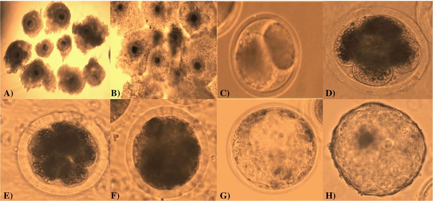
Fig. 5 Bright-field images of a Grade A and B oocytes aspirated from buffalo ovaries, b Matured cumulus-oocyte complexes after IVM, c 2-celled
stage, d 4-celled stage, e 8-celled stage, f 16-celled stage, g Morula and h Blastocyst stage of buffalo embryo observed during IVF studiesBatra et al. BMC Genomics (2021) 22:480 Page 9 of 18 Fig. 6 Scatter plots showing the Mean ± SD for cleavage rate a, morula b and blastocyst formation rates c in the control group and samples treated with three different concentrations of anti-BuBD-129 and. To elucidate such non-covalently bound (ionic) of the mammalian epididymis that help the spermatozoa sperm surface antigens, we used elevated salt (NaCl) in their survival and performance the male reproductive concentration i.e. 2x-DPBS. Although, it had previously tract (MRT) and FRT [64–66]. The identification of been documented that a population of such non- many BDs including the two CA-BDs viz. BuBD-129 and covalently bound sperm surface (glyco)proteins could be 126 indicated that the translated products of these genes released by exposing the macaque spermatozoa to 2x are applied as peripheral coats onto the buffalo sperm DPBS i.e. 300 mM NaCl [44] the proteomics profile of surface, as reported in primates and other ruminants these proteins was not available. Many (glyco) proteins, [29, 51]. Recently, many studies that link the BDs with e.g. the cattle PDC-109 and primate DEFB-126, are the regulation of male fertility have been reported in known to be released from the mammalian sperm- multiple mammalian species [30, 38, 45, 51, 67–69]. surface after elevated salt or PI-PLC extractions [45–47]. Interestingly, the number of BD genes and thus the epi- Thus, the other population of sperm-surface proteins is didymal secreted BDs found on sperm, are highly vari- GPI-linked and is known to be firmly integrated into able in different species at least partly, due to the particular microdomains on its plasma membrane. Most differences in microbial load, historical contingency, of the GPI-APs are laid down on the sperm-surface dur- genetic drift and disparate ecological niches occupied ing epididymal transit of the sperm and these (glyco) [64, 70, 71]. The epididymal BDs found on buffalo sperm proteins have aptly been addressed as ‘maturation anti- surface are among the class of proteins that are weakly gens [48–51]. We also removed the GPI-APs from the bound, presumably by non-covalent (ionic) interactions. buffalo sperm-surface using the enzyme PI-PLC and As observed in buffalo, many BDs e.g. DEFB-126 and subjected them to gene ontology and singular enrich- DEFB-15 are known to be expressed and secreted by the ment analysis. Our results revealed that most of the pro- epididymal epithelium cells which subsequently interact teins that adorn buffalo sperm-surface (e.g. the BDs) are with the traversing spermatozoa surface across mamma- immune response-related and reproduction-specific lian species [43, 51, 66, 72, 73]. The BDs are amongst (glyco) proteins, as observed in other species [48, 52– the dominant molecules of the buffalo sperm surface ap- 54]. parently due to their heavy glycosylation, their ability to Throughout nature, innate immunity and sexual interact with phospholipid membranes and their im- reproduction are tightly linked. As observed for buffalo munologic activity [9, 13, 29, 30, 39]. sperm-surface proteins, a growing body of evidence sug- The glycocalyx of the sperm is the molecular frontier gests that a major fraction of sperm-surface proteins be- that interacts directly with the hostile and immunologic long to the innate defence family and are required for milieu of the FRT. The flow cytometry experiments not surmounting various immunologic impediments in the only validated the presence of the cognate glycans for FRT [3, 48, 55–60]. Expectedly, the epididymis- the six lectins on the sperm-surface but also helped to specialized genes are thus known to be overrepresented monitor the reduction in the O-linked and N-linked gly- by the genes encoding secretory proteins which are in- cans from buffalo sperm-surface after protein extraction volved in the innate immune response and reproduction treatments. We have previously demonstrated that the [16, 56, 61–63]. Many molecules e.g. the highly glycosyl- BuBD-129 is an atypical BD, possessing a long C′ tail ated and negatively charged, antimicrobial peptides, and was predicted to be heavily O-glycosylated, similar beta-defensins (BDs) have been discovered in the lumen to what has been reported for the primate DEFB-126
Batra et al. BMC Genomics   (2021) 22:480                                                                     Page 10 of 18

[29, 32, 74]. Other epididymal secreted BDs like murine        TK Datta, Unpublished data). On the other hand, the
Defb-15 have also been predicted to be heavily O-              spatial distribution of BuBD-126 and BBD-126 [67] dif-
glycosylated and has a long 20 amino-acid extension in         fers from the observed binding pattern of DEFB-126 in
the C′ of its protein [42]. Our results thus suggested that    monkeys [78] or defb22 in rodents [79]. The BuBD-126
the glycocalyx barrier on the buffalo spermatozoa is ei-       was found to localize to the post-acrosomal region and
ther removed or greatly re-organized post the elevated         the tail rather than environing the whole sperm surface
salt or PI-PLC treatments. Therefore, the cognate gly-         like the primate DEFB-126 and defb22 in mice. These
cans for ABL, LEL and JAC appeared to be presented in          species-specific differences could be ascribed to the high
a way to become unavailable for lectin binding after           variability in the distribution pattern of glycoconjugates
these treatments. Interestingly, the recognition of nega-      and thus in the manifested Sperm Associated Glycan
tively charged, terminally positioned, sialic acids by         Topography (SpAGT) in various species [28].
MAL-II on the buffalo sperm surface pointed towards               IVF was used to predict the reproductive functional
their role as a protective coat. This is because the sialic    significance of the BuBDs. The blocking of BuBD-129
acid moieties are known to bestow upon the spermato-           reduced the cleavage as well as the morula and blasto-
zoa the ability to evade the elicited immune responses in      cyst formation rate of the fertilized buffalo oocytes dur-
the FRT [29, 32, 46]. Alternatively, they mask the tes-        ing IVF. It is well established that the antibodies
ticular protein components making the sperm invisible          directed against the sperm-specific antigens (ASAs) are a
to the FRT’s immune-surveillance [3, 29, 75]. Although         major cause of immunological infertility since they per-
the reduction in the MFI signal indicated either the dele-     turb the normal fertilization process [3, 50, 80]. Blocking
tion or conformational change in the available glycans         the BuBD-129 appeared to preclude successful
this approach can’t pinpoint individual sperm glycopro-        fertilization events which confirmed their presence on
tein rather identifies general shifts in the surface sugars    buffalo sperm-surface. Likewise, the ortholog of primate
on the sperm-surface.                                          DEFB-126 in mice (defb22) has been demonstrated to
   Immunofluorescence (IF) revealed a differential spatial     incorporate in the oolemma during gamete fusion which
distribution pattern of the heavily O-glycosylated periph-     subsequently floated and extended out from the fused
eral protein BuBD-129 and BuBD-126 on the buffalo              spermatozoa [81, 82]. Besides, its ortholog in cattle
sperm surface. The BuBD-129 spanned the entire buffalo         (BBD-126) has been reported to be retained on the
spermatozoa similar to what has been observed for the          sperm surface, after induction of in vitro capacitation
primate DEFB-126 and rodent defb22 [44, 76, 77]. Al-           [67]. This suggests that the BBD-126 too remains associ-
though the coats of maturation antigens and their inter-       ated with the spermatozoa during fertilization indicating
acting partners tend to localize to specific micro-            an additional role in fertilization [47]. It has been pro-
domains, nevertheless, few of them such as BuBD-129,           posed that these features may be true for all the BDs
rodent defb22 and the primate DEFB-126 are adsorbed            with high levels of O-glycosylation [83]. Our results indi-
globally around the entire spermatozoa [29, 40, 78, 79].       cated that the BuBD-129 is present on the buffalo
This similarity in the spatial distribution pattern hints at   sperm-surface even after induction of in vitro capacita-
the existence of functional orthology between the buffalo      tion. Similar to BuBD-129, antibodies against another
BuBD-129, rodent defb22 and primate DEFB-126, apart            member of the CAP superfamily, CRISP1, a sperm-
from other similarities like gene length, chromosome           surface protein have been demonstrated to obstruct
cluster, an extended amino-acid tail at C-terminus, a          fertilization by interfering in the sperm-oocytes fusion
high potential of O- and N-glycosylation and the prefer-       process, thus reducing fertility [78, 84]. The cysteine-
ential expression in the distal segments of MRT [74].          rich defensin-like peptides appear to be integral to the
The peripheral localization of BuBD-129 along the entire       reproductive success of organism ranging from inverte-
buffalo spermatozoa perimeter presumably establishes a         brates, plants to higher primates [85–88]. It cannot be
barrier between the buffalo spermatozoa and the im-            denied that the blocking of BuBD-129 changes the attri-
munologic milieu of the FRT as reported for other BDs          butes of sperm surface features which not only influence
in other mammalian species [3, 29, 50, 75]. Despite be-        fertility but also the developmental potential of the sub-
ing identified amongst the dominant proteins on the            sequent embryos. The BuBD-129 probably imparts sur-
buffalo sperm surface, the BuBDs were not deemed as            face properties that are essential for fertilization by the
antigenic sperm-surface proteins by the female immune          buffalo sperm due to its heavy O-linked glycosylation
system. This indicates their role in immune-protection         and uniform spatial distribution on the entire buffalo
because the antibodies were generated against other pro-       sperm. These results suggest multiple and putatively epi-
teins (e.g. Acrosin and Profilin) rather than the BuBDs        static roles of the BD genes in immune response and re-
when female Wistar rats were immunized with buffalo            productive physiology of buffalo, e.g.in the process of
sperm-surface extracted proteins (V Batra, R Kumar and         sperm-oocytes interaction.
Batra et al. BMC Genomics   (2021) 22:480                                                                           Page 11 of 18

Conclusion                                                        supernatant/acetone ratio), concentrated on a speed-vac
The buffalo sperm surface is heavily glycosylated and many        vacuum concentrator and subjected to SDS-PAGE after
glycoproteins are applied as peripheral coats on to the sur-      quantification by Quick Start™ Bradford protein assay.
face of mammalian spermatozoa. Many of these glycopro-
teins are immune-regulatory and have reproduction-                PI-PLC extraction of GPI-APs
specific functions. The molecular functions and biological        The PI-PLC enzyme specifically hydrolyzes the phosphodiester
roles of only a limited number of such proteins have been         bond of phosphatidylinositol to form a free 1, 2-diacylglycerol
studied regarding their role in male reproductive physi-          and glycopeptide-bound Myo-inositol 1,2-cyclic phosphate and
ology. The BuBDs like BuBD-129 and 126 are amongst the            is therefore used to release the GPI-APs from the surface of
dominant molecules of the buffalo sperm surface. It would         the membranes [44, 77]. The post-swim-up spermatozoa
be interesting to quantify the BuBD-129 abundance in LF           (100 × 106) in Sp-TALP were incubated with three different
and HF bulls with contrasting field CRs through a targeted        concentrations of Phosphoinositide-phospholipase C (PI-PLC)
proteomics approach. The effect of exogenous supplemen-           from Bacillus cereus (1 U/mL, 1.5 U/mL and 2 U/mL) in sili-
tation of recombinant BuBD-129 to the low fertile sperm to        conized tubes at 37 °C for 2 h. The siliconized tubes were
augment the current field fertility rates should also be eval-    shaken gently during the period of incubation. Thereafter, the
uated. Further investigations into the functional roles of this   samples were centrifuged at 1000 x g for 10 min and the super-
critical component of buffalo sperm are warranted.                natants were collected and then filtered through a 0.22 μm fil-
                                                                  ter. The supernatants from 10 to 12 ejaculates were pooled
Methods                                                           and then precipitated by acetone precipitation method and
Chemicals and plasticware                                         quantified before subjecting to SDS-PAGE.
Chemicals and media used in the present study were ob-
tained from Sigma Chemicals Co., St. Louis, Missouri, USA/        Mass spectrometry (LC-MS/MS) of sperm-surface
Qiagen/Fermentas/Invitrogen as mentioned for specific cases.      extracted proteins
                                                                  For the identification of sperm surface proteins, mass-
Semen collection and pre-processing                               spectrometry was performed using the method described
Freshly ejaculated normozoospermic semen samples                  by Gourinath et al. [89]. The extracted proteins (100 μg)
(mass motility ≥3, n = 5–12, aged between 3 and 5 years)          from the pooled elevated salt and PI-PLC extractions
were collected from mature Murrah buffalo bulls (of               were dissolved in 6 M guanidium hydrochloride. Subse-
proven fertility) using the artificial vagina at the Artificial   quently, 25 μL of the dissolved samples were reduced
Breeding Research Centre (NDRI, Karnal, India). Each              with 5 mM tris (2-carboxyethyl) phosphine (TCEP). The
ejaculate was collected in a 15 mL centrifuge tube con-           samples were then alkylated with 50 mM iodoacetamide
taining twice the volume of non-capacitating media                for 20 min in dark at room temperature (RT) and then
(NCM), Sp-TALP i.e. HEPES buffered Tyrode’s medium                digested with trypsin (1:50, trypsin/lysate ratio) for 16 h
(pH 7.4, 37 °C). The semen was transported to the la-             at 37 °C after re-suspension in digestion buffer. The di-
boratory at 37 °C and was washed thrice with Sp-TALP              gests were cleaned using a C18 silica cartridge to remove
by centrifuging at 280 x g for 6 min to remove the sem-           the salt and dried using a speed vac vacuum concentra-
inal plasma and its protein components. The motile                tor. The dried pellet was resuspended in buffer A (5%
spermatozoa were selected by subjecting the final pellet          acetonitrile, 0.1% formic acid). All the experiments were
to the swim-up technique. The upper 1.5 mL volume                 performed using EASY-nLC 1000 system (Thermo
was later collected and centrifuged to obtain the pellet          Fisher Scientific), which was coupled to a QExactive
of motile spermatozoa.                                            Mass Spectrometer (Thermo Fisher Scientific) equipped
                                                                  with a nano-electrospray ion source. One μg of the pep-
Surface protein extraction from motile spermatozoa                tide mixture was resolved using a 25 cm PicoFrit column
Elevated salt extraction of surface proteins                      (360 μm outer diameter, 75 μm inner diameter, 10 μm
The post-swim-up spermatozoa in Sp-TALP were washed               tip) filled with 1.8 μm of C18-resin (Dr Maisch, Ammer-
in DPBS and divided into four groups by re-suspending in          buch, Germany). The peptides were loaded with buffer
either 2x or 4x DPBS for 30 min and 60 min at 37 °C (viz.         A and eluted with a 0–40% gradient of buffer B (95%
2x-30, 2x-60, 4x-30 and 4x-60). The micro-centrifuge              acetonitrile, 0.1% formic acid) at a flow rate of 300 nL/
tubes were shaken gently during the period of incubation          min for 90 min. The MS data were acquired using a
and the samples were subsequently pelletized by centrifu-         data-dependent top 10 method dynamically choosing the
gation at 280 x g for 10 min. A pool of supernatants from         most abundant precursor ions from the survey scan
5 to 7 ejaculates was collected and subsequently filtered         (Fig. 7). All raw MS data have been deposited to the Pro-
through a 0.22 μm filter. The proteins in the filtrate were       teomeXchange consortium through the PRIDE partner
precipitated with acetone precipitation method (1:9,              repository (Identifier: PXD022114).
Batra et al. BMC Genomics (2021) 22:480 Page 12 of 18
Fig. 7 The overall research methodology followed for BuBD identification on the buffalo sperm surface by LC-MS/MS.
LC-MS/MS data processing relevant to the agricultural community. For SEA (Singu-
The generated raw files for the seven samples were analyzed lar Enrichment Analysis), the Bovine genome locus (Bo-
using Trans-Proteomic Pipeline TPP v5.1 (Syzygy) rev.0 [90], vine Genome Database): GLEAN_03528 was used as
against a database generated from UniProt knowledgebase background dataset and Fisher’s test was used to calcu-
(Bos taurus, Homo sapiens, Bubalus bubalis and beta- late the p-values. The Yekutieli (FDR under dependency)
defensin, downloaded January 3, 2019; www.uniprot.org) method [94] was used for Multi-test adjustment. The
using the Comet search engine [91]. The precursor and frag- minimum number of mapping entries for analysis was
ment mass tolerances were set at 10 ppm and 0.5 Da, re- set to 5 and values with p⩽0.05 were considered signifi-
spectively. The enzyme specificity was set to trypsin/P and a cant. The resulting long lists of Gene Ontology terms
maximum of two missed cleavages were allowed. Carbami- were summarized by REViGO webserver [95] which
domethyl on cysteine (C) was considered as fixed modifica- removes the redundant terms and the remaining terms
tion while oxidation of methionine and N-terminal were visualized in semantic similarity-based scatterplots.
acetylation were considered as variable modifications for the The SimRel was used as the semantic similarity measure
database search. The peptide spectrum match and protein (clustering algorithm) against the whole Uniprot data-
false discovery rates (FDR) were set to 0.01 to increase the base which employs a multidimensional scaling proced-
confidence and remove the false-positive identifications. The ure that initially places the terms using eigenvalue
use of iProphet tool in TPP results in an increased number decomposition of the terms’ pairwise distance matrix.
of correctly identified peptides at a constant FDR because it This was followed by a stress minimization step which
combines the evidence from multiple identifications of the iteratively improves the agreement between the GO
same peptide sequences across different spectra, experi- terms’ semantic similarities and their closeness in the
ments, precursor ion charge states, and modified states. Only displayed two-dimensional space.
the proteins with iProphet probability greater than 0.99 were
considered for further analysis [92]. Flow cytometry
The validation of the surface (glyco) protein removal
Singular enrichment and scatter plot analyses of the after elevated salt and PI-PLC treatments was done by
identified proteins flow cytometry using six lectins viz. ABL, LEL, JAC,
The gene-ontology and singular enrichment analysis for MAL-II, LCA and PNA (Table 3), which bind to various
identified proteins in all of the seven treatments were cognate glycans on the sperm surface, including an un-
performed using agriGO [93] which is a specialized GO stained sample as the negative control (n = 3). A proto-
analysis toolkit and database for livestock species col for sperm flow cytometry analysis standardized byBatra et al. BMC Genomics (2021) 22:480 Page 13 of 18
Table 3 The six lectins used to assess changes in the sperm- Aldrich, USA). The cDNA synthesis and RT-qPCR
surface after salt/PI-PLC treatments optimization were done as described by Batra et al. [74].
Lectin Major sugar recognized The MIQE [98] guidelines were followed at every step,
LEL (Lycopericon esculentum [GlcNAc]1–3, N-acetylglucosamine wherever possible. The relative quantification of the BD
lectin) genes was done on a Bio-rad CFX-96 Touch Deep Well
ABL (Agaricus bisporus lectin) Galactosyl (β-1,3) N-acetylgalactosamine Real-Time PCR system platform using the iTaq Univer-
JAC (Jacalin) Mono or di-sialylated ofT-antigen sal SYBR Green Supermix (Bio-Rad, USA) in a10 μL re-
MAL II (Maackia amurensis α-2,3 linked Sialic acid
action mix. The thermal profile was 95 °C for 5 min, 40
lectin II) cycles consisting of denaturation at 95 °C for 15 s, an-
LCA (Lens culinaris agglutinin) Mannose and Glucose moieties nealing at variable optimized temperatures for 20sand
extension at 72 °C for 20 s, followed by the melt curve
PNA (Peanut agglutinin) Asialylatedgalactosyl (β-1,3)N-
acetylgalactosamine protocol with 10 s at 95 °C and then 60 s each at 0.5 °C
increments between 65 °C and 95 °C. A no-template con-
trol (NTC) was run in each plate to confirm the absence
Batra et al. [28] was followed for signal acquisition. of nucleic acid contamination. A melt curve analysis was
Briefly, the sperm were washed as described earlier and performed to ensure a specific, unique product forma-
the concentration was adjusted to 3 × 10⁶ sperm/ml. tion and to ascertain minimal primer dimer formation.
Samples were incubated with lectins for 10 min at The mean sample Cq (Cycle of quantification) values for
38.6 °C under an atmosphere of 5% CO2 before the flow the BuBD-126 and 129 were calculated for duplicate
cytometry analysis which was performed with a standard samples and their relative expression was calculated as
bench-top BD Accuri C6 flow cytometer (Becton Dickin- described previously [41]. The GAPDH (glyceraldehyde-
son Biosciences, Ann Arbor, MI, USA, with BD Accuri 3-phosphate dehydrogenase) and eEF-2 (eukaryotic
C6 software v.1.0.27.1). The cytometer was calibrated elongation factor) were used as the reference genes, and
daily according to the manufacturer’s recommendations the blood was considered as the calibrator for the quan-
with 8 and 6 peak calibration beads and QC was per- tification of gene expression. The differential gene ex-
formed every second day using BD CS&T RUO beads. pression levels between the blood and the spermatozoa
The 488-nm laser was used for the excitation of FITC, were examined for normality of distribution and were
and its emission was filtered using a 533/30 bandpass fil- analyzed by an unpaired two-tailed t-test as imple-
ter. Filtered emissions were detected by photomultiplier mented in GraphPad Prism 7.0 (for Windows, GraphPad
tubes. A threshold of 80,000 in the forward scatter (FSC) Software, La Jolla California USA, www.graphpad.com)
signal was applied to remove electrical noise, and very and a p-value < 0.05 was considered to be statistically
small events and samples were acquired at a low flow significant.
rate (14 μl/min). For each sample, 20,000 events (single
cells) were acquired. Only the singlet population was Antibody development for determining the spatial
identified and used for analyses. The statistical analyses distribution pattern of BuBD-129 and 126
were performed on Prism Graphpad 7.0 (for Windows, A custom polyclonal antibody specific for BuBD-129 and
GraphPad Software, La Jolla California USA, www. 126 was commercially generated by Genei using a stand-
graphpad.com) by one way ANOVA to test the differ- ard protocol. Briefly, the amino acid peptide sequences
ence of means and the Brown-Forsythe test to test the from the secreted fragments of BuBD-129 and 126 were
differences in the standard deviations of the MFI values selected based on computational modelling on IEDB [99]
produced from various FITC-bound lectins. for being surface epitopes. After chemical synthesis and
conjugation to keyhole limpet hemocyanin (KLH), the epi-
Expression dynamics of the BuBD-126 and BuBD-129 in tope (500 μg mixed with Freund’s complete adjuvant) was
the buffalo spermatozoa inoculated subcutaneously into the back of the neck re-
Total RNA was isolated from the peripheral blood and gion of sexually mature nulliparous female New Zealand
the spermatozoa as described previously [96, 97] using white rabbits (n = 1, each group) weighing between 1.1–
the TRI Reagent, RNA isolation reagent (Sigma-Aldrich, 1.5Kg (1 animal per cage). The pre-immune sera (control)
USA) and was quantified using a NanoDrop ND-1000 were extracted from the blood taken from the central ear
UV–Vis spectrophotometer (NanoDrop Technologies vein by the skilled technicians in the presence of a veterin-
Inc., Wilmington, DE, USA). The A260/280 and A260/ ary doctor. For initial immunization, 500 μg of either
230 were close to 2.0 for all the samples used in the BuBD-129 or 126 KLH-conjugated peptide was mixed in
study. The quality of the extracted RNA was assessed by 500 μl of Freund’s complete adjuvant and was adminis-
running 200 ng of the RNA (heated at 70 °C for 1 min) tered subcutaneously in the back of the neck region of the
in non-denaturing TAE buffered 1.2% agarose (Sigma- animal. However, for booster doses 300 μg of the antigenBatra et al. BMC Genomics (2021) 22:480 Page 14 of 18
mixed in 500 μl of Freund’s incomplete adjuvant and was physiological saline (0.9%, w/v NaCl) containing strepto-
administered similarly. An immune response was con- penicillin (50 mg/l). Ovaries were washed 3–4 times in
firmed by binding of the serum to the antigen on an normal saline and the cumulus-oocyte complexes
enzyme-linked immunosorbent-type assay. The antibody (COCs) were aspirated from the follicles with the help of
titer was checked using western blot and seven booster a vacuum aspiration unit (K-MAR-5200 IN, USA) in
doses were deemed necessary to obtain working IgGs iso- Hepes-buffered hamster embryo culture (HH) medium
lated from the serum. The blood was collected by bleeding and the aspirated follicular fluid was placed in a dry bath
the rabbit through the central ear vein by trained individ- at 37.5 °C for 35–40 min. The quality of COCs was ob-
uals in the presence of a veterinary doctor. The whole served and graded into grade A (> 5 layer cumulus layer)
blood (5 ml) was subsequently collected from the central and B (3–5 cumulus layers) under the stereo-zoom
ear vein and allowed to clot at RT for 30 min. The serum microscope. The COCs of only grade A and B were se-
was isolated from the blood and Protein G based NAb™ lected for in vitro maturation (IVM) and in vitro culture
Spin Kits were used for isolation of IgG antibodies (IVC). To perform IVM, the selected COCs were washed
from the collected serum according to the manufac- 3–4 times with maturation media (HEPES buffered
turer’s instructions. The subsequent quantification of TCM199 modified with 10% (v/v) fetal bovine serum
BuBD-129 and 126 IgG antibodies from the serum (FBS), 0.005% (w/v) streptomycin, 0.01% (w/v) sodium
was performed by measuring the A280 of each eluate pyruvate and 0.005% (w/v) glutamine supplemented with
fraction on an Infinite® 200 NanoQuant microplate 5.0 μg/ml FSH and 10 μg/ml LH, 1 μg/ml estradiol 17-β
reader (Tecan). A confirmatory SDS-PAGE was per- and 50 ng/ml epidermal growth factor (EGF), 64 μg/ml
formed to validate the isolated IgGs. cysteamine and 50 μl ITS). After washing the COCs were
placed in a group of 15 in 100 μl of maturation medium
Immunofluorescence drops. Three such drops were placed in a 35 mm culture
The swim-up fraction of spermatozoa (40 x 106cells/ml) dish which was then overlaid with mineral oil dressing.
was re-suspended in NCM and was added onto poly-L- The dishes were cultured in duplicate for 24 h at 38.5 °C
lysine coated slides. The NCM was removed after 15 in a humidified atmosphere of 5% CO2 in an incubator.
min since the spermatozoa adhered to the slide surface The IVF was carried out in 100 μl droplets of BO
by that time. Subsequently, the cells were washed twice medium supplemented with 1% (w/v) bovine serum al-
in PBS and fixed in 2% paraformaldehyde and 0.1% glu- bumin (BSA), 1.9 mg/ml caffeine sodium benzoate, 0.14
taraldehyde for 20 min at RT. The movement of the mg/ml sodium pyruvate and 0.01 mg/ml heparin. For
GPI-APs is prevented by using low glutaraldehyde con- the IVF, the control group didn’t contain anti-BuBD-129
centration. The spermatozoa were then washed with antibody whereas, the three treatment groups comprised
PBS thrice, and the slide surface was then blocked with of three different concentrations of anti-BuBD-129 (0.5
blocking buffer (1% BSA in PBST-0.1% Tween 20 in 1X- mg/ml) antibody in the fertilization medium drops viz.
PBS) for 1 h at room temperature. The cells were later the 1:15000, 1:10000 and 1:5000 dilution groups were
incubated with the primary polyclonal antibody (1:1000 prepared. The matured COCs were washed thrice in BO
dilutions) against BuBD-129 and 126 overnight at 4 °C. medium and placed in BO medium droplets supple-
The sperm were washed with PBST thrice and were then mented with the anti-BuBD for treatment groups. The
incubated with FITC conjugated goat anti-rabbit IgG frozen buffalo semen was thawed simultaneously and
secondary antibody (1:5000 dilutions; Sigma-Aldrich) in subsequently processed for in vitro capacitation as per
dark for 1 h at RT followed by final washings with PBST the procedure described earlier by Jain et al. [97]. Fifty μl
(3x). After the final washing, a coverslip was mounted of BO media was removed from the respective IVF
onto a glass slide onto which one drop of mounting medium drops and 50 μl of the sperm suspension (1 ×
medium, Dabco® 33-LV was placed. The cells were then 106 spermatozoa/mL) was added to each fertilization
observed under a BX-51 Olympus fluorescence micro- drop with 15 COCs and was then incubated at 38.5 °C
scope. The loss of the BuBD-129 and 126 from the with 5% CO2 for 12 h. The presumptive zygotes were re-
sperm-surface after incubation of spermatozoa in 2x- moved from the fertilization drops after 12 h of insemin-
DPBS or exposing them to 2 U/ml of PI-PLC was ation and the adhered cumulus cells were mechanically
similarly monitored by immunofluorescence after the removed by vortexing and were washed five times in a
stipulated time. modified Charles and Rosenkrans 2 amino acid
(mCR2aa) medium. Following washing, the 15 presump-
IVF study tive zygotes were co-cultured with the monolayer of
Buffalo ovaries were collected from a local abattoir, granulosa cells in 100 μl drops of IVC-I medium
Delhi regardless of the oestrous cycle stage and were (mCR2aa supplemented with 0.8% (w/v) BSA, 1 mM glu-
transported within 2-3 h to the laboratory in cose, 0.33 mM pyruvate, 1 mM glutamine, 1 x MEMYou can also read