Machine Learning in Dermatology: Current Applications, Opportunities, and Limitations - eScholarship
←
→
Page content transcription
If your browser does not render page correctly, please read the page content below
Dermatol Ther (Heidelb) (2020) 10:365–386
https://doi.org/10.1007/s13555-020-00372-0

 REVIEW

Machine Learning in Dermatology: Current
Applications, Opportunities, and Limitations
Stephanie Chan . Vidhatha Reddy . Bridget Myers . Quinn Thibodeaux .
Nicholas Brownstone . Wilson Liao

Received: February 26, 2020 / Published online: April 6, 2020
Ó The Author(s) 2020

ABSTRACT                                                        clinical images; (2) disease classification using
                                                                dermatopathology images; (3) assessment of
Machine learning (ML) has the potential to                      skin diseases using mobile applications and
improve the dermatologist’s practice from                       personal monitoring devices; (4) facilitating
diagnosis to personalized treatment. Recent                     large-scale epidemiology research; and (5) pre-
advancements in access to large datasets (e.g.,                 cision medicine. The purpose of this review is to
electronic medical records, image databases,                    provide a guide for dermatologists to help
omics), faster computing, and cheaper data                      demystify the fundamentals of ML and its wide
storage have encouraged the development of                      range of applications in order to better evaluate
ML algorithms with human-like intelligence in                   its potential opportunities and challenges.
dermatology. This article is an overview of the
basics of ML, current applications of ML, and
potential limitations and considerations for                    Keywords: Artificial intelligence; Convolutional
further development of ML. We have identified                   neural network; Deep learning; Dermatology;
five current areas of applications for ML in                    Image classification; Machine learning; Mobile
dermatology: (1) disease classification using                   applications; Personal monitoring devices;
                                                                Precision medicine

Enhanced Digital Features To view enhanced digital
features for this article go to https://doi.org/10.6084/
m9.figshare.12006789.

S. Chan  V. Reddy  B. Myers  Q. Thibodeaux
N. Brownstone  W. Liao (&)
Department of Dermatology, University of
California San Francisco, San Francisco, CA, USA
e-mail: wilson.liao@ucsf.edu
366                                                              Dermatol Ther (Heidelb) (2020) 10:365–386

                                                  dermatology is already abundant, with data
 Key Summary Points                               from the genome, epigenome, transcriptome,
                                                  proteome, and microbiome, areas of research
 Machine learning (ML) has the potential          that are often referred to by the shortened term
 to improve the dermatologist’s practice          ‘‘omics’’ [2]. Recent advancements in faster
 from diagnosis to personalized treatment.        processing and cheaper storage have allowed for
                                                  the development of machine learning (ML)
 This review article is a guide for               algorithms with human-like intelligence that
 dermatologists to help demystify the             have numerous applications in dermatology
 fundamentals of ML and its wide range of         [3–5]. To assess the effectiveness of these
 applications in order to better evaluate its     emerging technologies, it is imperative that
 potential opportunities and challenges.          dermatologists have a basic understanding of
 We have identified five current areas of         artificial intelligence and ML. In this review, we
 applications for ML in dermatology: (1)          first provide an overview of artificial intelli-
 disease classification using clinical images;    gence and ML and how algorithms are devel-
 (2) disease classification using                 oped. Second, we examine the current
 dermatopathology images; (3) assessment          applications of ML that are relevant to derma-
 of skin diseases using mobile applications       tologists. Lastly, we explore potential challenges
 and personal monitoring devices; (4)             and limitations for the future development of
 facilitating large-scale epidemiology            ML. This review is a guide for dermatologists to
 research; and (5) precision medicine.            help demystify the fundamentals of ML and its
                                                  wide range of applications in order to better
 While ML models are powerful,                    evaluate its potential opportunities and
 dermatologists should be cognizant of the        challenges.
 potential limitations of ML (e.g.
 algorithmic bias and black box nature of
 ML models) and how to make these                 METHODS
 technologies inclusive of skin of color.
                                                  This review is based on a literature search per-
 Involving more dermatologists in the             formed in Medline, Embase, and Web of Science
 development and testing of ML models is          databases of articles pertaining to artificial
 imperative for creating useful and               intelligence and ML in dermatology. The search
 clinically relevant technology.                  was conducted in December 2019. Articles from
                                                  2000 to 2019 were included to focus on
                                                  emerging methods. Only articles written in
                                                  English were included, and articles that were
                                                  repeated were excluded. The following primary
INTRODUCTION                                      keywords were used: ‘‘artificial intelligence,’’
                                                  ‘‘machine learning,’’ and ‘‘dermatology.’’ After
In dermatology and medicine at large, the         the preliminary results were reviewed, the Boo-
abundance of data in clinical records, patient    lean operators ‘‘AND’’ and ‘‘OR’’ were used with
demographic information, results from imaging     the following secondary keywords: ‘‘personal-
examinations, and data collected from ques-       ized medicine,’’ ‘‘teledermatology,’’ ‘‘smart-
tionnaires represent a wealth of information      phone apps,’’ ‘‘skin cancer,’’ ‘‘nonmelanoma
that has the potential to revolutionize person-   skin cancer,’’ ‘‘melanoma,’’ ‘‘psoriasis,’’ ‘‘atopic
alized medicine [1]. Translational research in    dermatitis,’’ and dermatopathology.’’ Our liter-
                                                  ature search yielded a total of 899 articles,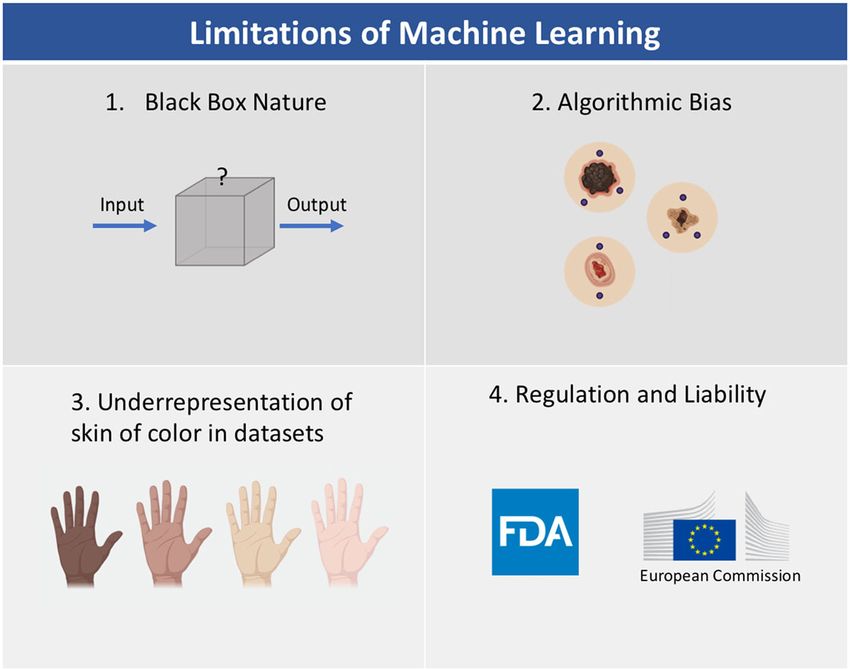
Dermatol Ther (Heidelb) (2020) 10:365–386                                                                    367

among which 70 articles were deemed relevant                 how to process data. However, the heuristics of
to this review.                                              human decision making in medicine were not
   This article is based on previously conducted             easy to program into explicit rules.
studies and does not contain any studies with                    ML is a tool comprising a subset of artificial
human participants or animals performed by                   intelligence that enables the goals of artificial
any of the authors.                                          intelligence to be achieved (Fig. 1). Recently,
                                                             ML has piqued attention for its broad range of
                                                             uses in daily life from personalized online rec-
OVERVIEW OF ARTIFICIAL                                       ommendations for videos and news to self-
INTELLIGENCE AND MACHINE                                     driving cars.
LEARNING                                                         ML covers a variety of algorithms and sta-
                                                             tistical methods, including logistic regression,
What is the Difference Between Artificial                    random forest, and deep learning. Although ML
Intelligence and Machine Learning?                           can seem enigmatic at first, it can be deeply
                                                             related to traditional statistical models recog-
Artificial intelligence is a branch of computer              nizable to most dermatologists.
science that uses machines and programs to
simulate intelligent human behavior. Artificial              Machine Learning Approaches
intelligence dates back to the 1950s to Alan
Turing’s question ‘‘Can machines think?’’ [6]. By            Machine learning approaches can be divided
the 1970s, software engineers had created                    into three broad categories: supervised learning,
algorithms with explicit rules for computers on              unsupervised learning, and reinforcement
                                                             learning [7]. Supervised learning requires a
                                                             dataset to be presented as inputs (called fea-
                                                             tures) and outputs (called labels) [7]. For exam-
                                                             ple, in an algorithm that classifies a pigmented
                                                             lesion as a melanoma or benign, images of
                                                             pigmented lesions are ‘‘features’’ and the cate-
                                                             gorical data of whether it is malignant or benign
                                                             are ‘‘labels.’’ The algorithm is first trained with
                                                             labeled images of melanoma and benign pig-
                                                             mented lesions and then the computer gener-
                                                             alizes this information to a new, unseen set of
                                                             images of skin. Supervised learning is the most
                                                             common type of learning used in dermatology.
                                                             In contrast, unsupervised learning only requires
                                                             inputs (unlabeled data), and this approach can
                                                             identify unknown clusters or anomalies in data
                                                             [7]. Reinforcement learning is a hybrid of both
                                                             supervised and unsupervised learning which
                                                             learns by trial and error and input from the
                                                             environment [7]. An example of reinforcement
Fig. 1 Artificial intelligence and machine learning.          learning is the algorithm in AlphaGo [8]. Rein-
Machine learning is a type of artificial intelligence. Some   forcement learning has yet to be explored in
common types of machine learning approaches used in          dermatology.
dermatology include convolutional neural network
(CNN), natural language processing (NLP), support vector
machine, and random forest. Notably, there are many
other possible machine learning approaches that are not
listed and out of the scope of this review
368 Dermatol Ther (Heidelb) (2020) 10:365–386
Machine Learning Algorithms for a series of inputs, the weights are fixed and
the NN can be applied to other datasets [14].
There are a variety of ML algorithms commonly Convolutional NNs (CNNs) are a special
used in dermatology. Most ML algorithms are subclass of ANNs that contain one or more
examples of statistical learning; for example, layers called convolutional units (pooling
some of the most common statistical learning units). CNNs take in two-dimensional or three-
methods are linear regression, logistic regres- dimensional inputs which are passed through
sion, k-nearest neighbor (k-NN), support vector multiple hidden layers. An image can be broken
machine (SVM), random forest (RF), and natural down into motifs, or a collection of pixels that
language processing (NLP). k-NN is used for data form a basic unit of analysis. The first few layers
classification and regression based on the of the CNN compare each part of an input
number of k neighbors [9, 10]. SVMs are used to image against some small sub-image [5]. Each
classify data by finding a hyperplane to differ- node is assigned a certain feature (e.g., color,
entiate between groups [11]. RFs generate a shape, size, etc.), and the node’s output to the
network of random decision trees to find the next layer depends on how much a part of the
most common outcome among all the ran- image resembles the feature, a process per-
domly generated decision trees [12]. NLP ana- formed by convolution [5]. After these convo-
lyzes large bodies of text in order to identify lutional layers, pooling layers, which are a
patterns [13]. standard NN, classify the overall image [5].
CNNs first showed promise for medical image
Neural Networks and Deep Learning classification at the historic 2012 ImageNet
Large Scale Visual Recognition (ILSVRC) con-
ference. A CNN, called AlexNet, was trained to
Deep learning is a subset of ML that uses sta-
classify 1.2 million images into 1000 different
tistical and mathematical models to mimic how
categories with a top-5 error rate of 15.3%,
neurons process information. Artificial neural
which is the percentage of images for which the
networks (ANNs), or neural networks (NNs), are
correct class was not among the top five pre-
based on a collection of connected units (e.g.,
dicted classes [15]. This was the first CNN to
nodes, neurons, or process layers) [14]. ANNs
display such a low error rate. Previous image
are inspired by the network of neurons in the
datasets were relatively small, comprising only
human brain. The neurons, or nodes, that make
of tens of thousands of images [16–18]. By 2016,
up the ANN are organized into linear arrays
all methods to classify medical images at the
called layers [14]. Each node receives inputs
2016 International Symposium on Biomedical
from other connections that have associated
Imaging used CNNs [19].
weights [14]. Creating an ANN includes choos-
Another subtype of CNNs is called a region-
ing the number of nodes in each layer, the
based CNN (R-CNN). R-CNN is a type of CNN
number of layers in the network, and the path
that can detect a desired object within an
of the connections among the nodes [14], and
image. In the case of dermatology, it can detect
the typical ANN has input layers, output layers,
the location of cutaneous lesions by combining
and hidden layers. ANNs are trained to perform
region proposal algorithms with CNNs.
specific tasks, such as classification, through a
learning process. Learning within ANNs can be
supervised or unsupervised; however, super- Transfer Learning and Ensemble Learning
vised learning is more common. In supervised
learning, a training set contains examples of Transfer learning utilizes the power of a pre-
input targets and output targets [14]. As the trained CNN. These pretrained CNNs are often
ANN is trained, the weights of the inputs are trained on databases that include millions of
adjusted to minimize the error between the images, so they are able to distinguish images
network output and the correct output [14]. with much higher accuracy than a CNN that is
Once the network produces the desired outputs only trained on databases of only a few hundredDermatol Ther (Heidelb) (2020) 10:365–386 369
or few thousand images. The last fully con- capturing the noise of the dataset [7]. Algo-
nected layer of a pretrained CNN is modified rithms that are trained and validated with the
and trained with images for the more specific same set of images or with the same dataset risk
classification task. Examples of common pre- overfitting to the data. However, current studies
trained CNNs are AlexNet, Google Inception are limited by the images available and may be
V3, ResNet-50, Xception, VGG-19, and VGG-16. trained and validated on images from the same
This layered architecture allows researchers to dataset. Validation or cross-validation to com-
use a pretrained network without its final layer pare the predictive accuracies of datasets can
as a fixed feature extractor for other tasks. The help prevent overfitting. In contrast, underfit-
learning process of a pretrained CNN can be ting occurs when a ML model cannot represent
faster because it relies on previously learned the training dataset and cannot generalize to a
tasks. Using a pretrained CNN that is trained on new dataset [7]. Overfitting is more common
1–2 million images is more accurate than a CNN than underfitting in ML models.
that is trained on a smaller number of images of
the more specific classification tasks. Ensemble
learning improves ML results by combining DERMATOLOGY AND MACHINE
several models (meta-algorithms) or the power LEARNING
of multiple of CNNs together [7].
There are many promising opportunities for ML
Metrics for Assessment and Validation in the dermatologist’s practice. The classifica-
of Machine Learning Models tion of images through CNN has garnered the
most attention for its potential to increase
accessibility of skin cancer screenings and
Machine learning models are assessed according
streamline the workflow of dermatologists.
to variety of metrics based on the number of
CNN has already proven successful in many
true positives (TP), true negatives (TN), false
other fields, such as ophthalmology, pathology,
positives (FP), and false negatives (FN) from a
and radiology. In 2018, the US Food and Drug
ML prediction. These metrics include sensitivity
Administration (FDA) approved a CNN in the
[TP/(TP ? FN)], specificity [TN/(TN ? FP)], pos-
IDx-DR diagnostic system (IDx Technologies
itive predictive value [TP/(TP ? FP)], negative
Inc., Coralville, IA, USA) [22] for independent
predictive value [TN/(TN ? FN)], and accuracy
use in diabetic retinopathy screening after being
[(TP ? TN)/(TP ? TN ? FP ? FN)]. ML models
validated in a pivotal prospective clinical trial of
are also evaluated on the area under the receiver
over 900 patients. This diagnostic system
operating characteristic (AUROC or AUC). The
achieved a sensitivity of 87.2% and specificity of
receiver operating characteristic (ROC) curve is
90.7% and was compared to an independent,
calculated by plotting the sensitivity versus
high-quality gold standard of imaging protocols
1-specificity [20]. The further the ROC curve
[23]. However, CNN for the screening of mela-
deviates from the diagonal and the larger the
noma or non-melanoma skin cancers has not
AUROC, the better the algorithm. An AUROC of
been validated in prospective clinical trials and
1.0 indicates perfect classification, and an
still has notable limitations. Beyond the sim-
AUROC of 0.5 indicates classification that is no
plistic task of differentiating malignant from
better than random chance.
benign lesions, ML can also be applied in dif-
Another important metric is the generaliz-
ferential diagnosis, dermatopathology, teleder-
ability of an ML model, or how well a ML model
matology, mobile applications, and
can learn concepts and apply these concepts to
personalized medicine. Understanding what
examples the model has never seen before [21].
type of deep learning methods are currently
There are two terms to describe the generaliz-
being used and how these methods are evolving
ability of an ML model: overfitting and under-
is crucial for regulating and optimizing these
fitting. Overfitting occurs when a ML model
technologies for patients.
represents the training dataset too well by370 Dermatol Ther (Heidelb) (2020) 10:365–386
Classification of Dermatological Diseases studies since then have leveraged transfer
Using Clinical Images learning to classify lesions into a number of skin
cancer classes and determine the probability of
The majority of studies implementing ML in malignancy; these studies showed comparable
dermatology focus on classifying skin lesions accuracy, AUROC, sensitivity, and/or specificity
for a variety of diseases, including melanoma, to board-certified dermatologists or dermatolo-
non-melanoma skin cancer (NMSC), psoriasis, gists in training [19, 31–39]. It is also important
atopic dermatitis, onychomycosis, and rosacea. to note the average dermatologist’s diagnostic
These studies primarily rely on CNN for image accuracy (e.g., sensitivity and specificity) when
recognition and classification. Initially, a pre- evaluating ML models for general screening.
trained CNN (i.e., AlexNet) was only used to The authors of a systematic review of prospec-
extract features, and these features were then tive studies note that regarding the diagnostic
classified by a more simplistic ML algorithm, accuracy of melanoma, the sensitivity for der-
such as k-nearest neighbors or SVMs [24–26]. matologists was 81–100% and that for primary
Currently, most CNNs can both extract features care physicians (PCPs) was 42–100% [40]. While
and classify images through end-to-end none of the studies in the review reported the
learning. specificity for dermatologists, one study did
report specificity for PCPs to be 98%. For biopsy
Melanoma or referral accuracy, the sensitivity for derma-
Melanoma is the fifth most common invasive tologists and PCPs ranged from 82 to 100% and
cancer in the USA, and its incidence is increas- from 70 to 88%, respectively, while the speci-
ing around the world [27, 28]. Melanomas are ficity for dermatologists and PCPs ranged from
also responsible for the vast majority of skin 70 to 89% and from 70 to 87%, respectively
cancer-related mortalities [28]. Skin cancer is [40]. Therefore, ML models demonstrating
screened visually with a total body skin exami- similar diagnostic and biopsy/referral accuracy
nation. Unfortunately, data from the National may prove useful for general screening of
Health Interview Survey indicate that screening melanoma.
rates are remarkably low (16% in men and 13% Studies by Brinker and colleagues have
in women) [29]. Consequently, the first deep demonstrated that CNNs can exhibit superior
learning algorithms focused on classifying sensitivity and specificity in melanoma classifi-
melanoma to address low screening rates and cation as compared to board-certified derma-
increase access. One of the first milestone tologists and dermatologists in training [41, 42].
studies to classify malignant melanoma with In one of their studies, these researchers used
notable accuracy was that of Esteva et al. in transfer learning on a pretrained ResNet50 CNN
2017 [30]. This historic study used a CNN called and trained it with 4204 biopsy-proven images
Google Inception V3 that was previously pre- of melanoma and nevi (1:1). They also asked
trained on 1.28 million images of general 144 dermatologists (52 board-certified and 92
objects. Using transfer learning, the authors junior dermatologists) to evaluate 804 biopsy-
trained the algorithm using 129,450 dermo- proven dermoscopic images for melanoma ver-
scopic and clinical images. This was the first sus nevi. The trained CNN achieved a higher
classifier to show comparable accuracy to der- sensitivity (82.3 vs. 67.2%) and specificity (77.9
matologists when classifying keratinocyte car- vs. 62.2%) than both the board-certified and
cinoma versus seborrheic keratosis, and junior dermatologists. Although these findings
malignant melanoma versus benign nevi. The are promising, images from the same overall
CNN achieved 72.1% accuracy while two der- dataset were used for both training and valida-
matologists achieved 65.56 and 66% accuracy, tion. This algorithm has also not been exter-
respectively. The CNN achieved an overall AUC nally validated; therefore, it is unclear whether
of [ 91%, which was similar to the average these results are generalizable to other datasets.
output predications of 21 dermatologists. Many This limitation is discussed by the authors who
suggest that future research could fine-tune theDermatol Ther (Heidelb) (2020) 10:365–386 371 CNN with a small sample of new images before However, the AUCs for basal cell carcinoma being applied to new datasets. Nevertheless, (0.96 for the Asian dataset vs. 0.90 for the further validation in prospective clinical trials Caucasian dataset) and melanoma (0.96 for the in more real-world settings is necessary before Asian dataset vs. 0.88 for the Caucasian dataset) claiming superiority of algorithm performance were slightly lower for the Caucasian dataset over dermatologists. While most of these stud- than for the Asian dataset [31], demonstrating ies pitted artificial intelligence algorithms that the accuracy of algorithms can be affected against dermatologists, a recent study by Hekler by differences in patient ethnicity. Both clinical et al. [43] found that combining human and presentation and prevalence of diseases can artificial intelligence accomplishes a better differ between ethnicities. For example, basal classification of images as compared to only cell carcinoma presents with a brown, glossy dermatologists or only classification by a CNN pigmentation in 75% of basal cell carcinomas in [43]. The mean accuracy increased by 1.36% Japanese patients but in only 6% of basal cell when dermatologists worked together with ML. carcinomas in Caucasian patients [46]. In addi- While the results were not statistically signifi- tion, melanomas have a low incidence in Asian cant, it is an important step toward finding the populations and are more common in Cau- best way to maximize combining human and casian populations [46]. Asians are more likely artificial intelligence. to develop a rare subtype melanoma called acral A few studies have also integrated clinical lentiginous melanoma, which accounted for metadata, such as patient demographics (i.e. 69.6% of melanomas in the Asian dataset that age, gender, etc.), with images of lesions and trained Han et al.’s algorithm [31, 46]. These have shown that a classifier based on both algorithms need to be trained and validated in patient metadata and dermoscopic images is more diverse population sets to better reflect the more accurate than a classifier based only on world’s population. Most algorithms are trained dermoscopic images [32, 44]. Other studies have on either Caucasian or Asian patients. combined dermoscopic images with macro- The algorithm published by Han et al. [31] is scopic images [45] or with a sonification layer one of the few that are publicly available and, [39] to increase the accuracy of its classifier. consequently, Navarrete-Dechent et al. were When diagnosing unknown skin lesions, clini- able to evaluate its generalizability [47]. These cians typically do not only visually inspect the authors tested the algorithm with 100 biopsy- skin, but they simultaneously take into account proven, high-quality images from the Interna- many points of clinical data, including patient tional Skin Imaging Collaboration Archive; all demographics, laboratory tests, etc. Accounting 100 images were lesions from Caucasians in the for clinical patient data into future CNNs has USA. They found that the top-1 error rate was the potential for creating more accurate algo- 71% and the top-5 error rate was 42%. Although rithms and significant biomarkers. the sample size of the images tested by Navar- It is difficult to validate the results of many rete-Dechent et al. [47] was small, these findings of these studies since their algorithms are not suggest that the sensitivity of the algorithm was publicly available. However, the authors of one significantly lower than had been presented in study made their algorithm easily accessible the original publication [31]. It is known that online [31]. In this study, Han et al. transferred published algorithms may underperform in less learning with the pretrained CNN, the Micro- than ideal conditions or when validated soft ResNet-152 model, to train 19,398 images through external testing. Therefore, just as with from four databases of Asian patients and then new drug treatments and other biomedical validated the model on both Asian and Cau- technologies, it is crucial to require more casian patients for 12 diagnoses, including basal external testing and validation of these cell carcinoma, squamous cell carcinoma, algorithms. intraepithelial carcinoma, and melanoma. The There has been only one prospective trial AUCs for the corresponding diseases were all examining the accuracy of a deep learning comparable to those of 16 dermatologists. algorithm for the accurate diagnosis of
372                                                                 Dermatol Ther (Heidelb) (2020) 10:365–386

melanoma [48]. This study used the artificial         Non-Melanoma Skin Cancer
intelligence algorithm Deep Ensemble for              New studies have focused on classifying NMSCs
Recognition of Malignancy, developed by Skin          or skin cancers that occur on specific regions
Analytics Ltd. (London, UK), to identify mela-        (i.e., lips or face). NMSCs, such as basal cell
noma in dermoscopic images of lesions taken           carcinoma and squamous cell carcinoma, are
with a smartphone and digital single-lens reflex      the most common cancers in Caucasians [51].
(DSLR) camera [48]. This project trained the          Diagnosing NMSC is a complex classification
algorithm on dermoscopic images of biopsy-            problem because the differential diagnosis for
proven and control lesions from 514 patients.         NMSC includes benign and malignant neo-
The inclusion of controls, or lesions believed to     plasms, cysts, and inflammatory diseases. In
be benign, was useful because the algorithm           contrast, determining the diagnosis of mela-
maintained a high specificity. Most studies have      noma versus benign pigmented nevus is a sim-
only been trained on datasets of lesions already      pler binary classification problem. In 2019,
considered to be suspicious of melanoma,              Tschandl et al. [52] demonstrated that a com-
which creates a biased dataset. The algorithm         bined CNN (cCNN) can classify dermoscopic
achieved comparable AUROCs for biopsied               and clinical images of nonpigmented lesions on
lesions and all lesions captured with Apple           par with experts, namely 95 human raters of
iPhone 6 s images (90.1% for biopsied lesions         whom 62 were board-certified dermatologists.
and 95.8% for all lesions), Samsung Galaxy S6         The authors combined the outputs of two
images (85.8% for biopsied lesions and 86.9%          CNNs, one trained with dermoscopic images
for all lesions), and DSLR camera images (86.9%       and the other with clinical images. While the
for biopsied lesions and 91.8% for all lesions).      AUROC of the trained cCNN was higher than
Specialists achieved an AUROC of 77.8% for all        that of the human raters (0.742 vs. 0.695), the
lesions; however, the type of specialist and the      cCNN did not achieve a higher percentage of
credentials of whether the specialist was board-      correct specific diagnoses when compared with
certified in dermatology were not specified.          experts (37.3 vs. 40%). The classifier exceeded
Previous studies have found significant differ-       the accuracy of human raters for common
ences in the accuracy of classification of malig-     nonpigmented skin cancers such as basal cell
nant versus benign melanoma between PCPs              carcinoma, actinic keratoses, squamous cell
and dermatologists.                                   carcinoma, and keratoacanthoma, but the clas-
    It is also important to be aware of potential     sifier was not as accurate as human raters for
diagnostic confounders before implementing            rare malignant nonpigmented lesions such as
these CNNs on a large scale. Artifacts, such as air   amelanotic melanoma and benign nonpig-
bubbles, skin hairs, or ruler markers, have been      mented lesions. This study is an important
noted to disrupt automated melanoma detec-            example of how the data input into a CNN can
tion [49]. Suspicious lesions are routinely           determine the accuracy of the CNN’s outputs, as
marked with gentian violet surgical skin mark-        the CNN used in this study was trained on very
ers. Ink markings have been found to be more          few images of the rare nonpigmented lesions.
prevalent among malignant lesions than among          Tschandl and colleagues admit that this algo-
benign lesions in test image datasets [47], and it    rithm is not ready to be implemented in the
has been reported that ink markings can sig-          clinic, but the results of the study do demon-
nificantly interfere with the CNN’s correct           strate that CNNs are capable of more complex
diagnosis of nevi by increasing both the likeli-      diagnoses and call for the collection of more
hood of the lesion being a melanoma and the           dermoscopic and clinical images of rare malig-
false-positive rate of the classifier [50]. There-    nant lesions. Marka et al. [3] carried out a sys-
fore, it is recommended to avoid ink markings         tematic review of 39 studies on the automated
in dermoscopic images analyzed by a CNN.              detection of NMSC and found that most studies
Such confounders are difficult to identify            report model performance greater than or equal
because ML algorithms cannot necessarily              to the reported diagnostic accuracy of the
explain their outputs.
Dermatol Ther (Heidelb) (2020) 10:365–386 373
average dermatologist, but that relatively few psoriatic patients [57]. CNNs have been created
studies have presented a high level of evidence. for other diseases, such as atopic dermatitis [58],
Some skin disorders occur more frequently or onychomycosis [56], and rosacea [59]. To clas-
exclusively on particular areas of the skin. For sify onychomycosis, Han et al. [56] used a
example, many skin cancers occur on sun-ex- R-CNN to generate a training datasets of 49,567
posed areas such as the face or neck. Early images of nails and found that a combination of
screening and detection of carcinomas that their datasets performed better than
occur on the face is crucial considering the sig- dermatologists.
nificant impact on quality of life and cosmetic When compared to the number of studies on
disfiguration if diagnosis is delayed. The tar- skin cancer, there is still a significant shortage of
geting of specific regions where skin cancers research conducted on classifying other types of
occur more frequently has been explored in two cutaneous diseases. These diseases may be
very recent studies [53, 54]. In one of these harder to classify because of greater clinical
studies, Cho et al. [53] focused on classifying lip heterogeneity (i.e., atopic dermatitis) [60], the
diseases at a similar level to dermatologists [53]. numerous subtypes (i.e., psoriasis), or more
The CNN performed on par with dermatologists variance in severity. Therefore, some studies
and outperformed non-dermatologists in clas- have focused on assessing specific features that
sifying malignant lip diseases. In the other contribute to the severity of a disease. For
study, a more recent study by Han et al. [54], example, for psoriasis, dermatologists use the
R-CNNs were used to detect keratinocyte cancer Psoriasis Area and Severity Index (PASI) as the
on the face [54], such that an R-CNN was used gold standard to assess severity based on body
to detect the location of the lesion and a tradi- surface area involved, erythema, induration,
tional CNN was used to classify the lesion. The and scaling. ML models have been developed to
R-CNN generated 924,538 possible lesions from singly assess body surface area [61, 62], scaling
182,348 clinical photographs. The CNN was [63], induration/color [64–67], and erythema
trained on 1,106,886 image crops of the possi- only [68] in patients with psoriasis.
ble lesions. The authors reported an AUC of
0.910, sensitivity of 76.8%, and specificity of Dermatopathology
90.6%. Again, the combined performance was
on par with dermatologists and outperformed Deep learning algorithms are useful for classi-
non-dermatologists. R-CNNs have been used in fying images of lesions and histopathological
fracture detection in radiology [55] and the images. Advancements in digital pathology,
detection of the nail plate in onychomycosis such as greater computing power and cheaper
[56]. Algorithms could potentially detect and data storage, have facilitated whole-slide imag-
diagnosis skin cancer without any preselection ing. Whole-slide images allow entire high-reso-
of suspicious lesions by dermatologists. lution slides to be stored permanently in a
digital format, making it easier to classify these
Other Dermatological Diseases images using an algorithm. The complexity of
Deep learning algorithms have also been examining histopathology was first captured in
implicated in classifying other important der- one study that developed a framework for an
matological diseases. Similar to the accuracy of unsupervised model to identify learned features
CNNs used to classify melanomas, a study using of basal cell carcinoma histopathology [69].
a CNN for psoriasis achieved an AUC of 0.981 Unsupervised models are not yet frequently
and outperformed 25 Chinese dermatologists. used in medicine and do not require the input
However, this model was limited to classifying of a clinician to label images for training the
psoriasis located on large areas of exposed skin model. The unsupervised learning model per-
due to the low quality and lack of scalp and nail formed with an AUROC of 98.1% [69]. How-
psoriasis images. This is a notable limitation ever, with this approach, explanations for why
considering that the incidence of scalp psoriasis certain patterns and features chosen by the
is 45–56% and nail psoriasis is 23–27% among374 Dermatol Ther (Heidelb) (2020) 10:365–386
algorithm that discriminate between cancer and problematic limitations to the methods of
healthy tissue are not always apparent. In some Hekler and colleagues’s paper. The first limita-
cases, patterns discovered that are thought to be tion was that dermatopathologists were only
specific for cancer actually identify cell prolif- given 20 min to assess 100 images (12 s per
eration patterns seen in healthy tissue [69]. image), which is unrealistic in a clinical setting.
Therefore, more recent studies have relied on In a survey of dermatopathologists, more than
supervised learning, in which a der- 70% of dermatopathologists noted that the
matopathologist labels images to help train the quality and clarity of clinical information has a
model [70, 71]. These studies highlight the ‘large’ impact on their diagnostic confidence
importance of the dermatopathologist in creat- and diagnostic accuracy, and some 44.7% of
ing models to classify melanocytic lesions. respondents spent 30 min or more searching for
Models trained with image curation done by a clinical information to assist with their
dermatopathologist were found to be 50% more histopathological interpretation [75]. Future
accurate and to take significantly less time to research could use natural language processing
train [70]. While most studies have focused on to comply relevant clinical information for each
using CNNs to classify whole-slide images, one set of histopathological images to reduce the
study has reported that basal cell carcinoma can time spent searching for clinical information.
also be identified by using microscopic ocular A second major limitation was that only one
images (MOIs) of histopathological tissue sec- histopathologist labeled the images to train the
tions [72]. MOIs are images taken on a smart- ML model. Between histopathologists there can
phone equipped with a microscope eyepiece. In be 25–26% discordance between diagnoses for
that study, using transfer learning onto a CNN, cutaneous melanoma and benign lesions [76].
the authors study achieved an AUC comparable Given this possibility of a significant amount of
to classification using whole-slide images [72]. disagreement between histopathologists, hav-
Hekler and colleagues claimed that a CNN ing only one histopathologist label the images
which they tested outperformed 11 pathologists used to train the model will magnify both the
in the classification of histopathological mela- accuracy and mistakes of that one histopathol-
noma images [73]. In this study, 695 lesions ogist. Géraud et al. [74] suggest that studies
were classified by one expert histopathologist should include a larger cohort of images that are
using tissue slides stained with hematoxylin labeled by at least three dermatopathologists.
and eosin. These slides were randomly cropped, Finally, a major limitation was that cropped
producing 595 images to train the algorithm images only allow a small, random portion of
and 100 images to validate the algorithm. A the lesion to be analyzed. Dermatopathologists
questionnaire with 100 randomly cropped make diagnoses by examining the entire
images was sent out to dermatologists, with the lesion—not by examining only a small part of
results showing that 157 dermatologists the lesion. Géraud et al. [74] found that 15% of
achieved a mean sensitivity of 74.1% and the images used in the trial had no recognizable
specificity of 60%. At a mean sensitivity of melanocytic lesion whatsoever, which indicates
74.1%, the CNN achieved a mean specificity of that these images only contained perilesional
86.5%. The 157 dermatologists ranged from normal skin tissue. Hekler et al. [77] also
junior to chief physicians. Chief physicians had released a similar study a few months earlier
the highest mean specificity of 69.2% and a with the same methods and model, claiming
mean sensitivity of 73.3%. At the same mean that their models achieved pathologist-level
specificity of 69.2%, the CNN had a mean sen- classification of histopathological melanoma
sitivity of 84.5% [73]. images, but this study still has the same pitfalls.
However, in a reply, Géraud et al. [74] cau- Most ML methods used to classify
tion us to evaluate these studies with the same histopathology have focused on skin cancer,
metrics that we would use on other double- but there are early studies starting to work on
blind peer-reviewed clinical trials or statistical classifying other diseases. CNN can differentiate
analyses [74]. These authors point out three dermis from epidermis in the histopathology ofDermatol Ther (Heidelb) (2020) 10:365–386 375
psoriasis lesions [78]. This is the first step toward reported improved results for an algorithm
developing a ML solution for automatic seg- trained on more than 130,000 images by more
mentation and diagnosis of psoriasis pathology. than 30,000 users [81]. This algorithm achieved
Further research will need to go beyond this a higher sensitivity (95.1 vs. 80.0%) and similar
basic segmentation and find more specific fea- specificity (78.3 vs. 78%) than previously
tures, such as detecting changes in the epider- reported [81, 82]. However, these results should
mis and the presence of immune and nucleated still be taken with caution given the lower
cells. specificity compared to other experimental
deep learning melanoma classifications. The
Mobile Applications and Personal lack of regulation of these applications and
Monitoring Devices potential for false negative/positives makes
these applications not adequate for patient use
Mobile applications and personal monitoring at the present time. In the future, use of these
combined with ML algorithms hold great applications under careful physician consulta-
potential due to their portability and conve- tion could allow for patients to better commu-
nience. Melanoma screening through a mobile nicate with their healthcare professionals about
application could ultimately increase the their skin concerns.
accessibility of screening, especially in rural Data from personal monitoring devices can
areas with limited availability to dermatologists. also be useful in dermatology for quantifying
There are currently two types of mobile appli- pruritus [83] or tracking skin over time [84].
cations for melanoma screening: (1) store-and- One scientific method for assessing pruritus is
forward teledermatology and (2) automated through video recording of patients, which is
smartphone apps. The store-and-forward teled- tedious and not practical outside an experi-
ermatology applications send pictures taken by mental setting [85]. Using a ML algorithm to
the patient to a remote dermatologist for eval- analyze data from a wrist actigraphy device to
uation. In contrast, automated smartphone quantify nocturnal scratching could lead to the
apps use a ML algorithm to determine the creation of novel therapies for atopic dermatitis
probability of malignancy on the spot without and other pruritic disorders [83].
consultation from a dermatologist. In one sur-
vey, 70% of the patients using a store-and-for- Facilitating Large-Scale Epidemiology
ward teledermatology program stated they Research
would not have seen a dermatologist without
the teledermatology program, indicating that ‘‘Big data’’ is defined by immense and complex
these applications can significantly impact datasets for which traditional data processing
outreach [79]. It is important to note that these methods may be inadequate. These large data-
applications are not a replacement for a face-to- sets can derive from electronic medical records
face consultation, especially since patients may (EMR), insurance claims, the internet, mobile
miss key lesions without a full body examina- applications, personal monitoring devices, and
tion. The authors of a 2018 systematic review omics databases. Big data in dermatology pre-
report that the sensitivity of these automated sents a promising hypothesis-generating
applications can range from 7 to 87% and that framework to conduct research by identifying
there is an overall lack of evidence regarding the unseen patterns within the data [1]. ML is the
safety for using these automated smartphone perfect tool to analyze and harness the power of
applications [80]. As of 2018, none of the this enormous amount of information. This
automated smartphone applications for mela- section will evaluate the use of ML in large
noma screening had been approved by the US datasets for epidemiology applications and for
FDA. Since the publication of the last systematic understanding patient experiences.
review of smartphone applications, a study on a Using EMR data, ML has been used to
smartphone application called SkinVision explore electronic health record-phenotyping376 Dermatol Ther (Heidelb) (2020) 10:365–386
[86–88], to conduct population-based analysis differential response to therapy based on these
[89], and to evaluate patient experiences [90]. biomarkers. However, by harnessing the power
NLP has been used to analyze EMR with the aim of ML, millions of datapoints can be analyzed,
to identify atopic dermatitis. This method is thereby expanding the power of its predictive
useful for phenotyping patients for a genome- results. Furthermore, genetic biomarkers can be
wide association study. Previous atopic der- discovered faster than previously possible. Here
matitis phenotyping done by human reviewers we discuss in more detail research being con-
of EMR had very high positive predictive value ducted on the following cutaneous diseases:
(95.2%) but low sensitivity (8.7%), which lim- psoriasis, psoriatic arthritis, and skin cancer.
ited the number of patients included [91]. Using
the ML algorithm for atopic dermatitis pheno- Psoriasis
typing, they achieved a similar positive predic- Despite the large amount of clinical trial data
tive value of (84.0%) with a much higher on the efficacy of 11 FDA-approved biologics,
sensitivity (75.0%), confirming that this choosing a biologic for a patient is still based on
approach may be effective for developing phe- trial and error. It often takes 12–16 weeks for a
notype algorithms. ML methods can also be clinical response to be meaningful, and the
used to phenotype rare diseases, such as sys- efficacy of the drug can range between a 30 and
temic sclerosis in EMR data. One study found 80% success rate [93]. This creates an ‘‘assess-
that the highest performing ML methods to ment gap’’ between a patient’s response to a
phenotype systemic sclerosis incorporated clin- treatment that may in part be biologically
ical data with billing codes [88]. Another study determined and when a response can be clini-
used NLP to develop the first population-based cally determined. Changes in biopsy or gene
estimates of melanocytic lesions from EMR expression profiles can show an improvement
pathology reports [89]. Further research can use and response to treatment faster than clinical
NLP to explore the epidemiology of other improvement. Therefore, ML can address this
cutaneous diseases using EMR. NLP can be used assessment gap by predicting the long-term
on other sets of unstructured data. In one study, outcomes of biologics in psoriasis patients.
social media data on Reddit were analyzed using Several studies have created ML prediction
NLP to evaluate dermatology patient experi- models to determine the long-term treatment
ences and therapeutics [90]. An examination of response to biologics [93–96]. The first study to
176,000 comments suggested the utility of assess this gap created two ML models to
social media data for dermatology research and examine gene expression data from skin biop-
engagement with the public. sies [93]. The models predicted the PASI 75
response (i.e., a C 75% improvement in PASI
Machine Learning and Precision Medicine score from baseline) after 12 weeks of treatment
by evaluating the molecular profile of the short-
The convergence of ML and stores of big data term (2–4 weeks) treatment. Both of these
has fueled the acceleration of the possibilities models predicted the PASI 75 response with
for precision (also known as personalized or high accuracy (AUC [ 0.80) and decreased the
individualized) medicine. The aim of precision psoriasis assessment gap by 2 months. Assess-
medicine is to develop targeted treatments ment of baseline samples of gene expression
based on data from multi-omics platforms or from skin biopsies may also be able to predict a
other phenotypic or psychosocial characteris- patient’s response to biologic therapies, a strat-
tics to improve clinical outcomes and reduce egy which was previously thought of as unre-
unnecessary side effects for those less likely to aliable [94]. Another study used multi-omics to
respond to a certain treatment [92]. Studies not examine patients with severe psoriasis on etan-
using ML have taken the first steps toward pre- ercept and found indications of treatment
cision medicine in dermatology by identifying response in genes and pathways associated with
new genetic biomarkers and showing tumor necrosis factor (TNF) signaling and the
major histocompatibility complex [94].Dermatol Ther (Heidelb) (2020) 10:365–386 377
Although the assessment of gene expression Psoriatic Arthritis
data from skin biopsies is a promising method Approximately 25% of patients with psoriasis
to predict treatment response, skin biopsies are also develop chronic inflammatory arthritis
still invasive and expensive. To address this called psoriatic arthritis [99]. There is currently
issue, Tomalin et al. [95] created a ML predictive no method to predict the development of pso-
model from blood biochemical measurements riatic arthritis in a patient with only psoriasis
rather than skin biopsies. These authors mea- before symptoms appear. ML could be useful in
sured the longitudinal profiles for 92 inflam- developing a quantitative assessment for psori-
matory and 65 cardiovascular disease proteins atic arthritis risk among psoriasis patients based
at baseline and 4 weeks following the respective on underlying genetic differences. Using data
treatment. The accuracy of the prediction of the from over 7,000 genotyped psoriatic arthritis
12-week efficacy endpoint following treatment and psoriasis patients, Patrick et al. 2018 iden-
with tofacitinib or etanercept was AUROC 78% tified 9 new loci for psoriasis or its subtypes
and AUROC 71%, respectively. Interestingly, [100]. They used ML methods to differentiate
simple models based on PASI scores performed psoriatic arthritis from psoriasis based on 200
better than the blood predictive model, which genetic markers and achieved an AUROC of
indicates that future studies may need to mea- 0.82 [100]. This is the first study to show a
sure more proteins. A very recent study used six robust prediction of psoriatic arthritis using
different ML models based on basic health only genetic data and presents the first step
information (i.e., drug discontinuations, toward a personalized approach to psoriatic
adverse events, etc.) to predict long-term arthritis management.
response to treatment [96]. The best model of
these authors predicted treatment outcomes Skin Cancer
with 18% classification error, demonstrating the Machine learning has been employed to
utility of basic clinical information. In addition develop cancer risk models for melanoma [101]
to identifying the treatment response for pso- and NMSC [102]. The dataset used to create the
riasis, ML models can also discover potential risk model of melanoma was unique because it
off-label treatments for psoriasis, atopic der- encompassed over four million dermatology
matitis, and alopecia areata [97]. This model patients in the USA from a cloud-based derma-
uses a combination of an unsupervised word tology-specific EMR called Modernizing Ana-
embedding model summarized drug informa- lytics for Melanoma [101]. Given the vast size of
tion from over 20 million articles and applica- the data, the authors used a hybrid method of
tion of classification of disease ML models to distributed computing (using multiple com-
identify potential drugs for immune-mediated puters to maximize efficiency) and nondis-
cutaneous diseases. tributed computing (using one computer) to
One step toward the effective treatment for analyze the data. The distributed computing
psoriasis is identifying the predictors of disease method was used for collecting and formatting
and its co-morbidities. Psoriasis is associated the data and nondistributed computing was
with an elevated risk of cardiovascular disease. used for ML. A good example of how ML and big
While coronary plaques can be characterized data can be used to examine a novel hypothesis
through coronary computed tomography is a study done by Roffman et al. in 2018 [102].
angiograph, identifying potential risk factors is While ultraviolet radiation exposure and family
important for predicting prospective cardiac history are major associated risk factors for
events. A recent study used ML to identify top NMSC, these authors aimed to create an ANN to
predictors of non-calcified coronary burden in predict personal NMSC risk solely based on 13
psoriasis [98]. These authors identified that parameters of personal health data: gender, age,
obesity, dyslipidemia, and inflammation are basal metabolic index, diabetic status, smoking
important comorbidities/risk factors in status, emphysema, asthma, Hispanic ethnicity,
atherosclerosis. hypertension, heart diseases, vigorous exercise
habits, and history of stroke. Given that the378 Dermatol Ther (Heidelb) (2020) 10:365–386
model performed with an AUROC of 0.81 models perform better with image curation
without any evaluation of the major associated done by dermatopathologists. Most of these
risk factors or images, it has the potential for studies were published without a dermatologist
improving the diagnosis and management of listed as an author, which suggests that more
NMSC. Another study predicted the likelihood dermatologists should be involved in the
of the development of NMSC by analyzing two development of these ML models. In the third
million randomly sampled patients from the area, mobile applications for classifying lesions
Taiwan National Health Insurance Research and skin cancer are unregulated and are not
Database [103]. A CNN analyzed 3 years of currently not sufficiently accurate or sensitive
clinical diagnostic information [i.e., Interna- to serve as useful screening tools. Once classifi-
tional Classification of Diseases, Ninth Revision, cation algorithms are more accurate and have
Clinical Modification (ICD-9-CM) diagnosis and been properly clinically validated, mobile
procedure codes and prescriptions; https:// applications can disseminate these screening
www.cdc.gov/nchs/icd/icd9cm.htm] and tem- tools to populations in need. In the fourth area,
poral-sequential information (i.e., dates of ML can analyze large datasets, such as EMR data
clinical visits and days of prescriptions) to pre- and insurance claims, that are useful for epi-
dict the development of NMSC of a given demiological studies. Lastly, ML can be useful as
patient within the next year and achieved an a tool to predict treatment response and sup-
AUROC of 0.89 [103]. plement diagnosis for patients with psoriasis,
psoriatic arthritis, and skin cancer.
DISCUSSION Machine Learning: Limitations
and Considerations
In this review, we have summarized the basic
principles of ML and its current applications in
dermatology. We have reviewed that ML has Before any of these technologies are imple-
numerous potential applications in the derma- mented in a real-life setting, it is critical to dis-
tologist’s workflow from diagnosis to treatment. cuss considerations and limitations for the
Based on the literature, we have identified five development of these technologies (Fig. 3).
areas of applications of ML in dermatology These algorithms are useful for making specialty
(Fig. 2). diagnoses more accessible in areas where there
All five areas have benefited from the advent is a paucity of dermatologists. However, the
of powerful deep learning algorithms that can accuracy of these algorithms is hard to deter-
analyze large datasets. In the first area, deep mine when they are used without any physician
learning algorithms are helpful for identifying input. A major limitation of ML is that it is hard
melanoma, NMSC, and other dermatological to explain how these algorithms come to their
diseases from dermoscopic, DSLR, and smart- conclusions. A ML algorithm can be compared
phone images. Classification algorithms for to a black box that takes in inputs and produces
melanoma and NMSC are the most well-studied outputs with no explanation of how it produced
ML algorithms in dermatology and can poten- the outputs. If an algorithm misdiagnoses a
tially address low skin cancer screening rates by malignant lesion, the algorithm cannot explain
increasing access. Classification of other dis- why it chooses a certain diagnosis. While the
eases is still in its nascency and will likely outputs can be helpful, if the model is unable to
involve more complex algorithms to grade dis- explain to a patient why it diagnosed a lesion as
ease severity and produce accurate differential malignant versus benign or how it chose a
diagnoses. In the second area, deep learning to particular therapy, it is potentially dangerous
classify histopathology is also still in its early and problematic for the patient. Physician
stages and has underscored the importance of interpretation is necessary to explain why a
involving dermatologists and dermatopatholo- diagnosis or treatment should be chosen. In
gists in the development of ML studies, as the addition to the black box nature of theseDermatol Ther (Heidelb) (2020) 10:365–386 379 Fig. 2 Applications of machine learning in dermatology. applications. Icons were created with the web-based Flowchart demonstrating the various sources of data in program BioRender (https://biorender.com) dermatology, machine learning models, and potential Fig. 3 Limitations of machine learning. Icons were created with the web-based program BioRender (https://biorender.com) algorithms, ML is also prone to the maxim images’ inputs are poorly labeled, then the ‘‘garbage in, garbage out.’’ This maxim indicates algorithm’s outputs will reflect these that the quality of the dataset input determines inaccuracies. the quality of the output. Therefore, if these
You can also read