ILD Nursing and Allied Health Guide - Pulmonary Fibrosis ...
←
→
Page content transcription
If your browser does not render page correctly, please read the page content below
ILD Nursing
and Allied
Health Guide

    I L D N U RS I N G GU I DE   |   2019   43
Table of Contents
Dear Nurses and Allied Health Professionals......................................................................... 2
Introduction ................................................................................................................................. 3
PART ONE: IDENTIFYING THE DISEASE ........................................................................ 4
Recognition of ILD .................................................................................................................... 4
The Diagnostic Process.............................................................................................................. 4
Idiopathic Interstitial Pneumonias ........................................................................................... 6
PART TWO: INTERPRETING TEST RESULTS ................................................................. 6
High-resolution computerized tomography scans (HRCT).................................................. 6
Pulmonary Function Tests (PFTs) ........................................................................................... 8
Pulmonary Function Test (PFT) Report................................................................................ 12
Oxygen Testing............................................................................................................................. 8
6-Minute Walk Test (6mwt)...................................................................................................... 11
PART THREE: COMORBIDITIES ........................................................................................ 11
Gastroesophageal Reflux Disease (GERD)........................................................................... 11
Pulmonary Hypertension.......................................................................................................... 15
Sleep Apnea ............................................................................................................................... 17
PART FOUR: DISEASE MANAGEMENT .......................................................................... 18
Progression of PF...................................................................................................................... 18
Supplemental Oxygen............................................................................................................... 20
Stationary Systems .................................................................................................................... 20
Portable Systems ....................................................................................................................... 21
Oxygen Safety............................................................................................................................. 22
Antifibrotic Therapies .............................................................................................................. 23
Pulmonary Rehabilitation......................................................................................................... 25
Non-Steroid Immunosuppressive Therapy........................................................................... 28
Palliative Care.............................................................................................................................. 29
Lung Transplantation................................................................................................................ 31
Clinical Trials.............................................................................................................................. 32
PART FIVE: ADVISING AND SUPPORTING PATIENTS
WITH PULMONARY FIBROSIS ...........................................................................................33
Monitoring Pulse Oximetry at Home..................................................................................... 33
Educating the Patient and Family............................................................................................ 34
Smoking Cessation..................................................................................................................... 34
The Importance of Vaccines................................................................................................... 34
Pulmonologist Testing Protocols and Follow Up................................................................. 35
Appendix..................................................................................................................................... 37

                                                                       ILD NURSING AND ALLIED HEALTH GUIDE                        |   2020     1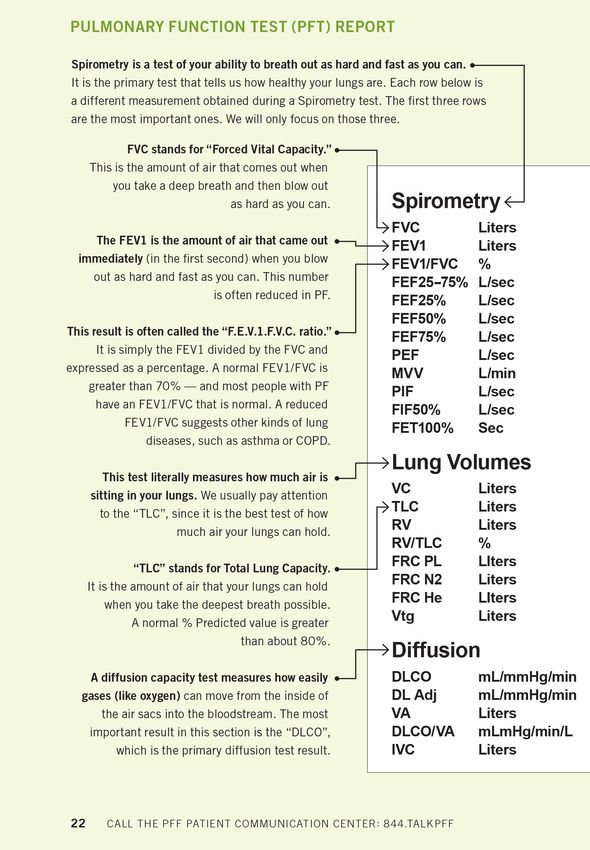
Dear Nurses and Allied
Health Professionals
                           This is a guide that was developed by the Pulmonary
                           Fibrosis Foundation in collaboration with several nurses
                           and allied health professionals who are experts in the
                           field of interstitial lung disease. We hope this is a valuable
                           tool for all of you who care for patients affected by these
                           challenging diseases.

                           As you know, the diagnosis and treatment of patients
                           with interstitial lung disease is complex and our goal is to
                           provide information identifying the disease, interpreting
                           test results, managing comorbidities, disease management,
                           and other supportive measures.

                           We recognize the important role that you play in the
                           care, support, and education of these patients and are
                           appreciative of all that you do.

                           We would especially like to recognize Jennifer Hayes,
                           RN, BSN; Shanna Hoskinson, RN; Kathleen O. Lindell,
                           PhD, RN; Wendi Mason, RN, MSN, ACNP; Tamra Perez,
                           BSN, RN; Ashleigh Rodriguez, NP; and Anne Turner,
                           BSN, RN, JD for the contributions they have made to this
                           important resource.

                           Thank you,

                           PAULINE BIANCHI BSN, RN
                           VICE PRESIDENT OF RESEARCH AND DEVELOPMENT
                           Pulmonary Fibrosis Foundation

2   ILD NURSING AND ALLIED HEALTH GUIDE   |   2020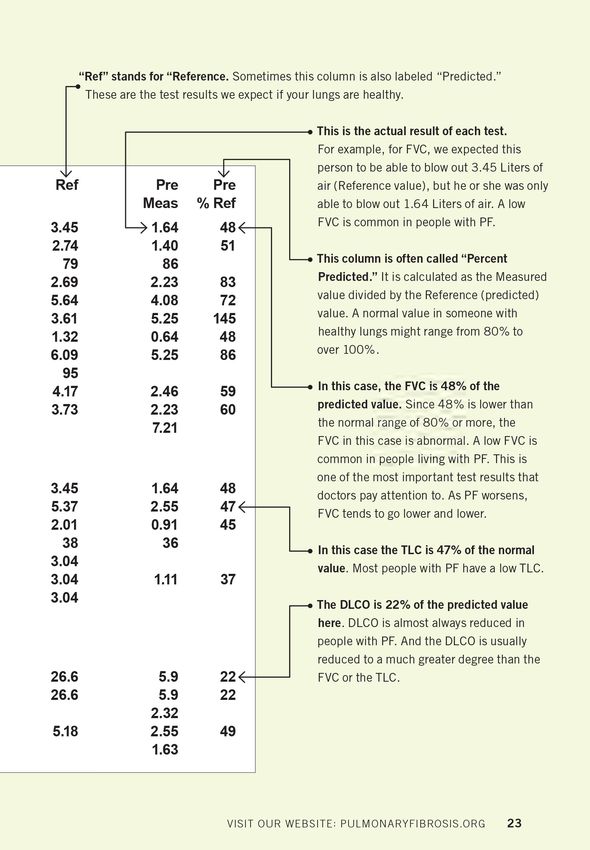
Introduction
The interstitial lung diseases (ILDs) are a family
of over 200 related conditions characterized
by inflammation, fibrosis (scarring), or other
abnormalities in the alveolar walls (also known as
the interstitium). These changes lead to thickening
of the alveolar walls, making the lung stiff and
making it difficult for oxygen to diffuse into the
bloodstream. ILDs are not cancer and they are
not infectious. In most cases, fibrosis is present.
We use the general term “pulmonary fibrosis”
(PF) to refer to any ILD in which fibrosis is
present. Fibrosis can be identified on a high-
resolution CT (HRCT) scan of the chest or on
lung biopsy. As noted below, there are many
different types of PF, but they may all share
common features: exertional dyspnea, dry bothersome cough, crackles on lung exam,
poor quality-of-life, supplemental oxygen requirement, and a progressive course and
high mortality rate.

Patients living with PF are different than those with chronic obstructive pulmonary
disease (COPD). They often require higher oxygen flow, experience more rapid
progression, and have few therapies that improve their quality-of-life or survival. Nurses
and allied health professionals play a central role in the care of patients with PF as
detailed throughout this guide.

The incidence (new cases per year) and prevalence (number of cases right now) of ILD
is not known. Based on available data, the prevalence of idiopathic pulmonary fibrosis
(IPF), the most common idiopathic form of PF, has been reported to be 58.7 per
100,000 people for the US population. Additionally, the prevalence of IPF in US adults
aged 60 and older has been reported to be approximately 200,000 and the prevalence of
IPF in US adults aged 70 and older has been reported to be 429.3 per 100,000. Based
on these prevalence estimates, up to 1 in 400 adults age 60 and older are living with IPF.
Up to 1 in 200 adults age 70 and older are living with IPF. PF can affect anyone. Men,
women, and children can all get PF. IPF is much more common in older adult males.

                                            ILD NURSING AND ALLIED HEALTH GUIDE   |   2020   3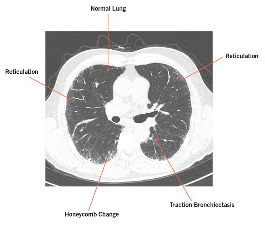
PART ONE:
Identifying the Disease
RECOGNITION OF ILD

Most patients with ILD will present with exertional dyspnea, chronic dry cough, or
both. Physical examination will show crackles at both lung bases in most cases. ILD
can then be identified by performing an HRCT scan of the chest. The initial work-up
should also include pulmonary function testing (PFT) detailed below.

THE DIAGNOSTIC PROCESS

ILDs can be divided into two groups: (1) those in which a cause can be identified, and
(2) those in which the cause is unknown, in which case we use the term “idiopathic
interstitial pneumonia.”

Identifying a cause always requires taking a careful history, performing a physical
examination, sending serologies for connective tissue diseases, and obtaining a high-
quality HRCT. In some cases, a lung biopsy is required. Here are some examples of
ILDs of “known cause”:

Connective Tissue Diseases: Autoimmune diseases such as rheumatoid arthritis,
scleroderma, Sjögren’s, and dermatomyositis/polymyositis can damage the lungs
and cause connective tissue disease-associated interstitial lung disease (CTD-ILD).
Sometimes, ILD is the first manifestation of a CTD. Patients should be carefully
questioned about any symptoms that could indicate underlying disease, such as joint
pain, stiffness, or swelling; skin thickening or tightening; rash; dry eyes; dry mouth;
Raynaud’s phenomenon; diffuse recurrent muscle pain or weakness; and severe
heartburn with gastric regurgitation. Patients should also undergo autoimmune serology
testing. In many cases, an ANA, RF and anti-CCP antibodies should be ordered.
Additional serologies, such as Scl-70, anti-Ro (SS-A), anti-La (SS-B), and myositis
antibodies should be ordered when indicated. If evidence is found of a possible
undiagnosed CTD, a referral for evaluation by a rheumatologist should be considered.

Medications: A number of medications (such as amiodarone, nitrofurantoin,
methotrexate, and certain chemotherapy agents) are known to have lung damage as a

4   ILD NURSING AND ALLIED HEALTH GUIDE   |   2020

possible effect. The ILD health professional must not only obtain a current medication
list, but also review past medications (including cardiology, autoimmune, and cancer
treatments). See Pneumotox www.pneumotox.com/drug/index/ for additional
information.

Environmental Factors (Hypersensitivity Pneumonitis): When gathering patient
history, it is important to consider current and past environmental factors that can
contribute to PF. Inhaled mold spores or bird proteins can trigger inflammation and
fibrosis. A history of dampness, mold, or bird exposure in the home or workplace
should immediately raise suspicion for hypersensitivity pneumonitis. When fibrosis is
present, we often call this “chronic” hypersensitivity pneumonitis. An exposure history
can be obtained by questionnaire. Involvement of an industrial hygienist or a home
inspection for mold is indicated in some cases. Avoidance of the exposure is a critical
part of the management of patients with hypersensitivity pneumonitis.

“Most patients with ILD will present
 with exertional dyspnea, chronic
 dry cough, or both.”
Occupational (Pneumoconiosis): A wide variety of workplace exposures are
potentially toxic to the lungs. “Black lung” in coal workers and asbestosis are two
well-known examples of pneumoconioses. The rate of occupational diseases in the
United States has declined due to improvements in occupational safety. However,
occupational factors remain an important cause of ILD and obtaining a thorough
occupational history is critical, including any time served in the armed forces, especially
naval shipyards. Patients may not know if they are or were exposed to any potentially
hazardous substances while on the job. They have a legal right to this information from
both current and former employers. Material Safety Data Sheets (MSDS), available from
their employer or online, can provide details about each substance, including whether it
is linked to respiratory toxicity. Larger companies often employ an industrial hygienist,
safety officer, or compliance manager who is responsible for maintaining safe working
conditions and can help answer these questions. Patients can also contact their regional
Occupational Safety and Health Administration (OSHA) office for help. OSHA is also
an excellent resource for the provider who is trying to determine potential lung toxicity
from an unfamiliar chemical or industrial process.

                                            ILD NURSING AND ALLIED HEALTH GUIDE   |   2020   5
Radiation: Patients should be asked if they have ever received radiation therapy to the
chest, which can cause PF.

IDIOPATHIC INTERSTITIAL PNEUMONIAS

The ILDs with no known cause make up a subgroup of ILDs called idiopathic
interstitial pneumonias (IIPs). Idiopathic pulmonary fibrosis (IPF) is the most common
disease in this group. Other IIPs include: idiopathic non-specific interstitial pneumonia
(idiopathic NSIP), desquamative interstitial pneumonia (DIP), cryptogenic organizing
pneumonia (COP), and unclassifiable ILD.

The IIPs are diagnosed after a complete history, physical exam, blood work, and HRCT
fail to identify a known cause of disease. In some cases, a confident diagnosis of an IIP
can be made. A good example of this situation is IPF, which has characteristic features
on an HRCT. But, in some cases, including some cases of IPF, a lung biopsy will be
performed to make a specific diagnosis.

PART TWO:
Interpreting Test Results
HIGH-RESOLUTION COMPUTERIZED TOMOGRAPHY SCANS (HRCT)

Results of the HRCT chest scan without contrast should be interpreted by the
radiologist, ideally with experience or expertise in reviewing ILD. The findings should
then be reviewed by the pulmonologist or a healthcare provider with training in ILD
to look for visual patterns of fibrosis and inflammation that can help point toward a
specific diagnosis.

On CT imaging, air is typically black as noted by the air above the chest and throughout
most of the lung in the normal patients. With abnormalities such as inflammation
of fibrosis in the lung, varying degrees of white opacities are noted in the lung with
structural changes such as honeycomb change or traction bronchiectasis.

On the next page, please see the diagram for a few examples of important findings on CT.

6   ILD NURSING AND ALLIED HEALTH GUIDE     |   2020
Reticulation

Irregular intersecting white lines. Reticulation indicates the presence of fibrosis. All
forms of PF have reticulation.

Traction bronchiectasis: Dilated airways due to fibrosis.

 Honeycombing

Thick-walled linear cysts, often occurring along the periphery of the lung. Due to
fibrosis. Common in, but not specific for, IPF.

 Ground-glass opacities/infiltrates

Hazy areas of the lung. Ground-glass is not specific for any one problem. Fluid (such
as from heart failure), inflammation, infection, and other pathology can all show up as
ground-glass. When “mosaic” ground-glass attenuation is present, it often indicates
diseases that involve the small airways as seen in hypersensitivity pneumonitis, forms
of bronchiolitis, and other conditions.

                                             ILD NURSING AND ALLIED HEALTH GUIDE   |   2020   7
PULMONARY FUNCTION TESTS (PFTS)

Pulmonary function test (PFT) results can be useful for any trained healthcare provider.
The presence of abnormal PFT findings can assist in making sure those that suggest
ILD would then obtain an HRCT. See below for basic interpretation of PFTs.

Spirometry gives many results, but the most important for the diagnosis of ILD are
forced vital capacity (FVC), forced expiratory volume in 1 second (FEV1), and the ratio
of FEV1 to FVC (FEV1/FVC ratio). In ILD, the FVC can be normal in early disease
and will decline as the disease progresses. This is one of the primary ways that we can
track progression of PF, and it is often measured at every clinic visit. A normal FVC is
often >80% of the predicted value, but each lab uses different thresholds.

The FEV1 often tracks with the FVC, and while it is extremely valuable in other
diseases like asthma and chronic obstructive pulmonary disease, it is often not followed
closely in PF. The FEV1/FVC ratio is typically normal (>70%) in PF.

Lung volume measurement also gives many results, but the most important one is
Total Lung Capacity (TLC), which is the volume of air the lungs can hold during the
deepest possible breath in. Just like FVC, the TLC can be normal in early disease and
will decline as the disease progresses. When TLC is reduced (

SPIROMETRY
FEV / FVC Ratio
70%
Airflow No Airflow
Obstruction Obstruction
(*less common in ILD) (Typical for ILD)
FVC
80%
Reduced Normal
BOTH CAN BE SEEN IN ILD
LUNG VOLUMES
TLC
80%
Restrictive Ventilatory Normal
Defect
BOTH CAN BE SEEN IN ILD
*Can occur in RA-ILD, HP, Sarcoidosis, and combined COPD/ILD
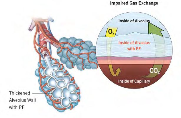
ILD NURSING AND ALLIED HEALTH GUIDE | 2020 9DIFFUSING CAPACITY
DIFFUSING CAPACITY OF THE LUNG FOR CARBON MONOXIDE
< 70%–80% > 70%–80%
Impaired Gas Normal
Exchange
Uncommon
Seen in almost all ILD in ILD
OXYGEN TESTING
Step 1 Measure SpO2 or PaO2 at Rest
SpO2 < 88% or SpO2 > 88% or
PaO2 < 55mmhg PaO2 > 55mmhg
Continuous Home No Resting Oxygen
Oxygen Needed Needed
Step 2 Measure SpO2 or PaO2 with Exertion
• Six Minute Walk Test
• Cardiopulmonary Exercise Test
• Oxygen Titration in a Hallway ot Treadmill
SpO2 < 88% or SpO2 > 88% or
PaO2 < 55mmhg PaO2 > 55mmhg
Oxygen with Exertion No Oxygen with
Indicated Exertion Indicated
10 ILD NURSING AND ALLIED HEALTH GUIDE | 2020Please note that when the SpO2 is 89% or the PaO2 is 56-59, oxygen can also be
prescribed if your patient has (1) dependent edema, (2) pulmonary hypertension or “cor
pulmonale” determined by measurement of right heart catheterization, echocardiogram,
or EKG (P waves greater than 3 mm in leads II, II, or aVF), or (3) a hematocrit > 56%.
It is important to determine and document how much oxygen (liter/minute flow
and type of interface) is required to maintain saturations over 88% at rest and during
ambulation when crafting the oxygen prescription.
Because of the higher cost of liquid oxygen systems, many home care companies no
longer offer or encourage the use of concentrators and tanks for portability. However,
if the ordering healthcare provider believes a specific modality of a system better
accommodates the patient, it should be specified in the prescription.
6-MINUTE WALK TEST (6MWT)
The 6-minute walk test (6MWT) is completed for a variety of reasons, including
measurement of distance walked in six minutes, nadir (lowest) saturation, end-walk
saturation, and heart rate recovery. These data–particularly any changes over time–can
help inform prognosis. Use the European Respiratory Society (ERS)/American Thoracic
Society (ATS) document “Field walking tests in chronic respiratory disease” for more
information. thoracic.org/statements/resources/copd/FWT-Tech-Std.pdf
PART THREE:
Comorbidities
GASTROESOPHAGEAL REFLUX DISEASE (GERD)
Gastroesophageal reflux disease (GERD) occurs as a result of the weakening of
the muscle tone of the lower esophageal sphincter (LES), a circular ring of muscle
connecting the esophagus and stomach, leading to reflux of gastric contents into the
esophagus. GERD has long been identified as a cause or trigger of pulmonary diseases
such as asthma and chronic cough. An increasing body of evidence also suggests an
association between the presence of GERD and IPF. IPF patients are also more likely
to have a hiatal hernia, a risk factor for GERD. The role of GERD in the etiology,
progression, and exacerbation of IPF remains uncertain.
ILD NURSING AND ALLIED HEALTH GUIDE | 2020 1112 ILD NURSING AND ALLIED HEALTH GUIDE | 2020
ILD NURSING AND ALLIED HEALTH GUIDE | 2020 13
Role of the Doctor’s Office
                                                  term oxygen therapy
Identify the oxygen needs of the patient          Depending on where the patient lives and
When indicated, the testing includes:             how much a patient travels, a local home
                                                  care company or one of the national
• 6-minute walk test (6MWT)                       companies may be better suited to the
• High Altitude Simulation Test (HAST)            patient’s needs
• Overnight oximetry/sleep study
• Arterial Blood Gas Test (ABG)                       Role of the Patient

Provide the appropriate prescription and          Using supplemental oxygen is something
qualifying documents                              the patient can control, though it can be
Chart notes and testing results for the           a difficult transition
home care company
                                                  Having an SpO2 in the normal range will
Work with the home care company                   help prevent dyspnea in many patients
• Submit Certificates of Medical
                                                  Using supplemental oxygen during
 Necessity (CMN) for insurance billing,
                                                  activities will allow the patient to remain
 primarily Medicare
                                                  more active, thus improving quality of life
Educate the patients on effective and
                                                  Avoiding low oxygen levels may help
safe use of oxygen
                                                  prevent pulmonary hypertension
 Role of the Home Care Company                    Being more active and able to participate
Receive the prescription                          in rehabilitation may help patients
                                                  maintain a healthy weight and improve
Verify insurance coverage and bill                their pre-transplant candidacy
insurance
                                                  Living longer by using supplemental
Provide a basic oxygen system, including          oxygen is not proven in ILD, but it has
any equipment and disposable supplies,            been shown to prolong survival in chronic
such as cannulas and humidifiers                  obstructive pulmonary disease (COPD)
Keep records of CMNs for Medicare                 Maintaining an SpO2 > 90% is
Provide 24-hour emergency service for             encouraged by many clinicians. Some
equipment malfunctions or power outages           patients find it useful to purchase
                                                  portable finger oximeters to monitor
Provide education on proper and safe use          levels at home during different activities,
of equipment                                      if recommended by their healthcare
Play an important role in a patient’s long        provider.

14   ILD NURSING AND ALLIED HEALTH GUIDE   |   2020
GERD Diagnosis
Some patients with ILD will have symptoms of GERD, such as heartburn or
regurgitation, while others will not have symptoms. This is referred to as “silent reflux.”
If clinically indicated, a diagnosis of GERD can be made using objective testing, such
as ambulatory 24-hour pH monitoring, esophageal manometry, upper endoscopy, or
barium swallow esophagram. However, the indication for these invasive tests without
heartburn remains uncertain.
GERD Treatment
Most clinicians will treat GERD when symptoms, such as heartburn, are present.
While current ATS/ERS/JRS/ALAT IPF Guidelines conditionally recommend
antacid treatment to treat IPF, the treatment of asymptomatic GERD in IPF patients
is controversial. While some non-randomized studies suggest that IPF patients treated
with antacid therapy have better outcomes, others suggest that antacid therapy may
increase the risk of infection. Anti-reflux surgery, the most common type being
fundoplication, involves a laparoscopic procedure where the surgeon wraps the top part
of the stomach around the esophagus in order to restrict the back-flow of stomach
contents. A clinical trial of surgical fundoplication to treat GERD in IPF has been
completed the results were suggestive of (but did not prove) a benefit.
GERD management can include lifestyle interventions including weight loss, elevating
the head of the bed by six inches, avoidance of late evening meals, and elimination
of food that can trigger reflux, such as chocolate, caffeine, alcohol, and spicy foods.
Medical management of GERD includes antacids, such as H2 blockers and proton
pump inhibitors (PPIs). Studies have shown that PPIs, most of which are now
available over the counter, are more effective than H2 blockers, and there are no major
differences in efficacy between the different PPIs. Both drugs suppress acid secretion
by the stomach. Common side effects occurring in 1-2% of patients taking PPIs
include nausea, constipation, diarrhea, headache, and skin rash. PPIs should be taken 30
minutes before eating.
PULMONARY HYPERTENSION
Pulmonary hypertension (PH) is defined as an elevation in the mean pulmonary arterial
pressure greater than 25 mm Hg. PH is considered severe if cardiac index is < 2 L/
min/m2 or if mPAP is ≥ 45 mm Hg. The World Health Organization (WHO) classifies
patients with PH into five groups based upon etiology. Patients diagnosed with ILD-
associated PH are classified as WHO Group 3: “Pulmonary hypertension due to lung
ILD NURSING AND ALLIED HEALTH GUIDE | 2020 15diseases and/or hypoxia.” PH is a recognized complication in patients with ILD. Pulmonary Hypertension PH has been reported to occur in 20-85% of patients with PF. In PF patients undergoing evaluation for lung transplantation, PH was associated with more severe fibrosis as measured by pulmonary function tests (PFTs) and resting oxygen saturation. Due to overlapping primary symptoms of shortness of breath with progressive dyspnea on exertion and exercise limitation, it may be difficult to detect PH in patients with PF. Clues may include exertional dyspnea or fatigue whose severity seems out of proportion to the degree of lung disease as demonstrated by a severely decreased DLCO. Other symptoms may include chest pain or syncope with exertion. PH can also lead to right heart failure, which manifests as peripheral edema, elevated jugular venous pressure, and severe exertional limitation. Although symptoms, an ECG (right ventricular hypertrophy), a chest CT scan (dilated main pulmonary artery, right ventricular dilation), and PFTs may hint at PH, an echocardiogram is the initial test of choice. Echocardiographic evidence of PH includes an estimated right ventricular (or pulmonary artery) systolic pressure greater than 40 mm Hg, reduced right ventricular ejection fraction, dilated right ventricle and/or atrium, and bowing of the interventricular septum. Echocardiography, while useful, cannot diagnose PH. The gold standard for confirming the diagnosis is right heart catheterization. PH is defined as a mean pulmonary artery pressure (mPAP) > 25 mm Hg at rest, as measured by right heart catheterization. However, since there are no proven therapies for ILD-associated PH, right heart catheterization is only indicated in a minority of patients. Some indications might include evaluation for lung transplantation and suspicion that pulmonary hypertension may be the dominant condition, with mild ILD playing a small role in symptoms (such as in some cases of connective tissue disease). Pulmonary Hypertension Treatment The mainstay of therapy for PH in patients with ILD is supplemental oxygen therapy when indicated. Oxygen can dilate pulmonary arterioles and decrease pulmonary artery pressure in some patients. 16 ILD NURSING AND ALLIED HEALTH GUIDE | 2020
The presence of PH has been associated with an increased risk of death in ILD.
Nevertheless, since there are no proven therapies to treat PH in patients with ILD,
pharmacological therapy is often not prescribed by clinicians. Medications approved
for other forms of PH are not approved by the Food and Drug Administration (FDA)
for patients with IPF-associated PH (WHO Group 3), due to lack of clinical trial
evidence of efficacy and safety. Per 2011 American Thoracic Society (ATS) Guidelines,
“Pulmonary hypertension should not be treated in the majority of patients with IPF, but
treatment may be a reasonable choice in a minority in patients with moderate to severe
pulmonary hypertension documented by right heart catheterization.”

SLEEP APNEA

Sleep Apnea Diagnosis
Apnea means “not breathing.” Obstructive sleep apnea (OSA) is the presence of
recurrent episodes of upper airway obstruction (due to closure of the throat and upper
airway) occurring at least five times per hour on average during sleep. OSA has been
found to be common among those diagnosed with IPF. Traditionally, symptoms of
sleep apnea include daytime sleepiness, snoring, gasping while sleeping, awakening and
feeling that you can’t breathe, falling asleep at inappropriate times (such as in a waiting
room or during a conversation) or at unsafe times (such as driving a car), dry mouth,
and headache upon awakening.

Risks and comorbidities associated with sleep apnea include obesity, GERD, and
coronary artery disease. Physical examination findings may show a large neck
circumference (men > 17 inches, women > 16 inches), a crowded airway (Mallampati III
or IV), a deviated nasal septum, patulous soft palate, and scalloped tongue. Many IPF
patients with OSA are not sleepy, may not be obese, and many do not have severe IPF.
Some data suggest that OSA might even contribute to early lung injury and pulmonary
hypertension. Polysomnography, a test used to diagnose OSA, should be considered in
patients who have symptoms of OSA.

Sleep Apnea Treatment
If sleep apnea is diagnosed, patients can be treated with continuous positive airway
pressure (CPAP) therapy or bilevel positive airway pressure (BiPAP) therapy if clinically
indicated.

                                            ILD NURSING AND ALLIED HEALTH GUIDE   |   2020   17
PART FOUR: Disease Management PROGRESSION OF PF Early PF is often symptom-free, and patients frequently do not receive a diagnosis until they have moderate or severe disease. “Progression” means that fibrosis has built up over time. Progression occurs over different time periods that vary from person-to- person. Typical patterns of IPF progression include: • Intermittent Step-Wise Progression: Most patients experience periods of stability alternating with periods of progression, as measured by increasing symptoms, decreasing FVC, decreasing DLCO, and increasing supplemental oxygen needs. • Rapid Progression: Other patients will progress rapidly, experiencing a significant decline within 6 months to a year of diagnosis. • Acute Exacerbations: Acute exacerbations are unpredictable, occur in about 10% of patients each year, and usually have a very poor outcome. While most are “idiopathic,” some are due to infection, aspiration, or drug toxicity. Patients with mild to moderate symptoms may believe they are stable. It is important to educate patients about the unpredictability of acute exacerbations and the inevitability of disease progression. This will enable them to engage in appropriate planning and make informed choices. Although disease course cannot be predicted, there is a staging system available for IPF called the GAP score. The GAP score is based on age, sex, and physiology (FVC and DLCO) with scores ranging from 0 to 9 which correspond to three Stages: Stage I (score 0-3), Stage II (4-5), and Stage III (scores 6-8). One-year mortality rates are 6%, 16%, and 39%, respectively (see Table 1). It is important to be familiar with this tool because patients can easily find “GAP Calculators” on the internet and compute the score for themselves. There are limits to how the information should be used. While patients may want to know their stage and their “risk”, you should communicate to your patient that even if they are “Stage III” with a 39% risk of death in 1-year, that means that 61% of people live longer than 1 year – and there is no way to know if your 18 ILD NURSING AND ALLIED HEALTH GUIDE | 2020
Predictor Points           Predictor Points           Predictor                   Points
 Gender		                   Age (y)		                  Physiology

					                                                  FVC, % predicted

 Female         0           ≤60          0             >75                         0

 Male           1           61-65        1             50-75                       1

			                         >65          2             55                                                                           0

					36-55                                                                         1

					≤35                                                                           2

					Cannot perform                                                                3

					                                                  Total Possible Points       8

patient is in the 39% group or
                                     Stage                     Mortality       % Risk
the 61% group. There are several
limitations that patients should     I (Points 0-3)            1-y             5.6
understand. Also remember that
                                    		                         2-y             10.9
the GAP score only applies to
IPF. Also, the GAP stage was        		                         3-y             16.3
developed before anti-fibrotic
                                     II (Points 4-5)           1-y             16.2
therapy was available in the
United States, and therefore        		                         2-y             29.9
your patients may have a better
                                    		                         3-y             42.1
“average” outcome than that
predicted by the GAP score. In       III (Points 6-8)          1-y             39.2
addition, other forms of PF may
                                    		                         2-y             62.1
have a better prognosis.
                                    		                         3-y             76.8
http://annals.org/data/Journals
AIM

                                         ILD NURSING AND ALLIED HEALTH GUIDE   |   2020     19
SUPPLEMENTAL OXYGEN Home Oxygen Delivery Systems It is vital to maintain appropriate oxygen levels as oxygen plays an important role in the energy metabolism of living organisms. Air is approximately 21% oxygen, 78% nitrogen, and about 1% argon and other trace gases. One of the most common symptoms ILD patients experience is dyspnea on exertion. Because of the nature of the disease, it is not unusual for a patient to have resting oxygen saturations within normal limits and then desaturate with activity. The increase in cardiac output during exertion means there is less time for oxygen to diffuse from the alveoli through the alveolocapillary membrane and into the bloodstream. As the disease progresses, desaturation becomes more severe and occurs with lesser degrees of exertion. Types of Oxygen Systems Basic oxygen delivery systems include an in-home stationary unit and a portable system that allows the patient to leave the home. “One of the most common symptoms ILD patients experience is dyspnea on exertion.” STATIONARY SYSTEMS Stationary Concentrator Oxygen concentrators work by extracting and separating oxygen from other gases and moisture in room air. Oxygen then accumulates in a reservoir, which provides a continuous stream of highly concentrated oxygen (> 94%). Stationary concentrators have the ability to produce enough oxygen to meet most needs up to 10 LPM. The unit stays in the room in which it is placed and patients use different lengths of oxygen tubing to move around. A hidden cost is the electricity required to power the concentrator, which is not reimbursable. The concentrator is a reliable and relatively inexpensive option, although noisy. A humidifier should also be used once a patient requires more than 4-5 LPM to prevent bloody noses, congestion, and irritation, as the oxygen flow tends to dry out nose membranes. 20 ILD NURSING AND ALLIED HEALTH GUIDE | 2020
Stationary Liquid System
When oxygen is cooled to a very low temperature (approximately 300 degrees below
zero Fahrenheit), it becomes a liquid. To remain in liquid form, oxygen must be held
in insulated canisters. Large amounts of oxygen can be stored in a container at low
pressure in liquid form. Stationary liquid systems provide not only a large storage
capacity, but also allow for filling and refilling smaller units. As the liquid oxygen leaves
the container, it warms up to room temperature and becomes a gas. Conventional liquid
oxygen systems are quiet, have no major moving parts, and require no power source to
operate. Depending on the patient’s consumption rate and liter flow, the canister needs
to be refilled approximately every two weeks.

PORTABLE SYSTEMS

Compressed Gas Tanks
Tanks in a variety of sizes are inexpensive and readily available.
Using them with a backpack allows for hands-free activities.
However, tanks can be heavy. Some patients use a wheeled cart
to be mobile, especially if larger tanks are required. No power
source is needed, and the patient does not need to refill as they
are delivered full. They can be used with conserving devices so
that patients who tolerate pulse dose delivery (where a breath
generates the oxygen to be delivered) can go longer on one tank. Patients are required
to store cylinders safely and learn how to change the conserving devices. It follows that
the higher the liter flow, the faster the tank will be depleted, so a variety of sizes based
on a patient’s liter flow and activity level may allow greatest flexibility.

Liquid Portable Units
This portable system is used in conjunction with the stationary
unit and requires the patient to refill as needed. The portable
unit is lighter than an oxygen E-cylinder tank, lasts longer, and
is worn over the shoulder with a strap. While lighter, it’s prone
to leaks if not kept upright. It requires dexterity to refill, and
has a danger of cold burning during refilling. Patients have
complained of evaporation and less tank life in humid weather.

Portable Oxygen Concentrators
Portable oxygen concentrators (POCs) are simply smaller versions of the stationary
unit. A portable concentrator runs on battery, allowing for increased portability. A

                                            ILD NURSING AND ALLIED HEALTH GUIDE   |   2020   21
patient carries additional batteries to allow for greater time away from an outlet. POCs can be plugged in to recharge and come with car adapters to allow for use during driving trips. POCs are the accepted mode of oxygen delivery on airplanes. Battery life depends on the patient’s liter flow and oxygen consumption. The higher the liter flow, the faster a battery is depleted. The main limitation, especially for ILD patients, is the limited liter flow. Many of the small units provide a maximum of 2 LPM, while ILD patients require a higher level of liter flow than many of the POCs provide. In many cases, POCs cannot deliver sufficient oxygen flows for ILD patients with severe exertional desaturation. Another issue is that patients who need more than 3 LPM on the 6-minute walk test in order to maintain SpO2 > 88% use continuous flow and cannot tolerate the pulse dose delivery system. Portable oxygen systems give patients greater freedom to participate in activities outside the home to maintain quality of life. The right system for each patient depends on his or her activity level, goals, and ability to utilize one system over another. Cannula Options With all systems, the general rule is the higher the patient’s required liter flow and use of continuous flow instead of pulse dose flow, the more limited the patient’s options. Some strategies for patients on a higher flow are to change from a regular nasal cannula to either a pendant or mustache oxymizer cannula. These cannulas are simple conserving devices that accumulate oxygen usually wasted during exhalation and store it in a 20 milliliter reservoir. The patient then receives a bolus of oxygen at the beginning of inhalation. It is important to note that humidifiers cannot be used along with the conserving cannula. OXYGEN SAFETY Oxygen itself is not flammable, but because it is an accelerant, patients should take precautions to prevent injury. Compressed tanks should be kept 8-10 feet away from any flame or spark, such as candles, stoves, fireplaces, electric razors, and people who smoke. Petroleum-based products should be avoided; patients should use aloe or cocoa butter products for dry nose instead. Unused cylinders should be secured to avoid falling and becoming projectiles. Home concentrators should be well-ventilated, and patients should be cautioned to avoid tripping on their oxygen tubing. 22 ILD NURSING AND ALLIED HEALTH GUIDE | 2020
Using supplemental oxygen can be a major adjustment for a patient. Many are
embarrassed to be seen in public wearing a cannula. Patients who were previously active
may perceive oxygen use as a signal to cease their activities. The health care team must
educate patients about the need for supplemental oxygen to aid in facilitating continued
activities that give them joy.

ANTIFIBROTIC THERAPIES

Two antifibrotic drug therapies, pirfenidone and nintedanib, are available to help slow
disease progression in IPF. One antifibrotic therapy, nintedanib, is available to help slow
the disease progression in systemic sclerosis-associated ILD (Ssc-ILD). With long term
follow up of patients on antifibrotic therapies, the decrease in disease progression is
associated with improved survival. For full prescribing information, see FDA.gov.

Pirfenidone (Esbriet®, Pirfenex®, Pirespa®): Pirfenidone is an antifibrotic and
anti-inflammatory drug approved to treat IPF in the US, Europe, Canada, and Asia. In
clinical trials, pirfenidone has been shown to slow progression of mild-to-moderate IPF.

    Dosing
    267 mg capsules or tablets; uptitrate to 3 capsules orally 3 times daily with meals
    over 14 days. MUST be taken with full meals. Once on a stable dose, an 801mg
    tablet (equivalent to 3 capsules) is available.

    Adverse Reactions
    The most common adverse reactions (≥10%) are nausea, rash, abdominal pain,
    upper respiratory tract infection, diarrhea, fatigue, headache, dyspepsia, dizziness,
    vomiting, anorexia, gastro-esophageal reflux disease, sinusitis, insomnia, weight
    decreased, and arthralgia.

    Warnings & Precautions
    Elevated liver enzymes: ALT, AST, and bilirubin elevations have occurred. Monitor
    ALT, AST, and bilirubin before and during treatment. Temporary dosage
    reductions or discontinuations may be required.

Photosensitivity and rash: Photosensitivity and rash have been noted. Avoid exposure to
sunlight and sunlamps. Wear sunscreen and protective clothing daily. Temporary dosage
reductions or discontinuations may be required.

                                           ILD NURSING AND ALLIED HEALTH GUIDE   |   2020   23
Gastrointestinal disorders: Nausea, vomiting, diarrhea, dyspepsia, gastroesophageal
reflux disease, and abdominal pain have occurred.
Elevated liver enzymes and drug-induced liver injury: In the post-marketing period,
non-serious and serious cases of drug-induced liver injury, including severe liver injury
with fatal outcome, have been reported. Inform patients about the need for periodic
monitoring. Monitor ALT, AST, and bilirubin prior to the initiation of therapy in all
patients, then monthly for the first six months and every three months thereafter,
and as clinically indicated. Measure liver function tests promptly in patients who
report symptoms that may indicate liver injury, including fatigue, anorexia, right upper
abdominal discomfort, dark urine, or jaundice. Temporary dosage reductions or
discontinuations may be required.
Drug Interactions
Moderate (e.g., ciprofloxacin) and strong inhibitors of CYP1A2 (e.g., fluvoxamine)
increase systemic exposure of pirfenidone and may alter the adverse reaction profile of
pirfenidone. Discontinue fluvoxamine prior to administration of pirfenidone or reduce
to 267 mg three times a day. Consider dosage reduction with use of ciprofloxacin.
The full prescribing information is available at: https://www.gene.com/download/
pdf/esbriet_prescribing.pdf
Nintedanib (OFEV®): Nintedanib is an anti-fibrotic drug that is approved to treat
IPF in the United States and Europe. In clinical trials, nintedanib has been shown to
slow progression of SSc-ILD and mild-to-moderate IPF. Nintedanib is also approved
to treat systemic sclerosis-associated ILD (SSc-ILD) (also known as scleroderma-
associated ILD) in the United States and Canada.
Dosing
150 mg capsules; one capsule orally every 12 hours taken with food
Adverse Reactions
Most common adverse reactions (≥5%) are: diarrhea, nausea, abdominal pain,
vomiting, liver enzyme elevation, decreased appetite, headache, weight decreased,
and hypertension.
Warnings & Precautions
Hepatic impairment: nintedanib is not recommended for use in patients with moderate
24 ILD NURSING AND ALLIED HEALTH GUIDE | 2020or severe hepatic impairment. In patients with mild hepatic impairment (Child
Pugh A), the recommended dosage is 100 mg twice daily approximately 12
hours apart taken with food. Consider treatment interruption, or discontinuation
for management of adverse reactions in these patients.
Elevated liver enzymes and drug-induced liver injury: ALT, AST, and bilirubin elevations
have occurred with nintedanib, including cases of drug-induced liver injury. In the post-
marketing period, non-serious and serious cases of drug-induced liver injury, including
severe liver injury with fatal outcome, have been reported. The majority of hepatic
events occur within the first three months of treatment. Liver enzyme and bilirubin
increases were reversible with dose modification or interruption in the majority of cases.
Monitor ALT, AST, and bilirubin prior to initiation of treatment, at regular intervals
during the first three months of treatment, and periodically thereafter or as clinically
indicated. Temporary dosage reductions or discontinuations may be required.
Gastrointestinal disorders: Diarrhea, nausea, and vomiting have occurred. Treat patients
at first signs with adequate hydration and antidiarrheal medicine (e.g., loperamide) or
anti-emetics. Discontinue nintedanib if severe diarrhea, nausea, or vomiting persists
despite symptomatic treatment.
Embryo-Fetal toxicity: Can cause fetal harm. Advise females of reproductive potential
of the potential risk to a fetus and to use effective contraception.
Arterial thromboembolic events have been reported. Use caution when treating patients
at higher cardiovascular risk including known coronary artery disease.
Bleeding events have been reported. Use nintedanib in patients with known bleeding
risk only if anticipated benefit outweighs the potential risk. nintedanib should also be
used with caution (if at all) in patients on anticoagulants or dual anti-platelet therapy.
Gastrointestinal perforation has been reported. Use nintedanib with caution when
treating patients with recent abdominal surgery, previous history of diverticular disease
or receiving concomitant corticosteroids or NSAIDs. Discontinue nintedanib in patients
who develop gastrointestinal perforation. Only use nintedanib in patients with known
risk of gastrointestinal perforation if the anticipated benefit outweighs the potential
risk.
ILD NURSING AND ALLIED HEALTH GUIDE | 2020 25Drug Interactions Co-administration of P-glycoprotein and CYP3A4 inhibitors may increase nintedanib exposure. Monitor patients closely for tolerability of nintedanib. The full prescribing information is available at: https://docs.boehringer-ingelheim.com/Prescribing%20Information/PIs/Ofev/ ofev.pdf PULMONARY REHABILITATION Exercise is an important component in the treatment and management of ILD. The health benefits of exercise are well-documented and can improve the patient’s quality of life. Benefits include improved dyspnea and quality of life, more efficient use of oxygen, weight loss, and creating an overall sense of well-being. Exercise does not improve lung function, but neither does it harm the lungs. A person with ILD experiences a loss of control with their lung condition, and exercise becomes a form of self-management. Pulmonary rehabilitation (PR) is specifically designed for people with lung diseases such as chronic obstructive pulmonary disease (COPD) and ILD. PR programs provide a safe, secure environment for exercising, as well as classes on controlling and improving symptoms and overall health in people with pulmonary disease. As there are differences in the lung mechanics and disease progression between a person with COPD and a person with ILD, modifications are necessary. The person with ILD will have oxygen desaturation during exertional activities, and oxygen supplementation is usually required. This desaturation can be severe; the person with ILD should be monitored closely with pulse oximetry and may require increased oxygen flow rates during exercise. Continuous oxygen delivery should be available to those with ILD, as opposed to oxygen conserving devices (pulse dose delivery), to maintain pulse oximetry values > 90% saturation. A person with connective tissue-related PF, such as rheumatoid arthritis, may benefit from a PR program that includes a physical therapy component to minimize joint pain and damage. For patients who are candidates for lung transplantation ongoing PR is an important part of the preparation for surgery and aftercare. PR programs are usually 8-12 weeks, with 2-3 sessions per week. A multidisciplinary 26 ILD NURSING AND ALLIED HEALTH GUIDE | 2020
team that includes an exercise physiologist provides an initial assessment, develops
goals, and prepares an individualized exercise program with progress reports throughout
participation. It is imperative that any comorbidities, such as hypertension or diabetes,
are stable before undergoing PR. Communication between the patient’s provider and
the program team is essential.

After completing the initial program, individuals are expected to continue the exercise
regimen on their own. Studies show that the benefit gained from PR will be lost if the
individual does not continue to exercise. Many PR programs offer a maintenance, or
“graduate,” program for a small fee to encourage people to continue to exercise. For
many, it provides a means to stay connected to people with the same lung condition and
serves as a support system.

PR was initially established for people with moderate to severe COPD per the Global
Initiative for Chronic Obstructive Lung Disease (GOLD) diagnosis guidelines, and
this can pose challenges for obtaining insurance coverage for ILD patients. A policy
statement issued by the American Thoracic Society (ATS) and updated in June 2016
includes additional local determination coverage guidelines for reimbursement.
     “Patients appropriate for PR must have a diagnosis of a chronic, stable respiratory
     disorder with disabling symptoms that impair the patient’s function. PFTs need
     to show FVC, FEV1, and/or DLCO of < 65% predicted on PFT within 1 year of
     PR. There must be the expectation of measurable improvement in a reasonable
     and predictable amount of time, and the patient must be able, motivated, and
     willing to participate.”

“Benefits include improved dyspnea and
 quality of life, more efficient use of
 oxygen, weight loss, and creating an
 overall sense of well-being.”
Local Medicare determination policies vary regionally; however, national policies
determine final coverage. Currently, no national policy for PR coverage exists for
persons diagnosed with ILD.

Patients with ILD may visit PR facilities prior to starting a program. There are several

                                           ILD NURSING AND ALLIED HEALTH GUIDE   |   2020   27
factors involved in selecting a PR facility, including location and program schedule. If the facility is close to home, attendance is higher. While overall functional improvement in ILD patients remains less than those with COPD, studies of a modified PR program have shown statistical improvement in quality of life for a lung condition that has few treatment options. Prednisone Therapy Prednisone is a pharmacological therapy that reduces inflammation. Some clinicians use prednisone to treat patients who have lung inflammation as part of their PF. However, long-term use in patients with IPF may be harmful. Selected side effects of prednisone: • Increased appetite and weight gain • Susceptibility to infection • Fluid retention • Mood changes • Insomnia • Osteoporosis, fractures, and avascular necrosis of the hip • Increased blood sugars and diabetes • Hypertension • Fragile skin and bruising Dose adjustments are typically on a case-by-case basis and should always be tapered down and not stopped abruptly. NON-STEROID IMMUNOSUPPRESSIVE THERAPY Immunosuppressive therapy can be effective in decreasing immune response and inflammation in patients. However, patients must be informed that there is a Black Box warning on some of these medication and pregnancy is contraindicated. Patients of childbearing age should use two forms of birth control to prevent pregnancy. These drugs are FDA-approved, but have not been FDA-approved to treat ILD. Common Medications: • Mycophenolate (CellCept®) • Azathioprine (Imuran®) • Cyclophosphamide (Cytoxan®) • Infliximab (Remicade®) 28 ILD NURSING AND ALLIED HEALTH GUIDE | 2020
• Rituximab (Rituxan®)
• Methotrexate

Possible Side Effects (vary by medication):
• Increased risk for infection (and tuberculosis for infliximab)
• GI symptoms (nausea, vomiting, diarrhea)
• Pancreatitis (azathioprine)
• Liver problems (azathioprine)
• Hematuria and bladder cancer (cyclophosphamide)
• Various cytopenias

Lab Monitoring & Prophylaxis
While taking these medications, it is important to monitor blood work. Most patients
taking a non-steroid immunosuppressant will require periodic monitoring of CBC,
LFTs, and basic metabolic panel.

Other prophylactic considerations while using immunosuppressive medications include:
• Age-appropriate vaccination (best performed prior to immunosuppression).
  Remember not to give live vaccines to immunosuppressed patients.
• Prophylaxis for Pneumocystis jiroveci (PCP; now abbreviated PJP) with
  sulfamethoxazole-trimethoprim, a sulfa antibiotic. For sulfa allergic patients, dapsone
  can be used if a G6PD level is normal. Another alternative is atovaquone.
• Osteoporosis treatment and prevention while on prednisone.

PALLIATIVE CARE

Palliative care (PC) is an approach that improves quality of life for patients facing
life-threatening illness and their families by preventing and relieving suffering. This
is achieved through early identification, assessment, and treatment of pain and other
physical, psychosocial, and spiritual problems. The focus is on managing symptoms and
addressing advance care planning to improve quality of life.

PC should be offered upon diagnosis of a serious illness. Several professional
organizations have developed consensus guidelines for implementation of PC. The
National Consensus Project for Quality Palliative Care evolved from the work of five
palliative care organizations: the American Academy of Hospice and Palliative Medicine;
Center to Advance Palliative Care; Hospice and Palliative Nurses Association; Last Acts
Partnership; and National Hospice and Palliative Care Organization. The mission of

                                           ILD NURSING AND ALLIED HEALTH GUIDE   |   2020   29
the National Consensus Project for Quality Palliative Care was to create guidelines that
improved the quality of palliative care in the United States.
“Palliative care involves management of
symptoms, which is relevant even in
patients with mild to moderate disease.”
Diagnosis
Life-prolonging therapy Medicare Death
of serious
hospice benefit
illness Palliative care
PC involves management of symptoms, which is relevant even in patients with mild
to moderate disease. Patients receive emotional and spiritual support to enable them
to live better with the consequences of an incurable illness. It can be a means to assist
patients and families through the process of reflection, discussion, and communication
of treatment preferences for end-of-life (EOL) care. This process, known as advance
care planning, is “a more deliberate, organized, and ongoing process of communication
to help an individual identify, reflect upon, discuss, and articulate values, beliefs, goals,
and priorities to guide personal care decisions up to and including EOL care.” The
mantra of “It is wise to hope for and expect the best, but it is also wise to prepare
for the worst” is a way to introduce advance care planning to ILD patients and their
caregivers.
An interdisciplinary team can deliver PC, known as specialty palliative care, or a
member of the clinical care team can deliver PC, known as primary palliative care. PC
practitioners are trained to conduct discussions regarding EOL planning and may be
helpful in initiating and facilitating such discussions.
Often confused with hospice, PC has different goals. Because PC focuses on assisting
patients and family caregivers to better manage symptoms associated with disease
progression, primary and/or specialty palliative care should ideally occur soon after
diagnosis. For ILD patients, this is particularly important as disease progression is
difficult to predict.
Many ILD patients represent a group of individuals with a chronic respiratory disease
who are without disease-reversing treatment options and, absent lung transplantation,
30 ILD NURSING AND ALLIED HEALTH GUIDE | 2020face progressive decline and death. The goals of PC are to prevent and relieve
suffering, support the best quality of life for patients and their families, and encourage
discussions regarding EOL preferences. Studies report that even when patients and
caregivers understood the terminal nature of the disease, they did not appreciate that
symptoms could escalate rapidly, resulting in death. Because the disease course of ILD
is unpredictable, early introduction of PC should be considered as a standard of care to
maximize benefits and improve quality of life.
“The goals of palliative care are to prevent
and relieve suffering, support the best
quality of life for patients and their
families, and encourage discussions
regarding end-of-life preferences.”
LUNG TRANSPLANTATION
Single (unilateral) or double (bilateral) lung transplantation is available to patients who
meet very stringent criteria. It is reserved as a mechanism to save the life of the patient,
not simply to improve quality of life. However, mortality with lung transplant is high,
with median survival rates of 5-6 years overall, and only 3.5 to 4 years for adults over
65 years old. Therefore, it is important to understand the totality of the risks of the
procedure and the complications that may result from the medications given following
transplant.
“It is important to determine whether
a patient would ever want a lung
transplant.”
There are many lung transplant centers in the United States. Outcomes and transplant
volume for each center can be found on the Scientific Registry of Transplant
Recipients website (srtr.org). Each center has its own criteria for transplant, based on
the agreements of those who serve on the transplant team (e.g., thoracic surgeons,
pulmonologists, pharmacists, psychiatrists, and social workers). These criteria may
change as patient outcomes change, insurance approvals are granted or denied, or
ILD NURSING AND ALLIED HEALTH GUIDE | 2020 31You can also read