Umbilical Cord Mesenchymal Stem Cells: The New Gold Standard for Mesenchymal Stem Cell-Based Therapies?
←
→
Page content transcription
If your browser does not render page correctly, please read the page content below
TISSUE ENGINEERING: Part B
Volume 20, Number 5, 2014
ª Mary Ann Liebert, Inc.
DOI: 10.1089/ten.teb.2013.0664

                    Umbilical Cord Mesenchymal Stem Cells:
                    The New Gold Standard for Mesenchymal
                          Stem Cell-Based Therapies?

            Reine El Omar,1,* Jacqueline Beroud,1,* Jean-Francois Stoltz, PhD,1,2 Patrick Menu, PhD,1
                           Emilie Velot, PhD,1 and Veronique Decot, PharmD, PhD1,2

Due to their self-renewal capacity, multilineage differentiation potential, paracrine effects, and immunosup-
pressive properties, mesenchymal stromal cells (MSCs) are an attractive and promising tool for regenerative
medicine. MSCs can be isolated from various tissues but despite their common immunophenotypic charac-
teristics and functional properties, source-dependent differences in MSCs properties have recently emerged and
lead to different clinical applications. Considered for a long time as a medical waste, umbilical cord appears
these days as a promising source of MSCs. Several reports have shown that umbilical cord-derived MSCs are
more primitive, proliferative, and immunosuppressive than their adult counterparts. In this review, we aim at
synthesizing the differences between umbilical cord MSCs and MSCs from other sources (bone marrow,
adipose tissue, periodontal ligament, dental pulp,.) with regard to their proliferation capacity, proteic and
transcriptomic profiles, and their secretome involved in their regenerative, homing, and immunomodulatory
capacities. Although umbilical cord MSCs are until now not particularly used as an MSC source in clinical
practice, accumulating evidence shows that they may have a therapeutic advantage to treat several diseases,
especially autoimmune and neurodegenerative diseases.

Introduction                                                          In this review, we will focus on the similarities and dif-
                                                                   ferences between WJ-MSCs and MSCs from other sources
M      esenchymal stromal cells (MSCs) are attractive
       cells due to their capacity of long-term ex vivo pro-
liferation, multilineage differentiation potential, and immu-
                                                                   with regard to their proliferation, their surface markers, and
                                                                   their transcriptome profiles. The controversy between their
                                                                   paracrine effects and trans-differentiation potential will be
nomodulatory properties. These cells were first identified
                                                                   discussed. In addition, we will particularly highlight their
and isolated from the bone marrow (BM) and have emerged
                                                                   roles as (a) immunomodulators and the mechanisms in-
as powerful tools in tissue engineering and regeneration.1 Al-
                                                                   volved in their immunosuppressive properties, as (b) anti-
though adult BM is the most common and best-characterized
                                                                   tumor agents, (c) migratory curative cells with (d) a special
source of MSCs, Wharton’s jelly (WJ) of the umbilical cord
                                                                   emphasis on their clinical and therapeutic applications in
provides a novel source of MSCs with higher accessibility
                                                                   autoimmune and neurodegenerative diseases.
and fewer ethical constraints than BM holding great promise
as an alternative. WJ in an extra-embryonic tissue that is
easily obtained after birth, and it has initially been described   Main Features of WJ-MSCs
by Thomas Wharton in 1656.2 While the isolation of MSCs
                                                                   Isolation methods
from BM requires an invasive procedure for the donor,
MSCs can be noninvasively isolated from WJ.3 These WJ-                Isolation of MSCs from WJ requires complex processing.
MSCs are believed to be more primitive than MSCs derived           Many isolation and expansion protocols have been demon-
from more mature tissue sources and to have intermediate           strated for a fast and efficient ex vivo generation of large
properties between embryonic and adult stem cells.4 More-          quantities of cells. Currently, ‘‘enzymatic digestion’’ and
over, WJ-MSCs are available in potentially large quantities,       ‘‘tissue explant’’ are the two types of methods used for the
have a fast proliferation rate, a great expansion capability,      isolation of WJ-MSCs.7
do not induce teratomas, and harbor strong immunomodu-                Those based on enzymatic digestion have mainly used
latory capacities.5,6                                              collagenase alone or in combination with other enzymes

  1
   CNRS UMR UL 7365, Bâtiment Biopôle, Faculté de médecine, Vandœuvre-lès-Nancy, France.
  2
   CHU de Nancy, Unité de Thérapie Cellulaire et Tissus, Vandœuvre-lès-Nancy, France.
  *Both authors contributed equally to this work.

                                                               523
524                                                                                                               EL OMAR ET AL.

(e.g., trypsine, hayluronidase) and were performed with              the early passages and have a cell doubling time (24 h) almost
or without the dissection of the umbilical cord into small           twice shorter than BM-MSCs (40 h) over the 1st passage.
pieces and with or without removing the blood vessels.8              These observations were confirmed by Abu Kasim et al.
Recently, Han et al. have suggested that using 0.2% colla-           showing that WJ-MSCs and dental pulp-mesenchymal stem
genase II at 37C for a digestion of 16–20 h is an effective         cells (DP-MSCs) were highly proliferative as compared with
and simple enzyme digestion method.7 Other groups have               BM and adipose tissue (AT)-MSCs.15 Furthermore, WJ-MSCs
found that enzymatic digestion can alter cell population             have a greater ability to form colony-forming unit-fibroblasts
and function and, thus, have developed explant approaches            colonies in vitro than BM-MSCs, and their formation’s fre-
without using any enzyme and taking advantage of the                 quency depends on seeding cell density.4,16
ability of MSCs to migrate from the tissue to adhere on the             Other studies focusing on DP-MSCs showed that cells
plastic.8–10 Hua et al. have, very recently, compared three          from both sources (WJ and teeth) initially grew slowly but
explant and three enzymatic methods with regard to time of           their proliferation rates were increased after the first sub-
primary culture, cell number, cell morphology, immune                culture.17 However, WJ-MSCs growth is influenced by the
phenotype, and differentiation potential of WJ-MSCs. They            number of culture passages in vitro, as amplifying these
have shown that the 10 mm-size tissue explant method was             cells until passage 10 will result in a slower cell growth
the optimal protocol for the isolation of MSCs.11                    compared with the same cell culture at passage 5.18
                                                                        A very recent study has evaluated the proliferation ki-
Morphology and proliferation capacity                                netics and phenotypic characteristics of MSCs derived
                                                                     from WJ and AT during prolonged in vitro expansion and
   WJ-MSCs cultured in vitro shared a similar fibro-blastoid
                                                                     found that WJ-MSCs were isolated with a high efficiency
shaped morphology to BM, amniotic fluid (AF), or teeth and
                                                                     and bore a substantially increased proliferation capac-
periodontal ligament (PDL)-MSC.12,13
                                                                     ity; whereas AT-MSCs exhibited a reduced proliferation
   The proliferation capacity of cells is important regarding
                                                                     potential showing typical signs of senescence at an early
their application potential in cell therapy and tissue engi-
                                                                     stage.19
neering. WJ-MSCs proliferation capacity seems to be dif-
ferent from other sources MSCs. Indeed, for instance, Yu
                                                                     Marker expression at protein level
et al. have shown that over a period of 7 days after seeding,
WJ-MSCs grew much faster than PDL-MSC and had a cell                    A large number of studies have analyzed the surface
doubling time of 22.23 h against 27.51 h for PDL-MSC.14              markers of WJ-MSCs and compared their expression pro-
Compared with BM-MSCs, WJ-MSCs grow much faster for                  files with other sources of MSCs such as BM, teeth, or AF.

               Table 1. Phenotypic Profile of WJ-MSCs Compared with MSCs from Other Sources
WJ-MSCs markers                                                             Compared with []                               References
Positive: CD29, CD105, HLA-ABC, Oct-4,                  [AF-MSCs]                                                                  18
  Gata-4, Cx43, a-actin, cTnt                           Similar marker expression except Oct-4: *25% for
Negative: CD34, HLA-DR                                    WJ-MSC vs. *51% for AF MSC
Positive: CD44, CD13, CD56, CD61, CD73,                 [dental pulp of milk and adult wisdom teeth-derived                        17
  CD105, CD90, CD166, CD29, HLA-ABC,                      MSCs]
  CD59                                                  Similar marker expression
Negative: HLA-DR
Positive: CD73, CD105, CD90,                            [PDL-MSCs]                                                                 14
Negative: CD34, HLA-DR, CD45, CD19,                     Similar marker expression
  CD11b
Positive: CD68                                          [promyelocytic cell line (HL-60): known to express                         20
Negative: CD34, CD45, CD163                                CD68]
                                                        Similar level expression
Positive: CD13, CD29, CD44, CD105,                      [Bone marrow MSCs]                                                  13,16,21
  CD106, CD73, CD166, HLA-ABC, CD90                     Similar marker expression except:
Negative: CD14, CD34, CD38, CD45 CD31,                      CD106: WJ < < < BM
  HLA-DR                                                    HLA-ABC: WJ < < BM
Positive: CD105, CD146, CD73, CD90                      [human MSCs from: tibial plateau (TP), trabecular                          22
Negative: CD14, CD34, CD31, CD45, CD3                      bone, iliac crest (IC), BM, and WJ umbilical cord]
                                                        Similar level expression for all markers except CD46
                                                           (twice more expressed for IC than for WJ and TP)
Positive: CD44, CD73, CD105, CD90,                      [Adult and fetal bone marrow (aBM-MSCs and fBM-                            23
  CD106, CD29, vimentin, laminin, Oct-4,                   MSCs) and adipose tissue-derived MSC (AT-
  Nanog,                                                   MSCs)]
Negative: CD34, CD14, CD45, CD31, vWF                   Similar marker expression except Oct-4 and Nanog
                                                           expressed only by BM-MSCs and WJ-MSCs
  AF, amniotic fluid; AT, adipose tissue; BM, bone marrow; IC, iliac crest; MSCs, mesenchymal stromal cells; PDL, periodontal ligament;
TP, tibial plateau; WJ, Wharton’s jelly.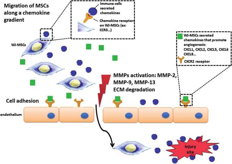
IN SEARCH OF A NEW SOURCE OF MESENCHYMAL STEM CELLS                                                                        525

The following table summarizes the phenotypic profiles of         opment compared with BM-MSCs, AT-MSCs, and skin-
these MSCs mentioned in the literature (Table 1).                 MSCs.
   WJ-MSCs, such as MSCs from other sources, positively
express the classical mesenchymal surface markers. How-             WJ-MSCs express genes involved in neural develop-
ever, Table 1 highlights the differences in the expression        ment. WJ-MSCs and DP-MSCs revealed a high expres-
levels of other markers:                                          sion of the neuro-ectoderm lineage markers.15 De Kock

                                                                  et al. have studied the whole gene expression profiles of
    Unlike BM-MSCs, WJ-MSCs weakly expressed en-
                                                                  four human mesoderm-derived stem cell populations: AT-
    doglin (SH2, CD105) and CD49e at passage 8.
   WJ-MSCs and AT-MSCs expressed CD106 at much
                                                                  MSCs, BM-MSCs, skin-MSCs, and WJ-MSCs. They have
                                                                  shown differences in gene expression between distinct
    lower levels than BM-MSCs.
   In comparison with BM-MSCs, HLA-ABC is very
                                                                  stem cell types. Skin-MSCs predominantly expressed
                                                                  genes involved in neurogenesis (NES), skin, and bone
    weakly expressed by WJ-MSCs, suggesting that these
                                                                  (RUNX2, BMP4).28
    cells could be good candidates for allogeneic cell
                                                                     Such a transcriptomic profile reveals a closer proximity
    therapy.
                                                                  between WJ-MSCs and BM-MSCs than between other
                                                                  combinations. Considering the genomic profile of WJ-
Transcriptomic profile                                            MSCs, WJ may be considered a reliable source of MSCs
                                                                  useful not only in cardiovascular regenerative medicine30,31
   Emerging data have compared the transcriptomic profile
                                                                  but also in neurodegenerative diseases. The latter will be
of WJ-MSCs with MSCs from other sources. The fol-
                                                                  discussed in the last part of the review.
lowing table gives an overview of the main comparisons
(Table 2).
   Some studies showing a high expression of embry-               Regenerative role of MSCs: differentiation potential
onic genes such as LIFR, ESG1, SOX2, TERT, NANOG,                 versus secretome
POUF1, OCT4, LIN28, DNMT3B, and GABRB3 by WJ-
                                                                     A summary of the various comparisons between sources
MSCs suggest that WJ could be a more primitive source
                                                                  of MSCs that have been already described in the literature is
of MSCs.4,6,24–26 Furthermore, as shown in Table 2, WJ-
                                                                  shown in Figure 1.
MSCs express genes encoding for proteins that are asso-
                                                                     WJ-MSCs differentiate into adipocytes slower than BM-
ciated with morphogenesis: SHH, neuregulin-1 and 4,
                                                                  MSCs.4 Bai et al. have shown that AF-MSCs and WJ-MSCs
SNA2, and WNT4.27
                                                                  could differentiate into myocardial-like cells with an im-
                                                                  portant expression of myocardial genes such as GATA-4, c-
   WJ-MSCs, compared with MSCs from other tissues,
                                                                  TnT, a-actin, and Cx43 after myocardial induction.18
differentially express genes involved in bone develop-
                                                                     More recently, Chen et al. have worked on in vitro dif-
ment. Transcription factors involved in osteoblast differ-
                                                                  ferentiation analysis of MSCs isolated from DP and WJ.
entiation such as RUNX2 were found to be expressed at
                                                                  They have shown that MSCs isolated from both sources ex-
comparative levels in BM-MSCs, skin-MSCs, AT-MSCs,
                                                                  hibited the capacity to differentiate into osteoblasts, chon-
and WJ-MSCs. However, Table 2 shows that skin-MSCs are
                                                                  drocytes, and adipocytes. However, they have noted some
characterized by a significantly increased expression of
                                                                  differences in their differentiation potentials. DP-MSCs and
genes (BMP4, BMP2) that are associated with bone and
                                                                  WJ-MSCs had a similar potential for osteogenic differentia-
cartilage development in comparison to the other MSCs.
                                                                  tion, but the chondrogenic and adipogenic differentiation
                                                                  potentials of WJ-MSCs were more important than those of
  WJ-MSCs reveal an important expression of genes in-             DP-MSCs.17 Meanwhile, according to Zhang et al., fetal
volved in liver and cardiovascular development. The tran-         human BM-MSCs have the highest potential of in vitro
scriptomic profile of WJ-MSCs and AF-MSCs reveals the             monolayer osteogenic differentiation, come after human WJ-
basal expression of several mature myocardial genes: GATA-        MSCs, human adult BM-MSCs, and then AT-MSCs.23 Baksh
4, c-TnT, and Cx43, which could be associated to the potential    et al. have found similar results as the previous study when
of differentiation into myocardial cells. Interestingly, a high   comparing the in vitro differentiation potentials of WJ-MSCs
expression of genes encoding for GATA-binding protein 6           and BM-MSCs.44
(GATA6), heart and neural crest derivatives expressed 1              Jo et al. have studied the in vivo osteogenic differentia-
(HAND1), inflammatory cytokine-induced intercellular ad-          tion, in a rat model, of human MSCs isolated from differ-
hesion molecule 1 (ICAM1), and vascular cell adhesion             ent sources. No differences were detectable in osteogenesis
molecule 1 (VCAM1) was detected in WJ-MSCs (Table 2).             between adult AT-MSCs, BM-MSCs, and WJ-MSCs.45
WJ-MSCs were also shown to express genes involved in              Controversial results have been described by Zhang et al. In
cardiovascular system development, including angiogenesis,        fact, after a subcutaneous implantation of MSCs scaffolds in
cardiogenesis, endothelial cell (EC) development, and vas-        mice, better results were obtained with scaffolds elaborated
culogenesis (Table 2). In addition, other genes involved in       with human fetal and adult BM-MSCs than those con-
cardiovascular development, including endoglin (ENG),             structed with WJ-MSCs and AT-MSCs.23 Differences in the
GJA1, VCAM1, and GATA6, were significantly increased in           results between the mentioned studies are probably due to
BM-MSCs.28                                                        the different experimental conditions. This explains that
   The trancriptomic profile also reveals that WJ-MSCs have       MSCs of various tissue origins have specific characteristics
a significantly increased expression of genes (AFP, DKK1,         of differentiation or require different conditions for os-
DPP4, and DSG2) which are associated with liver devel-            teoinduction.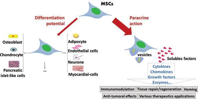
Table 2. Transcriptomic Profile of WJ-MSCs Compared with MSCs from Other Sources
      Gene                     Gene identification/function    WJ-MSCs     BM-MSCs     PDL-MSCs      AF-MSCs    AT-MSCs      Skin-derived MSCs   References
      Genes related to bone development and neurogenesis
        BMP4               Induce endochondral osteogenesis       ++           +           ND          ND           +             +++                28
        TGFBR1             Bone development                       ++           +           ND          ND          ND              ND                27
        OPN                Osteogenic marker                      ++          ++          +++          ND          ND              ND               14,27
        STAT1              Bone development                        +           +          ND           ND          ND              ND                22
        BSP                Osteogenic marker                       -          ND          ++           ND          ND              ND                14
        OSX                Osteogenic marker                       -          ND            -          ND          ND              ND                14
        CXCR4              Mesoderm marker                         +           +          ND           ND          ND              ND                27
        BMP2               Bone development                        +         ++           ND           ND           +             +++                28
        RUNX2              Osteogenic marker                      ++         +++          ND           ND          ++              ++                22
        CDH2               Neural gene                            ++           +          ND           ND           +               -                27
        NES                Neural development                     ++         ++           ND           ND           -             +++                28
      Gene related to liver and cardiovascular systems
        GATA-4             Mature myocardial gene                  +           +           ND           +           +              ++             18,28,29
        c-TnT              Mature myocardial gene                  +          ND           ND           +          ND              ND                18
        VEGF               Cardiovascular development              +           +           ND          ND          ND              ND                27
        Cx43               Mature myocardial genes                 +         ND            ND           +          ND              ND              18,20
        VCAM1              Cardiovascular development             ++         +++           ND          ND           -               -                28
        GJA1               Cardiovascular development             ++           +           ND          ND           +              ++                28
        AFP                Liver development                       +           -           ND          ND          ++               +                28
        DSG2               Liver development                      ++           -           ND          ND           +               -                28

526
        ENG                Cardiovascular development             ++         +++           ND          ND           -               -                28
        HAND1              Cardiovascular development             ++           +           ND          ND           +              ++                28
        GATA6              Cardiovascular development             ++           -           ND          ND           +               -                28
        DPP4               Liver development                       +           -           ND          ND          ++               -                28
        DKK1               Liver development                       +           -           ND          ND          ++               -                28
      Gene related to stemness
        ACTG2              Stemness-related genes                  +          ++           ND          ND          ND              ND                27
        TERT               Stemness-related genes                 ++         +/-           ND          ND          ND              ND               4,27
        ESG1               Stemness-related genes                 ++          ++           ND          ND          ND              ND                27
        Oct3/4             Stemness-related genes                  +          ++           ND          ND          ND              ND                27
        ABCG2              Stemness-related genes                +++          ++           ND          ND          ND              ND                27
        LIFR               Stemness-related genes                +++         +++           ND          ND          ND              ND                27
        SOX2               Stemness-related genes                  +         ND             +          ND          ND              ND               14,27
        Nanog              Stemness-related genes                  +         ND            ++          ND          ND              ND                14
        Oct-4              Stemness-related genes                ++         ++++           ++          ND          ND              ND                14
        THY1               MSCs marker                           ++           +            ND          ND           +               -                28
      Adipogenic and chondrogenic genes
        PPARc              Adipogenic marker                     +++         +++          +++          ND          ND              ND               13,14
        FABP4              Adipogenic marker                      +          ++            ND          ND          ND              ND                13
        LPL                Adipogenic marker                      -           ++            +          ND          ND              ND               13,14
        LEPR               Adipogenic marker                      -           +            ND          ND          ND              ND                13

                                                                                                                                                 (continued)
Table 2. Continued
Gene Gene identification/function WJ-MSCs BM-MSCs PDL-MSCs AF-MSCs AT-MSCs Skin-derived MSCs References
CD36 Adipogenic marker - ND + ND ND ND 14
CEBPA Adipogenic differentiation + ND ++ ND ND ND 14
COL2 Chondrogenic marker + + + ND + + 14
SOX9 Chondrogenic marker + + ND ND ND 14
Genes implicated in morphogenesis, adhesion, cell structure, and other mesodermal markers
ACTA Maintains the cytoskeleton + ND ND + ND ND 18
ACTB Cell motility, structure, and integrity +++ + ND ND ND ND 27
CDH5 Controls the cohesion and organization ++ + ND ND + + 28
of the intercellular junctions
in endothelial cells
ITGB1 Extracellular adhesion molecule ++ ++ ND ND ND ND 18
STAG1 Encodes for component cohesion + + ND ND +++ ++ 28
WT1 Kidneys and gonads development ++ -/+ ND ND -/+ -/+ 28
WNT4 Associated with morphogenesis + + ND ND ND ND 18
SNA2 Associated with morphogenesis ++ ++ ND ND ND ND 18
527
SHH Associated with morphogenesis -/+ + ND ND ND ND 18
Neuregulin 4 Associated with morphogenesis ++ ++ ND ND ND ND 18
COL1A1 Mesodermal marker +++ +++ ND ND - - 28
CXCR4 Mesoderm marker + + ND ND ND ND 18
ICAM1 Mesodermal lineage specification +++ + ND ND + + 28
CD44 Mesoderm marker ++ ++ ND ND ND ND 18
PECAM1 Cell adhesion marker ++ +/- ND ND +/- + 28
CD9 Implication in differentiation, + - ND ND ++ ++ 28
adhesion, and signal transduction
Collagen X Mesoderm marker - +++ ND ND ND ND 18
FlK-1 Mesoderm marker ++ - ND ND ND ND 18
CD68 Highly expressed by human monocytes +++ ND ND ND ND ND 20
and tissue macrophages
The table given next presents the difference of gene expression, for various markers, between the different MSCs sources. The difference of expression between the sources is presented
by + + + , + + , + , - , and ND corresponds to a nondetermined comparison.
PPIA, cyclophilin A, Homo sapiens peptidylprolylisomerase A; ITGB1, integrin, b1:fibronectin receptor, b polypeptide; WNT4, wingless-type MMTV integration site family, member 4; SHH, sonic
hedgehog homolog (Drosophila); SNAI2, snail homolog 2 (Drosophila); SNAI2, snail homolog 2 (Drosophila); TGFBR1, transforming growth factor, b receptor I; TERT, telomerase reverse
transcriptase; ESG1:ESTs, weakly similar to embryonal stem cell specific gene 1; Cx43, connexin-43; ACTA, alpha-actin; ACTB, beta actin; CDH2, cadherin 2; OPN, osteopontin; LIFR, leukemia
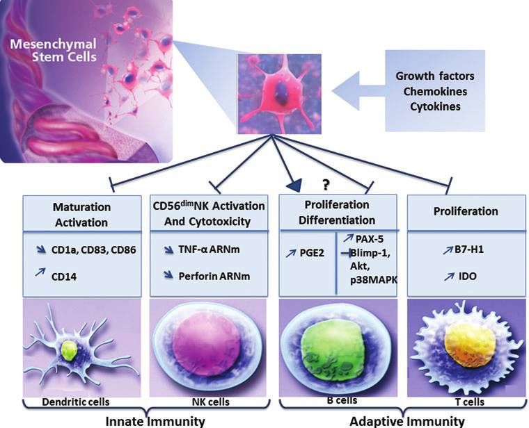
inhibitory factor receptor: SOX2, sex-determining region Y (SRY)-box; ND, nondetermined.528 EL OMAR ET AL. FIG. 1. Differentiation potential of WJ-MSCs compared with MSCs from other sources. This figure shows differences in the differentiation potential of MSCs from many sources toward a specific cell type, which is indicated by + + + , + + , + , - , and ND corresponds to a nondetermined comparison. For example, PDL-MSCs have the greatest potential to differentiate into chondrocytes, while DP-MSCs have the lowest differentiation potential toward this type of cells. DP, dental pulp; WJ-MSC, Wharton’s jelly-mesenchymal stromal cell. Color images available online at www.liebertpub.com/teb Very recently, Yu et al. have shown that WJ-MSCs are never been proved. Indeed, recent studies suggest that the not good alternatives for periodontal tissue generation benefits of MSCs transplantation may be associated to a compared with PDL-MSCs which have a much better osteo/ paracrine modulatory effect rather than the replacement of dentinogenic differentiation potential.12,14 Various studies affected cells, at the site of injury, by differentiated stem have demonstrated the capacity of WJ-MSCs to differentiate cells.49,50 Emerging data suggest that stem cells could be into pancreatic islet-like cells.46–48 Kim et al. have compared then considered as a reservoir of trophic factors which are this potential with other sources of MSCs and did not show released when needed to modulate and repair surrounding significant differences between BM-MSCs and AT-MSCs.46 damaged tissues, which leads to a paradigm shift in regen- Some authors have focused on the application of MSCs in erative medicine. Understanding the cell secretome has at- vascular engineering and more particularly their capacity to tracted much attention, and it has been demonstrated that differentiate into EC, or to acquire pericyte markers when trophic factors could have many effects such as modula- co-culturing with EC. Chen et al. were pioneers in studying tion of inflammatory reactions, immunomodulation, anti- the endothelial differentiation potential of BM-MSCs and apoptotic and pro-angiogenic capacities, and many others WJ-MSCs. Both sources of MSCs were able to differentiate (reviewed in Doorn et al.51). Vallone et al. have highlighted into EC but WJ-MSCs appear to have a greater differenti- in their review the exact mechanisms that would lead MSCs ation potential, as derived EC-like exhibited a higher ex- to damaged tissues after transplantation, where they will pression of endothelial markers.5 exert their remedial actions.52 Katsuda et al. have also de- For a long time, it has been considered that the regen- scribed a possible therapeutic mechanism of AT-MSCs, in erative potential of MSCs is due to their plasticity and Alzheimer disease, through a paracrine pathway. Vesicules differentiation capacity. However, the direct link between secreted by these cells could carry soluble factors that may their differentiation potential and their beneficial effects has treat this pathology. Results of this study will be discussed
IN SEARCH OF A NEW SOURCE OF MESENCHYMAL STEM CELLS                                                                       529

in the final part of this review. Therapeutic effects of BM-    BM-MSCs, making them a good candidate for allogeneic
MSCs in regenerative medicine (heart disease for example)       transplantation.
through paracrine/autocrine mechanisms have been re-
viewed by Pourrajab et al.53
                                                                MSCs-mediated immunosuppression
   The controversy between the implication of the differ-
entiation potential and the paracrine mechanisms of MSCs           MSCs show an absence or a low expression of MCH class
in their beneficial therapeutic actions is shown in Figure 2.   II and co-stimulatory molecules, so they can be considered
   During the next few sections of this review, we will         immunoprivileged cells, but they also interfere with differ-
highlight the effects of WJ-MSCs secretome involved in          ent pathways of the immune response.58 Their ability to
many processes such as immunomodulation, homing to              modulate the immune system was first recognized after the
damaged tissues and others.                                     fact that they could evade immunosurveillance after cell
                                                                transplantation.59 Especially, human MSC populations such
WJ-MSCs as Immunoprivileged Cells                               as BM-, AT-, or UC-derived MSCs selectively alter immune
                                                                cell function by suppressing T-cell proliferation, B-cell
Immunological features of MSCs                                  proliferation, and terminal differentiation,60 inhibiting NK
   In the last decade, MSCs have gained considerable atten-     cell proliferation and cytotoxicity, steering monocytes and
tion as candidates for tissue engineering, as modulators of     dendritic cells (DCs) to an immature DC state.61
immune responses in graft-versus-host disease, and as au-
toimmune diseases,54 as these cells, once administered             MSCs and immune cell population. Adaptive immunity:
therapeutically, may be able to evade the immune system of         MSCs and T cells: T cells recognize antigens and are
the host. They are currently being assessed as a novel anti-    critical for cell-mediated immune response. They mature
inflammatory therapeutic agent in numerous clinical trials.55   within the thymus into one of different subtypes with di-
Two outstanding features of MSCs are relevant to their          verse roles. These cells are involved in the maintenance of
immunomodulatory effects:                                       self-tolerance, activation of other lymphocytes, lysis of in-
                                                                fected cells, and interaction with cells of the innate immune
   Immunosuppression. MSCs-mediated immunosuppres-              system.
sion describes the fact that MSCs are able to suppress sev-        Currently, interactions of MSCs with T cells have been
eral functions (proliferation, production of soluble factors,   extensively studied. Graft versus Host Disease models
and cellular cytotoxicity) exerted by diverse immune cells      presented the first evidence that MSCs can regulate immu-
such as T-, B-, and natural killer (NK) cells. It has been      nosuppression in vivo.62 MSCs could reduce allograft re-
shown that immunosuppression is mediated by both cell–          jection, which is partly mediated by T cells.63,64 Shortly
cell contact and paracrine signals via soluble factors.         afterward, T-cell immunosuppression mediated by MSCs
                                                                was demonstrated in vitro. MSCs probably inhibit, via their
   Immunoprivilege. MSCs themselves are somehow pro-            induced or constitutively expressed secreted factors,
tected from immunological defense mechanisms.56 Indeed,         T-lymphocyte activation and proliferation induced by mi-
MSCs lack expression of major histocompatibility complex        togens and alloantigens65–68 as well as T-cell activation with
(MHC) class II, giving MSCs the potential to escape recog-      CD3 beads.66,69 MSCs have been shown to equally inhibit
nition by alloreactive CD4 + T cells but express MHC Class I    CD4 + , CD8 + , CD2 + , and CD3 + subsets.70 In addition,
molecules. This expression enables them to escape from NK       T-lymphocytes inhibited by BM-MSCs do not enter apo-
cell lysis, In addition, MSCs do not express co-stimulatory     ptosis, as they actively proliferate on re-stimulation with
molecules required for effector T-cell induction.57             cellular and humoral activators.65 Many other studies have
   Even if BM-MSCs, considered the gold standard in MSC         shown the ability of BM-MSCs to induce the expansion of
therapy, and UC-MSCs share many similarities, emerging          functional regulatory T cells (Tregs).70,71 Recently, it has
data suggest that WJ-MSCs could be less immunogenic than        been shown that adhesion molecules ICAM1 and VCAM1,

                                                                                      FIG. 2. Summary of the potential
                                                                                      therapeutic roles of MSCs. Beneficial
                                                                                      effects of MSCs have been attributed
                                                                                      to their differentiation potentials.
                                                                                      However, attention has been shifted to
                                                                                      their paracrine effects (via vesicles
                                                                                      and soluble factors) rather than their
                                                                                      plasticity. The role that MSCs will
                                                                                      play is determined by the microenvi-
                                                                                      ronment where they reside. MSCs,
                                                                                      mesenchymal stromal cells. Color
                                                                                      images available online at www
                                                                                      .liebertpub.com/teb
530 EL OMAR ET AL. which are required for a direct adhesion of MSCs to T and proliferation.82 They are directly responsible for the cells, are critical for subsequent MSC-mediated immuno- humoral immune response via the secretion of antibodies suppression, and are inducible by the parallel presence of against pathogenic or foreign antigens. A subset of B lin- interferon-gamma (IFN-g) and inflammatory cytokines.72 eage differentiates into memory B cells, which can medi- Another possible mechanism underlying the BM-MSC- ate a rapid response on secondary exposure to that same mediated suppression of T cells is to prevent their entry into antigen. the S phase of the cell cycle by mediating irreversible Corcione et al. demonstrated that BM-MSCs inhibited the G0/G1 phase arrest through the inhibition of cyclin D2 ex- proliferation of B cells and significantly decreased the pression.69,73 Similarly, it has been shown that the addition production of IgM, IgG, and IgA83; the same effect has been of DP-MSCs to phytohemagglutinin-stimulated T cells reported by Che et al. showing that UC-MSCs significantly mediated an inhibition of their response.74 Increased ex- suppressed the proliferation, differentiation, and immuno- pression of immunodulatory soluble factors (hepatocyte globulin secretion of B cells in vitro.84 To understand the growth factor [HGF]-b1, ICAM-1, IL-6, IL-10, trans- results of Che et al., it is essential to know that ‘‘B- forming growth factor-b1 [TGF-b1], VCAM1, and vas- lymphcote-induced matruration protein-1’’ (Blimp-1), ‘‘X- cular endothelial growth factor (VEGF)) secreted by box binding protein-1’’ (Xbp-1), ‘‘B-cell lymphoma-6’’ human DP-MSCs was detected in a co-culture system with (Bcl-6) and ‘‘paired box gene-5’’ (PAX-5) are known as the decreased expression levels of some pro-inflammatory main regulators of B-cell differentiation to immunoglobulin- cytokines and increased levels of some anti-inflammatory secreting cells. PAX-5 and Bcl-6 are required to keep B-cell ones. Induction of Treg markers by human DP-MSCs phenotypes. Blimp-1 inhibits the expression of both PAX-5 was also demonstrated.75 A very recent study has examined and Bcl-6 in order to let B cells differentiate. BCR signaling the in vivo and in vitro immunomodulatory effects of involves the MAPK signaling pathway and increases the human supernumerary tooth-derived mesenchymal stem transcriptional activity that is mediated by the transcription cells (SNT-MSCs). It has been shown that, in in vitro co- factor activator protein-1 (AP-1), which leads to Blimp-1 cultures, these cells suppressed the viability of T cells and expression. Che et al. have shown a suppression of Blimp-1 also the differentiation of Th17 cells. In vivo transplanta- expression and an induction of PAX-5 in the co-cultures of tion of SNT-MSCs in systemic lupus erythematosus model UC-MSCs and B-cells. Thay have also found that Akt and MRL/Ipr mice suppressed increased levels of peripheral p38 MAPK were inhibited by WJ-MSCs.84 Th17 cells and IL-17 as well as ex-vivo differentiation of However, these results have been contradicted by other Th17 cells.76 groups. Rasmusson et al. have shown an increase of B-cell Fetal MSCs have been reported to have similar inhib- immunoglobulin secretion when co-cultured with BM- itory effects on T-lymphocytes. It has been shown that MSCs; this effect varied depending on the type of stimulus mitogen-induced T-cell proliferation in an allogeneic used to trigger B cells.85 Likewise, Traggiai et al. have model transplant, as well as in a xenograft model, was reported that BM-MSCs could promote B-cell expansion effectively suppressed by WJ-MSCs with levels compa- and differentiation after treatment with an agonist of Toll- rable to BM-MSCs immunosuppression.77 In addition, like receptor 9.86 A recent study has demonstrated that UC- IFN-g and/or tumor necrosis factor-alpha (TNF-a) pro- MSCs promoted proliferation and differentiation of B cells duced by activated T cells stimulate the production of both in vitro and in vivo partially through prostaglandin E2 indoleamine 2,3-dioxygenase (IDO) by MSCs, which, in (PGE2) axis.82 turn, inhibited T-cell proliferation.78 Tipnis et al. have Contradictions in the effects of MSCs on B cells could be reported that the expression of B7-H1, a negative regu- associated to the differences in the B-cell source, the manner lator of T-cell activation constitutively expressed by WJ- of their purification and stimulation, the culture conditions, MSCs, is increased after IFN-g treatment. In addition, and many other factors. However, the microenvironment IFN-g treatment induced IDO secretion by WJ-MSCs, plays a decisive role in determining the role that the MSCs which inhibited T-cell proliferation.79 These results were will play. confirmed very recently by Manochantr et al. showing that MSCs from amnion, placenta, and WJ can potentially Innate response: substitute BM-MSCs in several therapeutic applications. MSCs and NK cells: NK cells are major effector cells of Indeed, these cells inhibited alloreactive T-lymphocytes innate immunity, because they lack antigen-specific cell in the mixed lymphocyte reaction in a similar degree as surface receptors.87 They mediate antibody-dependent cel- BM-MSCs.80 lular cytotoxicity as well as ‘‘spontaneous’’ killing of in- MSCs and B cells: The reasearch on T-cell immunosup- fected or transformed cells through the release of perforin pression mediated by MSCs has attracted most of the at- and granzyme from cytotoxic granules.88 tention in clinical applications and has been widely studied. MSCs and NK cells have been shown to interact in vitro. However, B cells and humoral immune responses are more The outcome of this interaction may depend on the state of and more known as important mediators of chronic allograft NK-cell activation and/or the cytokines present in the cul- rejection. Indeed, data about the influence of MSCs on B ture medium. IFN-g-activated MSC-escaped NK cells me- cells growth, differentiation, and production of immuno- diated lysis through the induction of HLA-E and NK globulins (Ig) are still scarce and controversial.81 inhibitory ligands.89,90 Previous studies have indicated that B cells play an essential role in adaptive immunity. These cytokine-induced proliferation of NK cells leads to the cells develop in the BM strictly after a close interaction up-regulation of HLA class I on MSCs.90 In response to this between B-cell progenitors and stromal cells that produce up-regulation, HLA class I molecules, including human cytokines which are capable of supporting B-cell survival leukocyte antigen-G5 (HLA-G5), expressed by MSCs, bind
IN SEARCH OF A NEW SOURCE OF MESENCHYMAL STEM CELLS                                                                          531

to the inhibitory receptor ILT2 expressed on NK cells.91            MSCs and DCs: DCs play a key role in the initiation of
Furthermore, other studies have shown that the suppression       primary immune responses and tolerance, depending on the
of NK cell functions is mediated by a down-modulation of         activation and maturation stage of DCs. Locally produced
some activating NK cell receptors (NKp30, NKp44, and             inflammatory cytokines or microbial components promote
NKG2D) and by the inhibition of NK cell lytic granule            the maturation of DCs from a processing to a presenting
formation.92 There is growing evidence that IDO, PGE2,           stage, characterized by the up-regulation of MHC-class II
and TGF-b1 may control MSC-mediated inhibition of NK-            and co-stimulatory molecules (CD80 and CD86), production
cell function.93                                                 of IL-12, and migration to lymphoid tissue. DCs maturation
   Boissel et al. evidenced that NK cells had a higher           is a prerequisite to induce immunogenic T-cell responses,
expansion when cultured with allogeneic and autologous           whereas tolerance is observed when antigens are presented
WJ-MSCs as feeders in the presence of NK growth factors.         by immature or semi-mature DCs. Therefore, DC matura-
WJ-MSCs feeders were rejected during the first week of co-       tion plays a key role in initiating T-cell responses.
culture. Expanded NK cells maintained an elevated cytotoxic         BM-MSCs were shown to block the generation of func-
profile and may be genetically manipulated.88 In a recent        tional antigen-presenting cells, including myeloid DCs from
study, Zhao et al. have been interested in elucidating the       both monocytes and CD34 + cell precursors.96–98 Most re-
effect of UC-MSCs on NK cell-mediated cytotoxicity against       sults supported the notion that DCs at early stages of dif-
DCs and the mechanism involved. They found that UC-              ferentiation are sensitive to their inhibitory effects, while at
MSCs can enhance this effect possibly by inhibiting DCs          later stages, they are resistant. However, WJ-MSCs in-
maturation and up-regulating the ligands for killer activator    hibited DC maturation and activation even when the contact
receptor on the surface of the DCs.94 When comparing the         happended during the mature or immature stage. Both cell
immunosuppressive activity of MSCs derived from UC, AT,          contact via surface ligands (B7H1) and soluble factors
and BM on lymphocytes, Ribeiro et al. have shown that all        (IDO) enhanced the efficiency of suppression.79 Very re-
the three types of MSCs exhibited a strong inhibitory effect     cently, Saeidi et al. showed that UC-MSCs and BM-MSCs
on CD56dimNK cell subset activation (cytotoxic NK cells).        strongly inhibited the differentiation and maturation of DCs
UC-MSCs were the only cells that were unable to inhibit the      with a more inhibitory effect on CD1a, CD83, CD86 ex-
activation of CD56bright NK cell subset (a subset that has the   pression, and DC endocytic activity. These cells also se-
capacity to produce abundant cytokines after activation but      verely up-regulate CD14 expression. Results have indicated
has a low natural cytotoxicity). Among all these MSCs, AT-       that UC-MSCs and BM-MSCs exerted their inhibitory effect
MSCs had a higher inhibitory capacity. A down-regulation         on differentiation, maturation, and function of DCs through
of perforin and TNF-a ARNm by MSCs from the three                the secreted factors and free of any cell-to-cell contacts.99
sources was observed, while only AT- and BM-MSCs in-                Immunomodulatory properties of WJ-MSCs in innate and
duced a minor reduction of granzyme B ARNm.95                    adaptive responses are resumed in the figure given next (Fig. 3).

                                                                                                   FIG. 3. Immunomo-
                                                                                                   dulatory effects of the WJ-
                                                                                                   MSCs on innate and adap-
                                                                                                   tive immunity. The effects
                                                                                                   can be summarized as fol-
                                                                                                   lows: inhibition of the mat-
                                                                                                   uration and activation of
                                                                                                   dendritic cells as well as the
                                                                                                   proliferation of T cells, ac-
                                                                                                   tivation of the expansion
                                                                                                   and cytotoxicity of NK
                                                                                                   cells. Effects of WJ-MSCs
                                                                                                   on B cells are still contra-
                                                                                                   dictory; they can stimulate
                                                                                                   or block the proliferation
                                                                                                   and differentiation of B
                                                                                                   cells and the secretion of
                                                                                                   immunoglobulin (Ig). NK,
                                                                                                   natural killer; WJ, Wharton’s
                                                                                                   jelly. Color images available
                                                                                                   online at www.liebertpub
                                                                                                   .com/teb
532 EL OMAR ET AL.
MSCs and immumodulatory paracrine factors. Multiple the WJ-MSCs has been reported; both are implicated in
reports have evidenced, first in vitro and then in vivo, the tolerogenic processes occurring at the fetal-maternal inter-
ability of MSCs to express molecules that interact with both face, along with HLA-G.114
innate and adaptive immunity, both through soluble fac-
tors65,100 and in a cell contact-mediated fashion probably
Homing of MSCs
through the interaction of membrane receptors, adhesion
molecules, or the cellular exchange of membrane vesi- Mantaining the function and the integrity of the human
cles.101 It is still a matter of debate whether the regulatory body, which is often subjected to injuries, is essentially due
effects are cell-to-cell contact -dependent, or whether solu- to tissue repair. Shortly after an injury, different types of
ble factors are sufficient.102 The MSCs immune modulating immune cells (neutrophils, monocytes, and lymphocytes)
effects will also depend on the ratio between MSCs and are conducted to the site of damage. These cells are re-
immune cells, and the state and stage of immune cell acti- sponsible for the secretion of various growth factors and
vation or maturation. Several factors that contribute to the cytokines that will attract other residing or circulating cells
MSCs-mediated effects have been identified, in particular such as MSCs. Endogenous MSCs present a pool of re-
growth factors, cytokines, chemokines, and hormones, all of generative cells, participate in tissue repair, and communi-
which exert paracrine effects on immune cells and enable cate with other cells in response to signals of cellular
homing, migration, and their attachment to injured cells. damage.115 Their ‘‘homing’’ can be defined as the arrest of
Soluble factors implicated in MSCs-mediated immune MSCs within the vasculature of a tissue than crossing the
modulation include nitric oxide (NO), indoleamine 2,3- endothelium.116. Thus, the homing of endogenous MSCs is
dioxygenase (IDO), heme oxygenase (Hmox1), secretion of being considered a therapeutic benefit, and studies are
anti-inflammatory cytokines such as IL-10, TGF-b, HGF, evaluating new methods for recruiting a sufficient number of
IL-6, and PGE2.68,70,103,104 A study comparing the immu- MSCs to exert their regenerative capacity. In cases where
nomodulatory properties of MSCs derived from many the reservoir of MSCs is depleted because of several dis-
sources showed that despite their similar cytokine profiles, eases or the age, exogenous MSCs could be administrated to
WJ-MSCs only secrete IL-12, IL-15, and platelet-derived compensate the lack of endogenous MSCs (reviewed in
growth factor-AA (PDGF-AA). They did not secrete VEGF Marquez-Curtis and Janowska-Wieczorek115 and Sohni and
similar to other adult MSC sources.78 The precise meaning Verfaillie117). It has been reported that in-vitro-expanded
of these differences, however, needs to be understood in MSCs preferentially home to sites of tissue damage, where
WJ-MSCs/immune cell co-cultures.105 they enhance wound healing, support tissue regeneration,
Specialized immune tolerance implicated at the maternal- and restore the BM microenvironment after damage by
fetal interfaces depends on the expression of many mole- myeloablative chemotherapy or integrate into tumors.118
cules, including galectin-1, B7 proteins, HLA-G,106 and the Since the precise molecular mechanisms by which MSCs
expression of immune suppressive cytokines such as leu- migrate into sites of injury are not yet fully defined, the
kemia inhibitory factor (LIF).107,108 Since WJ-MSCs are migration of leukocytes into sites of inflammation has been
isolated from a peri-natal source, they could exhibit immune taken as a model.119 Indeed, on delivery into the blood
evasion mechanisms that are dominant at the fetal-maternal stream, the MSCs keep close contact with EC whose role is
interface. In fact, Najar et al. showed that higher constitu- being extensively studied in MSCs migration. They engraft
tive as well as IFN-g inducible levels of LIF are expressed into the endothelium, and eventually pass and leave the
by WJ-MSCs than by BM-MSCs and the suppression of endothelium.120 The migration of MSCs is mediated by a
lymphoproliferation can be rescued by blocking LIF in co- wide variety of molecules that are expressed by MSCs, in-
cultures.109 Furthermore, Prasanna et al. reported higher cluding growth factors, chemokines, and receptors, and by
levels of both constitutive and IFN-g inducible HGF in WJ- chemotactic factors produced by immune cells.116 It has
MSCs compared with BM-MSCs.110 been demonstrated that human MSCs showed significant
Nonclassical type I HLA molecules are an interesting as chemotaxis responses to several factors (including PDGF,
yet only partly explored field in MSCs immune function. VEGF, IGF-1, IL-8, bone morphogenetic protein BMP-4,
Several reports showed that BM-MSCs and WJ-MSCs ex- and BMP-7)121 and express a variety of chemokine recep-
press the HLA-G molecule, at both mRNA and protein le- tors (such as CCR1, CCR4, CCR7, CXCR5, and CCR10)
vel, and its soluble form HLA-G5.29,77,111 Weiss et al. also which might be involved in their migration into injured
showed that WJ-MSCs constitutively express high levels of tissues along a chemokine gradient.122
the immune suppressive HLA-G6 isoform, while BM-MSCs In addition, specific proteolytic enzymes are required so
express the HLA-G5 isoform constitutively and its expres- the cells can traverse the protein fibers of the extracellular
sion is not induced by IFN-g.77 HLA-G5 secretion has been matrix (ECM) and reach the target sites.118,123 In particular,
directly implicated in the induction of regulatory cells the matrix metalloproteinases (MMPs), consisting of more
(CD4 + CD25 + FoxP3 + Tregs) that are characterized as key than 24 zinc-dependent endopeptidases, are physiologically
suppressors of effector responses to alloantigens.112 HLA- necessary for stem cell migration, degradation, and re-
G5 secretion has also been linked with the suppression of modeling of ECM components, and are crucial for devel-
NK cell production of IFN-g in BM-MSCs co-cultures.113 opmental events such as morphogenesis, cell proliferation,
Since the inhibition of maternal alloreactivity is due to the apoptosis, and differentiation.124–127
expression of high levels of HLA-G by the fetus, the exact Ries et al. were the first to show that human BM-MSCs
role of immune-suppressive HLA-G isoforms, such as HLA- use constitutively expressed MMP-2 (gelatinase A), mem-
G6 expressed by WJ-MSCs, needs to be evaluated in de- brane-type matrix metalloproteinase-1 (MT1-MMP), and
tail.105 Recently, the expression of HLA-E and HLA-F in tissue inhibitor of metalloproteinase 2 (TIMP-2) to migrateIN SEARCH OF A NEW SOURCE OF MESENCHYMAL STEM CELLS 533
through human recombinant basement membranes. In- CCRL12 (plays a role in the control of airway inflammatory
flammatory cytokines such as TGF-b1, IL-1b, and TNF-a response and in lung DC trafficking) were more expressed in
are able to exert chemoattractive potential on hBM-MSCs BM-MSCs. Moreover, results have reported a stronger ex-
and to up-regulate MMP-2, MT1-MMP, and/or MMP-9, pression, in WJ-MSCs, of many growth factors linked with
enabling cellular trafficking of MSCs across human ECM angiogenesis such as VEGF-D, PDGF-AA, TGF-b2, b-FGF,
barriers.118 MMP-2 has been also detected in WJ-MSCs in and HGF.116 Their chemokine gene profile suggests that WJ-
association with MMP-9 (gelatinase B), MMP-8, and MMP- MSCs may be useful in the healing and treatment of ische-
13 (respectively collagenase-1 and -2), as well as in dif- mic lesions such as the ischemic myocardium, with cerebral
ferent regions of full-term human umbilical cord and in ischemia for example. Moreover, they could be suggested as
cultured HUVEC. The wide expression of these enzymes in a treatment to reduce or prevent fibrosis and scarring in
the umbilical cord has been attributed to their role in the tissue lesions, as it has been shown that they secreted bFGF
degradation and remodeling of ECM and in other physio- and HGF (known to have an anti-fibrotic effect).
logical processes.128 Thus, ex vivo-expanded human MSCs with cytokines may
Recently, Balasubramanian et al. have compared che- be a useful method, in clinical applications, to increase their
mokine and receptor gene expression between WJ- and BM- migration/homing potential after transplantation into pa-
MSCs. Their results have shown that Chemokine (C-C tients, as well as the administration of cytokines to mobilize
motif ) receptor 3 (CCR3) was more expressed in WJ-MSCs MSCs to sites of injury.
than in BM-MSCs; whereas the latter have presented a
higher expression of CCR1, CCR7, CCRL2, Chemokine (C-
Anti-tumourigenic Effects of WJ-MSCs
X3-C motif ) receptor 1 (CX3CR1), and CXCR5. MSCs
from both sources had a similar expression of CCR5, CCR6, MSCs have the capacity to migrate to tumor sites and
and CCRL1. modulate their microenvironment. Thus, they have an im-
In addition, Chemokine (C-X-C motif ) ligand 1 (CXCL1), pact on tumour behavior.129 A great deal of evidence sug-
CXCL2, CXCL5, CXCL6, and CXCL8 (members of the gests that solid tumors generate a microenvironment similar
CXC chemokine family) were up-regulated in WJ-MSCs in to that associated with wound healing, as they apply phys-
comparison with BM-MSCs. These chemokines are known ical and chemical stress to neighboring tissues. Tumors can,
as potent promoters of angiogenesis and mediate their ac- therefore, be considered sites of tissue damage, which in-
tivity by binding CXCR2 receptor on the endothelium (Fig. duces the migration of MSCs.130
4). On the contrary, CXCL12 and CXCL13, also two Human MSCs have been intensively studied for their
members of the CXC chemokine family and known to potential use in cancer treatment. Their use has been limited,
contribute to immune and nonimmune cell homing, were up- however, by a general concern related to their biosafety.131
regulated in BM-MSCs. WJ-MSCs have shown a higher Many studies have reported pro- or anti-tumorigenic effects
expression of IL-1A (enhance the expression of CXCL8) and of MSCs on the progression of primary and metastatic tu-
TNF-a (angiogenic factor) than BM-MSCs; while IL16 (has mours. These contradictory results could be associated with
an immunomodulatory role in asthmatic inflammation) and differences in the MSC sources used, the type of tumour
FIG. 4. Proposed mecha-
nisms involved in the homing
of WJ-MSCs to sites of tissue
injury and their angiogenesis
capacity. MSCs home to site
of injury along gradients cre-
ated by inflammatory chemo-
kines and several factors.
They express a variety of
chemokine receptors that
might be involved in their
migration into injured sites.
The expression of a set of
MMPs by WJ-MSCs contrib-
utes to the extracellular matrix
(ECM) degradation, which
enables them to cross the
ECM and reach the site of
injury. They also express a
variety of chemokines that are
known as angiogenesis medi-
ators and exert their function
by binding to their receptor
CXCR2 on the endothelium.
MMPs, metalloproteinases.
Color images available online
at www.liebertpub.com/teb534 EL OMAR ET AL.
model, the method of administration, or other unknown extracts.152 In addition, Ma et al. have shown that WJ-MSCs
factors.130,132 significantly inhibited the growth of breast cancer stem cells
The tumor stroma consists of a complex ECM in which in vitro and in vivo, probably by inducing a cell cycle arrest
inflammatory and immune cells, fat cells, fibroblasts, and and tumor cell apoptosis and inhibiting the activities of
blood vessels reside. It plays a crucial role in tumor pro- phosphoinositide 3-kinase (PI3K) and AKT (also known as
gression, angiogenesis, and metastasis through its effects protein kinase B).149 A more recent study has reported that
on tumor-host interactions. Tumor-associated fibroblasts WJ-MSC conditioned medium as well as its cell lysate in-
(TAFs) are activated fibroblasts in the tumor stroma.131 hibits mammary carcinoma and osteosarcoma cell growth
Several reports have hypothesized that BM-MSCs selec- via apoptosis and autophagy in vitro and in xenograft
tively proliferate to tumors and contribute to the formation mice.153 In another study evaluating the tumorigenesis po-
of tumor-associated stroma by transforming into TAFs. tential of WJ-MSCs in comparison with ESCs, animals in-
They also promote tumor growth and metastasis by en- jected with ESCs developed teratomas with increased levels
hancing migration and angiogenesis and inhibiting apoptosis of pro-inflammatory cytokines; whereas those injected with
of tumor cells.133–137 WJ-MSCs developed no tumors or inflammatory reactions at
On the other hand, the immunosuppressive effects of the injection sites and exhibited increased production of
MSCs can impair the function of a variety of immune cells anti-inflammatory cytokines.154 A very recent study on the
(directly or through paracrine signals). This may be an im- effects of WJ-MSCs on intrahepatic cholangiocarcinoma
portant mechanism enabling MSCs to promote tumor growth (ICC, a common form of primary liver cancer) has shown
or to increase the incidence of tumor formation. For instance, that these cells can inhibit the proliferation and induce the
by increasing Tregs and reducing the activity of NK cells and apoptosis of human ICC cells. Apoptosis of tumour cells is
cytotoxic T lymphocytes (CTL) (known to kill tumour cells), related to the inhibition of PI3K/Akt and the Wnt/b-catenin
BM-MSCs can protect breast cancer cells.138 They also have signaling pathways.155 The effects of WJ-MSCs have also
been linked to osteocarcinomas,139 prostate tumors,140,141 been studied in hematopoietic tumours. Results obtained by
breast tumors,138,140,142 colon cancer,137 and others. A recent Tian et al. have provided a new insight on how these cells
study has also demonstrated a fusion between MSCs and may modulate leukemic tumour growth in vitro. According
gastrointestinal epithelial cells, suggesting the formation of a to this study, p38 MAPK, a suppressor of tumor develop-
more cancer-prone cell type.143 ment, was required for leukemic tumor suppression by WJ-
Very recently, in order to examine the possible anticancer MSCs.156
therapeutic applications of MSCs from different sources, These studies, taken together, indicate that WJ-MSCs are
Akimoto et al. have studied the inhibitory effects of MSCs nontumorigenic, anti-tumorigenic, and hypoimmunogenic;
from umbilical cord blood (UCB) and AT on ‘‘glioblastoma do not transform to the TAF phenotype that is associated
multiforme (GBM)’’ (the most aggressive type of primary with enhanced growth of solid tumors; and suppress he-
brain tumor in humans). They found that, both in vitro and matopoietic tumor development. WJ-MSCs appear to be a
in vivo, GBM growth was inhibited by UCB-MSCs but safe and promising tool for future cancer therapy and clin-
promoted by AT-MSCs. UCB-MSCs induced apoptosis ical applications, but more pieces of evidence are needed to
through the tumor necrosis factor-related apoptosis-inducing further characterize their anti-tumorigenic mechanisms and
ligand (TRAIL), which is more strongly expressed by UBC- to confirm this hypothesis.
MSCs than by AT-MSCs.144 Furthermore, it has been shown
that naı̈ve WJ-MSCs are able to produce factors suppressing Therapeutic Applications
cancer cell growth and inducing apoptosis, and so may be a
Treatment of autoimmune diseases
novel tool for cancer therapy in contrast to MSCs from some
other sources.145 Other reports have likewise shown that The immune properties of WJ-MSCs suggest that they
WJ-MSCs can abrogate certain solid tumors.146–149 These may be a therapeutic option to treat autoimmune diseases
cells decreased the growth of human breast cancer in vitro such as type 1 diabetes or Crohn’s disease (CD).
and stopped its growth when intravenously injected in an
SCID mouse model.150 Later, Fan et al. showed that WJ- Type 1 diabetes. Diabetes is a metabolic disease listed
MSCs do not induce teratomas in immunodeficient SCID among the leading causes of death in some countries. It is
mice, nor do they induce tumors when transplanted into characterized by absolute or relative insulin deficiency.
diseased animal models.151 In a recent study, Subramanian Type 1 diabetes is characterized by an absolute insulin
et al. have examined whether WJ-MSCs, such as BM- decrease due to T-cell-mediated destruction of insulin-
MSCs, transform to the TAF phenotype in the presence of producing pancreatic b cells.157 This autoimmune destruc-
ovarian and breast cancer conditioned medium. Results have tion of pancreatic islet b-cells reduces the patient’s ability
shown no expression of tumor-associated markers for hWJ- to regulate blood glucose, leading to a high frequency of
MSCs with a low expression of TAF-related genes, con- vascular complications that compromise the quality and
firming that these cells are not associated with enhanced expectancy of life.158
growth of solid tumors.3 In order to determine whether WJ- Transplantation of pancreatic islet cells (PICs) as a
MSC-mediated inhibition of cancer cell growth was not potential cure for type I diabetes has been hampered by
specific to breast cancer cells, the same group compared the immune rejection and recurrent attacks against islets by the
effects of WJ-MSC extracts and cell lysate on three other underlying autoimmunity. Studies have shown the capacity
types of solid tumors: breast adenocarcinoma, ovarian car- of WJ-MSCs to differentiate into mature islet-like cell
cinoma, and osteocarcinoma. They observed the same ef- clusters. These islet-like cell clusters have been shown to
fects, which were probably mediated via agents in WJ-MSC contain human C-peptide and to release insulin in vitro andIN SEARCH OF A NEW SOURCE OF MESENCHYMAL STEM CELLS 535
in vivo in response to physiological glucose levels. Real- et al. performed a clinical study on patients suffering from
time PCR analysis has shown the enhancement of insulin Crohn’s enterocutaneous fistulas in which they compared
and other pancreatic b-cell-related genes, such as pancreatic the therapeutic effects of autologous expanded AT-MSCs
and duodenal homeobox 1 (pdx1), homeobox HB9 ou and unexpanded cells corresponding to the stromal vascular
MNX1 (hlxb9), NK2 homeobox 2 (nkx2.2), NK6 homeobox fraction (SVF) when cells of each type were implanted in
1 (nkx6.1), and glucose transporter 2 (glut-2) in these the fistulas. Three out of four cases treated with expanded
cells.159 Various publications have confirmed the pancreatic AT-MSCs were healed, compared with only one out of four
islet-like cell differentiation potential of WJ-MSCs.47,48,160 cases treated with SVF. The authors have suggested that the
Kim et al., comparing the capacities of MSCs from various use of expanded AT-MSCs would be more advantageous,
sources (WJ, BM, AT, and periosteum) to differentiate into and that the immunosuppressive properties of these cells
PICs, have confirmed that all cell lines were well differen- were responsible for their healing effects in the treatment of
tiated with an increased insulin mRNA expression, but only CD. Other studies in progress will enable us to better un-
PICs derived from periosteum progenitor cells showed in- derstand the link between the expansion of AT-MSCs and
sulin secretion to a high glucose concentration.160 their beneficial effects.163 In another study, Ciccocioppo
More recently, Hu et al. have studied the therapeutic et al. examined the effect of ex-vivo-expanded BM-MSCs
potential of WJ-MSCs in patients with type 1 diabetes, in CD. All 10 cases in which BM-MSCs were injected into
evaluating the effects of these cells over a longer treatment the fistula exhibited signs of healing. In addition, a pro-
time. They followed two groups of patients, the first of apoptotic effect of BM-MSCs on mucosal T cells has been
which received a basic treatment combined with WJ-MSC observed.164
implantation; while the second received a basic treatment Recently, studies have focused on two granulomatous
combined with normal saline therapy. Patients were fol- disorders: intestinal tuberculosis (ITB) and CD. Both dis-
lowed for two years after the operations. During the follow- eases present similar clinical signs and are difficult to dis-
up period, patients treated with WJ-MSCs showed better tinguish. The current challenge is the early identification of
Hba1 and C-peptide expression levels than patients in the the correct disease in order to treat it efficiently and quickly
second group. Although the precise mechanisms involved to avoid complications or death. Working from the fact that
are unknown, WJ-MSC therapy appears to have a promising recruited MSCs within granulomas in ITB can evade the
effect on type 1 diabetes patients and to be a good strategy host immune response, Banerjee et al. have been pursuing
for treatment of this disease.158 the possibility of analyzing MSC markers in the two types of
granulomas (i.e., those derived from patients with CD and
Type 2 diabetes. Type 2 diabetes mellitus (T2DM) is those derived from patients with ITB). Their results have
the most common form of diabetes and is characterized by shown that the mesenchymal marker CD73 is expressed
insulin resistance and pancreatic b-cell dysfunction. Hu only in MSCs within tuberculous granulomas, identifying
et al. studied the effect of intravenous infusion of human CD73 as a possible marker of ITB. This would explain the
WJ-MSCs as therapy, administering them alone and in essential pathogenic mechanisms in ITB as being based
combination with sitagliptin (a dipeptidyl-peptidase IV in- on the recruitment of MSCs with high CD73 expression.
hibitor known to increase insulin release and decrease glu- These observations suggest that MSCs with increased CD73
cagon levels by having an impact on a and b cells in the expression could be a future candidate for therapeutic
pancreatic islets) in a T2DM rat model. Compared with the intervention in CD. Given their phenotypic profile, WJ-
control groups (a diabetic control group and a sitagliptin- MSCs could have real potential for therapeutic applications
only group), rats treated with WJ-MSCs only and those in CD.165
treated with a combination of WJ-MSCs and sitagliptin
exhibited increased numbers of b cells. Glucagon level
Treatment of neurodegenerative diseases
was decreased in the sitagliptin-only group and the WJ-
MSCs + sitagliptin group compared with the WJ-MSCs- Neurodegenerative diseases are chronic and progressive
only group and the diabetic control group. These results disorders of the central nervous system (CNS) which are
suggest the therapeutic potential of WJ-MSCs in b-cell re- characterized by a steady loss of neurons in the region of the
generation.161 brain and spinal cord that affects the mental and motor
abilities of affected people. According to the World Health
Crohn’s disease. CD is an inflammatory chronic disease Organization, in 2040, the devastating diseases known as
caused by a dysregulation of immune tolerance and char- Alzheimer’s and Parkinson’s will represent the second
acterized by an idiopathic inflammation of the gastrointes- leading cause of death worldwide. Multiple sclerosis (MS)
tinal tract. Frequent complications in CD are abscess and is another neurodegenerative disease.
stricture formation, intestinal obstruction, and fistulas (ab-
normal connective passages from the epithelial lining of the Parkinson’s disease. Parkinson’s disease (PD) is a neu-
intestines to another organ or to the skin caused by in- rodegenerative disorder that is more common in the elderly.
flammation). Anti-TNF-a therapy is the first choice in the Its symptoms (tremor, rigidity, bradykinesia, and postural
treatment of patients with perianal fistulas. Even with this instability) are caused primarily by the degeneration of
treatment, however, perianal fistulas often lead to physi- dopamine (DA) neurons in the substantia nigra.166 Current
cal and emotional distress, and only 46% of cases heal therapies mostly relieve symptoms but do not restore the
completely.162 function of the lesioned side of the brain or the efficacy lost
Therapeutic effects of AT-MSCs and BM-MSCs on CD due to disease progression. Embryonic stem cells have been
have already been proved.163,164 For instance, Garcia-Olma investigated as a renewable source of DA-producing cells.You can also read