The Late triassic ischigualasto formation at cerro Las Lajas (La Rioja, Argentina): fossil tetrapods, high resolution chronostratigraphy, and ...
←
→
Page content transcription
If your browser does not render page correctly, please read the page content below
www.nature.com/scientificreports
OPEN The Late Triassic Ischigualasto
Formation at Cerro Las Lajas
(La Rioja, Argentina): fossil
tetrapods, high‑resolution
chronostratigraphy, and faunal
correlations
Julia B. Desojo1,2*, Lucas E. Fiorelli1,3, Martín D. Ezcurra1,4, Agustín G. Martinelli1,4,
Jahandar Ramezani5, Átila. A. S. Da Rosa6, M. Belén von Baczko1,4, M. Jimena Trotteyn1,7,
Felipe C. Montefeltro8, Miguel Ezpeleta1,9 & Max C. Langer10
Present knowledge of Late Triassic tetrapod evolution, including the rise of dinosaurs, relies heavily
on the fossil-rich continental deposits of South America, their precise depositional histories and
correlations. We report on an extended succession of the Ischigualasto Formation exposed in the
Hoyada del Cerro Las Lajas (La Rioja, Argentina), where more than 100 tetrapod fossils were newly
collected, augmented by historical finds such as the ornithosuchid Venaticosuchus rusconii and the
putative ornithischian Pisanosaurus mertii. Detailed lithostratigraphy combined with high-precision
U–Pb geochronology from three intercalated tuffs are used to construct a robust Bayesian age model
for the formation, constraining its deposition between 230.2 ± 1.9 Ma and 221.4 ± 1.2 Ma, and its
fossil-bearing interval to 229.20 + 0.11/− 0.15–226.85 + 1.45/− 2.01 Ma. The latter is divided into a
lower Hyperodapedon and an upper Teyumbaita biozones, based on the ranges of the eponymous
rhynchosaurs, allowing biostratigraphic correlations to elsewhere in the Ischigualasto-Villa Unión
Basin, as well as to the Paraná Basin in Brazil. The temporally calibrated Ischigualasto biostratigraphy
suggests the persistence of rhynchosaur-dominated faunas into the earliest Norian. Our ca. 229 Ma
age assignment to Pi. mertii partially fills the ghost lineage between younger ornithischian records and
the oldest known saurischians at ca. 233 Ma.
1
Consejo Nacional de Investigaciones Científicas y Técnicas (CONICET), Godoy Cruz 2290, C1425FQB Ciudad
Autónoma de Buenos Aires, Argentina. 2División Paleontología Vertebrados, Facultad de Ciencias Naturales y
Museo, Universidad Nacional de La Plata, Paseo del Bosque s/n, B1900FWA La Plata, Argentina. 3Paleontología
de Vertebrados, Centro Regional de Investigaciones Científicas y Transferencia Tecnológica de La Rioja
(CRILAR). Gobierno de La Rioja, UNLaR, SEGEMAR, UNCa, CONICET., Entre Ríos y Mendoza s/n, CP5301 Anillaco,
Provincia de La Rioja, Argentina. 4Sección Paleontología Vertebrados, Museo Argentino de Ciencias
Naturales “Bernardino Rivadavia”, Av. Ángel Gallardo 470, C1405DJR, Ciudad Autónoma de Buenos Aires,
Argentina. 5Earth, Atmospheric and Planetary Sciences, Massachusetts Institute of Technology, Cambridge,
MA 02139, USA. 6Laboratório de Estratigrafia e Paleobiologia, Departamento de Geociências, Centro de Ciências
Naturais e Exatas, Universidade Federal de Santa Maria, Santa Maria, RS 97.105‑900, Brasil. 7Departamento
de Biología, Departamento de Geología, Instituto de Geología (CIGEOBIO), Universidad Nacional de San Juan,
Av. Ignacio de la Rosa 590 (oeste), San Juan J5402DCS, Argentina. 8Laboratório de Paleontologia e Evolução
de Ilha Solteira, Universidade Estadual Paulista, 15385‑000 Câmpus de Ilha Solteira, SP, Brasil. 9Centro de
Investigaciones en Ciencias de la Tierra (CICTERRA), Universidad Nacional de Córdoba, Av. Vélez Sársfield 1611,
Ciudad Universitaria, Córdoba X5016GCA, Argentina. 10Departamento de Biologia, FFCLRP, Universidade de São
Paulo, Av. Bandeirantes, 3900 Ribeirão Preto, SP, Brasil. *email: julideso@fcnym.unlp.edu.ar
Scientific Reports | (2020) 10:12782 | https://doi.org/10.1038/s41598-020-67854-1 1
Vol.:(0123456789)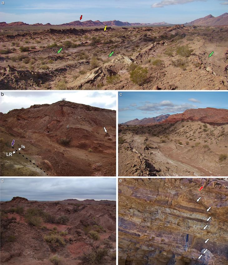
www.nature.com/scientificreports/
Figure 1. Geological map of the Bermejo Valley, La Rioja Province, northwestern Argentina. Yellow arrow
points to the Hoyada del Cerro Las Lajas palaeosite. Map generated and designed by one of the authors (L.E.F.)
using Corel Draw X5 software based on Google Earth imagery and our own observations and geological studies
in western La Rioja. Abbreviations: Co, Cerro (hill).
With one of the richest land biotas recorded worldwide, the Ischigualasto Formation of north-western Argen-
tina represents a unique “window” into Late Triassic biodiversity and evolution. This stratigraphic unit is well
known from the Ischigualasto Provincial Park (IPP), San Juan Province, with a fossil record composed of plants,
fishes, and most of the known tetrapod groups of the time, i.e., temnospondyls, rhynchosaurs, archosauriforms
(including dinosaurs), dicynodonts, and c ynodonts1–3. Radioisotopic dates of various vintages have given the
Ischigualasto fauna a temporal context, elevating its global significance in understanding the Triassic land eco-
systems, as well as the early evolution of d inosaurs3. Nevertheless, exposures of the Ischigualasto Formation
outside the IPP have only been briefly explored, delivering only subordinate fossil r ecords3. One exception is the
site known as Hoyada del Cerro Las L ajas4–6 in La Rioja Province, where the northernmost known outcrops of
the formation are exposed (Fig. 1; see also fig. 1 in Baczko et al.7). Explored by several expeditions starting in the
early sixties (see Historical background and motivation in the Supplementary Information), the fossil record of
the area appears meagre compared to that of the IPP and it has been described as “a poorly fossiliferous outcrop”
(p. 20 in Martínez et al.3), but includes key specimens, such as the holotypes of the ornithosuchid Venaticosuchus
rusconii and the probable ornithischian Pisanosaurus mertii.
Aiming to expand the fossil collections of the Hoyada del Cerro Las Lajas and to investigate the chron-
ostratigraphic context of previous fossil collections, our team explored the Cerro Las Lajas area in the course of
four expeditions from 2013 to 2019. Here, we report on more than 100 new tetrapod fossil specimens collected
form the Ischigualasto Formation at Cerro Las Lajas. Detail stratigraphy of its over 1,000 m-thick succession,
integrated with high-precision U–Pb zircon geochronology of three interlayered tuffs, provide a high-resolution
chronostratigraphic framework for the Ischigualasto Formation in the the Hoyada del Cerro Las Lajas. In this
context, we discuss palaeobiologic aspects of the Ischigualasto fauna and their implications for Late Triassic
tetrapod evolution.
Scientific Reports | (2020) 10:12782 | https://doi.org/10.1038/s41598-020-67854-1 2
Vol:.(1234567890)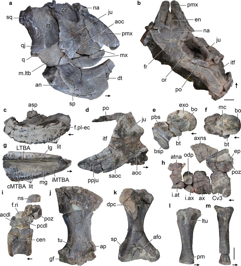
www.nature.com/scientificreports/

                            Figure 2.  Detailed stratigraphic column of the Hoyada del Cerro Las Lajas, including positions of tetrapod
                            fossils and dated tuff beds. Stratigraphy generated by some authors (A.A.S.D.R., J.R., M.E., and L.E.F.) using
                            Corel Draw X5 software based on our own observations and geological studies in the Hoyada del Cerro Las
                            Lajas.

                            Stratigraphy.       The section studied here is located in the “hoyada” (depression) positioned to the east of
                            Cerro Las Lajas, southwest of the town of Villa Unión in the western La Rioja Province (Fig. 1). The lowlands
                            of Cerro Las Lajas expose a 1,059 m-thick succession of predominantly greyish to tan siliciclastic rocks with an
                            average dip of 30° east that belong to the Ischigualasto Formation. Floodplain siltstones and (mottled) mud-
                            stones, channel sandstones and conglomerates, and a variety of tuffs and tuffaceous sediments form the bulk
                            of the formation. To the east, the top of the Ischigualsto succession is in transitional contact with the overlying
                            red sandstones of the Los Colorados Formation (Figs. 2 and 3). The base of the succession to the west is juxta-
                            posed against outcrops of the (younger) Los Colorados Formation via a steep, N–S trending fault. Discontinu-
                            ous exposures of the underlying, greenish-grey Los Rastros Formation rocks occur along the fault zone, with
                            complex stratigraphic relationships to the basal Ischigualasto strata at our measured section (see Supplementary
                            Information). Elsewhere in the area and away from the faults, the conformable contact between the Los Rastros
                            and Ischigualasto formations is well documented. These confirm the near-complete nature of the Ischigualasto
                            succession exposed at the Hoyada del Cerro Las Lajas.
                                The succession of the Ischigualasto Formation exposed at Cerro Las Lajas is subdivided into three sections
                            based on lithologic characteristics and alluvial depositional facies (Fig. 2) (see Supplementary Information).
                            The lower section extending from the contact with the Los Rastros Formation—at 11 m above base (mab) of
                            the profile in Fig. 2—to 310 mab was formed by a meandering fluvial system developed in a broad floodplain
                            depositional setting. This section exposes an isolated, greyish-white, tuff marker bed (‘Toba-2’) up to 2 m-thick
                            at 107 mab (i.e., 96 m above the base of the Ischigualasto Formation), which could provides a direct correlation
                            point to the Ischigualasto Formation at the IPP (Herr Toba bentonite; see below). The majority of the fossils
                            discovered in the Hoyada del Cerro Las Lajas occur above the tuff marker and are restricted to the lower section,
                            with the largest concentration of fossils (main fossil zone) occurring between 240 and 300 mab (Figs. 2 and 3).
                            The middle section (310–740 mab) marks an increase in accommodation together with fewer fossiliferous beds
                            and more pedogenic features indicative of higher humidity conditions. The upper section (740–1,070 mab)
                            is characterized by the transition from a meandering to a braided fluvial system concomitant with increased
                            pyroclastic activity in the form of prominent welded tuff (ignimbrite) beds (Fig. 3e and Fig. S3d–e). The upper
                            section is essentially currently fossil-free and extends to the base of the overlying Los Colorados Formation.
                            The well-recognized and conformable contact between the two formations provides another correlation point
                            between the Cerro Las Lajas succession and that of the IPP. See details of the stratigraphy of the Ischigualasto
                            Formation at Cerro Las Lajas in the Supplemental Information file.

                            Geochronology.       Three samples of tuff (volcanic ash) collected from various stratigraphic levels of the
                            Ischigualasto Formation (Fig. 2) at the Hoyada del Cerro Las Lajas (Sample Toba-2: 107 mab, Sample LL041219-
                            2: 160 mab, and Sample LL041219-6: 1,035 mab) were successfully analysed by the U–Pb isotopic method using

Scientific Reports |   (2020) 10:12782 |               https://doi.org/10.1038/s41598-020-67854-1                                            3

                                                                                                                                        Vol.:(0123456789)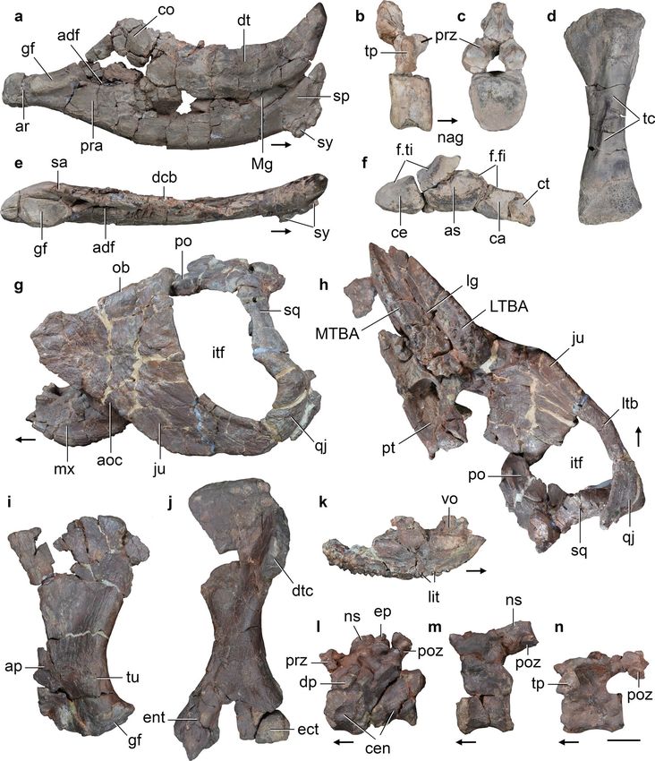
www.nature.com/scientificreports/
Figure 3. Ischigualasto Formation in the Hoyada del Cerro Las Lajas locality. (a) Panoramic view of Hoyada del
Cerro Las Lajas, with the Los Colorados Formation (red arrow) in the background, and the Teyumbaita (yellow
arrow) and Hyperodapedon (green arrows) main levels marked. (b) Contact between the Los Rastros Formation
(olive-green) and the Ischigualasto Formation (reddish-brown) marked by dotted line and purple arrow. White
arrow indicates a bentonite level at ca. 50 mab. (c) Strata typical of the Hyperodapedon biozone (ca. 150–200
mab) with the red Los Colorados Formation in the background. (d) Main level of Teyumbaita biozone, ca. 300
mab. (e) Interbedded tuffaceous mudstone and welded tuff (white arrows) at ca. 1,030 mab; red arrow indicates
welded tuff Sample LL041219-6 (see Supplementary Fig. 3).
the chemical abrasion isotope dilution thermal ionization mass spectrometry (CA-ID-TIMS) technique at the
Massachusetts Institute of Technology Isotope Laboratory. Details of sampled tuffs, analytical procedures, iso-
topic data, and age interpretations are given in the Supplementary Information. A Bayesian age-stratigraphic
model has been employed to interpolate statistically robust ages for the stratigraphic levels of interest.
Our new age model based on a set of high-precision U–Pb dates from the Hoyada del Cerro Las Lajas
(Fig. 4; Table 1 and Table S2) provides a reliable chronostratigraphic framework for the Ischigualasto Forma-
tion and its Late Triassic vertebrate fauna. Accordingly, the base of the Ischigualasto Formation is constrained
at 230.23 + 1.88/− 0.86 Ma. Caution must be made, however, in interpreting this age, as the limited outcrops of
the underlying Los Rastros Formation at the measured section have a complex stratigraphy due to faulting (for
further information, see above and the Supplementary Information). Our age model places the conformable
Ischigualasto-Los Colorados formation boundary with a high degree of confidence at 221.36 + 0.44/− 1.31 Ma.
Vertebrate fossils at the Hoyada del Cerro Las Lajas have so far been limited to the lower part of the
Ischigualasto Formation (lower and middle sections), from ~ 115 to 400 mab. The fossil record starts shortly
above the dated ‘Toba-2’ tuff marker at 229.25 ± 0.10/0.16/0.30 Ma; a stratigraphic relationship that mimics that
of the Herr Toba bentonite at IPP3, 8. The main fossil concentration, however, occurs between 240 and 300 mab
Scientific Reports | (2020) 10:12782 | https://doi.org/10.1038/s41598-020-67854-1 4
Vol:.(1234567890)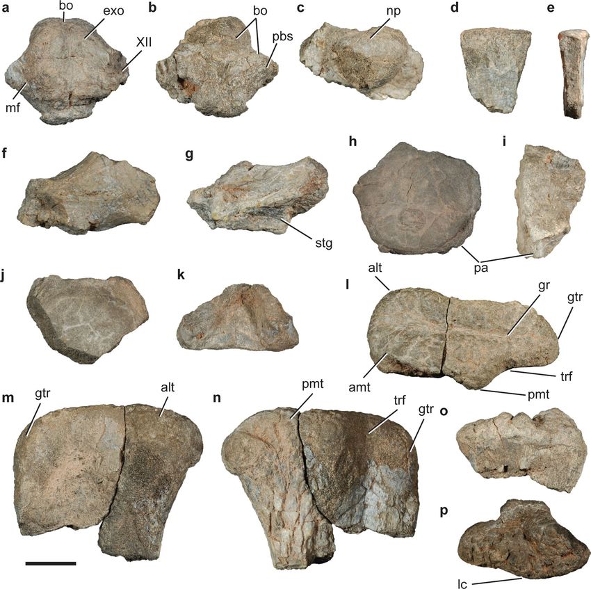
www.nature.com/scientificreports/
Figure 4. Date distribution plot of zircon CA-ID-TIMS U–Pb analyses from tuff samples of Cerro Las Lajas.
Black bars are individual zircon analyses used in weighted mean age calculation. Horizontal shaded band
represents the weighted mean 206Pb/238U date and its 95% confidence level internal uncertainty. Arrows point to
older (detrital) analyses that fall outside the plot area. See Table 1 and Table S2 for complete U–Pb data and for
details of calculated dates and their uncertianites.
206
Pb Uncertainty (2σ)
Sample Formation Height above base (m) Latitude (S) Longitude (W) 238
U date X Y Z MSWD n #
LL041219-6 Ischigualasto 1035 29°28’43.29" 68°20’12.81" 221.82 0.10 0.12 0.27 0.63 4 7
LL041219-2 Ischigualasto 160 29°28’42.53" 68°20’57.64" 228.97 0.22 0.23 0.33 1.9 4 8
Toba-2 Ischigualasto 107 29°28’33.00" 68°21’1.04" 229.25 0.10 0.16 0.30 1.2 6 6
Table 1. Summary of calculated U-Pb ages and their uncertainties. Notes: Latitude/Longitude relative
to WGS84 datum. X internal (analytical) uncertainty in the absence of all external or systematic errors. Y
incorporates the U-Pb tracer calibration error. Z includes X and Y, as well as the uranium decay constant
errors. MSWD mean square of weighted deviates. n number of analyses included in the calculated weighted
mean date, out of a total number of # analyses.
(Figs. 2 and 5), which is constrained in time between 228.10 + 0.72/− 1.50 and 227.61 + 1.04/− 1.79 Ma, based
on our Bayesian age model. As such, it roughly coincides with the extrapolated age of the Carnian-Norian stage
boundary, which has been indirectly calibrated at ~ 227 Ma (updated)9 or ~ 228.4 M a10.
Fossil provenance. Consistent with observations in José Bonaparte field notes and photographs (see details
in Supplementary Materials), the fossiliferous beds at the Hoyada del Cerro Las Lajas are mostly concentrated
in the lower third of the studied succession, between 100 and 400 mab (Figs. 2 and 5). The lowermost recovered
fossil corresponds to the aetosaur Aetosauroides scagliai (CRILAR-Pv 580) that occurs at 115 mab (Figs. 2 and
5). Further upsection, a specimen representing a new species of the rhynchosaur Hyperodapedon (CRILAR-Pv
585) was collected in brown mudstones, near a specimen of the cynodont Exaeretodon sp. (CRILAR-Pv 672) at
about 120–125 mab. Hyperodapedon sanjuanensis also occurred as isolated finds at 140 (CRILAR-Pv 646) and
175 (CRILAR-Pv 584) mab.
The zone of main fossil concentration (240–300 mab) starts with indeterminate rhynchosaur records (CRI-
LAR-Pv 675, 687) and H. sanjuanensis (CRILAR-Pv 583), with the uppermost record of that genus and species
(CRILAR-Pv 650) occurring near 260 mab. This is slightly below the lowest record, at 265 mab, of Teyumbaita
(CRILAR-Pv 592, 643) in the succession. The latter genus accounts for fourteen out of the twenty fossil specimens
identified at the genus level in this 60 m interval, also occurring as high as 350 mab. The range of Exaeretodon
spans across that main fossil zone, but with a lower abundance (three specimens). The only record of Paracroco-
dylomorpha (CRILAR-Pv 581) occurs at 260 mab, whereas that of Proterochampsa barrionuevoi (CRILAR-Pv
579) is at 290 mab (Fig. 5).
Few fossils have been collected from above 300 mab. Teyumbaita (CRILAR-Pv 597, 642) was found at 310
and 350 mab, along with Exaeretodon (CRILAR-Pv 649). This cynodont marks the stratigraphically highest
fossil identified to the genus level (CRILAR-Pv 644) at 400 mab, but an indeterminate rhynchosaur tooth plate
(CRILAR-Pv 662) was found at the same level. A complete list of specimens with their geographic provenance
and stratigraphic positions are provided in the Supplementary Material Table S1.
Petrographic analyses of rock matrices from legacy fossil collections without exact location data have been
used to infer their stratigraphic positions in the context of our new chronostratigraphic framework (see details
in Supplementary Materials). Accordingly, Pi. mertii is speculated to have been recovered from between 110
Scientific Reports | (2020) 10:12782 | https://doi.org/10.1038/s41598-020-67854-1 5
Vol.:(0123456789)
www.nature.com/scientificreports/
Figure 5. Stratigraphic column of the Ischigualasto Formation at the Hoyada del Cerro de Las Lajas with
ranges of occurrence of its main taxa concentrated between 100 and 350 m above base. Figure generated by
some authors (A.A.S.D.R., J.R., M.E., and L.E.F.) using Corel Draw X5 software based on our own geological
studies in the region.
and 180 mab in the lower section of the Ischigualasto Formation and below the main fossil zone. Similarly, the
positions of V. rusconii and Tri. romeri are inferred to be ~ 270 mab, within the main fossil zone (Fig. 5).
Systematic palaeontology.
Cynodontia Owen, 1 86111.
Eucynodontia Kemp, 1 98212.
Traversodontidae Huene, 1 93613 sensu Kammerer et al., 2 00814.
Exaeretodon Cabrera, 194315.
Exaeretodon sp.
Material. CRILAR-Pv 640, portion of right lower postcanine, portion of humeral distal condyle, and indetermi-
nate bone fragments. CRILAR-Pv 644, badly preserved partial skull with two upper postcanine teeth. CRILAR-Pv
647, partial postcranium, including several presacral, sacral, and caudal vertebrae, ribs, and most of the fore- and
Scientific Reports | (2020) 10:12782 | https://doi.org/10.1038/s41598-020-67854-1 6
Vol:.(1234567890)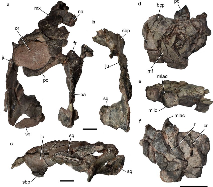
www.nature.com/scientificreports/
Figure 6. Exaeretodon sp. CRILAR-Pv 649, Ischigualasto Formation, Cerro Las Lajas, La Rioja Province. (a)
Partial skull with (b) its isolated right zygomatic arch in dorsal view, and (c) detail of the left zygomatic arch in
lateral view. Fragment of right dentary with two posterior postcanines in (d) lateral, (e) occlusal, and (f) medial
views. Abbreviations: bcp, base of coronoid process; cr, crypt; fr, frontal; ju, jugal; mf, masseteric fossa; mlac,
mesiolabial cusp; mlic, mesiolingual cusp; mx, maxilla; na, nasal; or, orbit; pa, parietal; pc, lower postcanine
tooth; po, postorbital; sbp, suborbital process of jugal; sq, squamosal; r, root. Scale bars: 2 cm.
hindlimbs, plus unprepared plaster jackets. CRILAR-Pv 649, partial skull in three parts bearing some badly pre-
served postcanine teeth, lacking most of the snout and braincase, posterior fragment of right dentary with two
postcanine teeth, right postcanine tooth, fragment of left dentary with one postcanine tooth and part of a root,
two partial incisors, two partial upper canines, and some indeterminate bone fragments. CRILAR-Pv 663, partial
skull still in plaster jacket. CRILAR-Pv 672, fragment of bone with natural cast of quadrangular tooth mixed
with rhynchosaur remains. Three other cynodont specimens (i.e., vertebral centra, long bones fragments) were
collected during the field works reported here, but their incompleteness hampers a more detailed classification.
Description. Of the specimens referred to Exaeretodon, the most complete cranial material belongs to CRILAR-
Pv 647 (Fig. 6), but it is badly preserved. It corresponds to a small-sized skull, with an estimated skull length
of ~ 20 cm. It is fragmented in three pieces, including: (1) part of the left portions of snout, orbit, the postorbital
bar, the zygomatic arch and the parietal crest (Fig. 6a); (2) part of the right zygomatic arch, with the anteroven-
tral edge of the orbit and the ventral base of the postorbital bar (Fig. 6b); and (3) part of the palate including
portions of maxillae with poorly preserved posterior postcanines, palatines, and pterygoids. As in other speci-
mens of Exaeretodon (e.g., MACN-Pv 18125; Bonaparte16), the zygomatic arch is deep with the anterior process
of the squamosal anteriorly extended almost to the level of the postorbital bar and there is a well-developed
descending process of the jugal below the orbit (Fig. 6c). The orbit (preserved only in the left side) is relatively
Scientific Reports | (2020) 10:12782 | https://doi.org/10.1038/s41598-020-67854-1 7
Vol.:(0123456789)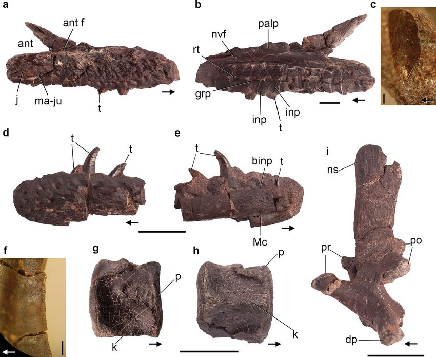
www.nature.com/scientificreports/
large, in accordance with the small, sub-adult size of the skull, and it faces mostly dorsally due to taphonomic
dorsoventral compression (Fig. 6a). The parietal crest is incomplete, but appears to have been well-developed.
The palatal area is poorly preserved, with the primary palate exposed lateral to the last postcanine teeth as in
other traversodontids16,17, due to the presence of an axially short secondary palate. The crown morphology of
the upper postcanines is difficult to access, but it is typically divided in two (labial and lingual) lobes, with an
occlusal basin, and an extensive “V” shaped shouldering.
The mandibular elements and lower dentition are also poorly preserved (Fig. 6d–f). A right dentary fragment
preserves two postcanines and the crypt for a posterior one (possibly in process of eruption at the moment of
death). That dentary preserves part of its straight ventral edge, the anterior end of the masseteric fossa, and the
base of the coronoid process (Fig. 6d). The masseteric fossa seems to be well-developed and reaches the level of
the last functional postcanine. The base of the coronoid process is robust and laterally covers the crypt in the
area where postcanines are added to the tooth row during ontogeny (Fig. 6f). Another dentary fragment only
holds a left postcanine and part of the root of the posterior one.
The lower postcanine teeth of CRILAR-Pv 647 have the typical shape observed in Exaeretodon (e.g., MACN-
Pv 18125)18,19. They are subquadrangular in occlusal view, with a deep central occlusal basin surrounded by four
cusps placed at each corner (Fig. 6e). The mesiolabial cusp is the largest and both mesial cusps (mesiolabial and
mesiolingual) are in a higher position relative to the distal ones, forming a mesial cutting edge slightly apico-
distally inclined (Fig. 6e,f). The crown is relatively low and surrounded by a thick layer of enamel, which does
not cover the occlusal surface. The limit between crown and root is not marked by a neck, but by the edge of the
enamel layer. The root of each tooth is conical and tapers toward the apex. The apex is distally curved (Fig. 6f)
as typical of Exaeretodon (UFRGS-PV 1096-T)20, as the result of the tooth replacement.
There are two partially preserved canines of small size in CRILAR-Pv 647, which are interpreted as lower
elements. Their crown is sub-conical, slightly labio-lingually flattened, and well curved distally. The incisors are
represented by three crown fragments of small size. One is attached to the palate fragment and the other two are
isolated. They have the labial surface strongly convex and the lingual one flat to slightly convex, with the crown
slightly curved lingually.
Comments. The specimens of Exaeretodon from the Hoyada del Cerro Las Lajas are fragmentary and poorly
rovince2, 3,16. Only CRI-
preserved in comparison to those discovered at the Hoyada de Ischigualasto, in San Juan P
LAR-Pv 647, which is still under preparation (and will be described elsewhere), includes a well preserved post-
cranium, but apparently lacks cranial elements. Moreover, cynodonts are not as abundant as archosauromorphs
(mainly rhynchosaurs) in the area and are taxonomically restricted so far to the traversodontid Exaeretodon.
Exaeretodon was first d escribed25 based on specimens collected in the Hoyada de Ischigualasto. After several
taxonomical revisions21,22, Ex. argentinus is regarded as the only valid species known from the Ischigualasto
Formation. The genus is also recognized in the Hyperodapedon Assemblage-Zone of the Candelária Sequence,
Santa Maria Formation, Brazil, represented by Ex. riograndensis17,19, and in the lower Maleri Formation of India,
represented by Ex. statisticae23.
Presently, the Hoyada del Cerro Las Lajas specimens can be clearly assigned to the genus Exaeretodon.
Nonetheless, the lack of complete skulls precludes the evaluation of features that may distinguish between Ex.
argentinus and Ex. riograndensis, such as the prootic crests and the postcanine variation along ontogeny. Also,
amongst the specimens traditionally referred to Exaeretodon from the Hoyada de Ischigualasto, two different
morphotypes were recently briefly c ommunicated24, one of which seems to be closely related to the recently
described traversodontid Siriusgnathus niemeyerorum from the Candelária Sequence of Rio Grande do Sul,
Brazil25. The specimens collected in La Rioja do not have the combination of features observed in Sir. niemeyero-
rum (CAPPA/UFSM 0032), e.g., a very reduced suborbital process of the jugal25. Consequently, the traversodontid
material from the Hoyada del Cerro Las Lajas fits better with the genus Exaeretodon and only further material
will allow elucidating their taxonomy at a specific level.
Rhynchosauria Osborn, 1 90326 sensu Ezcurra27.
Hyperodapedontinae Lydekker, 1 88528 sensu Langer & S chultz29.
Hyperodapedon Huxley, 1 85930.
Hyperodapedon sanjuanensis Sill, 197031.
Material. CRILAR-Pv 583, fairly complete left hemimandible, left tibia, probable distal tarsal, and probable left
metacarpal (Fig. 7a, d, e). CRILAR-Pv 584, left maxilla without anterior portion, right dentary with damaged
posterior end, left articular and partial prearticular, an anterior dorsal vertebra, two dorsal centra, a fragment of
probable caudal centrum, a right and a left postzygapophysis, articulated left centrale, astragalus, and calcaneum,
two distal tarsals, proximal end of right metatarsals II and III, five non-ungual phalanges, an ungual phalanx
lacking its distal end, and indeterminate bone fragments (Fig. 7b, c, f). CRILAR-Pv 646, partial left dentary,
missing its posterior edge and anterior half, and left tibia. CRILAR-Pv 650, right dentary and partially prepared
partial cranium.
Description. The occlusal surface of the maxillary tooth plate is subdivided by a single, medially displaced
longitudinal groove, as occurs in H. mariensis (MCN 1867-PV), H. huxleyi32, the morphotype 1 of H. tikiensis33,
Supradapedon stockleyi34, Isalorhynchus genovefae35, unnamed hyperodapedontines from Zimbabwe36 and Nova
Scientific Reports | (2020) 10:12782 | https://doi.org/10.1038/s41598-020-67854-1 8
Vol:.(1234567890)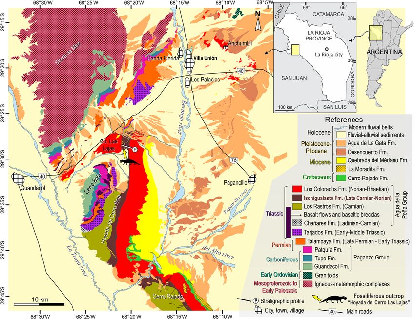
www.nature.com/scientificreports/

                            Figure 7.  Hyperodapedon spp. Selected bones of specimens (a, d, e) CRILAR-Pv 583, (b, c, f) CRILAR-Pv 584, and (g–n) CRILAR-Pv
                            585 referred to (a–f) H. sanjuanensis, and (g–n) Hyperodapedon sp. nov. collected in the outcrops of the Ischigualasto Formation
                            immediately east to Cerro Las Lajas. (a, e) Left hemimandible, (b, c) anterior dorsal vertebra, (d) left tibia, (f) proximal tarsals, (g, h)
                            partial skull, (i) left scapula, (j) left humerus, (k) left maxilla, (l) two articulated anterior postaxial cervical vertebrae, (m) posterior
                            cervical vertebra, and (n) anterior-middle dorsal vertebra in (a, k) medial, (b) right lateral, (c, d, f, j) anterior, (e) dorsal, (g, l–n)
                            left lateral, (h) ventral, and (i) lateral views. Arrows indicate anterior direction. Abbreviations: adf, adductor fossa; aoc, anguli oris
                            crest; ap, acromial process; ar, articular; as, astragalus; ca, calcaneum; ce, centrale; cen, centra; co, coronoid; ct, calcaneal tuber; dcb,
                            dentary cutting blade; dp, diapophysis; dt, dentary; dtc, deltopectoral crest; ect, ectepicondyle; ent, entepicondyle; ep, epipophysis; f.fi,
                            facet for fibula; f.ti, facet for tibia; gf, glenoid fossa; itf, infratemporal fenestra; ju, jugal; lg, longitudinal groove; ltb, lower temporal
                            bar; LTBA, lateral tooth bearing area; Mg, Meckelian groove; mx, maxilla; MTBA, medial tooth bearing area; nag, non-articular gap;
                            ns, neural spine; ob, border of orbit; po, postorbital; poz, poszygapophysis; pra, prearticular; prz, prezygapophysis; pt, pterygoid; qj,
                            quadratojugal; sa, surangular; sp, splenial; sq, squamosal; sy, symphysis; tc, tibial crest; tp, transverse process; tu, tuberosity. Scale bar:
                            2 cm.

Scientific Reports |   (2020) 10:12782 |                      https://doi.org/10.1038/s41598-020-67854-1                                                                   9

                                                                                                                                                                    Vol.:(0123456789)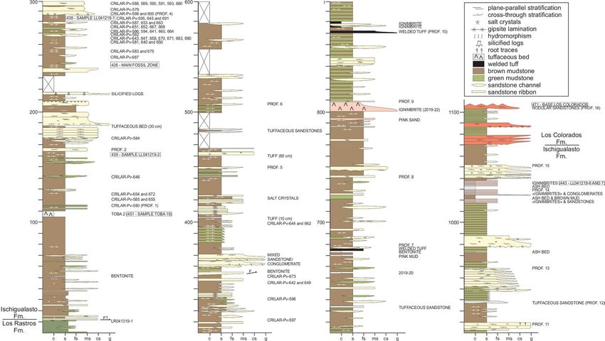
www.nature.com/scientificreports/
S cotia37, and other specimens of H. sanjuanensis38. By contrast, the single longitudinal groove is approximately
centred on the tooth plate in H. gordoni39 and an indeterminate hyperodapedontine from the Ischigualasto
Formation40, whereas there are two longitudinal grooves in H. huenei29 and Teyumbaita sulcognathus41. The
lateral tooth bearing area (LTBA) has five longitudinal tooth rows and the crowns in the two medialmost rows
(L1 and L2) are worn to the root, resembling the condition of most hyperodapedontines (e.g. H. mariensis,
UFRGS-PV 0149T, 0408T; H. huxleyi32). By contrast, only one or two longitudinal tooth rows are present in the
LTBA of North American hyperodapedontines (Wyoming form42; Nova Scotia form37), Te. sulcognathus41, and I.
genovefae35. The number of rows in the medial tooth bearing area (MTBA) and presence of lingual teeth cannot
be determined in CRILAR-Pv 584 because of damage.
The dentary forms more than half of the hemimandible, has a tapering anterior end, and does not form part
of the mandibular symphysis, which are character-states retained by all h yperodapedontines43–45 (Fig. 7a, e). The
dentary has a single, transversely thin cutting blade with one row of mesiodistally compressed teeth. There is
no lingual teeth and no medially bulged area in the dentaries of CRILAR-Pv 583, 584, 646, 650, as typical of H.
sanjuanensis38 and also reported for an unnamed hyperodapedontine from Nova Scotia37. By contrast, all other
hyperodapedontines have lingual teeth on the dentary34,38. A well-developed coronoid prominence is formed by
the dentary, surangular, and coronoid bones. A deep and lateroventrally opened posterior surangular foramen
is located at level with the glenoid fossa. The retroarticular process is short and its dorsal surface is damaged in
CRILAR-Pv 583.
The anterior dorsal vertebra possesses a spool-shaped, taller than long centrum that lacks a ventral keel
(Fig. 7b, c). The neural spine is restricted to the posterior two-thirds of the neural arch and does not extend
between the bases of the prezygapophyses. The tibia has transversely expanded proximal and distal ends (Fig. 7d).
The shaft possesses a well-developed, proximolaterally to distomedially oriented tibial crest on its anterior surface,
as occurs in other r hynchosaurids43,46,47. The distal articular surface of the bone is transversely convex and slants
proximolaterally. The proximal row of tarsals is composed of a centrale, astragalus, and calcaneum, as occurs in
other rhynchosaurs48 (Fig. 7f). The centrale is not fused to the astragalus and its proximal surface extensively
contributes to the tibial facet. The proximal surface of the astragalus has tibial and fibular facets separated from
one another by a non-articulating gap. The posterior surface lacks a posterior groove and the autapomorphic
transverse boss present in Te. sulcognathus46. The medial half of the proximal surface of the calcaneum is occu-
pied by the fibular facet and the lateral half of the bone is developed as a laterally projected calcaneal tuber. The
calcaneal tuber is anteroposteriorly narrower than proximodistally tall and the latter axis is rotated approximately
45° from the proximodistal plane of the proximal tarsus.
Comments. These specimens can be referred to Hyperdapedontinae because of the presence of the following
synapomorphies34: mandible dorsoventral depth > 0.25 times its total length (CRILAR-Pv 583); dentary with
mesiodistally compressed teeth (all specimens); posteriormost dentary teeth on the posterior half of the lower
jaw (CRILAR-Pv 583); and astragalus with centrale facet greater than the tibial facet (CRILAR-Pv 584, although
this condition is unknown in the immediate sister-taxa to Hyperodapedontinae). In addition, CRILAR-Pv 584
can be included in the Hyperodapedon clade44 because of the presence of a maxillary tooth plate with more than
two tooth rows in the LTBA and four or more tooth rows on its anterior half. Within Hyperodapedontinae, these
specimens can be referred to H. sanjuanensis because the absence of lingual teeth in the dentary has been consid-
ered an autapomorphy of this s pecies38. Nevertheless, the unnamed Nova Scotia hyperodapedontine apparently
also lacks lingual dentary t eeth37 and this feature may be an apomorphy of a more inclusive clade of hyperoda-
pedontines (i.e. H. sanjuanensis + North American forms34). We preferred here to maintain this character-state
as diagnostic of H. sanjuanensis until more information of the Nova Scotia hyperodapedontine is published. In
any case, the maxillary tooth plate of CRILAR-Pv 584 differs from those of the Nova Scotia hyperodapedontine
in the presence of a higher number of tooth rows in the LTBA and, at least for this specimen, such combination
of character-states still supports its referral to H. sanjuanensis.
Hyperodapedon sp. nov.
Material. CRILAR-Pv 585, articulated partial left side of cranium missing lacrimal, prefrontal, anterior region
of palate and almost entirely the skull table; fragment of right maxilla; partial braincase; at least 14 postaxial
cervical and anterior-middle dorsal vertebrae; several ribs and gastralia; both scapulae; left humerus; distal end
of right humerus; and multiple indeterminate bone fragments (Fig. 7g–n).
Description. The overall morphology of the skull of CRILAR-Pv 585 resembles that of other hyperodape-
dontine rhynchosaurs in the presence of a ventral border of the orbit positioned dorsal to the mid-height of
the infratemporal fenestra, a massive and anterodorsally-to-posteroventrally sloping jugal, and a closed lower
temporal bar (e.g. I. genovefae45; H. mariensis, MCN 1867-PV; H. huenei29; H. sanjuanensis, MACN-Pv 18185;
Te. sulcognathus41) (Fig. 7g,h). The infratemporal fenestra is kidney-shaped, with a notched posterior border.
This outline is a result of the strongly concave anterior margin of the ascending process of the quadratojugal, as
occurs in H. huenei29 and H. huxleyi32. By contrast, this margin is approximately straight in the holotype of H.
sanjuanensis (MACN-Pv 18185), Te. sulcognathus41, and I. genovefae45, and convex in H. gordoni39.
Scientific Reports | (2020) 10:12782 | https://doi.org/10.1038/s41598-020-67854-1 10
Vol:.(1234567890)www.nature.com/scientificreports/
The occlusal surface of the maxillary tooth plate of CRILAR-Pv 585 is divided into equally broad LTBA and
MTBA by a longitudinal groove (Fig. 7h), as also occurs in H. gordoni39 and an indeterminate hyperodapedon-
tine from the Ischigualasto Formation40. By contrast, the maxillary tooth plate has a broader LTBA in all other
hyperodapedontines with a single groove34. The maxillary tooth plate of CRILAR-Pv 585 also differs from those
of Te. sulcognathus, H. huenei, and the morphotype 2 of H. tikiensis, which possess two longitudinal grooves that
define a third, central tooth bearing a rea29, 33,41. CRILAR-Pv 585 has four longitudinal tooth rows at the posterior
end of the LTBA and three rows in the MTBA. In addition, there is a row of six lingual teeth, well-spaced from
one another and located on the medial surface of the maxilla, dorsally to the MTBA (Fig. 7k), resembling the
condition in H. huenei29 and a Zimbabwean h yperodapedontine36. However, CRILAR-Pv 585 differs from these
two forms in the presence of lingual tooth crowns that are mainly oriented ventrally rather than perpendicular
to the occlusal surface, and from all rhynchosaurs in the presence of lingual teeth restricted to the anterior half
of the tooth plate.
The jugal forms the ventral border of the orbit and bears an anterodorsally-to-posteroventrally oriented anguli
oris crest that overhangs laterally the maxilla (Fig. 7g). The lateral surface of the jugal is coarsely ornamented
by low ridges and bulges on its main body and striations adjacent to the orbital edge. No secondary anguli oris
crest is present on the main body of the jugal, contrasting with Te. sulcognathus41 and I. genovefae45. The lateral
surface of the posterior process of the jugal of CRILAR-Pv 585 lacks the deep and posterodorsally well-rimmed
depression located on the ventral half of the base of this process in H. huxleyi (ISIR 01), H. huenei (UFRGS-PV
0132T), and referred specimens of H. mariensis (UFRGS-PV 0149T). The posterior process of the jugal forms
the entire ventral border of the infratemporal fenestra in CRILAR-Pv 585, as occurs in H. huenei29 and Te.
sulcognathus41. By contrast, the anterior process of the quadratojugal contributes to the ventral border of the
opening in I. genovefae45, the holotype of H. sanjuanensis (MACN-Pv 18185), H. mariensis (UFRGS-PV 0149T),
H. gordoni39, and H. huxelyi (ISIR 01).
The palatine of CRILAR-Pv 585 contacts the ectopterygoid posterolaterally and, as a result, excludes the max-
illa from the border of the infraorbital foramen. The pterygoid possesses a cup-shaped, dorsomedially projected
process that received the basipterygoid process of the parabasisphenoid. This facet indicates the presence of a
basal articulation two times dorsoventrally taller than transversely broad, as occurs in Te. sulcognathus41 and
other species of Hyperodapedon45.
The basioccipital possesses a long occipital neck and basal tubera broadly separated from one another. The
exoccipital contacted its counterpart on the floor of the endocranial cavity, as occurs in several other hypero-
dapedontines (e.g. H. huenei, UFRGS-PV 0132T; H. mariensis, UFRGS-PV 0149T; H. sanjuanensis, MACN-Pv
18185), but contrasting with the absence of such contact in Te. sulcognathus41. The occipital surface of the base of
the paroccipital process possesses a ventrally well-defined depression on its dorsal half, resembling a condition
previously reported as autapomorphic of Te. sulcognathus41.
The postaxial cervical vertebrae have a spool-shaped centrum that lack a ventral keel and possess a shallow
depression on its dorsolateral surface (Fig. 7l). By contrast, the anterior-middle cervical vertebrae of Te. sulcog-
nathus have a median ventral keel46. There is a tall, crest-shaped (i.e. conical) epipophysis on the dorsal surface
of the postzygapophysis, perhaps absent only in the posteriormost cervical vertebrae (Fig. 7m). The neural spine
is restricted to the posterior half of the neural arch. The centra of the anterior and middle dorsal vertebrae are
generally longer and slightly more transversely compressed than the cervical centra (Fig. 7n). The anterior-
middle dorsal neural arches possess comma-shaped transverse processes in cross-section and lack laminae. The
postzygapophyses lack an epipophysis.
The scapula is anteroposteriorly expanded at both the proximal and distal ends (Fig. 7i). The very base of the
acromial process is thick, ridge-like and distinctly laterally raised, resembling the condition in most hyperoda-
pedontines (e.g., H. sanjuanensis, MACN-Pv 18185; H. huxleyi, ISIR 01; H. tikiensis33). By contrast, this process
is sub-circular and blunt in Te. sulcognathus46. The scapular blade has distinctly divergent anterior and posterior
margins, as occurs in most hyperodapedontines (e.g., H. huxleyi32; H. mariensis, MCN 1867-PV), but the scapular
blade possesses a tab-like, poorly developed posterior expansion in Te. sulcognathus (UFRGS-PV 0232T). The
proximal and distal ends of the humerus are distinctly transversely expanded and their main axes rotated approxi-
mately 40° from one another (Fig. 7j). The deltopectoral crest is mainly anteriorly oriented. The distal end has
a very deep, subtriangular, and concave anterior fossa and a shallower and more proximally extended posterior
fossa. The lateral surface of the distal end possesses a deep longitudinal ligament groove (= ectepicondylar groove)
that is anteriorly delimited by a supinator ridge, resembling the condition in other hyperodapedontines (e.g., H.
tikiensis33; H. huxleyi32; H. gordoni39; H. sanjuanensis, MACN-Pv 18185; Te. sulcognathus, UFRGS-PV 0232 T).
Comments. CRILAR-Pv 585 is identified as a hyperodapedontine rhynchosaur because of the presence of
the following synapomorphies of the clade34: jugal without an elevated orbital rim; fully closed lower temporal
bar; anguli oris crest extended onto the anterior process of the jugal, but not the maxilla; maxilla well laterally
overlapped by the jugal; maxillary tooth plate with cushion-shaped LTBA; and maxillary teeth with conical and
‘pyramidal’ crowns. In addition, CRILAR-Pv 585 shares with other members of the Hyperodapedon clade the
following synapomorphies34: maxillary tooth plate with more than two tooth rows in the MTBA; maxillary tooth
plate with four or more tooth rows of occlusal teeth on its anterior half; parabasisphenoid with a basipterygoid
process wider than long (inferred from the shape of the basal articulation on the pterygoid); and postaxial cer-
vical postzygapophyses with crest-shaped epipophysis. Among hyperodapedontines, CRILAR-Pv 585 differs
from other taxa in the presence of an autapomorphic row of ventrally oriented lingual teeth restricted to the
anterior half of the maxillary tooth plate. This new species will be formally named and described in detail in a
future contribution.
Hyperodapedon sp.
Scientific Reports | (2020) 10:12782 | https://doi.org/10.1038/s41598-020-67854-1 11
Vol.:(0123456789)www.nature.com/scientificreports/
Material. CRILAR-Pv 582, isolated right partial maxilla, fragment of right occlusal blade of right dentary (origi-
nally in occlusion with the maxilla), and an indeterminate partial bone.
Description and comments. The maxilla of CRILAR-Pv 582 is represented by the middle portion of the tooth
plate. The occlusal surface of the bone is subdivided by a single, medially displaced longitudinal groove, as occurs
in H. sanjuanensis, H. mariensis, the holotype of H. tikiensis, H. huxleyi, Su. stockleyi and I. genovefae32,34,38,49. By
contrast, the longitudinal groove is centred on the tooth plate in H. gordoni, the above Hyperodapedon sp. nov.,
and an indeterminate hyperodapedontine from the Ischigualasto F ormation40. The longitudinal groove narrows
anteriorly, resembling the condition in some other hyperodapedontines (e.g. UFRGS-PV 0149T, 0408T). The
longitudinal groove bows slightly laterally and is very deep, with a V-shaped cross-section. The LTBA possesses
four longitudinal tooth rows and the MTBA has three rows. The presence of more lateral longitudinal tooth
rows than medial ones is consistent with the condition in H. sanjuanensis (MACN-Pv 18185, MCP-PV 1693),
H. mariensis (UFRGS-PV 0149T, 0408T), H. huxleyi (ISIR 01), and the holotype of H. tikiensis33. By contrast,
the MTBA has more longitudinal tooth rows than the lateral one in H. gordoni39, Su. stockleyi (SAM-PK-11705),
and the unnamed hyperodapedontine from Nova S cotia37. The preserved L1 and M1 tooth crowns are strongly
worn on the walls of the longitudinal groove, exposing the root in coronal section. The teeth of both tooth-
bearing areas are relatively small and closely packed, as occurs in most hyperodapedontines with the exception
of I. genovefae35,45. The preserved tooth crowns of both tooth-bearing areas have a circular cross-section, but it
is not possible to determine the presence of pyramidal teeth because the posterior region of the tooth plate is
not preserved. The preserved portion of the medial surface of the bone lacks lingual teeth, but it not possible
to determine if they were present more posteriorly or anteriorly in the tooth plate. The fragment of dentary of
CRILAR-Pv 582 possesses a V-shaped cross-section as a result of the presence of a transversely thin and sharp
occlusal cutting blade. It is not possible to observe teeth in the dentary fragment.
The presence of a single longitudinal groove and more than two tooth rows in the MTBA of the maxillary
tooth plate of CRILAR-Pv 582 allows referring this specimen to the Hyperodapedon clade34. Its maxilla dif-
fers from those of hyperodapedontines with a centrally located single longitudinal groove (i.e. H. gordoni, an
hyperodapedontine from the Ischigualasto Formation, and the above Hyperodapedon sp. nov.), more tooth rows
in the MTBA than in the LTBA (Su. stockleyi), less than two tooth rows in the LTBA (I. genovefae, the unnamed
hyperodapedontines from Nova Scotia and Wyoming) or with two longitudinal grooves (H. huenei and Te. sul-
cognathus). Instead, the morphology of CRILAR-Pv 582 is congruent with that of H. sanjuanensis, H. mariensis,
H. huxleyi, the holotype of H. tikiensis, and the unnamed Zimbabwean hyperodapedontine.
Teyumbaita Montefeltro, Langer & Schultz, 2 01041.
Teyumbaita sp. nov.
Material. CRILAR-Pv 586 (Fig. 8), partial cranium lacking most of the skull roof and right side, partial left
hemimandible, a median segment of the right hemimandible, atlas, axis and third cervical vertebra, four middle-
posterior cervical vertebrae, a fragment of humeral shaft, a probable metacarpal lacking the distal end, eight
non-ungual phalanges, and indeterminate bone fragments (Fig. 8h). CRILAR-Pv 587, partial left premaxilla,
right maxilla, left nasal, basicranium, right atlantal neural arch, five postaxial cervical vertebrae, a posterior dorsal
vertebra, right scapula, coracoid, clavicle, humerus, ulna and femur, proximal and distal ends of fibula, a right
metacarpal probably from digit II, two non-ungual and two ungual phalanges, and several ribs (Fig. 8c,e–g,i).
CRILAR-Pv 588, partial left maxilla lacking its lateral edge and anterior tip, fragment of the medial dentary crest
and indeterminate bone fragments. CRILAR-Pv 595, partial skull with almost complete right side and missing
the left orbital and temporal regions, braincase and post-dentary bones with exception of the angulars (Fig. 8a,
b). CRILAR-Pv 642, partial left maxilla lacking most of the ascending process, anterior tip, and occlusal surface
of the LTBA, and—still in the field—partial postcranium. CRILAR-Pv 643, middle third of right maxilla, six
postaxial centra, ventral end of clavicle, proximal and distal ends of tibia and indeterminate bone fragments of
bones of a very small-sized individual. CRILAR-Pv 645, articulated right quadrate, squamosal and paroccipital
process, probable partial left postfrontal, partial right parietal and pterygoid, partial parabasisphenoid, posterior
two-thirds of left dentary, articulated posterior half of right surangular, prearticular and fragment of articular,
six postaxial cervical vertebrae, two articulated probable anterior dorsal vertebrae, two dorsal or anterior caudal
vertebrae, right scapula and humerus, partial scapular blade of another right scapula, multiple fragments of
ribs and gastralia and several indeterminate bone fragments. This specimen consists of at least two individuals
found in association with CRILAR-Pv 595. CRILAR-Pv 651, partial left premaxilla and dentary, posterolateral
corner of right maxilla, partial right jugal, postorbital, quadrate, pterygoid and ectopterygoid, axial centrum,
at least three postaxial centra, right scapula, humerus, ulna and radius, fragment of left scapula, distal third of
metacarpal or metatarsal, three non-ungual phalanges, several rib fragments, and several probable cranial and
postcranial indeterminate bone fragments (Fig. 8d,j–m).
Description. The skull of Teyumbaita sp. nov. is broader than long, with a jugal representing the main compo-
nent of its lateral surface, a fully closed lower temporal bar, and a dorsolaterally facing orbit (Fig. 8a,b), as occurs
yperodapedontines41,44,45,50. The nasals form a straight posterodorsal border in the single external naris,
in other h
contrasting with the diagnostic notched border at the median line present in Te. sulcognathus41. The lateral surface
of the main body of the jugal is deeply concave and lacks a secondary anguli oris crest (CRILAR-Pv 595, 651),
contrasting with I. genovefae45 and the holotype and a referred specimen of Te. sulcognathus41. Nevertheless, the
Scientific Reports | (2020) 10:12782 | https://doi.org/10.1038/s41598-020-67854-1 12
Vol:.(1234567890)www.nature.com/scientificreports/
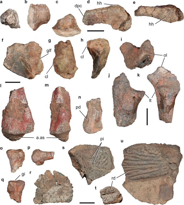
Figure 8. Teyumbaita sp. nov. Selected bones of specimens (a, b) CRILAR-Pv 595, (c, e, g–i) CRILAR-Pv 587, (d, j–m)
CRILAR-Pv 651, and (h) CRILAR-Pv 586 collected in the outcrops of the Ischigualasto Formation immediately east to
Cerro Las Lajas. (a, b) Skull, (c, g) right maxilla, (d) articulated right jugal and postorbital; (e, f) articulated exoccipitals,
basioccipital and parabasisphenoid, (h) articulated atlas, axis and third cervical vertebra, (i) posterior dorsal vertebra,
(j) right scapula, (k) right humerus, (l) right ulna, and (m) right radius in (a) right lateral, (b, f) dorsal, (c) medial, (d, j,
l, m) lateral, (e, h, i) left lateral, and (k) anterior views. Arrows indicate anterior direction. Abbreviations: acdl, anterior
centrodiapophyseal lamina; afo, anterior fossa; aoc, anguli oris crest; ap, acromial process; asp, ascending process; atna,
atlantal neural arch; ax, axis; axns, axial neural spine; bo, basioccipital; bsp, basipterygoid process; bt, basal tuber; cen,
centrum; cMTBA, central tooth bearing area; Cv3, third cervical vertebra; dpc, deltopectoral crest; ep, epipophysis; exo,
exoccipital; f.ri, facet for rib; f.pl-ec, facet for palatine-ectopterygoid; gf, glenoid fossa; i.at, intercentrum of atlas; i.ax,
intercentrum of axis; iMTBA, inner medial tooth bearing area; itf, infratemporal fenestra; ju, jugal; lg, lateral groove; lit,
lingual teeth; LTBA, lateral tooth bearing area; ltu, lateral tuber; mc, median contact between exoccipitals; mg, medial
groove; ns, neural spine; odp, odontoid process; pbs, parabasisphenoid; pcdl, posterior centrodiapophyseal lamina; pm,
process for muscle attachment; po, postorbital; poz, postzygapophysis; ppju, posterior process of jugal; saoc, secondary
anguli oris crest; sp, supinator process; tu, tuberosity. Scale bar: 2 cm.
Scientific Reports | (2020) 10:12782 | https://doi.org/10.1038/s41598-020-67854-1 13
Vol.:(0123456789)www.nature.com/scientificreports/
base of the posterior process of one specimen (CRILAR-Pv 651) has a thick, rugose ridge that does not extend
further anteriorly on the bone (Fig. 8d: saoc). A very similar structure was interpreted as a posteriorly restricted
secondary anguli oris crest in another referred specimen of Te. sulcognathus41. The presence of this latter ridge
and the absence of a lateral depression at the base of the posterior process differs from the condition present in
species of Hyperodapedon (e.g. H. sanjuanensis, MACN-Pv 18185; H. huenei29; H. huxleyi, ISIR 01; H. gordoni39;
H. mariensis, UFRGS-PV 0149T). The anterior margin of the ascending process of the quadratojugal is slightly
convex in lateral view, as occurs in Te. sulcognathus41, but contrasting with the concave margin of H. huenei29
and Hyperodapedon sp. nov. (CRILAR-Pv 585).
The maxillary tooth plate of the currently largest specimen of Teyumbaita sp. nov. has a transverse width of
47.6 mm (CRILAR-Pv 587), which is ca. 84% smaller than the largest specimen of Te. sulcognathus (transverse
width = 56.7 mm, UFRGS-PV 290T). The maxillary tooth plate possesses two longitudinal grooves that define
three tooth bearing areas (Fig. 8g), as occurs in Te. sulcognathus41, H. huenei29, the morphotype 2 of H. tikiensis33,
and several non-hyperodapedontine rhynchosaurids43,51. The lateral groove is considered homologous to the
single longitudinal sulcus of most hyperodapedontines (Chatterjee52 and subsequent authors). As a result, the
MTBA is subdivided into a central medial tooth bearing area (cMTBA) and an inner medial tooth bearing area
(iMTBA). The entire MTBA is broader than the LTBA, as is the case in Te. sulcognathus and H. huenei29,41, but
it contrasts with the distinctly broader LTBA present in both morphotypes of H. tikiensis33. Both longitudinal
grooves converge anteriorly at the anterior third of the maxilla of Teyumbaita sp. nov., resembling the condition
in Te. sulcognathus and the morphotype 2 of H. tikiensis33,41, but the medial groove is restricted to the posterior
third of the tooth plate in H. huenei29.
The number of tooth rows in the LTBA, cMTBA, and iMTBA show minor variation among preserved speci-
mens. The LTBA and cMTBA have two longitudinal tooth rows posteriorly, and the iMTBA has two (CRILAR-Pv
587, 642, 643) or three to four (CRILAR-Pv 588) tooth rows on the posterior half of the tooth plate. The arrange-
ment of these occlusal tooth rows resembles that of Te. sulcognathus41. In addition, there is a row of well-spaced
lingual teeth immediately medial to the cushion-shaped iMTBA (Fig. 8c,g). This row of lingual teeth extends from
the posterior end up to the posterior third (CRILAR-Pv 588, 642) or approximately the mid-length (CRILAR-Pv
587, 595) of the tooth plate and is composed of up to eleven teeth (CRILAR-Pv 587). By contrast, Te. sulcogna-
thus possesses a single lingual tooth positioned on the posteromedial corner of the b one41. The lingual teeth of
Teyumbaita sp. nov. are ventrally oriented, resembling the condition in Hyperodaperdon sp. nov. (CRILAR-Pv
585), but contrasting with H. huenei29 and the Zimbabwean h yperodapedontine36.
In the braincase, the ventral ends of the exoccipitals contact their counterparts on the floor of the endocranial
cavity (Fig. 8e,f), as occurs in most other hyperodapedontines (see above), but contrasting with the absence
of such contact in Te. sulcognathus41. The parabasisphenoid has an oblique, posterodorsally to anteroventrally
oriented, main axis in lateral view. The basipterygoid processes are dorsoventrally taller than transversely broad,
as in other hyperodapedontines with the exception of I. genovefae45.
The dentary has a lateral cutting blade and a lower and transversely thicker medial cutting blade, as occurs
in Te. sulcognathus and non-hyperodapedontine r hynchosaurids41. Multiple lingual teeth are located on the top
of the medial blade, immediately medial to it, and on a medially bulged border, being disposed in a crowded
pattern, resembling the condition in Te. sulcognathus41, but differing from the well-spaced lingual teeth of H.
huenei29 and several other hyperodapedontines (e.g. H. mariensis, MCN 1867-PV).
The morphology of the atlas (Fig. 8h) closely resembles that of Te. sulcognathus46 and other hyperodapedon-
tines (e.g. H. huxleyi32). The dorsal margin of the neural spine of the axis possesses a strongly convex central
portion in lateral view that becomes concave at the level of the postzygapophyses, resembling the condition in H.
gordoni39. By contrast, the posterior portion of the dorsal margin of the axial neural spine of Te. sulcognathus46
(UFRGS-PV 0232T, 0298T) is convex in lateral view. The postaxial cervical vertebrae have a relatively short cen-
trum with a thick, ventral keel. The postzygapophyses have a stout, crest-like epipophysis that vary in the series
from short structures that do not extend posteriorly beyond the postzygapophyseal facet to substantially longer
epipophyses, as occurs in Te. sulcognathus46. The best preserved dorsal vertebra has a spool-shaped centrum with-
out a ventral keel (Fig. 8i). The neural arch possesses short and thick anterior and posterior centrodiapophyseal
and postzygodiapophyseal laminae. There is no epipophysis, nor a hyposphene or hypantrum.
The scapula has a broad and fan-shaped blade, more anteriorly than posteriorly expanded (Fig. 8j). By con-
trast, the posterior margin of the scapular blade is nearly straight in Te. sulcognathus46. The acromial process
is well-raised from the rest of the bone and mainly laterally projected, resembling the condition in some other
hyperodapedontines (e.g. H. huxleyi, ISIR 01), whereas in Te. sulcognathus this process is shorter and b lunt46. The
humeral entepicondyle lacks the autapomorphic well-developed longitudinal groove of Te. sulcognathus46. The
ectepicondyle has a tall supinator ridge and shallow ligament groove, which are mainly proximodistally oriented
(Fig. 8k), as is the case in Te. sulcognathus46, H. gordoni (Benton 1983), and Hyperodapedon sp. nov. (CRILAR-Pv
585). By contrast, these ridge and groove are oblique, posteroproximally to anterodistally oriented, in H. sanjuan-
ensis (MACN-Pv 18185) and H. huxleyi32. The ulna lacks an olecranon process and has a subtriangular lateral
tuber in proximal view (Fig. 8l). The femur has a well-developed internal trochanter and a tibial condyle with a
posteromedially oriented apex, as in some other hyperodapedontines (e.g. Te. sulcognathus and I. genovefae45,46).
Comments. The new species of Teyumbaita from the Ischigualasto Formation is identified as a hyperodapedon-
tine rhynchosaur because it bears the following synapomorphies of the c lade34: orbit mostly dorsally oriented
(CRILAR-Pv 595); orbit without an elevated rim along the jugal, postorbital, frontal, prefrontal and lacrimal
(CRILAR-Pv 595, 651); fully closed lower temporal bar (CRILAR-Pv 586, 595, 651); anguli oris crest extended
onto the anterior process of the jugal, but not the maxilla (CRILAR-Pv 595); maxilla well laterally overlapped by
Scientific Reports | (2020) 10:12782 | https://doi.org/10.1038/s41598-020-67854-1 14
Vol:.(1234567890)You can also read