Circulating Tumor Cell Separation of Blood Cells and Sorting in novel Microfluidic approaches: a review
←
→
Page content transcription
If your browser does not render page correctly, please read the page content below
Preprints (www.preprints.org) | NOT PEER-REVIEWED | Posted: 29 October 2020 doi:10.20944/preprints202010.0622.v1

 Circulating Tumor Cell Separation of Blood Cells and Sorting in novel
 Microfluidic approaches: a review

 A.Farahinia1*, W.J. Zhang2, I. Badea3

 ABSTRACT

 Separation and interpretation of rebellious Circulating Tumor Cells (CTCs) originating from the
 primary tumor or cancer tissue plays a significant role in diagnostics, cancer progression analyses,
 suitable medicine exploration, and treatment proficiency examination. Cancer metastasis occurs
 when CTCs spread throughout the body and invade healthy tissues, leading to new tumors in that
 area. Although a dramatic rate of deaths begins from spreading CTCs around the body, valuable
 measures have been made to control their development. However, the first step is separating these
 harmful cells from the bloodstream and investigating their features. Having examined the
 characteristics of CTCs as cancer’s main strength, researchers can introduce complementary
 treatments that can affect cancerous cells without damaging the healthy cells. Therefore, according
 to their unique characteristics, numerous techniques have been established for continuous and fast
 separation and sorting of CTCs. Nevertheless, few separators enjoy the efficient performance and
 appropriate accuracy and can be produced in mass numbers due to the available fabrication
 equipment. Microfabrication advancements enable separators to combine the advantages of active
 and passive methods in a small-scale platform for probing individual cells and separation purposes.
 Reduction in reagents, sample volume, analysis time, and less harmfulness to patients are some of
 the motivations that encourage researchers to employ microfluidic instruments for CTCs separation
 from other blood cells over the last two decades. However, microfabrication limitations mean
 effective separators, and the diagnostic option they provide, are not readily available. Addressing
 these limitations requires optimizing the design and fabrication of separators such that they are
 reduced in their size and fabrication cost, while also maintaining high-throughput separating
 capability. The emergence of the Lab-On-a-Chip (LOC) and then Lab-On-a-CD (LOCD) technologies,
 having more inherent benefits than conventional microfluidic devices, has created new opportunities
 and become increasingly widespread in recent years. Evidence suggests that employing single
 methodologies or integrating approaches without sufficient understanding of potential outcomes is
 unlikely to result in successful diagnostic results. This paper contributes an extensive review of
 several separation systems, including fundamental theories and experimental details, and describes
 detailed operating principles and device performance.

 KEYWORDS:

 Cancerous Cell, Tumors, Cancer Metastasis, Circulating Tumor Cells (CTCs), Microfluidic Devices, lab-
 On-a-Chip (LOC), Lab-On-a-CD (LOCD)

 1 Department of Mechanical Engineering, University of Saskatchewan, Saskatoon, Canada (Corresponding
 Author Email: alf712@mail.usask.ca)
 2 Department of Mechanical Engineering, University of Saskatchewan, Saskatoon, Canada
 3 Drug Design and Discovery Group, College of Pharmacy and Nutrition, University of Saskatchewan

 1

 © 2020 by the author(s). Distributed under a Creative Commons CC BY license.
Preprints (www.preprints.org) | NOT PEER-REVIEWED | Posted: 29 October 2020 doi:10.20944/preprints202010.0622.v1

 1- INTRODUCTION

 Finding a cure for cancer is among the major challenges facing human health today because more
 than 200 types of cancer threaten human life [1]. Based on the World Health Organization (WHO)
 bank, approximately 14.1 million new cancer cases, and 8.2 million deaths from cancer were reported
 in 2012 worldwide [2]. This amount accounts for 14.6% of all global deaths that year [3]. This
 organization had also estimated the annual financial costs of cancer treatment in 2010 at
 approximately $ 1.6 trillion.

 In recent years, the total number of patients dying from cancer has escalated. Among them, the
 highest prevalence of cancer, belonging to the United States, is 1.5 percent of its population over five
 years. One percent of Japanese, 7% of Eastern Europeans, and 4% of Latin Americans have been
 affected by cancer for the past five years or more and kept living with it [4]. According to the
 American Cancer Society’s predictions for 2025 [5], 16 million people will be affected by cancer, and
 10 million will succumb to the disease. That is roughly 28,000 deaths each day due to cancer.

 Cancer is the second most frequent cause of mortality in the United States after heart disease,
 accounting for one in every four deaths. Unless strictly essential measures are taken into account to
 prevent the disease from escalating and spreading over the next ten years, 85 million people will die
 of cancer [6]. Therefore, numerous attempts have been made to control the disease because
 physicians believe that timely diagnosis is the most vital factor that makes cancer treatment possible
 in the majority of cases. Therefore, it is conceivable to prevent an increase in the incidence of this
 deadly disease because more than 40% of cancer cases can be preventable in case of early detection,
 and a third of such patients can be definitively treated. For other non-curable patients, supportive
 therapies will drastically benefit them to improve their life quality. Various methods have been
 devised to diagnose cancer. A number of them can detect cancer by examining cancerous tissue and
 imaging, recently known as destructive procedures. In the following section, we will introduce the
 new approaches in this field.

 The human body consists of millions of cells that create tissues such as muscles, bones, and skin. Most
 natural cells grow and reproduce in response to stimuli received from the inside and outside of the
 body and eventually die. If this process occurs properly, the body will remain healthy and maintain
 its normal function, but problems initiate when a natural cell mutates and turns into a cancer cell [7].
 The number of cells in a tissue is determined by the balance between cell division and cell death.
 When the body needs it, the cells split into two and replace those defective cells or end their lifespan.

 2
Preprints (www.preprints.org) | NOT PEER-REVIEWED | Posted: 29 October 2020 doi:10.20944/preprints202010.0622.v1

 Such a phenomenon makes it possible for the tissues to preserve their shape and their respective
 functions over time.

 Cancer refers to a group of diseases involving abnormal cellular growth that can potentially spread
 and threaten other tissues. Cancer starts from damage in the cell’s genetic material or Deoxyribo
 Nucleic Acid (DNA), responsible for the cell shape and function [8]. This DNA damage or alteration in
 a gene is called “mutation” [9, 10]. When a cell’s DNA changes, that cell varies from its adjacent
 healthy cells and no longer behaves like normal cells in the body. This altered cell separates from its
 neighboring cells, and the cessation of growth and its death is unpredictable. The aforementioned
 cell neither follows the internal instructions supervising the other cells nor acts in harmony with
 other cells. In other words, the appearance and function of cancer cells are different from those of
 healthy cells [11].

 There are many different things that can cause mutations. An ordinary cell may turn into a cancer
 cell for no obvious reason; however, it is mainly caused by repeated exposure to toxic agents,
 chemical products, radiation, unhealthy behaviors, and poor diet [12, 13]. These damages are usually
 detected and repaired before the cell can divide, but some may be ignored and transferred to
 daughter cells. These cells do not obey any rules and will divide even if they do not receive
 appropriate signals. So, cancer results from the accumulation of many mutations of genes, and
 happens over a long period, over many rounds of cell divisions, which explains why cancers are more
 common in older people.

 When the mutant cell divides into two new mutated cells, the uncontrolled process continues in the
 same way until the earlier cancer cell ultimately multiplies to a mass of cells called tumors.
 Sometimes these tumors are benign and do not develop. Benign tumors are slow-growing and
 constrained by surrounding connective tissues, so they do not spread to other organs. Conversely, in
 case the tumor cells grow, divide, and destroy the natural cells around themselves and invade other
 parts of the body through the vessels, it is considered a malignant tumor.

 The critical hazard of malignant tumors is their ability to invade healthy tissues and spread
 throughout the body. This multi-stage process in which cancer spreads inside the patient’s body and
 involves several parts of the body is known as cancer metastasis. The more the tumors grow and
 enlarge, the more they block the supply of nutrients and oxygen to healthy cells. So, healthy cells will
 die when cancer progresses, and then the patient’s function and health are compromised. Failure to
 stop this process can lead to death. Therefore, cancer refers to a set of diseases originating from
 uncontrolled cell reproduction.

 3
Preprints (www.preprints.org) | NOT PEER-REVIEWED | Posted: 29 October 2020 doi:10.20944/preprints202010.0622.v1

 Meanwhile, isolated cells from the original tumor or cancer tissue are scattered throughout the body
 by blood flow or lymph, also called Circulating Tumor Cells (CTCs). These cells can then invade other
 parts of the body, multiply and produce new tumors in the new location. These cells can travel to the
 body’s vital tissues through the bloodstream, which is one of the most general ways cancer can lead
 to death [14]. In fact, these cells cause cancerous tumors in different locations of the patient’s body
 [15]. Because they can generate new tumors with similar or different properties from the primary
 tumor as soon as moving to a new location [16], diagnosis and interpretation of these unruly cells
 can define the incidence and the rate of cancer progression in the body. In other words, the outbreak
 of cancer in the body and its progression percentage is feasibly determined by monitoring CTCs. As
 expected, by analyzing the CTCs’ genetics and observing drugs' treatment efficiency, valuable
 information can be attained to prevent cancer from spreading and eliminating the mentioned above
 cells [17, 18].

 CTCs were first identified by Thomas Ashworth in 1869 in the patient’s foot vein, far away from the
 primary tumor. According to his findings, these cells cause cancerous tumors at different spots in the
 patient’s body [15]. However, the importance of studying cancer cells goes back to the 1990s. In this
 decade, Allard and his colleagues [19] observed CTCs in metastasis’s primary stages. The critical
 point that should be reminded again here is that CTCs play a fundamental role in beginning the
 metastasis process.

 The conventional technique for investigating cancers is sampling tumor and patient tissue. Patient
 sampling suffers from disadvantages so that it is crucial to discover new approaches in this domain.
 Whereas, this approach is a destructive procedure, and the patient cannot experience multiple
 sampling. Besides, this method cannot provide a measure of the treatment usefulness and the disease
 progression. In contrast, blood sampling holds many positive aspects, including measuring disease
 progress, verifying the treatment approach, the repeatability of the test, and non-destructive testing.
 In addition, unlike tumor sampling, this repeatable procedure is much easier for specialists and
 sufferers. Because of the CTCs’ presence in the bloodstream, a simple blood test can accurately detect
 them. Despite counting and identifying CTCs, separating them is not as easy as it may seem. The
 problem with assessing such cells in the circulatory system is their much lower number than other
 cells [20]. Whereas one milliliter of adult human blood contains 5 billion red blood cells and 4,000 to
 11,000 white blood cells [21], approximately 10 to 15 CTCs are only available in the same amount of
 patient’s blood suffering cancer at the metastasis stage [22, 23]. This issue complicates their counting
 and makes it extremely difficult to separate these limited numbers from the large population of the
 red and white blood cells.

 4
Preprints (www.preprints.org) | NOT PEER-REVIEWED | Posted: 29 October 2020 doi:10.20944/preprints202010.0622.v1

 The CTCs’ promising point is possessing different biochemical and physical properties than blood
 cells. Most CTCs have different surface proteins in comparison to the blood cells and are larger as
 well. While blood platelets and red blood cells are about 5 and 8 μm in diameter, respectively, and
 white blood cells estimated between 10 and 15 μm, the average size of CTCs is 15 to 25 μm. Because
 of these differences and features, microfluidics devices have been considered a precise and
 appropriate instrument for CTCs separation from other blood cells over the last two decades [24].
 Such systems can operate with a small volume of fluid (10-8-10-9 liter) for various experiments in the
 fields of biotechnology, engineering, chemistry, physics, and so on. Microfluidic systems are
 extensively adopted in capillary electrophoresis, isoelectric centralization, immunoassay, flow
 cytometry, sample injection in mass spectrometry, PCR amplification, and DNA analysis, cell isolation
 and manipulation, and cellular modeling. The research applications of microfluidic systems are
 chiefly related to the study of resistant bacteria to antibiotic drugs, the transfer of nanoparticles in
 the blood, and the kinetic investigation of chemical reactions. These devices measure the molecular
 diffusion coefficient, viscosity, fluid alkalinity, and chemical bonding coefficients. Diagnostic usages
 of microfluidic systems are cancer and pathogen detections. In the pharmaceutical industry,
 microfluidic systems have many analytical applications in biological products, such as improving and
 controlling medicinal protein products and experiments containing human cells [25].

 The employment of the micro-scaled devices originates from the fact that the fluids behave
 differently on small scales than macro orders [26-28]. This different behavior is due to the
 domination of forces such as viscosity and surface tension, generally ignored on macro scales [29-
 31]. Microfluidic devices enjoy positive attributes such as low cost, low sample size, portability, high
 accuracy and sensitivity, the ability to integrate multiple technologies, and short experiment time for
 cell separating, classifying, and sorting, and so forth [32-34]. Furthermore, microfluidic devices, due
 to their small size, allow us to efficiently manage and observe the testing fluid. However, such devices’
 main advantage of consuming a small sample volume elevates their usage in medicine [1]. For
 example, the size reduction of conventional drug delivery devices to micro and nano scales makes it
 possible to insert them into the patients’ bodies. As such, we can check the patients’ health situation
 meticulously and devise preventive remedies from outside the body and apply them inside.

 These devices can inject drugs at different doses continuously or intermittently over prescribed time
 intervals. Whereas in conventional drug delivery types, the drug may enter the body in higher or
 lower doses than required or at an inappropriate rate, which breed toxicity or low efficiency. Another
 most important use of microfluidic devices in the medical industry is separating, classifying, and
 sorting cells [27]. The measurement of stem cells in a microfluidic chip is much more accurate than

 5
Preprints (www.preprints.org) | NOT PEER-REVIEWED | Posted: 29 October 2020 doi:10.20944/preprints202010.0622.v1

 in the conventional culture vessels. Miniaturizing measurement enhances the overall efficiency
 because it allows fluid control and precise biological simulation to work with stem cells in
 microfluidic devices [35]. In biological concepts, cell separation and sorting are divided into two
 types: the mass classification of cells and single-cell categorization. In the first type, cells are assorted
 into masses with common characteristics. While in the single-cell sorting, each cell is individually
 chosen and examined.

 The first step in any categorization, whether in traditional or new technologies, such as lab-on-a-chip,
 is to screen cells based on one or more of the key features underlying the classification. Cells can be
 distinguished according to their physical properties such as size, density, or behavior in force fields
 such as electric or magnetic fields, or their resistance to chemical decomposition.

 Classification can also be positive or negative. The target cells are separated based on the desirable
 traits and collected for subsequent positive categorization applications. Positive sorting is usually
 effective in isolation through flow cytometry. Positive sorting of individual cells using flow cytometry
 can accomplish high purity cell separation; still, each cell must be carefully examined prior to
 separation, and the most appropriate method is selected. This method has good efficiency and
 function and separates the cells with high purity as long as the target cell population is not infected
 with undesirable cells. The objective population’s infection by unwelcome cells is common, especially
 when the favorable cells are in the minority. In the negative category, undesirable cells have
 characteristics to absorb and collect based on them. As a result, the target cells pass and exit without
 absorption [36]. Negative categorization is usually used in connection-based approaches such as
 antibody binding.

 The target cells must be separated from the other cells in the sample once they have been detected.
 There are many ways to isolate cells based on microfluidics, which will be elaborated in the next
 section. The most significant breakthrough in microfluidic fields is the emergence of the lab-On-a-
 Chip (LOC) concept. The LOC is a set of micro-channels, micro-pumps, micro-valves, micro-mixers,
 and other components that a macro-scaled lab demands to perform and analyze the trials. As a
 minimal, portable, inexpensive, and disposable laboratory, this system is meaningful in medical fields
 [3]. In other words, if we consider the microchannel dimensions of about one-thousandth of a
 laboratory tube, the required material amount for testing through this system has been reduced by
 about one million times. For example, while a macro-dimension experiment consumes one liter of
 raw material, only one micro-liter of the sample is spent in microsystems. Therefore, this dramatic
 reduction minimizes both the amount of test material and the material added to the testing sample.

 6
Preprints (www.preprints.org) | NOT PEER-REVIEWED | Posted: 29 October 2020 doi:10.20944/preprints202010.0622.v1

 On the other hand, the fabrication of these systems are inexpensive; the livability and
 biodegradability of applied material in such devices make them disposable. This property is an
 advantage in many experiments. For instance, in many blood tests, the sample contamination is
 probable; hence the test results will not be precisely defined [5], but the usage of a disposable device
 dramatically reduces the possibility of infection.

 A particular type of LOC called Lab-On-a-CD (LOCD) has become increasingly widespread in recent
 years. In these systems, the microfluidic components are established on a rotating disc platform or
 CD. Therefore, the generated forces in a rotational platform (i.e., centrifugal forces) can be used as
 flow actuators inside microchannels; consequently, the system will no longer require any external
 accessories as a pump. Comparatively, the rotary devices controlling fluid flow have more inherent
 privileges than conventional LOC that employs micro-pumps to drive fluid. This subject reduces the
 design and fabrication cost, and the microfluidic scheme compression, which subsequently enables
 the implementation of more sophisticated operating units on LOCD that illustrates the difficulty and
 importance of working on such systems.

 Since the centrifugal forces (due to the system rotation) link directly to the drive system rotational
 frequency, by changing the drive engine angular velocity, we can regulate these forces, promoting
 such devices’ advantages. On the other hand, since the velocities and forces practiced to the fluid
 depend on the rotational frequency’s second power, a much more extensive range of the required
 force for driving or stopping fluid can be chosen by changing the angular velocity. Additionally,
 centrifugal forces can be enlarged enough to overcome other forces, such as surface tension and
 capillary forces, and cause fluid flow. Moreover, by only using channel geometry, this force can be
 reduced to a point where other forces override it and prompt the fluid flow to stop. To clarify, we
 create a micro-scaled valve that is easily controllable and operational [37].

 Furthermore, because of the disc’s inertia, its rotational motion is self-stabilizing; therefore, the
 generated vibrations in the fluid pumping methods using pressure difference will vanish. Another
 advantage of LOCD is having centrifugal force independence of parameters such as viscosity,
 conductivity, PH, and surface tension. These systems are substantially able to create and conduct the
 fluid flows with different physical properties, especially for biological liquids and laboratory
 reagents. The rotational motion generated by an ordinary, inexpensive rotary motor also eliminates
 the need for additional devices such as pumps and their other microfluidic connections.

 Although the principles of centrifugal analyzer systems were established in the late 1960s, their
 implementation in microfluidic domains was first carried out by a few commercial organizations in

 7
Preprints (www.preprints.org) | NOT PEER-REVIEWED | Posted: 29 October 2020 doi:10.20944/preprints202010.0622.v1

 the early 1990s [38]. Following the activities of these pioneering commercial companies, research
 and academic groups have developed numerous microfluidic processes, including volume
 measurement [39], gating [37, 40-42], mixing [43], flow orientation [44], and pumping of various
 fluids [37, 40], which have been mounted on centrifuge-based platforms and a CD.

 In addition to the merits of fluid flow control, centrifugal microfluidic systems have other inherent
 advantages, including optical detection, over conventional lab-on-a-chip systems. This optical
 detection can be useful for counting particles or cells separated by a microfluidic separator. The fluid
 flow controller and detector systems can be assembled on a powerful macroscopic unit, such as an
 optical disc. As a result, an ordinary LOC device converts to a passive unit (optically and
 microfluidically).

 Despite all the above advantages, these centrifugal microfluidic systems also have disadvantages. The
 most undesirable drawback, originating from the low volume and small quantity of the sample, is
 that this method is not suitable for isolating scarce cells from the testing instance. For example, it is
 difficult to directly detach CTCs in a patient’s blood sample through this approach. To tackle this
 problem, a fixed-substrate separator is initially used to concentrate the target cells, and then the
 condensed sample will be placed on a disc for the next procedures.

 2- CELL SEPARATION TECHNIQUES

 As mentioned in the previous section, various methods have been developed to diagnose and treat
 cancer disease due to the increasing incidence of this illness. However, the problem is that because
 cancer is not a common illness and is a human body malfunction, it is difficult to reach one universal
 medicine. Each person is different, and they have different genes, different environmental influences,
 and habits. In other words, lung cancer in one person can be radically different from lung cancer in
 another. One of the most prominent of the recently introduced techniques is to discern cancer by
 isolating specific cells from one milliliter of human blood. When a living tissue suffers from cancer,
 some cancerous cells named circulating tumor cells (CTCs) separate from that tissue and enter the
 bloodstream. For diagnostic purposes, these cells can be counted to determine the stage of cancer
 growth. Hence, these cells must first be separated from the bloodstream. There are numerous
 reasons why separating CTC from the bloodstream for investigational purposes is necessary:

 1- About ninety percent of the deaths due to cancer involves tumors that have spread around
 the body. Cancer’s main strength can turn into a weakness by investigating the CTCs and

 8
Preprints (www.preprints.org) | NOT PEER-REVIEWED | Posted: 29 October 2020 doi:10.20944/preprints202010.0622.v1

 removing them by a suitable approach. For example, CTCs examination can help fight cancer
 more effectively by purposely changing DNA in the immune system [45].

 2- Scientists want to monitor how a patient’s tumor responds to the treatment; if a blood sample
 shows signs of tumor after treatment, it means that the patient needs another type of treatment.
 Ultimately, CTC investigation can open up new ways to personalize each patient’s care and help
 their doctors stay one step ahead of the disease.

 3- Before choosing a suitable treatment, doctors must know everything about the cancer cells to
 control their development, because the complementary treatments will be used alone or in
 combination according to the type of cancer and its status. The conventional treatment acts not
 only on cancerous cells but also healthy cells and makes them die. Better knowledge of
 cancerous cells’ characteristics makes it possible to develop therapists that target the cancer
 development mechanisms more specifically.

 Having understood the importance of CTC separation, the problems for CTC isolation must be
 introduced. The conventional technique for investigating cancers is sampling tumor and patient
 tissue, but it has some negative aspects; for example, it is a destructive procedure so that the patient
 cannot experience multiple sampling, and it also cannot show the treatment success and the disease
 progression. In contrast, as another technique for investigating cancers, blood sampling holds many
 positive aspects, including the disease progress, the treatment verification, repeatability, and non-
 destructive procedure.

 One of the questions might be why blood sampling has more advantages than tissue sampling. The
 answer would be because of the CTCs’ presence in the bloodstream so that a simple blood test can
 accurately detect and demonstrate cancer types. Although examining CTC will help researchers
 distinguish cancer and find its treatment, counting and identifying CTCs and separating them from
 the patient’s blood is not as easy as it seems. The problem is their much lower number compared to
 the other cells. There are 5 billion red blood cells and 4,000 to 11,000 white blood cells in one
 milliliter of human blood, and approximately 10 to 15 CTCs even at the metastasis stage. So the low
 number makes it too complicated to separate these limited numbers from the large population of the
 red and white blood cells. Nevertheless, different biochemical and physical properties of CTCs than
 the other blood cells can be regarded. CTCs are usually larger than the others; while red blood cells
 are about 6 to 8 μm in diameter, and white blood cells are estimated between 10 and 15 μm, the
 average size of CTCs is 15 to 25 μm. Such different feature provokes researchers to employ
 microfluidic devices to separate such cancerous cells from the whole bloodstream.

 9
Preprints (www.preprints.org) | NOT PEER-REVIEWED | Posted: 29 October 2020 doi:10.20944/preprints202010.0622.v1

 This review article presents papers and research that have already been employed to fabricate cell
 separation devices. According to the common points among the methods and their integration, it is
 possible to classify them differently. Firstly, cell labeling methods are expressed. Then, the traditional
 non-micro-scaled separation methods are described. In the following, devices made for the
 separation of CTCs in micro dimensions using microfluidics concepts are divided into two categories:
 active classification (applying external force field on cells) and passive classification (separation
 during fluid motion based on the generated hydrodynamic forces) [46].

 All these methods focus chiefly on enhancing accuracy and increasing the cancer diagnosis speed.
 Three essential parameters are usually noted for the isolation of cancer cells:

 1- Method efficiency is defined as the number of isolated cancer cells to the total number of cancer
 cells in the blood.

 2- Isolation speed determines the maximum inflow to the microchannel so that the device’s
 efficiency does not decrease significantly and can isolate cancer cells. In these methods, if the flow
 rate exceeds a certain amount, either the cancer cells are damaged due to high shear stress, or
 the device’s efficiency is low.

 3- Separation resolution: The first definition is the number of different groups of particles (based
 on their sizes) separated from different outlets. The second definition states that separation
 resolution means the distance of isolated particles at the microchannel exit.

 2-1- Cell labeling methods

 Cell labeling methods for separating distinctive cells are complementary and not specific to the micro
 or macro scale. Regardless of the dimensions (micro/macro) of the separation devices, this method
 can be used in all separators along with other isolation techniques. Cell labeling is employed to mark
 the particular cells and facilitate their identification and separation. Such isolation is performed
 based on the cell’s biochemical properties. To be specific, separation can proceed based on different
 surface markers of the cells.

 In this method, particles with desired electrical and magnetic properties are coated by an absorber
 of cell surface characteristics. These absorbers interact with the cell surface markers; as such,
 particles are attached to the cells. The external field is then applied to the sample according to the

 10
Preprints (www.preprints.org) | NOT PEER-REVIEWED | Posted: 29 October 2020 doi:10.20944/preprints202010.0622.v1

 particles’ property. Therefore, the cells can be controlled and transferred by such particles in
 electrical, magnetic, or acoustic fields [47].

 The separation of cells with functionalized particles’ aid depends on the particle material, size, and
 ability to connect to the desired or undesirable cells. In some cases, the channel’s inner surface is
 coated with these functionalized particles so that the isolated cells are trapped when they pass
 through the channel [48]. This method’s application will be elaborated more in the separation section
 by applying magnetic and electric fields. Cell labeling is valuable in cell separation techniques;
 however, this method should be more cautiously employed due to the Cancer Stem Cell (CSC)
 hypothesis and similar cell surface features. The CSC hypothesis states that particular cells in the
 tumor can multiply, develop, and turn into various cancer cells. CSCs have properties similar to stem
 cells and can produce a new tumor even after various treatments have removed the primary tumor.
 In the traditional approaches, the tumor was eliminated without regarding CSC, while CSCs can cause
 the recurrence of disease after tumor removal. Oppositely, CSCs are first targeted in modern
 methods, and the rest of the tumor will then be treated [49].

 2-2- Common non-microfluidic cell separation devices

 Among all the non-microfluidic methods designed to separate cells, only a few of those have a suitable
 performance and are still in use. Some of the most important ones, such as flow cytometry, membrane
 filter, and separation of cells through the centrifugal forces and magnetic field, which are still usable
 and applicable, are elaborated in subsequent sections.

 2-2-1- Fluorescence-activated cell sorting (FACS)

 The usage of flow cytometer devices is the most well-known technique by which cells with low
 frequency are differentiated. Cytometry means measuring and analyzing different cellular
 components in a sample. The flow cytometer device identifies various cell components and counts
 them simultaneously. In this method, cells are first labeled with monoclonal antibody attached to
 fluorescent or fluorochromes connecting to cell components. These painted cells with fluorescent
 colors are then arranged and concentrated in the center of a microchannel by hydrodynamic
 techniques and individually transmitted in front of the laser beam. Subsequently, the light is

 11
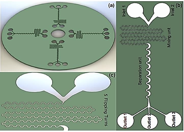
Preprints (www.preprints.org) | NOT PEER-REVIEWED | Posted: 29 October 2020 doi:10.20944/preprints202010.0622.v1

 scattered directly (Forward scatter) or perpendicular to the laser beam axis (Side scatter) and is then
 collected by the detectors (Figure 2-1).

 Figure 2-1: Overview of diagnosis section of the flow cytometry device, Sheath fluid focuses the cell
 suspension, forcing cells to pass through a laser beam one cell at a time. The forward and side
 scattered light is detected, and fluorescence is emitted from stained cells.

 Optical signals are obtained due to the laser beam to the cells and their size and internal structure.
 The collected light can then be converted to the equivalent electrical signals by the Photomultiplier
 tube (PMT). Receiving and detecting such electrical signals is carried out by employing detectors
 [48]. Light scattering at various angles can differentiate cells based on differences in size and internal
 complexity. In contrast, the scattered fluorescent light from a fluorescently labeled antibody can
 differentiate cells based on differences in surface and cytoplasmic antigens. Consequently, cells are
 distinguished based on characteristics such as volume, granulation, and colorability rate. Although
 the cells move concentratedly and individually inside the microchannels in front of the light beam,
 they can be isolated up to 100,000 cells per second through this method [50].

 FACS, based on the flow cytometer, was first invented in 1960 and has undergone many changes to
 date [48]. In basic FACS methods, each cell is trapped in an aerosol droplet having a specific electrical
 charge. Detectors then separate the droplets based on their electrical charge [47]. Many attempts
 have been made to eliminate this step due to droplet creation problems. These efforts led to the

 12
Preprints (www.preprints.org) | NOT PEER-REVIEWED | Posted: 29 October 2020 doi:10.20944/preprints202010.0622.v1

 design of the first FACS device adopting a microfluidic chip in 1999. The μFACS device (Figure 2-2)
 [43] has a lower manufacturing cost than large-dimension devices and has higher accuracy.

 Figure 2-2: The cells are settled in an electroosmosis solution and controlled by platinum
 electrodes. A Laser beam radiating on the solution stimulates the fluorescent. Fluorescent
 radiations are then collected by a microscope and measured by photomultiplier tubes (PMT). The
 computer receives signals from the PMT and adjusts the voltage to control the electroosmosis fluid
 [51]

 This device was first employed to separate painted E.Coli bacteria from its colorless type [52]. The
 pulsed lasers have been used in the device fabrication to produce propellant bubbles that push cells
 towards the separation channel, increase the FACS operation speed, and enhance the efficiency [53].

 Isolation of CTCs has been performed by applying the FACS method for patients with bladder cancer
 through their urine samples [54]. FACS has also been recently used in infertility tests for sperm
 separation [55]. This method also has some drawbacks, such as expensive devices, required materials
 for each test, microchannel clogging, and sample contamination. Although flow cytometry suffers
 from the disadvantages mentioned above, it is the most common procedure in laboratories. Some of
 the main reasons for utilizing such a method are:

 1- Flow cytometry devices help identify and count a small number of cells, even among many
 other cells.

 2- This device is time-efficient to count a large number of cells in a short time. The collected
 information about each of these cells can be beneficial in subsequent studies.

 Furthermore, this method has also been recently applied in some microfluidic devices. These
 microfluidic devices usually consist of a bifurcation region separating the detected cells by optical

 13
Preprints (www.preprints.org) | NOT PEER-REVIEWED | Posted: 29 October 2020 doi:10.20944/preprints202010.0622.v1

 forces [52], electroosmosis [56], or hydrodynamic forces [57, 58]. However, such microfluidic
 devices have not been sufficiently developed to supersede the conventional flow cytometry methods.

 2-2-2- Centrifugal devices

 The most conventional method for isolating cells and blood components is the centrifugal
 approaches. The basis of separation in this method originates from the different density of cells.
 Density gradient centrifugation can be used as a separation technique to isolate the CTCs and as a
 measurement method to estimate the densities of particles or molecules in a mixture.

 In this method, the patient blood sample is first blended with an anticoagulant solution to not
 coagulate during the separation process. The blood is then transferred to a centrifugal device and
 spun at a specific speed. The centrifugal force causes the cells with high density to move further away
 from the rotation center. As a result, having a higher density, the red blood cells are placed at the
 lowest point, and the white blood cells and plasma are respectively settled on the hematocrit (Figure
 2-3) [59].

 Figure 2-3: Blood components after centrifugation

 One of the most well-known techniques in separating blood cells through the centrifugal concept is
 the Ficoll method. In this method, red blood cells are deposited on the bottom of the tubes after
 centrifuges, while nucleated cells are placed on the red blood cell surface in the form of rings. Plasma
 also appears on the highest layer. Ficoll method has also been practiced to separate CTCs. Meanwhile,
 the centrifugal devices have some limitations as well. For instance, due to stress and shear stress on

 14
Preprints (www.preprints.org) | NOT PEER-REVIEWED | Posted: 29 October 2020 doi:10.20944/preprints202010.0622.v1

 cells during rotation, there is a possibility of cell loss or changes in their properties. It is also
 impossible to detach blood cells with a very small frequency through this method [60].

 One of the most crucial things that facilitated centrifugal separation was reaching the RosetteSep®
 solution. The RosetteSep® solution containing a series of antibodies is added to the blood and causes
 the accumulation of white and red blood cells and a clot formation. Subsequently, the clots are
 deposited owing to the centrifuge, and the target cells can be easily separated (Figure 2-4) [32].

 Figure 2-4: Blood separation through centrifugation 1) RosetteSep antibody solution is added to the
 blood 2) The adequate time is considered for the blood cells to coagulate with the help of the
 antibody solution 3) The sample is centrifuged, and the different layers of blood are then separated
 4) Target cells can be extracted [54]

 2-2-3- Separation through membrane filters

 Separation of cells based on the size and flexibility is one of the oldest methods due to its visibility
 and easy process controllability. One of these methods is filtering. Among these, membrane filters
 are one of the most authentic models of filters. The principle of cell separation from the main flow
 through membrane filters is quite simple. The membrane acts as a filter that will let blood flow
 through, while it catches CTCs and isolates them individually based on their size and deformability.
 However, it is worth noting that the membrane filtration method is less applicable than the centrifuge
 method.

 In this method, similar to the centrifuge method, the blood sample is first mixed with an anticoagulant
 solution to not coagulate during the separation process. The diluted blood next passes through a

 15
Preprints (www.preprints.org) | NOT PEER-REVIEWED | Posted: 29 October 2020 doi:10.20944/preprints202010.0622.v1

 membrane. This membrane causes larger cells than a threshold to be trapped while smaller ones
 continue on their path and are separated (Figure 2-5). Membranes are also made of various materials.

 Figure 2-5: Overview of membrane filtration method

 The common problems in using membrane filters are the possibility of clogging, the need for periodic
 maintenance and regular cleaning, and the cells’ inability to recover after filtration. Such a method’s
 most undesirable drawback is that it cannot separate more than one particle type in one step.

 A number of them have already been commercialized, such as Cell Microsieves® and ISET®. For
 example, the filter of Cell Microsieves® is manufactured of pure nylon and has cavities measuring 5
 to 200 microns. The possibility of sticking cells and proteins to the filter and its clogging is reduced
 because the filter’s extractants and detergents are paint-free. ISET® stood for Isolation by Size of
 Epithelial Tumor cell was developed to separate isolated Epithelial cells from tumors. Its name briefly
 refers to the isolation of epithelial cells based on size. This device, having cavities as small as 8
 microns, can work with a small volume of patient blood samples (only 1 ml) [61].

 2-2-4- Separation of cells through the magnetic field

 As an essential method for cell separation, the magnetic field (magnetophoresis) usage, while having
 the same fundamental principles and similar separation considerations, applies to microfluidic and
 non-microfluidic devices. Due to the cells’ low magnetic permeability, the Magneto-static forces can
 highly selectively separate cells tagged with magnetic nanoparticles without interference from either
 the surrounding electrolyte solution used for cell suspension or the rest of the cells [62].

 In this method, magnetic particles coated with an antibody are used. These particles are attached to
 the specific antigens at the cell surface. First, the magnetic particles are well covered with antibodies

 16
Preprints (www.preprints.org) | NOT PEER-REVIEWED | Posted: 29 October 2020 doi:10.20944/preprints202010.0622.v1

 and then mixed with the cells and incubated. The mixture of the cell and the magnetic particles then
 enter the separator, and the particles containing target cells are also collected by a magnet [32].

 The first commercial magnetic separator was the MACS1 (Magnetic Activated Cell Sorter) developed
 by Miltenyi in 1990, which used magnetic gradient columns. The target cells accumulate near the
 column walls where the magnets are located, and the flow carries the rest of the cells. Then the
 accumulated cells on the walls can be easily washed and collected from the device. At the output of
 this device, the target cells’ concentration is 105 times higher than the primary sample [63]. The
 device was equipped with a magnetic broom, which was a magnet that moved slowly in the sample,
 in subsequent research to increase efficiency [64]. The most significant achievement in the
 magnetophoresis field is fabricating the Cell Search® device. Figure 2-6 is one of the most well-
 known methods of identifying, distinguishing, and separating CTC.

 Figure 2-6: Cell Search® device and the process of its positive and negative separation, CTCs are
 absorbed by magnetic particles containing two types of antibodies, and another antibody absorbs
 white blood cells. Ultimately, the cells can be isolated with high purity by applying the magnetic
 field [65]

 This device, which detects CTCs by immunohistochemistry, is currently the first and only device
 approved by the US Food and Drug Administration (FDA). Cell Search® device, commercialized in
 2000, is used for various cancers such as breast, intestine, and prostate. After entering the 7.5 ml
 patient’s blood samples to the device, samples with at least 5 CTCs for breast and prostate cancers,
 and at least 3 CTCs for intestinal cancer are detected [65].

 17
Preprints (www.preprints.org) | NOT PEER-REVIEWED | Posted: 29 October 2020 doi:10.20944/preprints202010.0622.v1

 2-3- Microfluidic cell separation devices

 This section is an overview of cell separation devices made in the microfluidic field. Microfluidic
 devices generally enjoy some significant advantages: low cost, low sample size, portability, high
 accuracy and sensitivity, the ability to integrate multiple technologies, and short experiment time for
 cell separating, classifying, and sorting, so on. It should be noted that the general principles and
 physical fundamentals of separation are quite similar to the non-microfluidic devices in many cases.
 However, the system’s dimensions have been decreased to the micron to reduce fabrication costs and
 the required sample, while increasing the accuracy.

 2-3-1- Active Cell Separation Devices

 In active separation systems, external forces are used to control and manage the cells. According to
 the applied force nature for isolation, active separation systems are sorted into four general
 categories:

 A. Isolation adopting electric fields (Dielectrophoresis) [66-71]
 B. Isolation employing acoustic waves (Acoustophoresis) [72-74]
 C. Isolation by applying magnetic fields (Magnetophoresis) [75-78]

 These devices not only can control the cells precisely but also possess a relatively high operational
 speed.

 2-3-1-1- Isolation through electric field applications

 Isolation with the help of the electric field can be done in two ways: electrophoresis and
 dielectrophoresis. In the Electrophoresis approach, the particle displacement is caused by the electric
 field effects on their net free charge. Therefore, this method is based on the surface properties of the
 cell membrane. Most cells have a negative surface charge in physiologic PH; this charge density is
 different from one cell type to another and results in different electrophoretic mobility in an electrical
 field. Among several cell electrophoresis methods, free-flow electrophoresis (FFE) is the most widely
 used method. In FFE, the cells running in a buffer with a controllable flow rate are deflected based on
 their charge and collected into tubes. Although this method has been used to isolate viruses and
 bacteria from eukaryotic cells, the surface net charge is not a good criterion for separating a mass of
 cells with relatively similar properties. Hence, Dielectrophoresis was introduced to address the

 18
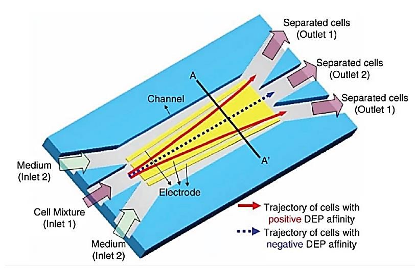
Preprints (www.preprints.org) | NOT PEER-REVIEWED | Posted: 29 October 2020 doi:10.20944/preprints202010.0622.v1

 electrophoresis’s constraints. Dielectrophoresis refers to the net migration of polarized particles due
 to interactions with an electric field gradient and depends on the cell wall, membrane, and
 cytoplasmic electrical properties [79].

 As a separation technique, Dielectrophoresis lies in differences in particle size and polarizability.
 Particles become polarized when electrical fields are applied to them. In turn, this induced
 polarization interacts with the applied field, resulting in each particle experiencing a unique net
 electrical force. This net force's magnitude depends on the particles’ dielectric characteristics (how
 easily the cells are polarized and how big they are), the electrical field frequency and strength, and
 the fluid medium’s electrical properties. Therefore, Dielectrophoresis (DEP) utilizes the difference in
 healthy and cancerous cells’ polarizability degrees and their size for separation [66-71]. The sample
 is placed in a non-uniformly electric field to achieve such a polarizability difference [66]. When a non-
 uniform electrical field exposes to dielectric particles, a force in the direction or opposite direction of
 the electric field gradient is applied.

 The most significant advantage of such a method is no need for particles to be charged so that any
 dielectric particle in a non-uniform field becomes dipole and oriented. However, the strength and
 direction of the generated force by the electric field depend on the particles’ dielectric properties
 (their polarization when exposed to a current), their size and shape, the electric field’s frequency and
 intensity, and the electrical properties of the fluid (Figure 2-7) [80-82]. Dielectrophoresis devices
 make the CTCs separate through their different properties, such as their size and dielectric properties
 compared to the other blood cells.

 The electrodes usually absorb and attach the CTCs to themselves while the rest of the blood cells
 continue moving along with the flow to leave the device. As shown in figure 2-7, cells were injected
 into the chamber by syringe and allowed to settle before the flow was initiated from the gear pump.
 Also, an AC electrical signal was applied to the electrode to influence the cell elution characteristics.
 This method’s valuable benefit over the other conventional methods is that the cells can be captured
 without being damaged during the separation process.

 19
Preprints (www.preprints.org) | NOT PEER-REVIEWED | Posted: 29 October 2020 doi:10.20944/preprints202010.0622.v1

 Figure 2-7A: Dielectrophoresis Separator, The cells are injected into the device by a syringe pump
 and allowed to be deposited. An electric current is then applied; Figure 2-7B: the cross-section of
 the channel has been identified, where the forces acting on the particle ultimately determine its
 equilibrium position) [83].

 Due to the difference in the particles’ electrical conductivity and their surroundings, some particles
 move towards increasing the field density, called positive electrophoresis. In contrast, the others
 start transferring towards decreasing the field density, named negative dielectrophoresis. The view
 of such separation is shown in Figure 2-8.

 Figure 2-8: Overview of positive and negative dielectrics in a non-uniform electric field [84]

 Therefore, when an electrical field is applied to particles, they become dipole. The difference in the
 polarization degree of healthy and cancerous cells makes the net electric force on each particle
 unique; thus, cancerous and healthy cells are located in different places in the presence of an electric
 field. For example, as shown in Figure 2-7B, all cells are subjected to two forces, a lift force and a force

 20
Preprints (www.preprints.org) | NOT PEER-REVIEWED | Posted: 29 October 2020 doi:10.20944/preprints202010.0622.v1

 originated from an electric field. Under such a circumstance, more electrical force is applied to the
 cancerous cells; therefore, they are more willing to move towards the lower plate and settle at a lower
 height than healthy cells. The next method, shown in figures 2-9, is a combination of the separation
 methods originated from the differences in size and the polarization degree in electric fields. As
 illustrated in this figure, fluid first passes through several pores to initially separate some of the
 cancerous cells, and the remaining cancerous and healthy cells exit through two different outputs
 under an electric field [85].

 Figure 2-9: Separation using differences in the particles’ polarization characteristics under an
 electric field

 The separation method’s main advantage is the difference in dimensions and sizes in the output rate
 or so-called test speed. The primary dielectrophoresis method’s benefit is the relatively high purity
 percentage of the isolation cells. Therefore, the advantage of both methods is applicable by this
 method so that the device enjoys both the adequate output advantage and a high purity percentage.
 Furthermore, such a device can separate different types of cells from each other. For example, this
 method can accurately isolate the bacteria or cancerous cells from the bloodstream [86, 87].

 The dielectrophoresis device can profit from the continuous and alternative electric current. For the
 lab on the chip, which alternating current (AC) is favorable, various electrodes arrangement is used.
 Electrodes are angled and placed in different convergent and divergent arrangements to prevent
 them from stopping the flow [88]. It is worth mentioning that this method’s main advantage
 compared to flow cytometry methods and separation by means of the magnetic field is that there is
 no longer a need to label cells; Hence, it has less difficulty and is more affordable [24, 89].

 Gascoyne et al. at the University of Texas in 2008 developed a device that can detect and separate
 breast cancer cells in the MDA-MB-468, MDA-MB-435 MDA-MB-231 categories from a sample having

 21
Preprints (www.preprints.org) | NOT PEER-REVIEWED | Posted: 29 October 2020 doi:10.20944/preprints202010.0622.v1

 30 million blood cells. Their device had larger electrodes than previous devices; consequently, a
 larger sample could be entered and processed at a higher rate [83].

 As mentioned earlier, dielectrophoresis techniques make it possible to collect and re-culture cells for
 the next usages, including drug testing. In addition, the electric current has also been applied to
 examine and test drug effectiveness and efficiency. For example, the effectiveness of Arsenic trioxide
 (As2O3), which is currently used in blood cancer treatment, is accurately measured. Therefore,
 different amounts of As2O3 are injected into the Dielectrophoresis activated cell sorter (DACS)
 containing the K562 class blood cancer cells. Subsequently, the drug’s effect on the cells is
 determined by isolating living cells from other cells and examining their death rates. The appropriate
 concentration and time intervals for having satisfactory drug effectiveness are determined in this
 way [90].

 The most prominent commercial cell separation device enjoying dielectrophoresis benefits is the Apo
 cell® device. Apo cell®, introduced in 2009, can separate CTCs from blood without labeling
 requirements because it can work based on their dielectric property differences [91]. One of the
 challenging issues while using this method for separating cells from each other is that the
 dielectrophoresis force is highly dependent on cell size. Most blood cells are only about 10% different
 in size and usually have similar electrical properties. Besides, the dielectrophoresis phenomenon is
 highly dependent on the electrical conductivity coefficient of fluid; however, the separation of
 particles from each other in physiological buffers is difficult due to their high electrical conductivity.
 Heat effects should also be considered in applying this method to isolate cells because a fluid
 temperature with a high permeability coefficient increases when exposed to an electric field. This
 temperature increase, known as the Joule heating effect, can damage cells [89]. Doh and Cho
 succeeded in isolating live yeast microorganisms from the dead ones by this method (Figure 2-10)
 [84].

 22
Preprints (www.preprints.org) | NOT PEER-REVIEWED | Posted: 29 October 2020 doi:10.20944/preprints202010.0622.v1

 Figure 2-10: Overview of the designed microfluidic device to take advantage of the
 dielectrophoresis phenomenon [84]

 Table 2-1: Advantages and disadvantages of separation under an electric field

 Benefits: Limitations:

 Large sample needs larger electrodes, Not
 Increasing accuracy, Precise control, Relatively suitable for rare cell capture, Limited stability,
 high-speed operation, Unchanged functional Time-consuming, Demanding sophisticated
 state of the cells, Contactless and expensive equipment, Not applicable on a
 rotational platform, Medium throughput, No
 fair resolution, The sample must be diluted
 before the operation

 2-3-1-2- Acoustic Cell Sorting

 This section explains how acoustophoresis forces separate the CTCs from peripheral blood samples
 obtained from cancer patients through an acoustic-based microfluidic device. Acoustic devices
 operate based on the separation of particles exposed to the acoustic field and acoustic waves [92-94].
 In this method, a stationary wave is formed inside the microchannel. A stationary wave is a wave
 whose fluctuation amplitude is a constant value at any specific point along the wave axis. The
 stationary wave occurs when two waves with similar frequency move in opposite directions of each
 other. The overlap of these two waves forms such a wave. A Pressure Node is formed at both ends of
 this stationary wave. The node in stationary waves refers to the points that are fixed.

 In contrast, Pressure Antinode develops at some points. The Pressure Antinode in stationary waves
 refers to the points experiencing the greatest displacement. These waves originate from the

 23
You can also read