A perfusable vascularized full-thickness skin model for potential topical and systemic applications
←
→
Page content transcription
If your browser does not render page correctly, please read the page content below
Biofabrication

PAPER • OPEN ACCESS

A perfusable vascularized full-thickness skin model for potential topical
and systemic applications
To cite this article: Sacha Salameh et al 2021 Biofabrication 13 035042

View the article online for updates and enhancements.

                               This content was downloaded from IP address 46.4.80.155 on 27/09/2021 at 02:06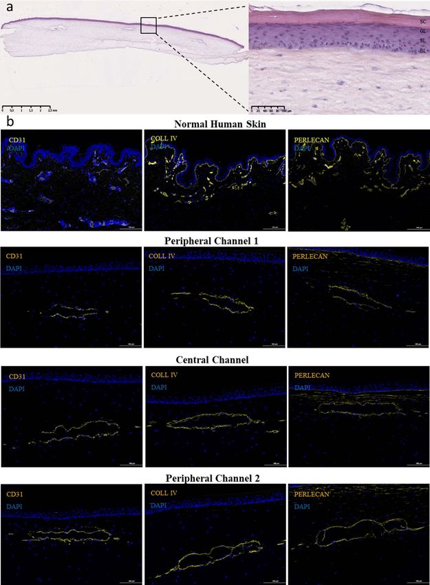
Biofabrication 13 (2021) 035042 https://doi.org/10.1088/1758-5090/abfca8
Biofabrication
PAPER
A perfusable vascularized full-thickness skin model for potential
OPEN ACCESS
topical and systemic applications
RECEIVED
16 December 2020 Sacha Salameh1,2,∗, Nicolas Tissot1, Kevin Cache1, Joaquim Lima1, Itaru Suzuki1, Paulo André Marinho1,
REVISED Maité Rielland1, Jérémie Soeur1, Shoji Takeuchi4, Stéphane Germain3,5 and Lionel Breton1,5
20 April 2021
1
L’Oréal Research and Innovation, Aulnay-sous-Bois, France
ACCEPTED FOR PUBLICATION 2
28 April 2021 Sorbonne Université, Collège Doctoral, F-75005 Paris, France
3
Center for Interdisciplinary Research in Biology (CIRB), College de France, CNRS, INSERM, PSL Research University, Paris, France
PUBLISHED 4
14 May 2021 Department of Mechano-Informatics, Graduate School of Information Science and Technology, The University of Tokyo, 7-3-1 Hongo,
Bunkyo-ku, Tokyo 113-8656, Japan
5
Equal contribution.
Original content from ∗
Author to whom any correspondence should be addressed.
this work may be used
under the terms of the E-mail: sacha.salameh@rd.loreal.com
Creative Commons
Attribution 4.0 licence. Keywords: reconstructed skin, tissue engineering, vascularization, vasculogenesis, angiogenesis, perfusion
Any further distribution
of this work must
Supplementary material for this article is available online
maintain attribution to
the author(s) and the title
of the work, journal
citation and DOI. Abstract
Vascularization of reconstructed tissues is one of the remaining hurdles to be considered to
improve both the functionality and viability of skin grafts and the relevance of in vitro applications.
Our study, therefore, sought to develop a perfusable vascularized full-thickness skin equivalent that
comprises a more complex blood vasculature compared to existing models. We combined molding,
auto-assembly and microfluidics techniques in order to create a vascularized skin equivalent
representing (a) a differentiated epidermis with a physiological organization and correctly
expressing K14, K10, Involucrin, TGM1 and Filaggrin, (b) three perfusable vascular channels with
angiogenic sprouts stained with VE-Caderin and Collagen IV, (c) an adjacent microvascular
network created via vasculogenesis and connected to the sprouting macrovessels. Histological
analysis and immunostaining of CD31, Collagen IV, Perlecan and Laminin proved the integrity of
vascular constructs. In order to validate the vascularized skin potential of topical and systemic
applications, caffeine and minoxidil, two compounds with different chemical properties, were
topically applied to measure skin permeability and benzo[a]pyrene pollutant was systemically
applied to evaluate systemic delivery. Our results demonstrated that perfusion of skin reconstructs
and the presence of a complex vascular plexus resulted in a more predictive and reliable model to
assess respectively topical and systemic applications. This model is therefore aimed at furthering
drug discovery and improving clinical translation in dermatology.
1. Introduction of active compounds and drugs’ efficacy and tox-
icity in cosmetic and pharmaceutical research, espe-
Tissue engineering and regenerative medicine hold cially after the ban of animal testing in cosmetic
high promises for organ replacement and develop- research in 2013 (European Community directive
ment of novel therapies. Being the largest human 93/35/EEC) [3–5].
organ, the loss of skin integrity due to injury or In the ‘70s Rheinwald and Green [6] managed
illness results in a substantial physiologic imbal- to culture epidermal human keratinocytes. Since
ance and ultimately in severe disability or death. then, many skin substitutes have been developed
The skin was the first reconstructed tissue gener- to reproduce physiological functions and get closer
ated by tissue engineering approach [1] for the treat- to in vivo skin. For instance, endothelial cells [7],
ment of burns, surgical scars, and plastic surgery immune cells [8], and melanocytes [9] have been
[2]. Skin models are also used in the evaluation included in 3D skin equivalents. Recently, more
© 2021 The Author(s). Published by IOP Publishing Ltd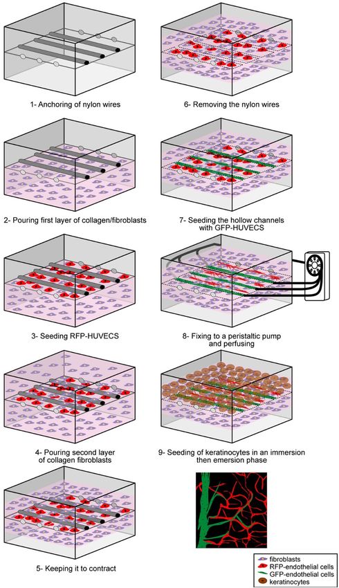
Biofabrication 13 (2021) 035042                                                                          S Salameh et al

advanced and complex skin substitutes were pub-              structures [42–45], bioprinting [46–48], and vascular
lished by the integration of hair follicles [10],            network patterning [49–52] to be able to perfuse the
mature adipocytes in a three-layered skin equival-           construct.
ent [11], sebocytes organoids derived from human                  However, the generation of a vascularized recon-
induced pluripotent stem cells (hIPS) [12], IPS              structed full-thickness skin that gathers a properly
derived sensory neurons, and Schwann cells with              differentiated epidermis as well as in vivo-like per-
endothelial cells in an innervated engineered skin           fused vasculature still represents a major challenge.
sponge model [13], etc. Moreover, recent interest has        Several studies reported vascularized skin substitutes
focused on transferring various micro-physiological          that cannot be perfused [30, 40, 41, 53–55] which
organ models, including skin, onto microfluidic              is a limitation for systemic applications. Some oth-
platforms [14–18]. These skin-on-a-chip models               ers developed perfusable vessel-like constructs in cell-
enable physiologically relevant transport of nutri-          free hydrogels that better mimic blood flow in vas-
ents and exogenous substances to the skin tissue,            cular networks, however do not allow the study of
better control over physical and chemical factors            the vascularization role or systemic exposure effect
in the cell microenvironment [19], and more reli-            on surrounding tissues [47, 50–52, 55, 56]. On the
able evaluation of drug candidates in terms of               other hand, vascular networks were also generated in
toxicity, efficacy, and delivery compared to static          a perfusable dermis substitute but these constructs
conditions [20].                                             still lacked an epidermal layer which is a limita-
    Nevertheless, reconstructing thick or complex tis-       tion for topical applications [57]. Nonetheless, the
sues that faithfully recapitulate all the functions of the   few existing models of perfusable vascularized full-
in vivo skin is still a challenge [21]. One of the main      thickness skin are limited. Today, and to our know-
limitations is the lack of vascularization and perfusion     ledge, there are only four developed in vitro models of
in reconstructed tissues.                                    perfusable vascularized reconstructed full-thickness
    Given that the diffusion coefficient of oxygen           skin. Groeber et al [43] used a porcine jejunum as
is limited to 200 µm in human tissues [22, 23],              a scaffold but this model cannot be used in large-
>200 µm thick skin constructs might encounter nec-           scale production as it relies on the use of an animal
rosis because of hypoxia [24, 25]. Therefore, devel-         organ. Abaci et al [58] used bioprinting to engin-
oping perfusable vascular networks in order to blunt         eer a spatially controlled vascular pattern in a 3D
hypoxia and improve viability is mandatory. For              skin reconstruct but their skin lacks microvasculariz-
instance, prevascularization in skin graft for clinical      ation and epidermal differentiation layers. Mori et al
application accelerates anastomosis in vivo with the         [28] developed a device with anchoring structures
host vascular network and thereby improves oxygen            and connectors allowing the reconstruction of a per-
and nutrient supply [26]. in vitro, generating a skin-       fusable reconstructed skin. However, this model dis-
equivalent with hair follicles as dense as what we find      played one macrochannel seeded with endothelial
in vivo could result in a necrotic core if the tissue        cells without a microvascular network in the dermis
is not prevascularized [10]. Additionally, vascular-         and with thin layers of keratinocytes that did not
ized skin reconstructs represent invaluable tools for        recapitulate full epidermis differentiation. Using the
basic research (e.g. study vascular remodeling dur-          same device with stretchable material, the authors
ing wound healing, aging, cancer, etc) but also for          cultured this skin-equivalent under stretching and
topical and systemic evaluation (e.g. screening pro-         perfusion conditions. They showed that perfusion
and anti-angiogenic drugs, evaluation of systemic            and stretch stimuli improved skin-equivalent mor-
toxicity, percutaneous absorption of chemicals and           phology [59]. Kim et al [60] used 3D printing to
drugs) [27–30]. Certainly, having a perfusable vas-          engineer perfusable vascularized skin with a hypo-
cular network in skin equivalents makes more relev-          dermis, dermis, and epidermis. However, the vas-
ant the evaluation of systemic delivery of substances,       culature of the skin equivalent remained a unique
but also the study of systemic exposure or inflamma-         macrochannel lacking the physiological cutaneous
tion responses which could not be properly assessed          microvascularization.
with existing skin models. Improving vascularization              In this study, by combining molding and auto-
of tissue-engineered constructs has therefore gained         assembly techniques as well as microfluidics, we
interest in the last decade [31]. Many strategies have       developed for the first time a fully vascularized per-
been developed to improve or accelerate vascular             fusable reconstructed skin that comprises a differen-
growth in engineered tissues either in vivo relying          tiated epidermis containing all the layers of the in vivo
on improving the angiogenesis process from the host          structure, perfusable macrovessels with angiogenic
tissue into the implant [32–35] or by prevascular-           sprouts, and a microvascular network with capil-
ization of the engineered tissue. Different in vitro         laries organized by vasculogenesis in the dermis
techniques were thus developed including sparse              part. We further showed that this model is relevant
cells seeding into scaffolds or hydrogels [36–38], cell      for studying both systemic drug delivery and skin
sheet strategy [39–41], reuse of biological vascular         permeability.

                                                2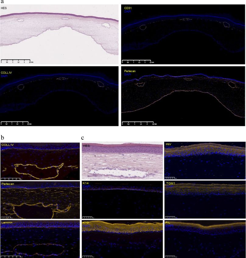
Biofabrication 13 (2021) 035042                                                                    S Salameh et al

2. Materials and methods                                  type I collagen and 2 × 10⁶ ml−1 NHDFs prepared
                                                          as previously described [61, 62] and incubated at
2.1. Cell culture reagents                                37 ◦ C for 30/60 min until the collagen gel formed a
DMEM high glucose GlutaMAX, Ham’s F-12 Nutri-             dermis-like layer. The final number of NHDFs was
ent Mix GlutaMAX, MEM supplemented with non-              0.5 × 10⁶ cells per lattice. Dermal substitutes were
essential amino acid, L-glutamine, sodium pyr-            incubated in NHDF 3D medium (DMEM contain-
uvate, phosphate-buffered saline without Mg2+ and         ing 10% FBS, 100 U ml−1 penicillin-streptomycin,
Ca2+ (PBS-), Trypsin-EDTA, antibiotic–antimycotic         0.25 µg ml−1 amphotericin B, and 70 µg ml−1 ascor-
solution (100 U ml−1 penicillin-streptomycin;             bic acid) for 5 d to allow contraction. The medium
0.25 µg ml−1 amphotericin B) were purchased from          was then changed to ECGM-2. Nylon wires were
Invitrogen (Carlsbad, CA, USA). L-Ascorbic acid 2-        removed and hollow channels were filled with 200 µl
phosphate sesquimagnesium salt hydrate (Vitamin           ECGM-2 containing 8 × 10⁵ HUVECs using a syr-
C), human recombinant epidermal growth factor             inge pump at a flow rate of 6.5 ml h−1 (KD Sci-
(EGF), adenine hydrochloride, hydrocortisone 21-          entific, Holliston, MA, USA). At this time point, once
hemisuccinate sodium, (-) isoproterenol hydrochlor-       nylon wires are removed, the contraction of the col-
ide, 3.3′ .5-triiodo-l-thyronine sodium 95%, apo-         lagen gel is enough to maintain a tubular structure
transferrin, were purchased from Sigma (St. Louis,        without causing damage or collapsing upon removal.
MO, USA). VEGFA-165 , Endothelial Cell Growth             The device was inverted for 30 min to coat the upper
Medium 2 (ECGM2), endothelial basal medium                half of the channels with HUVECs and then inver-
(EBM), Supplement Pack endothelial cell GM2 were          ted again and incubated for 24 h in ECGM-2 to
purchased from Promocell (Heidelberg, Germany).           allow the endothelial cells adhesion. Subsequently,
Fetal bovine serum (FBS) was purchased from PAN-          6 × 10⁵ keratinocytes were seeded on the dermal layer
Biotech GmbH (Aidenbach, Germany). Bovine type            and immersed in DMEM/F12 immersion medium
I collagen solution (ACI-100) was purchased from          (DMEM and Ham’s F12 at a ratio of 3:1 supple-
Symatese (Chaponost, France).                             mented with 180 µM adenine, 10 ng ml−1 hEGF,
                                                          0.4 µg ml−1 hydrocortisone, 5 µg ml−1 insulin, 2 nM
2.2. Cell culture                                         triiodo-L-thyronine, 1 µM isoproterenol, 5 µg ml−1
Normal human dermal fibroblasts (NHDFs) and nor-          apo-transferrin, 100 U ml−1 penicillin–streptomycin,
mal human epidermal keratinocytes (NHEKs) were            0.25 µg ml−1 amphotericin B, 1 nM VEGF-A165 and
provided by Episkin (Lyon, France) and used at pas-       10% FBS). The central channel was then perfused at
sage 8 and passage 2, respectively. Human umbil-          2–3 ml h−1 using a peristaltic pump (SJ-1211II-L,
ical vein endothelial cells (HUVECs) were purchased       ATTO Corp., Tokyo, Japan). After 3 d of immer-
from PromoCell GmbH (Heidelberg, Germany) and             sion, the skin equivalent was lifted to the air–liquid
used at passage 4. HUVECs were transduced at              interface for 7 d to induce stratification of the epi-
passage 2 using EGFP-Puro or Turbo-RFP lentivir-          dermal layer using DMEM/F12 emersion medium
uses (Vectalys, France) with a titer of 4.2 × 10⁹ or      (DMEM and Ham’s F12 at a ratio of 3:1 supple-
5.4 × 10⁹ TU ml−1 respectively and used at pas-           mented with 0.4 µg ml−1 hydrocortisone, 5 µg ml−1
sage 5 in 3D cultures. NHDFs were maintained in           insulin, 1 nM VEGF, 100 U ml−1 de penicillin-
DMEM 10% FBS, 1% antibiotic-antimycotic solu-             streptomycin, 0.25 µg ml−1 amphotericin B and 1%
tion, and HUVECs in ECGM-2 supplemented with              FBS). The flow rate was increased to 6.5 ml h−1 and
1% antibiotic–antimycotic solution. NHEKs were            perfusion was maintained until the end of the cul-
grown on irradiated primary mouse embryonic fibro-        ture (17 d in total). The media was changed every
blasts, using keratinocyte medium containing DMEM         other day.
and Ham’s F12 at a ratio of 3:1 supplemented with              To induce vasculogenesis, half the volume of the
180 µM adenine, 10 ng ml−1 epidermal growth               neutralized collagen/fibroblasts mix was poured until
factor, 0.4 µg ml−1 hydrocortisone, 5 µg ml−1 insulin,    it reached the nylon wires. The device was then incub-
2 nM triiodo-L-thyronine, 1 µM isoproterenol, 1%          ated at 37 ◦ C for 20–30 min until collagen gelation
antibiotics–antimycotic solution, and 10% FBS. All        and 6 × 10⁵ HUVECs transduced with Turbo-RFP
cells were grown at 37 ◦ C in 5% CO2 . The medium         lentiviruses (Vectalys, France) (RFP-HUVEC) were
was changed three times a week.                           then seeded as a monolayer and incubated for 1 h to
                                                          allow cell adhesion. After 20–30 min of incubation,
2.3. Reconstruction of the skin equivalent                an equal volume of collagen/fibroblasts was poured
The culture device was as previously described [28].      to cover the RFP-HUVEC layer. The dermal layer
Here, three nylon wires (0.55 mm diameter) were           was incubated in NHFD 3D/ECGM2 mix medium
strung across the connectors before sterilization using   (44% DMEM glutamax and 47% Endothelial Basal
70% ethanol and UV light. Devices were then treated       Medium EBM supplemented with Supplement Pack
for 15 min using a plasma etcher (Harrick Plasma,         endothelial cell GM2, 6.3% FBS, 1% antibiotic–
Ithaca, NY USA). The devices were then filled with        antimycotic, 70 µg ml−1 ascorbic acid) for 5 d.
1.5 mg ml−1 of a neutralized solution of bovine           Then the same protocol as described above was

                                             3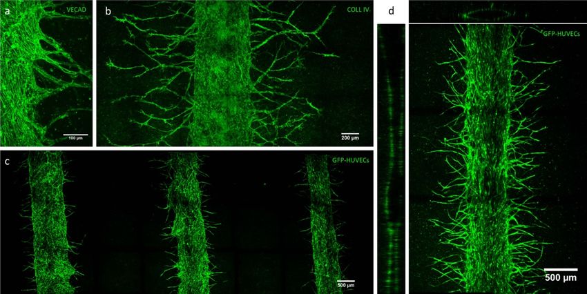
Biofabrication 13 (2021) 035042                                                                                       S Salameh et al

          Figure 1. Schematic illustration showing the development of the perfusable vascularized reconstructed skin model.

followed, except that hollow channels were seeded                       Immunofluorescent stainings were performed on
with HUVECs transduced with EGFP lentiviruses                       7 µm frozen sections fixed in cold acetone, blocked
(EGFP-HUVEC) (figure 1).                                            in bovine albumin serum (BSA)-10% normal goat
                                                                    serum (SP-004-vx10, Diagomics, France) and incub-
2.4. Histological analyses                                          ated 1 h at room temperature (RT) or O/N at 4 ◦ C
Morphologies of the skin equivalent and vascular                    with diluted primary antibodies in 1% BSA: Laminin
channels were analyzed using hematoxylin, eosin, and                alpha 5 (1:20 Dako M0638,Agilent, USA), Collagen
saffron (HES) staining or immunostaining.                           IV (COLL IV) (1:50 Dako M0785, Agilent, USA),

                                                     4
Biofabrication 13 (2021) 035042                                                                    S Salameh et al

Perlecan (1:100 ab26265, Abcam, United kingdom),          and secondary microvascularization (SandP). Three
CD31 (1:20 Dako M0823, Agilent, USA), Alpha               different batches of experiments were tested except
Actin (1:100 sc32251, Santa Cruz,USA), Keratin 14         for samples with secondary vascular plexus (SandnoP
(K14) (1:20 Progen 10003, Progen, USA), Keratin           and SandP) where only one batch of three devices was
10 (K10) (1:100 Dako M7002, Agilent, USA), Invol-         evaluated.
ucrin (INV) (1:50 BTI J64013-AB,Biotechnology Ltd,            Around 25 mg of each chemical were dissolved
UK), transglutaminase 1 (TGM1) (1:500 NBP2-               in 10 ml of PBS containing 0.25% (w/w) Tween 80
34062, Novus Biologicals, USA), filaggrin (FIL)           (723M-156-84, Fisher scientific, USA). This surfact-
(1:200 sc66192, Santa-Cruz, USA). Sections were           ant was added to guarantee chemical solubility in the
washed with PBS- and incubated with secondary anti-       buffer. Before application, the solution was filtered
body solution (A21422 or A21428, Thermo Fisher            through Millex HV 0.45 µm and then quantified to
Scientific, USA) diluted 1:500 in 1% BSA for 1 h          determine Caffeine and Minoxidil concentrations.
at RT. Sections were then mounted using Pro-                  To control the volume deposited on the skin equi-
long with DAPI (P36931, Life Technologies, USA)           valent, an 8 mm PTFE ring was designed. The ring
and observed with Nanozoomer S60 (Hamamatsu,              was sealed on the epidermis using DERMABOND
Japan) or Nikon Microscopy Eclipse 80i (Nikon,            ADVANCED® (AHVM12, Ethicon, USA) to avoid
Japan).                                                   leakage. Devices were placed in a static condition on
    For whole-mount staining, samples were fixed          a magnetic stirrer for homogenizing to respect sink
in 4% formaldehyde (VWRC9713, VWR, USA) for               conditions. An infinite dose of 300 µl cm−2 of the
20 min. The samples were then washed with PBS- and        buffered solution was topically applied on the skin in
vascular channels were filled with PBS-1% BSA using       the inserted ring. Each hour for the next 6 h, 300 µl
a syringe pump. After 1 h of saturation, vascular chan-   were sampled from the receptor fluid under the skin
nels were filled with Collagen IV primary antibody        equivalent. The same amount of fresh media was
diluted 1:50 in PBS-1% BSA (Dako M0785, Agilent,          added in order to maintain a constant volume in each
USA) and incubated for 2 h at RT. The primary anti-       device. Samples were then diluted (1/10) in a fresh
body solution was washed out with PBS- three times        culture medium and quantified with standards curves
for 10 min. The vascular channels were filled with        prepared in the same culture medium (DMEM/F12
secondary antibodies targeting mouse IgG coupled to       emersion medium).
Alexa Fluor 488 (A11029, Thermo Fisher Scientific,            The same study was performed on 8 split-
USA) diluted 1:100 in PBS-1% BSA and incubated            thickness pig ear skin set up on a Franz cell hav-
for 1 h at RT. For VE-Cadherin, primary antibody          ing 2 cm2 square areas and 3 ml receptor com-
already coupled to Alexa Fluor 488 secondary anti-        partment volume. Mean thickness was measured at
body (1:100 53-1449, Thermo Fisher Scientific, USA)       1056 ± 62 µm. The receptor compartment was filled
was incubated 2 h at RT in the channels. Channels         with PBS +0.25% (w/w) to guarantee sink con-
were then observed using two-photon microscopy            ditions and homogenized using a magnetic stirrer.
(Leica SP8 coupled to a Spectra Physics Insight femto-    After 1 h equilibration time, skin integrity was con-
second laser, Leica Microsystems, Germany). Fluores-      trolled with Trans Epidermal Water Loss measure-
cence signal emitted by the Alexa Fluor 488 or EGFP       ment (Vapometer, Delfin Technologies, UK). Accept-
and by the Turbo-RFP were detected with an object-        ance criteria was defined (i.e.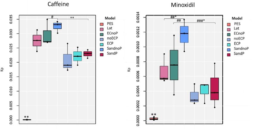
Biofabrication 13 (2021) 035042                                                                       S Salameh et al

with the following transition: Caffeine 195.0 (m/z)         and Emission λ (450 nm) were used for fluorescence
→ 138.1(m/z) and Minoxidil 210.0 (m/z) → 164.0              detection of BaP.
(m/z). The lower limit of quantification (LLOQ)
and upper limit of quantification (ULOQ) were at            3. Results
0.60 and 2130 ng ml−1 , respectively for Caffeine and
0.51 and 1990 ng ml−1 respectively for Minoxidil.           3.1. Development of a full-thickness skin
The cumulative amount of Caffeine and Minoxidil             equivalent with three vascular channels and
through the skin was plotted as a function of time.         angiogenic sprouts
According to Fick’s first law of diffusion, the slope of    This study was aimed at creating a full-thickness skin
the linear part of the curve defined the flux at a steady   equivalent model with a complex perfusable vascular
state. The permeability coefficient (Kp) was obtained       network as well as a mature epidermal layer. To do so,
by the ratio of flux and the concentration (C) of the       we used a culture device that allows the reconstruc-
tested compound in the solution applied on the skin:        tion of a skin equivalent model [28].
    (        ) Flux(µg cm−2 h−1 )
Kp cm h−1 = C(µg cm−3 ) [64].                                    First, three nylon wires were strung across con-
                                                            nectors to form molds inside this dermis-like layer
     The statistic model used took into account the
                                                            composed of a mix of human fibroblasts and colla-
batch effect as a random variable and skin models as
                                                            gen hydrogel. Once the hydrogel contracted, nylon
a fixed effect. This allowed the evaluation of different
                                                            wires were removed, thereby providing tubular struc-
sources of variability i.e. inter- and intra-batch and
                                                            tures used for further perfusion. Knowing that colla-
intra-model). Considering the low number of rep-
                                                            gen contraction depends on cell number and collagen
licates (8 for pig ear skin, 3–12 for different recon-
                                                            concentration [61], we adapted fibroblasts concen-
structed skin models), a non-parametric method was
                                                            tration to 7.1 × 10⁴ ml−1 in order to overcome
used as a statistical model as variability of the data
                                                            lattice contraction and maintain three opened vas-
did not follow a normal distribution. The relevance
                                                            cular channels. When normal human keratinocytes
of the comparison was evaluated according to the size
                                                            (NHK) were seeded on the perfused dermis, the cul-
of the effect (i.e. very small, small, moderate, strong,
                                                            ture medium was optimized to improve epidermal
or very strong). Resulting P-value was adjusted using
                                                            differentiation. In these conditions, histological ana-
the Bonferroni method, where a statistical signific-
                                                            lyses using HES staining demonstrated that reprodu-
ance was reached if P < 0.05.
                                                            cible full-thickness skins were obtained representing
                                                            a differentiated epidermis and three open microves-
2.6. Systemic exposure of benzo[a]pyrene (BaP)              sels (figure 2). The epidermis displayed all the lay-
pollutant                                                   ers of the differentiation process: (a) organization of
To evaluate the systemic transport of BaP to the            cubic cells (proliferative basal keratinocytes) at the
dermis and epidermis of skin substitutes, tissues           basal layer, (b) flattening of these cells in the spin-
either in static or perfused conditions were exposed        ous layer, and (c) development of granular keratino-
to 10 µM BaP (B1760, Sigma). BaP was added in               cytes in the granular layer followed by the formation
the DMEM/F12 emersion medium at Day 3 of the                of the stratum corneum (the outermost layer of the
emersion phase. A sample of fresh media contain-            epidermis) (figure 2(a)). These histological observa-
ing the 10 µM BaP was taken as a control to meas-           tions were confirmed by immunostaining the follow-
ure the initial BaP concentration. After 48 h, the          ing epidermal markers: keratin 14 (K14), keratin 10
media was changed to fresh media containing 10 µM           (K10), involucrin (INV), and filaggrin (FLG) were
BaP. Samples were cultured for an additional 48 h           localized at the basal layer, intermediate layers, and
before harvest. At the end of the culture period,           upper layers respectively confirming a proper differ-
media of each device were sampled to evaluate the           entiation of the epidermis (figure S1 (available online
remaining BaP after skin culture. The dermis and            at stacks.iop.org/BF/13/035042/mmedia)).
the epidermis parts were separated and proteins                  The presence of endothelial cells lining the chan-
were precipitated using acetonitrile (ACN) (271 004,        nels was confirmed by positive CD31 immunos-
Sigma). The supernatant was then extracted and ana-         taining (figure 2(b)). Immunostaining of the main
lyzed using liquid chromatography with fluorescence         components of the vascular basement membrane,
detection HPLC-FLD (Shimadzu Prominence- RF-                collagen IV and perlecan, showed a proper deposition
20A-XS). The analytical system was managed by the           of the vascular basal lamina (figure 2(b)).
software Empower® version 3.0. The analytical guard              Then, we sought to promote angiogenic sprout-
column and analytical column were Pinnacle II PAH           ing from these perfusable channels. The potential
(10 × 4,0 mm) and Pinnacle II PAH (150 × 3,2 mm;            of endothelial cells to sprout and organize into a
4 µm) (Restek, France), respectively. Analyses were         vascular network highly depends on the mechanical
carried out with an isocratic elution with mobile           properties of the surrounded environment. It was
phases of ACN/Water 95/5 (v/v). The volume of the           shown that matrix densities from 1.2 to 1.9 mg ml−1
injection was variable from 5 to 30 µl and the flow         improves coordinated migration and proliferation
rate was fixed at 1.2 ml min−1 . Excitation λ (275 nm)      of endothelial cells to form stable sprouts, while

                                               6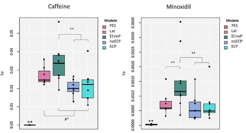
Biofabrication 13 (2021) 035042                                                                                        S Salameh et al

   Figure 2. Histological analysis and immunostaining of the skin equivalent with perfusable vascular channels. (a) HES staining of
   the skin equivalent showing a dermis, an epidermis, and three open microvessels of two skin equivalents. BL = basal layer,
   SL = spinous layer, GL = granular layer, SC = stratum corneum. (b) Immunostaining of CD31 in endothelial cells and COLL IV
   and Perlecan as markers of the basal lamina of the three microvessels. Scale bar = 100 µm.

lower densities (0.3–0.7 mg ml−1 ) or higher densit-                 Using anti-VE-cadherin (VECAD) immunostaining,
ies (2.7 mg ml−1 ) resulted in uncoordinated migra-                  angiogenic sprouts emanating from vascular chan-
tion or cell clusters, respectively [65]. Therefore, col-            nels were characterized (figure 3(a)). These newly
lagen concentration (1.5 mg ml−1 ), as well as the                   formed capillaries could reach up to 800 µm. These
number of endothelial cells seeded in the channels                   sprouts were also marked with collagen IV show-
(8.105 cells/channel), were optimized in order to pro-               ing the establishment of a mature basement mem-
mote efficient vascular morphogenesis and sprouting.                 brane (figure 3(b)). Sprouts were observed both in

                                                      7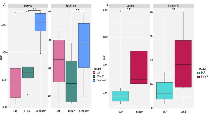
Biofabrication 13 (2021) 035042                                                                                     S Salameh et al

   Figure 3. Two-photon microscopy images of angiogenic sprouts from vascular channels. (a) VE-Caderin and (b) COLL IV
   expression of the main vascular channel and angiogenic sprouts. (c) GFP-HUVECs showing the angiogenic sprouts of the three
   main vascular channels. (d) Orthogonal sections of the GFP-HUVECs channel showing luminal structures.

peripheral and central channels (figure 3(c)). Ortho-             expressing CD31, perlecan and collagen IV at the
gonal sections showed opened tubular structure of the             location of the microvascular bed (figure 5(a)). The
main vessel (figure 3(d)).                                        three channels were preserved opened and prop-
    These results demonstrate that our full-thickness             erly expressed CD31, collagen IV, and perlecan
skin model recapitulates a mature epidermis and per-              (figure 5(a)). The dermo-epidermal junction (DEJ)
fusable vascular channels.                                        was also marked with collagen IV, perlecan, and
                                                                  laminin proving the deposition of basement mem-
3.2. Generation of a secondary capillary network                  brane proteins (figure 5(b)). Moreover, epidermal
by vasculogenesis                                                 differentiation was well-maintained and each layer
Our next goal was to further increase the complex-                was expressing its specific markers: K14 for the basal
ity of the vascular network in the skin equivalent.               layer, K10 for the spinous layer, INV, TGM1, and
Taking advantage of the auto-assembly properties of               FIL for the granular layer and stratum corneum
endothelial cells seeded into 3D collagen matrices,               (figure 5(c)).
HUVECS were then seeded between two layers of                         Hence, by combining optimal culture conditions,
fibroblasts embedded in a collagen matrix at the same             molding technique using nylon wires, and vasculo-
level as the perfused vascular channels. To identify              genesis, we here developed a full-thickness skin equi-
the capillaries formed by angiogenic sprouting from               valent representing a complex perfusable vascular
the fluidic channels from those developed by vas-                 network with a properly differentiated epidermis.
culogenesis in the lattice, we used GFP-HUVECs to
reconstruct the vascular channels and RFP-HUVECs                  3.3. Evaluation of skin permeability
for the microvascular network between the channels.               Reconstructed skin models can be used for safety and
A mixed media of 1:1 NHDF 3D:ECGM2 was used                       efficacy evaluation of chemicals systematically or top-
for the first 5 d of contraction of the vascularized              ically applied to skin tissues. For topical treatments
dermis while maintaining the same concentration of                and bioavailability evaluation, skin barrier function
growth factors present in both media (figure 1). Using            is critical for the percutaneous absorption. However,
these conditions, a complex RFP-HUVECs microvas-                  skin equivalents were proven to have lower epidermal
cular network was formed between the three GFP-                   barrier function therefore higher permeability com-
HUVECs perfusable vascular channels (figure 4(a)).                pared to normal human skin [66]. Additionally, these
This dense capillary network displayed connected                  models lack vascular networks and do not allow capil-
branches (figure 4(a)) that could also reach and                  lary clearance which plays an important role during
connect to the angiogenic sprouts emanating from                  ADME process [67, 68].
the fluidic channels (figure 4(b); Supp Video). The                    Here, we evaluated the effect of vascularization
formation of a lumen could be identified by hol-                  and perfusion on epidermal barrier function by
low spaces between vascular borders (figure 4(c);                 studying skin permeation of Caffeine and Minoxi-
Supp Video). The two layers of collagen and fibro-                dil, two chemicals with different structures and pen-
blasts were connected with a visible line of cells                etration potentials. Four different skin models were

                                                    8
Biofabrication 13 (2021) 035042 S Salameh et al
Figure 4. Vasculogenesis of a secondary vascular plexus. (a) Two-photon fluorescence microscopy showing RFP-HUVECs
microvascular bed was formed between three GFP-HUVECs perfusable vascular channels. (b) Anastomosis of RFP-HUVECs
capillary network and GFP-HUVECs angiogenic sprouts from main channels. (c) High magnification shows hollow space
between the borders of the capillaries.
evaluated: control samples with no channels (Lat), bioavailability [69]. Thus, a model with a closer Kp
channels with endothelial cells without perfusion to PES would be a more appropriate alternative for
(ECnoP), perfused channels with endothelial cells the assessment of skin permeation and penetration
(ECP), and perfused channels without endothelial in vitro.
cells (noECP). These models were also compared to Results showed that for either Caffeine or Minox-
porcine ear skin (PES) traditionally used as a surrog- idil, Kp is lower in perfused samples (noECP and
ate for human skin in the assessment of topical drug ECP) compared to non-perfused samples (ECnoP
9Biofabrication 13 (2021) 035042 S Salameh et al
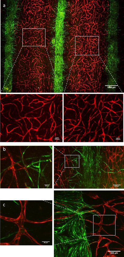
Figure 5. Histological analysis and immunostaining of the skin equivalent with perfusable vascular channels and a secondary
vascular plexus. (a) HES staining of the skin equivalent with three perfusable vessels and microvascular bed. Immunostaining of
CD31 marker of the endothelial cells, COLL IV, and Perlecan markers of the basal lamina. (b) Immunostaining of COLL IV,
Perlecan, and Laminin markers of the vascular basal lamina and the DEJ. (c) HES staining showing epidermal differentiation
layers. Immunostaining of K14 marker of the basal layer, K10 marker of the intermediate differentiation, Involucrin, Filaggrin,
and TGM1 markers of the late differentiation.
and to Lat). This decrease is significant between per- flow conditions (SandP) (figure 7). As the results
fused samples and ECnoP (P < 0.05) whereas it is between Lat, ECnoP, NoECP, and ECP were similar
a tendency with a moderate effect-size between Lat to previous ones, we only compare them to SandnoP
and perfused samples (noECP and ECP) (P < 0.1) and SandP in this experiment. In the static condi-
(figure 6). Interestingly, in static conditions, the pres- tion, even if the differences were not significant, the
ence of vascularized channels (ECnoP) significantly presence of a plexus (SandnoP) tended to increase
increased Kp for Minoxidil (P < 0.05) and tended to the Kp of Caffeine and Minoxidil with a moderate or
increase Kp with a moderate effect size for Caffeine strong effect-size compared to Lat and ECnoP. Sim-
compared to Lat model) (P < 0.1) (figure 6). While ilar to previous results, samples’ perfusion signific-
Kp obtained with the PES remained statistically lower antly decreased Kp either for Caffeine or Minoxidil
than all skin equivalents, these results showed that in SandP compared to SandnoP (P < 0.05). However,
perfused reconstructed skin has a lower Kp than non- no differences were observed between noECP, ECP,
perfused samples. and SandP (figure 7).
We then evaluated the effect of the vascular plexus Therefore, for both chemicals, we showed that the
on epidermal permeability in static (SandnoP) and perfusion of skin equivalents improved the epidermal
10Biofabrication 13 (2021) 035042 S Salameh et al
Figure 6. Evaluation of skin permeability. Permeability coefficient Kp (cm h−1 ) of each of the two chemicals and each skin model.
Lat = Control samples with no channels (N = 7), ECnoP = Channels with endothelial cells but without perfusion (N = 9),
ECP = Perfused channels with endothelial cells (N = 9), noECP = Perfused channels without endothelial cells (N = 7),
PES = Porcine ear skin (N = 8). ∗ P < 0.1, ∗∗ P < 0.05, #moderate effect size.
Figure 7. Evaluation of skin permeability. Coefficient of penetration Kp (cm h−1 ) of each of the two chemicals and each skin
model. Lat = Control samples with no channels, ECnoP = Channels with endothelial cells but without perfusion,
ECP = Perfused channels with endothelial cells, noECP = Perfused channels without endothelial cells, SandnoP = Channels with
endothelial cells and secondary microvascularization without perfusion, SandP = Perfused channels without endothelial cells and
secondary microvascularization. PES = Porcine ear skin ∗ P < 0.1, ∗∗ P < 0.05, #moderate effect size, ##strong effect size, ###very
strong effect size. N = 3 for each condition.
barrier or overall skin permeability independently of To do so, 10 µM of BaP, a well-known PAH and
the presence of ECs and the complexity of the vascular an extremely widespread environmental and indus-
network. trial pollutant [72], was added to the culture media of
each skin model. After 5 d of exposure, we measured
3.4. Evaluation of systemic exposure of a pollutant BaP concentration in epidermis, dermis, and culture
Besides topical applications, having a perfusable vas- media. In order to observe the impact of a vascu-
cularized skin model holds many interests for sys- lar plexus on BaP distribution, five different mod-
temic exposure either for inflammation studies [70] els were evaluated: Control samples with no channels
or drug screening and toxicity assessment [58, 71]. (Lat), non-perfused channels with endothelial cells
We, therefore, explored the advantage of having, for (ECnoP) or with endothelial cells and the secondary
the first time, perfusable vascular channels, but also a vascular plexus (SandnoP), perfused channels with
complex microvascular networked in a full-thickness endothelial cells (ECP) or with endothelial cells and
skin equivalent, to estimate dermal and epidermal the secondary vascular plexus (SandP) (figure 8).
transport of systemic compounds, specifically, poly- Either in static (figure 8(a)) or fluidic conditions
cyclic aromatic hydrocarbons (PAH) pollutants. (figure 8(b)), the presence of a secondary vascular
11Biofabrication 13 (2021) 035042 S Salameh et al
Figure 8. Evaluation of systemic delivery of pollutants. Graphical box plot presentation of BaP concentrations (ng ml−1 ) in the
dermis or epidermis either in static models (a) or perfused models (b). Lat = Control samples with no channels,
ECnoP = Channels with endothelial cells but without perfusion, ECP = Perfused channels with endothelial cells,
SandnoP = Channels with endothelial cells and secondary microvascularization without perfusion, SandP = Perfused channels
without endothelial cells and secondary microvascularization. ∗ P < 0.1, ∗∗ P < 0.05, #moderate effect size. N = 6 for each
condition.
plexus (SandnoP and SandP) increased BaP delivery cosmetic companies. Commercialized skin models
either in the dermis or the epidermis compared to which resulted in major advances in the field of skin
samples without channels (Lat) or with the three vas- engineering are available (e.g. EpiSkin®, SkinEthic®
cular channels only (ECnoP and ECP). This increase and T-Skin® (L’Oreal, France), Epi-Derm® (MatTek
of distribution is significant in the static dermal layer Corporation, USA), epiCS® (CellSystems, Germany),
and is a tendency with moderate effect-size in the Phenion® (Henkel, Germany), NeoDerm® (Tegos-
epidermal layer (static or perfused conditions) and cience, Korea), LabCyte® (J-TEC, Japan)). These
the perfused dermal layer. However, with (SandP) models have the advantage of being highly stable and
or without (ECP) a secondary vascular plexus, BaP reproducible as they represent mono or bi-layered
concentrations found in perfused samples were lower constructs with simple cell composition. However,
than in static conditions (figure 8(b)). These res- the lack of complexity of the in vivo skin as append-
ults are consistent with the concentrations found in ages, vasculature, cells’ variety, and distribution, is a
the culture media (figure S2(a)). Previous internal limitation for the predictivity and relevance of in vitro
studies showed that being highly hydrophobic, BaP applications. In this report, we developed a vascular-
is extremely adherent to plastic (data not shown). ized full-thickness skin model that comprises for the
Therefore, the low concentrations found in perfused first time a differentiated epidermis, perfusable ves-
samples could be due to BaP absorption by silicon sels, and a secondary microvascular network.
tubes used for peristaltic perfusion. To measure the As reconstructed skin models are specifically
loss of BaP in the tubing, a control experiment was used for safety and efficacy evaluation of chemicals,
set with all the setups of the classic culture (devices, applied systematically or topically to skin tissues, the
culture dishes, silicon tubes, and media) but without functionality of the vascularized skin equivalent for
the skin. We found that in fluidic culture either with in vitro assessments was evaluated with topical and
2.4 or 6.5 ml h−1 , BaP was ten times lower than static systemic applications.
culture (figure S2(b)). One important readout for topical application
Thus, in this experiment, fluidic and static con- is the estimation of new compounds’ skin absorp-
ditions could not be compared as the skin is exposed tion including skin distribution, metabolism, and
to different concentrations of BaP during the culture. elimination from the skin into systemic circulation.
However, these results proved that the presence of a However, existing skin models have a higher per-
complex vascular plexus increases systemic diffusion meation than in vivo skin [73] which could be related
of the pollutant either in the dermis or the epidermis. to a barrier function default. We, therefore, evalu-
ated the effect of vascularization and perfusion on
4. Discussion epidermal barrier integrity by evaluating the per-
cutaneous absorption of caffeine (high permeability
Currently, 2D and 3D in vitro skin models are used chemical compound) and minoxidil (low permeab-
to replace animal testing both in pharmaceutical and ility chemical compound). For both chemicals, we
12Biofabrication 13 (2021) 035042 S Salameh et al
showed that percutaneous absorption is lower in per- platforms were used to mimic blood flow for top-
fused samples. This could be due to an improve- ical and systemic studies [14, 18, 20]. However, in
ment of epidermal barrier function or the overall these platforms, fluidic media passes beneath the
skin permeability. In any case, these results suggest skin-equivalent whilst in vivo, it circulates through
that perfused skin models are a better alternative the dermis in confined tube-shaped microvessels.
for the assessment of skin permeability and penetra- Most of the time, culture media is also separated
tion in vitro compared to non-perfused models. This from the skin by a permeable membrane that could
is consistent with other studies showing the benefi- introduce a supplemental and non-physiological bar-
cial effect of fluidic cultures on the epidermal bar- rier for substance diffusion. On the other hand, the
rier function [18, 74]. Indeed, besides improving few existing skin models representing perfusable ves-
nutrients and oxygen supply, media perfusion was sels for systemic delivery of drugs were limited to
shown to enhance the deposition of DEJ proteins [18] macro-channels located in the dermis [28, 58, 60].
which is crucial for the basement membrane integ- Lack of micro-vascularization could result in mis-
rity and epidermal differentiation. It was also proven leading and/or irrelevant drug delivery estimation
to increase cell viability and tight junction forma- compared to the in vivo situation. To show the advant-
tion [74] as well as the expression of epidermal mark- age of having a complex vasculature, we exposed our
ers and stratum corneum thickness [75]. Addition- vascularized skin model to BaP in order to mimic
ally, the shear stress induced by dynamic culture can systemic exposure to pollutants. Pollutants like BaP
also affect epidermal maturation and barrier function can promote photoreactivity and phototoxicity with
[14, 18, 75]. However, in our experiments presence nanomolar range concentrations [72]. Various stud-
of ECs in non-perfused conditions seemed to lower ies have shown that a combined exposure of pollut-
the efficacy of the barrier function. We hypothesized ants and UVs could be toxic and aggravate sunlight-
that the presence of endothelial cells may delay the induced skin damage [72, 77–79]. However, these
maturation of the epidermis as both cell types share studies were investigated either in vivo, in 2D cell
common growth factors like EGF, insulin or hydro- culture, or 3D epidermal equivalents whereas UVs
cortisone. In fact, in static conditions, media diffuses and especially UVA (UVA1 and UVA2) wavelength
through the dermis. Therefore, growth factors could are capable to reach the dermal layer [80]. Moreover,
gradually be depleted by endothelial cells. This could BaP was measured in the blood of smokers and indi-
also explain why additional ECs in the secondary viduals living in polluted areas [81, 82]. This suggests
vascular plexus further increased the penetration of that the skin could be exposed to these pollutants
chemicals. To confirm this hypothesis, samples with from the surface but also the systemic circulation.
ECs could be cultured for a longer period to meas- Therefore, having a complex vascular structure in a
ure whether we find the same permeability as non- dermal equivalent allows a better understanding of
vascularized samples. BaP transport and its effects on the dermis before
In this study, we measured the concentration of reaching the epidermal layer. Indeed, we found that
chemicals in static media beneath the skin. However, either in static or fluidic conditions, the presence
we could also evaluate percutaneous penetration and of the secondary vascular plexus increased the pres-
systemic concentration of compounds in the vascular ence of benzo[a]pyren BaP in the dermis and the
channel underflow which could better mimic capil- epidermis compared to samples without vessels or
lary clearance and improve safety evaluation. In fact, with the three vascular channels alone. This sug-
after topical application, some chemicals are absorbed gests that having a finer vascular structure makes the
and/or metabolized and reach the systemic circula- systemic delivery of substances to or from the skin
tion. Under certain concentrations, these chemicals more efficient. This could apply not only to pollut-
could cause side effects on other distant organs [76]. ants but to any drugs and compounds systemically
Therefore, a skin model with a closer permeation to delivered.
in vivo physiology would allow a better prediction of We, therefore, demonstrated that our vascular-
systemic toxicity. ized full-thickness skin is a valuable model for top-
Besides topical applications, having a perfusable ical or systemic assessments. In this study, we focused
vascularized skin model holds many interests for sys- on the systemic delivery and transdermal absorption
temic applications. Most of the existing skin mod- of compounds. However, a broad range of applica-
els are cultured in static conditions which is not rep- tions is possible either for knowledge studies or effic-
resentative of the in vivo reality [58]. Additionally, acy and safety evaluations. For instance, vascular-
substances are usually added to static culture media ized skin models could also serve to study wound
in contact with all the surface of dermis equivalents healing, skin regeneration as well as the molecular
whereas, in vivo, systemic substances are delivered via mechanisms involved. In fact, 3D skin reconstructs
the vascular network meaning that they are exposed were already used to mimic the wound healing pro-
to blood flow and have the vessel wall as an addi- cess in in vitro trauma models [83]. However, these
tional diffusion barrier. In that sense, skin-on-a-chip models do not represent the local microenvironment
13Biofabrication 13 (2021) 035042 S Salameh et al
of in vivo wounds and cannot predict the side effect 5. Conclusion
of the therapeutic treatment due to the lack of vas-
cularization, systemic circulation, and inflammation We developed for the first time a full-thickness
mediators, widely reported as critical components human skin model with a mature epidermis and three
during the wound healing process [84]. Therefore, tubular structures with angiogenic sprouts that can
including a vascular network and systemic perfusion be perfused, associated with a complex microvascular
to reconstructed skin models could improve the effi- network. The integrity of each compartment was con-
ciency of screening therapeutic compounds and the firmed by histological immunofluorescence analysis
prediction rate of patient’s responses to such treat- and compared to normal human skin. We proved that
ments. Besides screening therapeutic compounds for having a perfusable vasculature closer to the in vivo
wound healing, having both angiogenesis and vas- vascular plexus resulted in a more reliable model for
culogenesis processes in the same skin model could topical and systemic assessments. This includes a wide
improve the screening of pro and antiangiogenic range of applications either for knowledge or efficacy
drugs. Indeed, vascularization plays an important role and safety studies.
in tumor growth and metastasis [85]. Therefore, this
model could be used to better understand tumor, Data availability statement
stroma, and endothelial cells’ interactions in non-
melanoma skin cancer. In addition, this complex vas- The data that support the findings of this study are
cularized skin model could also be beneficial for the available upon reasonable request from the authors.
integration of skin appendages that require blood
supply. For instance, without prevascularization and Acknowledgments
perfusion, generating an in vitro skin-equivalent with
hair follicles density equivalent to the in vivo situation We greatly appreciate Nobuhito Mori for his advice
could result in necrosis [10]. Indeed, fluidic systems and technical support. We thank Florence Berthelot
have proven to prolong the culture period of ex vivo for her help in HUVECS transduction, Samia Boudah
hair follicles and postpone the initiation of the hair for her support in HPLC-Fluo analysis and France
follicle regression phase (catagen) compared to static Maloumian for her support in schematizing the pro-
conditions [14]. tocol. We are grateful to Sébastien Grégoire for fruit-
Our skin model represents an advanced step com- ful discussions and analysis of skin permeability
pared to existing models but it also includes limita- assays. We also thank Julien Demaude and Alexandre
tions. Indeed, numerous parameters might further be Nicolas for their contribution in the initiation of this
improved. HUVECs were used as they are quite easy project.
to access and their culture condition is well-known.
Nevertheless, HUVECs could be replaced by human ORCID iDs
dermal microvascular endothelial cells which would
be more relevant for a skin model. In fact, HUVECs Sacha Salameh https://orcid.org/0000-0002-3162-
are isolated from large vessels whereas dermis vas- 994X
culature mainly consists of microvessels [31]. Vessel Shoji Takeuchi https://orcid.org/0000-0001-6946-
diameter is not the only difference, but this para- 0409
meter could influence cell behavior as it was already
shown that endothelial cells from different blood ves- References
sels or different tissues have distinct gene expres-
sion profiles [86]. Bioprinting could also be used as a [1] Meyer U 2009 The History of Tissue Engineering and
replacement of the molding technique to create func- Regenerative Medicine in Perspective Fundamentals of
Tissue Engineering and Regenerative Medicine (Berlin:
tional a vascular network that would contain a more
Springer) pp 5–12
complex architecture (smaller vessel diameters and [2] Albanna M and Holmes J H IV 2016 Skin Tissue Engineering
improved spatial control of their distribution) com- and Regenerative Medicine (Boston: Academic Press)
pared to tubular channels [87]. Moreover, arteries, pp 1–443
[3] Groeber F, Holeiter M, Hampel M, Hinderer S and
veins, and lymphatic vessels could be created using
Schenke-Layland K 2011 Skin tissue engineering—in vivo
multi-channels. Indeed, engineering reconstructed and in vitro applications Adv. Drug. Deliv. Rev. 63 352–66
tissues with both blood and lymphatic capillaries was [4] Yousuf Y et al 2018 2—Overall perspective on the clinical
previously reported [88–90]. Finally, besides optim- importance of skin models Skin Tissue Models ed
A P Marques (New York: Academic) pp 39–54
ization of skin reconstruction, the device could also
[5] Pellevoisin C et al 2018 1—Cosmetic industry requirements
be improved to be able to separate the culture media regarding skin models for cosmetic testing Skin Tissue
surrounding the skin equivalent from the one perfus- Models ed A P Marques (New York: Academic) pp 3–37
ing the vascular channels. This improvement would [6] Rheinwald J G and Green H 1975 Serial cultivation of strains
of human epidermal keratinocytes: the formation of
better mimic skin physiology and open the possib-
keratinizing colonies from single cells Cell 6 331–43
ilities for new applications requiring immune cells [7] Black A F, Berthod F, L’Heureux N, Germain L and
perfusion. Auger F A 1998 In vitro reconstruction of a human
14Biofabrication 13 (2021) 035042 S Salameh et al
capillary-like network in a tissue-engineered skin equivalent [28] Mori N, Morimoto Y and Takeuchi S 2017 Skin integrated
Faseb. J. 12 1331–40 with perfusable vascular channels on a chip Biomaterials
[8] Regnier M, Staquet M-J, Schmitt D and Schimdt R 1997 116 48–56
Integration of Langerhans cells into a pigmented [29] Massa S et al 2017 Bioprinted 3D vascularized tissue model
reconstructed human epidermis J. Invest. Dermatol. for drug toxicity analysis Biomicrofluidics 11 044109
109 510–2 [30] Dai N-T et al 2018 Development of a novel pre-vascularized
[9] Bertaux B, Morliere P, Moreno G, Courtalon A, Masse J M three-dimensional skin substitute using blood plasma gel
and Dubertret L 1988 Growth of melanocytes in a skin Cell Transplant. 27 1535–47
equivalent model in vitro Br. J. Dermatol. 119 503–12 [31] Bourland J et al 2018 8—Strategies to promote the
[10] Abaci H E, Coffman A, Doucet Y, Chen J, Jacków J, Wang E, vascularization of skin substitutes after transplantation Skin
Guo Z, Shin J U, Jahoda C A and Christiano A M 2018 Tissue Models ed A P Marques (New York: Academic)
Tissue engineering of human hair follicles using a pp 177–200
biomimetic developmental approach Nat. Commun. 9 5301 [32] Li W, Lan Y, Guo R, Zhang Y, Xue W and Zhang Y 2015
[11] Huber B, Link A, Linke K, Gehrke S A, Winnefeld M and in vitro and in vivo evaluation of a novel collagen/cellulose
Kluger P J 2016 Integration of mature adipocytes to build-up nanocrystals scaffold for achieving the sustained release of
a functional three-layered full-skin equivalent Tissue Eng. C basic fibroblast growth factor J. Biomater. Appl. 29 882–93
22 756–64 [33] Joshi V S, Lei N Y, Walthers C M, Wu B and Dunn J C Y 2013
[12] Albouy M, Tanguy M, Onteniente B, Thepot A, Maruotti J Macroporosity enhances vascularization of electrospun
and Dos Santos M 2018 Development of A 3D full-thickness scaffolds J. Surg. Res. 183 18–26
skin equivalent model containing sebocyte organoids [34] Griffin M, Palgrave R, Baldovino-Medrano V, Butler P and
derived from human ips cells J. Invest. Dermatol. 138 B24 Kalaskar D 2018 Argon plasma improves the tissue
[13] Muller Q, Beaudet M-J, De Serres-Bérard T, Bellenfant S, integration and angiogenesis of subcutaneous implants by
Flacher V and Berthod F 2018 Development of an innervated modifying surface chemistry and topography Int.
tissue-engineered skin with human sensory neurons and J. Nanomed. 13 6123–41
schwann cells differentiated from iPS cells Acta Biomater. [35] Cam C, Zhu S, Truong N F, Scumpia P O and Segura T 2015
82 93–101 Systematic evaluation of natural scaffolds in cutaneous
[14] Atac B, Wagner I, Horland R, Lauster R, Marx U, wound healing J. Mater. Chem. B 3 7986–92
Tonevitsky A G, Azar R P and Lindner G 2013 Skin and hair [36] Suh W et al 2005 Transplantation of endothelial progenitor
on-a-chip: in vitro skin models versus ex vivo tissue cells accelerates dermal wound healing with increased
maintenance with dynamic perfusion Lab Chip 13 3555–61 recruitment of monocytes/macrophages and
[15] Wagner I et al 2013 A dynamic multi-organ-chip for neovascularization Stem Cells 23 1571–8
long-term cultivation and substance testing proven by 3D [37] Sieveking D P, Buckle A, Celermajer D S and Ng M K C 2008
human liver and skin tissue co-culture Lab Chip 13 3538–47 Strikingly different angiogenic properties of endothelial
[16] Lee S, Jin S-P, Kim Y K, Sung G Y, Chung J H and Sung J H progenitor cell subpopulations: insights from a novel human
2017 Construction of 3D multicellular microfluidic chip for angiogenesis assay J. Am. Coll. Cardiol. 51 660–8
an in vitro skin model Biomed. Microdevices 19 22 [38] Cao Y, Sun Z, Liao L, Meng Y, Han Q and Zhao R C 2005
[17] Jeong Song H et al 2017 Development of 3D skin-equivalent Human adipose tissue-derived stem cells differentiate into
in a pump-less microfluidic chip J. Ind. Eng. Chem. endothelial cells in vitro and improve postnatal
60 355–59 neovascularization in vivo Biochem. Biophys. Res. Commun.
[18] Sriram G, Alberti M, Dancik Y, Wu B, Wu R, Feng Z, 332 370–9
Ramasamy S, Bigliardi P L, Bigliardi-Qi M and Wang Z 2018 [39] Jean J 2013 Bioengineered skin: the self-assembly approach J.
Full-thickness human skin-on-chip with enhanced Tissue Sci. Eng. 03 S5:001
epidermal morphogenesis and barrier function Mater. Today [40] Hikimoto D, Nishiguchi A, Matsusaki M and Akashi M 2016
21 326–40 High-throughput blood- and lymph-capillaries with
[19] Sung J H, Esch M B, Prot J-M, Long C J, Smith A, open-ended pores which allow the transport of drugs and
Hickman J J and Shuler M L 2013 Microfabricated cells Adv. Healthcare Mater. 5 1969–78
mammalian organ systems and their integration into models [41] Miyazaki H, Tsunoi Y, Akagi T, Sato S, Akashi M and
of whole animals and humans Lab Chip 13 1201–12 Saitoh D 2019 A novel strategy to engineer pre-vascularized
[20] Abaci H E, Gledhill K, Guo Z, Christiano A M and 3-dimensional skin substitutes to achieve efficient,
Shuler M L 2015 Pumpless microfluidic platform for drug functional engraftment Sci. Rep. 9 7797
testing on human skin equivalents Lab Chip 15 882–8 [42] Schultheiss D et al 2005 Biological vascularized matrix for
[21] Abaci H E, Guo Z, Doucet Y, Jacków J and Christiano A 2017 bladder tissue engineering: matrix preparation, reseeding
Next generation human skin constructs as advanced tools for technique and short-term implantation in a porcine model J.
drug development Exp. Biol. Med. 242 1657–68 Urol. 173 276–80
[22] Lovett M, Lee K, Edwards A and Kaplan D L 2009 [43] Groeber F et al 2016 A first vascularized skin equivalent as an
Vascularization strategies for tissue engineering Tissue Eng. B alternative to animal experimentation ALTEX 33 415–22
15 353–70 [44] Gershlak J R et al 2017 Crossing kingdoms: using
[23] Min S, Ko I K and Yoo J J 2019 State-of-the-art strategies for decellularized plants as perfusable tissue engineering
the vascularization of three-dimensional engineered organs scaffolds Biomaterials 125 13–22
Vasc. Spec. Int. 35 77–89 [45] Uygun B E et al 2010 Organ reengineering through
[24] Rouwkema J and Khademhosseini A 2016 Vascularization development of a transplantable recellularized liver graft
and angiogenesis in tissue engineering: beyond creating using decellularized liver matrix Nat. Med. 16 814–20
static networks Trends Biotechnol. 34 733–45 [46] Skardal A, Zhang J and Prestwich G D 2010 Bioprinting
[25] Liu J, Zheng H, Poh P, Machens H-G and Schilling A 2015 vessel-like constructs using hyaluronan hydrogels crosslinked
Hydrogels for engineering of perfusable vascular networks with tetrahedral polyethylene glycol tetracrylates
Int. J. Mol. Sci. 16 15997–6016 Biomaterials 31 6173–81
[26] Auger F A, Gibot L and Lacroix D 2013 The pivotal role of [47] Hinton T J et al 2015 Three-dimensional printing of complex
vascularization in tissue engineering Annu. Rev. Biomed. Eng. biological structures by freeform reversible embedding of
15 177–200 suspended hydrogels Science Advances 1 e1500758
[27] Amann A et al 2017 Development of a 3D angiogenesis [48] Zhang Y, Yu Y, Akkouch A, Dababneh A, Dolati F and
model to study tumour—endothelial cell interactions and Ozbolat I T 2015 In vitro study of directly bioprinted
the effects of anti-angiogenic drugs Sci. Rep. 7 2963 perfusable vasculature conduits Biomater. Sci. 3 134–43
15You can also read