Nerve Sheath Tumors in an Isolated Goldfish Population
←
→
Page content transcription
If your browser does not render page correctly, please read the page content below
Nerve Sheath Tumors in an Isolated Goldfish Population*

                                             HANS G. SCHLUMBERGER

                     (Dept. of Pathology, Collegeof Medicine, The Ohio State Unirersity, Columbus 10, Ohio)

                 INTRODUCTION                                      dom found, and none of them bore tumors; it is
    During the past 6 years studies have been car                  probable that only rarely did the tumors interfere
ried out on the morphology, growth, and possible                   with the normal life of the fish.
etiology of nerve sheath tumors observed in gold                       It is regrettable that an ecological survey of the
fish, Carassius auratus. The fish were all inhabit                 pond has not been made, for it presents a rather
ants of a large lagoon in the city of Cleveland.1                  unique opportunity to study a restricted yet essen
The tumors were neurilemomas and neurofibro-                       tially natural environment of a large number of
mas; in seven instances they were histologically                   tumor-bearing animals. The lagoon was stocked 26
malignant. The question of the neoplastic char                     years ago, and no goldfish have been added since
acter of the melanophores found in several deeply                  then. Though the possibility that visitors may
pigmented tumors will be considered.                               have introduced an occasional one must be enter
   After an account of the fish and their environ                  tained, no instance of this is known to the care
ment, the morphology of the tumors will be de                      takers or patrolmen on the grounds.
scribed, followed by data on transplantation and                       In addition to the goldfish, blue gill sunfish
transmission experiments which were carried out.                    (Lepomis incisor) and large-mouth black bass
The possible relation of the tumors and certain                     (Micropterus salmoides) are living and breeding in
abnormalities in the fish to constitutional factors,               the lagoon. Of those caught in the seine none
and the similarity of the lesions to those encoun                  showed visible evidence of a tumor, nor was there
tered in von Recklinghausen's neurofibromatosis                    any sign of ocular injury or copepod infestation so
will be presented in the discussion.                               common in the goldfish.
                                                                      The lagoon has a surface area of 3-4 acres; ex
           MATERIALS AND RESULTS                                   cept for three small sandy beaches it is enclosed by
           THEFISHANDTHEIBENVIRONMENT                              low limestone embankments. The water is largely
   Goldfish visible from the shore of the pond were                supplied by surface drainage and fountain over
large, measuring 20-35 cm. in over-all length. An                  flow, supplemented by municipal water during pe
age estimate of 5-8 years was based on studies of                  riods of drought. The surrounding lawns are well
the scales made at the Institute of Hydrobiology                   fertilized, which should aid in maintaining a rich
of the Ohio State University. Fish that bore tu                    plankton population. There is no evidence of pol
mors 1 or more centimeters in diameter were read                   lution.
ily spotted as they swam slowly about. They could                     The pond is drained to one-third its area each
be lured to within reach of the net by dropping                    spring, and the marginal zone cleaned for a dis
pieces of soda cracker into the water. Occasionally                tance of 20 feet. It requires at least 10 days of con
an entire school of fish was caught in a large seine               stant flow from a city hydrant to refill the lagoon.
when normal as well as tumor-bearing specimens                     During the summer months the temperature of the
were needed. Inspection of these fish showed that                  water averages 65°     F. (18.3°C.) near shore. In
8-10 per cent bore one or more tumors. This inci                   winter the entire surface is frozen, often to a depth
dence did not change appreciably during the years                  of 12 inches, but very few fish die of suffocation.
of observation. The neoplasms occurred with equal
frequency in both sexes. Dead goldfish were sel-                                 THE NERVESHEATHTUMORS
   * This investigation was supported in part by a research           Tumors of the peripheral nerves are among the
grant from the National Cancer Institute, U.S. Public Health       least common neoplasms found in fishes (30). Of
Service.                                                           seven papers on the subject, three describe a gan-
   1Mr. Arnold M. Davis, director of the Garden Center of          glioneuroma (12, 34, 35) and one a neuroepithelio-
Greater Cleveland, kindly gave permission for the discriminate     ma (36). Picchi (24) reported a pea-sized tumor
use of the fish and supplied data about the lagoon.                that he identified as a schwannoma in the region of
   Received for publication July 26, 1952.                         a caudal vertebra in a large goldfish. Young and
                                                             890

Downloaded from cancerres.aacrjournals.org on January 19, 2021. © 1952 American Association for Cancer
                                              Research.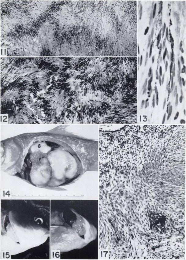
ScHLUMBERGER—Nene Sheath Tumors in Goldfish                                        891

Olafson (42) found multiple lesions in the auto            however, most were found on the dorso-lateral as
nomie nerves of 25 young brook trout. Although             pect of the head and trunk and on the caudal fin
the authors describe the pathologic changes as              (Chart 1). Although this is in the area of the lateral
characteristic of neurilemomas, their evidence for         line sensory organs and the larger nerve trunks, no
the neoplastic nature of the lesions is not entirely       tumor attachment to such a nerve could be demon
convincing.                                                strated.
   A comprehensive study of tumors of the nerve               The neoplasms ranged in size from 4 mm. to 4.5
sheaths in fishes was made by Lücke(15), who              cm. in diameter (Figs. 1, 3,4,8). The small tumors
published a detailed description of the gross and          were flat, orange-yellow in color, and firm in con
microscopic appearance of these neoplasms in 76            sistency. The larger lesions were hemispherical in
specimens of the snapper family (Lutianidae). The          shape and broadly sessile; only one was peduncu-
affected fish were of three species: L. griseus, L.        lated (Fig. 5). The surface of the tumors was
apodus, and L. jocu; most were caught near the Dry         smooth, sometimes gently lobulated; scales were
Tortugas in the Gulf of Mexico. The tumors gen             absent. The neoplasms were usually quite soft,
erally occurred along the course of the larger sub         pink in color, and occasionally hemorrhagic (Fig.
cutaneous nerves and had the histologie appear             2). All tumors were very vascular and bled pro
ance characteristic of neurilemomas seen in man.           fusely after injury. A few were pigmented (Fig.
                                                           23). Three tumors were cystic ; one of these overlay
        GROSS MORPHOLOGY  ANDDISTRIBUTION                  a 2-mm. defect in the skull, but the underlying
  The 144 tumors in the 53 goldfish examined               brain and meninges were intact.
were widely scattered over the surface of the body ;          None of the tumors was encapsulated, although
            LEFT

            RIGHT

   CHART1.—Distribution of 144 tumors found in 58 goldfish. The tumors occur most frequently on the dorso-lateral
aspect of the head and trunk, and on the caudal fin.

Downloaded from cancerres.aacrjournals.org on January 19, 2021. © 1952 American Association for Cancer
                                              Research.
892                                         Cancer Research

most were well circumscribed and did not pene          Schwann are similar to those found in other verte
trate the compact layer of the dermis. However,        brates (21). The normal histology of the goldfish
invasion of the underlying musculature (Fig. 31)       skin has been described by Graupner and Fischer
was observed twice. Metastasis did not occur, but      (10).
multiple primary tumors were often seen (Figs. 4,         Neurilemoma.—The palisading of nuclei so char
8, 23). Of the 53 fish examined, 31 had two or more    acteristic of the Antoni type A tissue in the neuri
tumors; the largest number found on any one fish       lemoma was prominent in 25 per cent of the tu
was eleven.                                            mors (Figs. 11, 17) and less apparent in several
   One goldfish bore no surface lesions, but the       others (Fig. 7). Although this nuclear arrangement
right operculum was elevated by a 4 X 4 X 3.5-         is most often found in nerve-sheath tumors, it oc
cm. tumor that arose from the second gill arch on      casionally occurs in leiomyomas (33), and in the
the right (Figs. 15. 16). Another fish had, in addi    early stages of this study several tumors were so
tion to three skin tumors, a markedly distended        identified (27). However, further investigation and
abdomen. After death of the animal, two large          the accumulation of additional material indicate
contiguous tumors measuring 6.5 X 4.5 X 5 cm.          that these, too, were neurilemomas.
were found to occupy most of the abdominal cav            Organoid structures such as the Verocay bodies
ity (Fig. 14). The viscera had been displaced, the     that resemble tactile corpuscles were not seen.
ovaries compressed and distorted. The tumors           Trichrome stains showed only occasional narrow
were loosely adherent to the parietal peritoneum;      collagen bundles; silver stains revealed numerous
despite careful dissection no other site of origin     reticulin fibers, but only rarely were neurites en
could be found.                                        countered. The cells of all type A tissue, whether
   In many tumor-bearing fish careful inspection       they showed nuclear palisading or not, were very
revealed 1-mm. thick elevations at the tip of iso      elongate; cell boundaries were indistinct or absent.
lated scales; the surface was mammilated, orange-      The nuclei were uniform in size, long and oval in
yellow in color (Fig. 21). Occasionally, many con      shape, and contained one to three prominent nu
tiguous scales or a large area of the scaleless head   cleoli. Mitotic figures were uncommon.
were similarly affected (Fig. 18). The limbus of one      The Antoni type B tissue with its irregularly ar
or even both eyes was also often the site of diffuse   ranged stellate cells and occasional microcysts
neoplastic thickening (29). Frequently, the lesion     (Fig. 6) was less prominent in these tumors than
encroached upon the cornea (Fig. 9).                   the more compact type A. In several instances
                                                       there appeared to be a transition between the two
              MICROSCOPIC
                        MORPHOLOGY
                                                       types characterized by cells which, though still
   Since the experimental production of neurilemo-     elongated, had an abundant cytoplasm in which
mas by Masson (16) in 1932, much work has been         the appearance of vacuoles coupled with intercel
done on the histogenesis of peripheral nerve tu        lular microcysts seemed to foreshadow the loosely
mors. These studies, among which those of Stout        arranged meshwork of the fully developed type B
(31, 33) and Murray and Stout (19, 20) must be         tissue (Fig. 10). The blood vessels had thin walls
reckoned particularly significant, led to the con      without the collagen sheaths described in some of
sensus that the chief cellular component of both       the human tumors. Multiple thrombi were only
the neurilemoma and the neurofibroma is the            encountered in the single pedunculated tumor
nerve-sheath cell of Schwann rather than the peri-     mentioned above.
neural fibroblast. The study of the histology as          Tissue cultures of these tumors were prepared
well as of the growth and development of the gold      according to a technic similar to that described
fish tumors gave results that are compatible with      elsewhere (27). There was often considerable dif
this concept of their histogenesis.                    ference in the appearance of the cells from various
   The tumors apparently arose from the small          expiants. In some the cells were stellate in shape
superficial nerves that are very numerous in the       with an abundant cytoplasm ; in other areas of the
corium of the goldfish. Many of the nerves end as      explant or in other expiants of the same tumor the
naked neuntes in the epidermis, others supply end      cells approached the extremely long spindle shape
organs known as terminal buds that structurally         (Fig. 36) shown by Murray and Stout (19) to char
resemble the taste buds of mammals. These are          acterize the human Schwann cell. The great pleo-
abundant on the head and trunk of goldfish and         morphism of the nerve-sheath cell growing in vitro
other fish of sluggish habit that live in mud (13).    has been emphasized by Weiss (39, 40), who ob
The only other specialized end organs in fish are      served their transformation into macrophages.
the neuromasts—organs of the lateral line system         Neurofibroma.—The thickenings observed at
(1). Histologically, the myelin sheath cells of        the tip of the scales or diffusely involving the

Downloaded from cancerres.aacrjournals.org on January 19, 2021. © 1952 American Association for Cancer
                                              Research.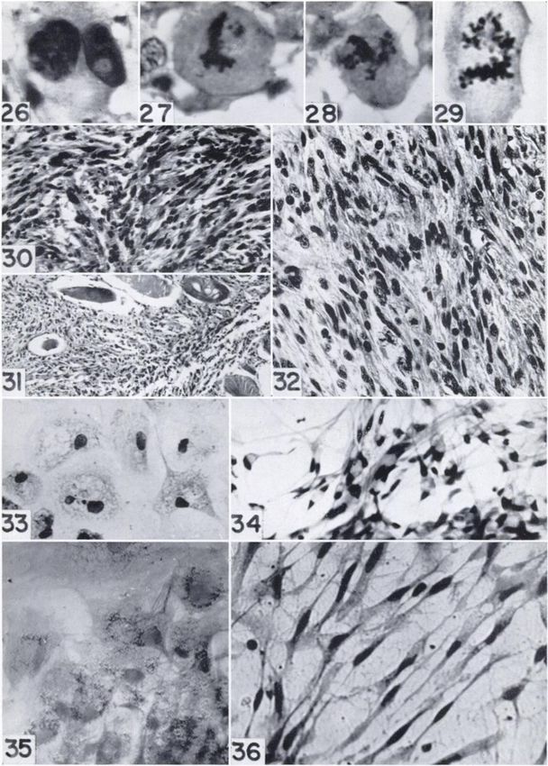
ScHLUMBERGER—NerveSheath Tumors in Goldfish                                    893
dorsum of the head have a histological appearance      that these findings do not rule out the origin of
that corresponds well with that currently accepted     melanophores during normal development from
for the neurofibroma (Figs. 19 and 22). The neo-       cells corresponding to those of the neural crest in
plastic cells, which were cytologically indistin       other vertebrates.
guishable from those of the type A tissue in the           Skin pigmentation in the goldfish is subject to
neurilemoma, were unlike the latter in their hap       considerable change; e.g., it is often increased in
hazard arrangement. Silver stains showed neuntes       areas of inflammation. Goodrich (7) has presented
were present (Fig. 20), a feature characteristic of    evidence of the existence in the goldfish dermis of a
human cutaneous neurofibromas (18). Neurofibro-        reservoir of melanoblasts (unpigmented precursors
mas and neurilemomas often co-existed on the           of melanophores). From these melanoblasts clus
same fish. When bleached, the histologie appear        ters of melanophores can be derived. Nevertheless,
ance of two pigmented tumors resembled that of a       the possibility exists that in the pigmented neuro
neurofibroma.                                          fibromas and neurilemomas of the goldfish the
   Pigmented nerve sheath tumors.—Although many      melanophore is derived from the same stem cell as
tumors had small areas of pigmentation, the four       is the Schwann cell and that it is an integral part of
selected for extensive histologie study were gray or   the tumor. This intimate relation of the two cell
black throughout. They occurred on four fish that      types recalls Masson's concept of pigmented moles
also bore one or more nonpigmented neurilemo           (17) which he believes are composed of a down
mas. Two involved the base of the left pectoral fin,   ward proliferation of intraepidermal melanoblasts
one grew on the caudal fin, and another was found      and upward migration of Schwann cells from the
on the dorsum of the trunk (Fig. 23).                  dermal nerves.
    In one the pigment was so dense that nuclear          The pigment-producing ability of Schwann cells
and cytoplasmic detail could be made out only at       has been described in certain ocular melanomas by
the periphery of the tumor or after bleaching.         Reese (26), who labeled the tumors "neurogenic
Growth in tissue culture was poor, but after 13        melanomas." A similar histologie picture was ob
days in roller tubes small numbers of pigment-         served by the writer in a malignant melanoma that
bearing cells had migrated out of the explant (Fig.    arose in the body wall of a python (30).
35). They did not have the branched appearance            In the light of Weiss's studies on the in vitro
of melanophores and resembled the pigment-laden        transformation of the Schwann cell into a macro
macrophages seen in cultures of fish, mouse, and       phage (39,40), the possibility should be considered
human melanomas by Grand and Cameron (9).              that the melanin-bearing macrophages described
    In another neurofibroma the pigmented cells        above (Fig. 35) are melanophores similarly altered
were less numerous and the melanin granules more       rather than macrophages that have phagocytized
widely scattered in the melanophores. This per         the pigment of disintegrating melanophores in the
mitted a comparison of the nuclei of the pigment       expiant.
cells and of the Schwann cells in the untreated           Malignant neurilemoma.—The histology of the
slide (Fig. 24). No distinguishing characteristics     nerve sheath tumors thus far considered has been
were recognizable, and in bleached sections it         that of benign neoplasms characterized by the
could no longer be determined which was the pig        absence of any profound cellular abnormality.
ment cell and which the Schwann cell (Fig. 25).        However, in seven fish there were rapidly growing
    In a neurilemoma which grossly was diffusely       tumors that showed histological evidence of malig
pigmented, histological sections revealed less mela    nancy. All were large, three were solitary, neuro
nin than was anticipated ; typical nuclear palisad     fibromas accompanied the other four. One of the
ing was present (Fig. 12). Within the palisades        tumors apparently arose in the right eye which it
were cells which differed in no way from their com     had destroyed, another grew on the dorsum of the
panions, except that the cytoplasm contained           head, a third occupied a large area of the opercu-
large numbers of melanin granules (Fig. 13).           lum behind the right eye (Fig. 8). The remaining
   The data on the origin of the vertebrate pigment    four were located on the dorsolateral aspect of the
cell (melanophore) has been reviewed by Rawles         trunk (Fig. 40).
(25), who concluded that in all classes they were         The histologie picture presented by these tu
derivatives of the neural crest. More recently,        mors was quite variable. It differed essentially
however, Oppenheimer (23) has shown experimen          from that of the benign tumors in the more abun
tally that in embryos of the fish Fundidus hetero-     dant cytoplasm of the cells and the frequent ap
clitus pigment cells can arise from cells that nor     pearance of giant forms with bizarre nuclei and
mally do not contribute to the teleostean counter      prominent nucleoli (Fig. 26). Normal and abnor
part of the neural crest. Nevertheless, she states     mal mitotic figures were often seen (Figs. 27 and

Downloaded from cancerres.aacrjournals.org on January 19, 2021. © 1952 American Association for Cancer
                                              Research.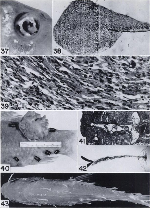
894                                           Cancer Research

28). In several instances the chromosomes, which         tured in vitro, where growth was poor. During the
normally are very small with a diploid number of         remaining 8 months of life there was no recurrence
90, were large, resembling the diplochromosomes          of the tumor at the primary site. The best in vitro
described by Biesele (2) in an ovarian tumor of the      growth of a benign tumor was obtained with a
goldfish (Fig. 29).                                      neurilemoma that arose from the second gill arch
    The neoplastic cells were arranged as interlacing    (Figs. 16 and 36).
bundles in which there was occasionally a sugges             The seven tumors that showed histological evi
tion of nuclear palisading (Fig. 32). Areas similar      dence of malignancy all grew rapidly during the
to the Antoni type B tissue of the benign neuri-         32-203 days they were under observation (Figs. 2
lemoma were encountered. Silver stains failed to         and 3). In one instance the tumor appeared to out
reveal the presence of neurites, and reticulin stains    strip its blood supply, for the bulk of it became
did not show the encircling reticulin fibers deemed      necrotic and sloughed off. Viable tumor continued
characteristic of fibrosarcoma (32). The histologie      to grow about the margins of the resultant ulcer. A
appearance of these tumors in the goldfish closely       tumor that was observed for 203 days displayed
resembles that of malignant        neurilemomas     re   alternating   periods of slow and rapid growth.
cently reported in man (8, 38).                          Biopsies taken from four of these seven tumors
    Invasion of the underlying musculature         was   yielded good growth in tissue culture.
only observed twice (Fig. 31); no métastases were
                                                                      TRANSPLANTATION
                                                                                   EXPERIMENTS
found. The only instance of the successful trans
plantation of a tumor in this entire series was an           Numerous     auto-, homoio-, and heterologous
autotransplant    in one of these seven cases (Figs.     transplants of the benign as well as of the histologi-
37-39).                                                  cally malignant tumors were made. Only a single
    Several of the malignant tumors grew well in         one, an autotransplant      to the anterior chamber of
tissue culture. Although they rarely displayed the       the eye, showed evidence of progressive growth.
very elongate form of human Schwann cells in             This failure of the transplants        to grow, coupled
vitro, many had the interesting "flask" shape (Fig.      with the absence of métastases, would indicate
34) and monocytoid character (Fig. 33) observed          that none of the goldfish tumors had reached the
by Weiss in Schwann cells from the nerves of adult       stage of autonomous growth (11).
rats. (Compare with Weiss's Figures 5 and 6,                 The technic used in transplanting         tissue to the
Plate I [40].) However, whether the monocytoid           anterior chamber of fish eyes is similar to that em
cells represent  morphological  variants of the          ployed with other animals. One difficulty should be
Schwann cells or macrophages present in the tu           noted; viz., the anterior chamber fluid of the gold
mor expiants could not be determined.                    fish is very viscid and escapes through the wound
                                                         for several minutes after the sclero-corneal incision
            RATEANDMANNEROF GROWTH                       has been made, often carrying the transplant with
    The benign tumors grew slowly; e.g., a neurile-      it. This was overcome by introducing the tumor
moma on the caudal fin (Fig. 1) snowed no demon          with a Bashford needle rather than with forceps
strable increase in size over a period of 5 months       and by placing the transplant at the most inferior
and no growth of expiants in tissue culture. A pig-      portion of the chamber. During the procedure,
mented neurilemoma at the base of a pectoral fin         which only lasted 3-4 minutes, each fish was
manifested no change during the 7j months the            wrapped in a damp cloth with only the head pro
fish was under observation. A pigmented neuro            truding. No ill effects from the resultant partial
fibroma in an identical location on a second gold        asphyxia were observed.
fish showed regrowth after portions were excised             Autotransplants    in the anterior chamber.—A bit
for tissue culture but only a slight enlargement of      of the primary neoplasm was transferred              to one
the entire tumor. Very poor growth was obtained          eye of the tumor-bearing         fish in five instances,
in vitro. During the 14 months that this animal was      Nos. 1, 5, 6, 8, and 28. All but the last were his-
in the laboratory a partly pigmented 4-mm. tumor         tologically malignant neurilemomas; No. 28 was a
on the operculum regressed until only a flat pig         benign neurilemoma         arising from a gill arch.
ment spot remained. A small tumor on the trunk           Growth occurred only in goldfish No. 1; the im
showed no change. In another case a pedunculated         plants in Nos. 6 and 28 were resorbed within 4
neurilemoma (Fig. 5) was amputated 2 days after          weeks. No. 8 lived only 6 days after inoculation,
capture of the fish. Tissue cultures of the tumor        and, in the case of No. 5, failure of growth may
were contaminated.    After 57 days there was evi        have been due to ocular trauma incurred during
dence of recurrence, but the mass measured only          the operation.
4X2X6        mm. This was removed and pieces cul             Growth of the autotransplant        in fish No. 1 was

Downloaded from cancerres.aacrjournals.org on January 19, 2021. © 1952 American Association for Cancer
                                              Research.
ScHLUMBERGER—Nene Sheath Tumors in Goldfish 895
not apparent until 3 months after inoculation; dur A small incision was made at the base of a scale
ing this period the bit of tumor was visible in the just lateral to the dorsal fin; through this a Bash-
lower half of the anterior chamber where it rested ford needle was inserted and the tumor transplants
upon the iris. In the 3 weeks before the animal's
placed in the subcutaneous tissue. Material
death, when the primary tumor was growing rap from two malignant neurilemomas was used. That
idly, the transplant began to increase in size and of goldfish No. 5 was inoculated into seventeen
grew into the pupillary space (Fig. 37). Histologi- fish obtained from dealers and that of goldfish No.
cal sections showed that the transplant had be 29 was transferred to eight goldfish from the
come attached to the iris which supplied it with Cleveland pond. No growth of the transplants took
blood vessels (Fig. 38). In their structure and ar place; all were completely resorbed.
rangement the cells of the transplant resembled Heterologous transplants in the anterior cham
those of the primary tumor (Fig. 39). ber.—Pieces of a neurilemoma (G. F. 28) were
TABLE 1
HOMOIOTKANSPLANTS IN THE ANTERIOR CHAMBER
ofCleve.«No.
No.
ofCleve.»fish8S11fUhwithtumor439No.
ofdealerfiahIt1212::.-.247208 Growth of
Identity trans
of donor Type of tumor Remarks plant
G.F. 6 Malignant neurilemoma In right eye 0
In left eye 4 wk. after inoc. of tumor G.F. 6 in right eye 0
Fish inoc. subcut. with tumor G.F. 10 one month before 0
G.F. 21 Malignant neurilemoma Inflammation in eyes of tumor-bearing fish 0
G.F. 29 Malignant neurilemoma 0
G.F. 44 Malignant neurilemoma 0
G.F. 28 Benign neurilemoma 0
Fish inoc. 1 wk. before in other eye with tumor G.F. 27 0
10 Fish inoc. 12 days before in other eye with tumor G.F. 27 0
10 Transplants were tissue cultures 0
G.F. 27 Neurofibroma 16 0
Total 66 10 138
* "Cleve." in all tables refers to the Cleveland lagoon.
Homoiotransplants in the anterior chamber.— transplanted to the anterior chamber of the left
Tissue from several rapidly growing tumors was eye in 24 frogs. A moderately severe inflammation
transplanted to the eyes of goldfish obtained from characterized by haziness of the cornea set in after
the Cleveland lagoon ; these may have been geneti 24-48 hours. A week later the cornea partly
cally related to the donors. In a few instances the cleared and the tumor tissue was visible in the
recipients bore spontaneous nerve-sheath tumors, anterior chamber. It had become white and opaque
a situation which in similar studies with rodents and subsequently was completely resorbed. Ho
has increased the number of takes (11). Additional moiotransplants of this tumor had also failed to
host fish were purchased from dealers to provide grow (Table 1), although growth in tissue culture
animals in which any genetic relation between was excellent (Fig. 36).
donor and recipient would be extremely unlikely
(Table 1). TRANSMISSION
EXPERIMENTS
In several instances one eye was inoculated with In view of the high incidence of these tumors
the same or a different tumor 1-4 weeks before among goldfish in the Cleveland lagoon, it ap
transplants were placed in the other eye. No effect peared that they might be induced by a transmis
was noted upon the rate of growth or regression sible agent. To test this possibility the following
that could be compared to the XYZ phenomenon procedures were carried out.
of Casey (4). In one experiment expiants from tis Subcutaneous inoculation of tumor suspensions.
sue cultures were used, but these too regressed. —Avirus or other carcinogenic agent might not be
The inflammatory response to the transplant liberated from the intact cells of the tissue used in
was very slight; clouding of the cornea seldom oc transplantation experiments. Pieces of the tumor
curred, and infection was rare. The majority of the were therefore ground with sand in isotonic saline
animals were observed for 3 months or more; in (0.8 per cent). After the sand and other coarse par
none did the transplants grow, nor did tumors de ticles had settled out, the supernatant fluid with
velop elsewhere in the inoculated fish. its suspended tissue debris was injected intra
Homoiotransplants in the subcutaneous tissue.— muscularly near the dorsal fin of healthy goldfish.
Downloaded from cancerres.aacrjournals.org on January 19, 2021. © 1952 American Association for Cancer
Research.89Ü Cancer Research
Tissue from the malignant neurilemomas of by Tidd (37). Other species of these minute para
goldfish Nos. 1, 10, and 27 was prepared as out sitic crustaceans, colloquially known as "anchor
lined and injected respectively into ten, twelve, worms," are often observed on fresh water or
and eight dealer's goldfish. In addition material marine fishes where they may produce prominent
from fish No. 27 was similarly injected into sixteen inflammatory tumefactions (22).
normal goldfish obtained from the Cleveland la The larvae of L. carassii pass through five
goon. nauplius stages in which they are free-swimming,
Although over half of the 46 inoculated animals then follow six copepodid stages during which the
survived more than 5 months, none developed tu larvae move about over the surface of the fish, oc
mors at the site of injection or elsewhere. casionally leaving one and transferring to another.
Association of normal with tumor-bearing fish.— At the end of the sixth copepodid stage the ani
Transmission of a carcinogenic virus or other mals molt and develop into the adult form. The
agent by accidental inoculation is unlikely in natu young adult burrows through the epidermis into
ral surroundings. To determine the existence of the corium of the fish, after which six hooks rap
other modes of transmission, tumor-bearing fish idly develop about the copepod's rostrum and
were placed in large tanks with healthy goldfish serve as anchors. Meanwhile the abdominal seg
obtained from the Cleveland pond and from ments elongate and dangle freely in the surround
dealers (Table 2). ing water. In the female two egg sacs, in which the
TABLE 2 early stages of development occur, grow from the
caudal segment (Fig. 42).
TUMOR-BEARING
FISH IN TANKS
The adult crustacean produces a considerable
WITHNORMALFISH
degree of local inflammation with hyperemia and
oftumor
No. ofCleve. ofdealer'i
fishin
small hemorrhages in the corium. At 25°C. the
fishin of (¡si,in
tank(gal.)14020140140No.
tank5165Size tank121211No.tank1581580 entire life cycle requires about 2 weeks, but the
adult parasite remains imbedded in the host for
several weeks. If infestation occurred in the fall it
may still be found on the fish the following spring.
This life cycle of the parasite, coupled with the
The aqueous environment of fish in the same frequent presence of the adults on tumor-bearing
tank lends itself to the transmission of disease fish and occasional appearance in the tumors them
agents. The urine and feces excreted into the water selves (Fig. 41), suggested that the parasite might
are taken up by the fish in the process of forcing be important in the genesis of the tumor. The cope-
water over its gills during respiration. That some pod could transmit a virus or a chemical carcino
of the water is also swallowed was demonstrated in gen, or by producing a chronic inflammatory lesion
these goldfish by visualization of the intestine in provide a stimulus that in a congenitally suscep
roentgenograms made after exposure of the fish to tible strain would lead to tumor formation.
water containing thorium dioxide, with the meth To test these possibilities mature female cope-
od of Frank and Allee (6). In addition, the excreta, pods were removed from tumor-bearing fish, the
other body discharges, and exfoliated cells sus egg masses collected, and the larvae reared to the
pended in the water come in contact with the en first copepodid stage, when they were brought in
tire skin surface of the fish. contact with the fish to be parasitized.2 Although
Although many normal fish, both from the some fish became heavily infested, an attempt was
Cleveland pond and from dealers, were kept in made to keep the number down to 10—15adults
tanks with tumor-bearing fish for over 8 months, per fish to facilitate mapping their location and
none of the healthy fish developed a tumor. It is noting changes at those sites during the following
possible that a carcinogenic agent present in the months.
water of the lagoon is not transferred with the In the first experiment 24 dealer's goldfish were
tumor-bearing fish. This can perhaps be deter heavily infested with copepods (Fig. 43). When the
mined by placing normal goldfish, restrained in infestation was at its height and there was danger
wide-mesh screen cages, in the Cleveland lagoon that the fish might die, they were treated with po
where they may be examined at intervals for the tassium permanganate. The infestation promptly
appearance of tumors. subsided, and after a week the parasites had dis
Copepod infestation and neoplasia.—Many tu
mor-bearing as well as healthy goldfish from the appeared. Although half the fish survived more
Cleveland pond were parasitized by copepods 1Grateful acknowledgment is made for valuable advice
(Fig. 40) of the species Lernaea carassii described given by Prof. Wilbur M. Tidd on the technique employed.
Downloaded from cancerres.aacrjournals.org on January 19, 2021. © 1952 American Association for Cancer
Research.ScHLUMBERGER—NerveSheath Tumors in Goldfish 897
than 6 months after infestation, none developed GENETIC FACTORS INTUMOR GENESIS
tumors. The possibility that genetic factors may play a
Further studies were then carried out using role in the development of these peripheral nerve
both tumor-bearing and nontumor-bearing fish tumors must be seriously entertained. A considera
from the Cleveland pond as well as dealer's gold tion of this problem will be presented in the discus
fish (Table 3). The fish with tumors were used in sion.
A pair of tumor-bearing fish were bred; of the
TABLE3
offspring about 150 are living and are now 2 years
COPEPOD
INFESTATIONS old. No tumors have been seen on any of the fish,
Length of
Animali No. of Water temp. survival No. of
and, except for the occasional occurrence of a
shortened or "pug" head, no somatic abnormalities
parasitized animals (mo.) survivors
Dealer's fish 60 25 6+ 23 were encountered. Since most of the tumor-bearing
Dealer's fish 60 15-25 6+ 25
15-25 0- 1 fish were over 5 years old when captured, it is pos
Cleve. fish 40 2
1- 2 5 sible that tumors will yet appear in the young
2- 3 3 goldfish.
3- 4 5
4- 5 6
5- 6 DISCUSSION
3
ft- 7 1 Neurofibromatosis of von Recklinghausen in
7- 8 S
&-10 man is characterized by the occurrence of multiple
12
Cleve. fish 15 15-25 0- 1 2 cutaneous and visceral neurofibromas and neu-
with tumors 1- 2 3 rilemomas with focal areas of pigmentation in the
2- 3 8
3- 4 8
skin. Associated with these classic lesions there
4- 5 0 may be a variety of neuroectodermal tumors and
5- 6 3 developmental anomalies (14).
ft- 7 1
In some measure the features of human neuro-
the event that a virus might be present in the tis fibromatosis are reproduced in these goldfish. In
sues of the fish that would be activated by the the fish as in man the common neurogenic tumors
wound, as is true of some plant tumor viruses (3). are cutaneous neurofibromas and neurilemomas;
Because these experiments were carried out in the visceral nerve sheath tumors are occasionally
fall of the year, it was possible to cool the water found. Malignant neurilemomas are encountered
in about 10-15 per cent of the cases in both man
sufficiently to check the infestation without resort
to potassium permanganate. and the goldfish. The melanoblasts found in some
Although approximately half of the animals human neurofibromas are often abundant in the
survived over 6 months after infestation, no tu tumors of the goldfish. The possible relation of
mors were observed to develop at the site of injury these pigmented neurilemomas of the fish to the
produced by the copepod. Histologically, an acute pigmented moles of man (quite frequent in von
Recklinghausen's disease) has already been noted.
inflammatory response in the host tissue enveloped
the parasite. After several weeks the body of the The diffuse involvement of the corium found in
parasite sloughed out and the wound healed, leav some patients with neurofibromatosis produces a
ing no trace of previous injury. great thickening of the skin called elephantiasis
neuromatosis. A histologically similar though
CHRONIC IRRITATIONANDTUMOR GENESIS grossly much less pronounced lesion has been seen
All the fish used in the copepod experiments and on the head and anterior trunk of several goldfish
listed in Table 3 were identified by a numbered (Figs. 18 and 19).
metal clip attached to the operculum (Fig. 4). In addition to the nerve-sheath tumors two in
This served as a chronic irritant to the delicate gill teresting abnormalities were observed in these
filaments immediately beneath the tag, to the goldfish; viz., poly cystic kidneys (28) and buph-
body surface behind it, and to the operculum in thalmos (29). Of eleven fish with polycystic kid
which the tag was fastened. The gill tissues re neys, three bore neurofibromas or neurilemomas.
sponded with chronic inflammatory changes fol The cysts varied in size and in one of the three
lowed by repair in which hyperplasia of the pave tumor-bearing fish filled and greatly distended the
ment epithelium was a prominent feature. Granu abdominal cavity (Fig. 23). In this instance the
lation tissue formed about the clip in the opercu fish had five cutaneous neurofibromas, of which
lum, but nothing that suggested neoplastic change two were pigmented. Renal malformations have
could be recognized. No evidence for activation of also been described in patients with von Reckling
a virus or other carcinogenic agent was obtained. hausen's disease (41).
Downloaded from cancerres.aacrjournals.org on January 19, 2021. © 1952 American Association for Cancer
Research.898 Cancer Research
Neurofibromas at the sclero-corneal junction Growth in tissue culture was often abundant.
are of frequent occurrence in these fish (29). The Acquisition of the morphological characteristics of
tumors were nearly always unilateral and in ten of a macrophage by nerve sheath cells growing in
twelve cases were associated with nerve-sheath tu vitro as reported by Weiss is also suggested in cul
mors elsewhere on the body. Other ocular abnor tures of the neoplastic Schwann cells of the gold
malities resembling keratoconus and buphthalmos fish.
in man were observed in 20-30 per cent of the The auto-, homoio-, and heterologous trans
goldfish from the Cleveland lagoon. These con plants of this tumor failed to grow, with the excep
genital abnormalities and ocular neurofibromas tion of a single autotransplant to the anterior
have also been seen in human cases of neuro- chamber of the eye. Attempts to determine the
fibroma tosis (5). presence of a carcinogenic agent by transmission
In conclusion, the high incidence of nerve sheath experiments, including infestation with a parasitic
tumors, some intimately associated with pigment copepod, were likewise negative.
cells, as well as the occurrence of developmental Congenital anomalies including polycystic kid
abnormalities in these isolated and probably in neys, keratoconus, and buphthalmos occurred in
bred goldfish, resembles von Recklinghausen's some of the tumor-bearing fish as well as in other
neurofibromatosis in man. A genetic background, wise normal goldfish from the same pond. The fre
recognized in the human patient with this syn quency of congenital lesions served to emphasize
drome, may also be present in the goldfish. the fact that the fish were an isolated population
SUMMARY probably subjected to considerable inbreeding and
Approximately 8-10 per cent of the goldfish in therefore had some genetic homogeneity. Although
an urban lagoon exhibit nerve-sheath tumors. Of 2-year-old offspring of a pair of tumor-bearing fish
53 tumor-bearing fish, 31 had two or more tumors. are still free of tumors, a possible genetic back
These were classified as neurilemomas and neuro ground for both the tumors and the anomalies
fibromas; seven were histologically malignant. should be considered and a comparison drawn with
von Recklinghausen's neurofibromatosis in man.
Melanophores were present in several of the neo
plasms; the question of the neural crest origin of
ACKNOWLEDGMENTS
both the pigment cells and the Schwann cells was
considered. All the tumors were subcutaneous with I am indebted to Mrs. Leonard Paul for technical assistance
in the preparation of histologie sections and tissue cultures,
two exceptions : in one fish two large neurilemomas and to Joe D. Humphrey for the photographs. The Ohio Divi
occupied the abdominal cavity, in another a neu- sion of Wildlife, Section of Fish Management, loaned valuable
rilemoma arose from the second right gill arch. equipment for use in these studies.
Flos. 1-7: Representative tumors. sembling Antoni type B tissue of human neurilemoma. Hema-
Fio. 1.—Aneurilemoma on the caudal fin; the tumor, which toxylin and eosin (H. and E.) stain. Mag. X200. (Goldfish 38.)
measured 3X2X2 cm., slowly increased in size over a period FIG. 7.—Adifferent area from the same tumor, showing
of 123 days. (Goldfish 7.) fasciculate arrangement of cells and a suggestion of nuclear
Fio. 2.—Ahemorrhagic and histologically malignant neu palisading. This may be compared to Antoni type A tissue of
human neurilemoma. H. and E. stain. Mag. X250. (Goldfish
rilemoma that arose on the trunk immediately behind the
38.)
operculum. (Goldfish 5.) FIGS. 8-10: Ocular neurilemomas.
Fio. 3.—Photograph of fish shown in Figure 2 taken 25 FIG. 8.—Alarge, crescentric, very soft, hemorrhagic tumor
days later; rapid increase in size of the tumor is apparent. involving most of the right cheek directly behind the eye. The
Tissue removed at this time grew well in vitro, but attempts tumor measured 3X2X1.5 cm. The cornea of the eye is
at auto- and homoiotransplantation were unsuccessful. (Gold thickened at its periphery by tumor tissue. Attempted homoio
fish 5.) transplantation of the cheek tumor was unsuccessful although
FIG. 4.—Aneurilemoma on the snout and another on the it had the histological character of a malignant neurilemoma.
dorsum of the tail anterior to the caudal fin; histologically both (Goldfish 44.)
Fio. 9.—Saggital section through the eyes of the goldfish
tumors showed nuclear palisading. The fish was observed for
110 days; during that interval there was only slight increase shown in Figure 8. The cornea of each eye is greatly thickened
in size of the tumors. Growth in tissue culture was poor. by a neoplastic growth. (Goldfish 44.)
FIG. 10.—Tumor replacing normal connective tissue of
(Goldfish 30.)
cornea shown in preceding figure. The corneal epithelium is in
FIG. 5.—A pedunculated neurilemoma. During the 10 the upper left corner. The cytoplasm of the neoplastic cells is
months following amputation of the tumor at the base of the abundant, often vacuolated. Small intercellular vacuoles
pedicle there was only slight local recurrence. Growth in tissue (microcysts ?) suggest a transition between tissue of Antoni
culture was poor. (Goldfish 38.) type A and type B in the neurilemoma. H. and E. stain. Mag.
FIG. 6.—Loosereticulated area from preceding tumor re X200. (Goldfish 44.)
Downloaded from cancerres.aacrjournals.org on January 19, 2021. © 1952 American Association for Cancer
Research.*.;•-.„:
^.7*.
v- •
•/-•
--',/:->
. >--v.¿:.-•>.
v* , ^
Downloaded from cancerres.aacrjournals.org on January 19, 2021. © 1952 American Association for Cancer
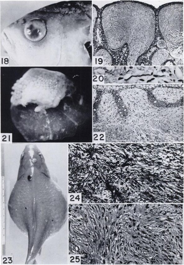
Research.FIGS. 11-18: Neurilemomas with prominent nuclear
palisading.
FIG. 11.—Sectionof a 6X8X4 mm. neurilemoma that grew
at the base of the caudal fin. Palisading of nuclei is very
prominent. Van Gieson stain. Mag. X105. (Goldfish 32.)
FIG. 12.—Apigmented tumor measuring 10X8X7 mm.
grew at the base of the left pectoral fin. Section shows an area of
nuclear palisading; branched pigment cells (melanophores) are
absent. Bodian stain. Mag. X135. (Goldfish 36.)
FIG. 13.—Portion of an area of nuclear palisading shown in
the preceding figure. Several of the sheath cells contain large
numbers of melanin granules in their cytoplasm. See also
Figures 23-25. Bodian stain. Mag. X335. (Goldfish 36.)
FIGS. 14-17: Visceral neurilemomas.
FIG. 14.—Twolarge neurilemomas occupy most of the ab
dominal cavity. The intestine is displaced forward by the
tumors and the swim bladder "B" lies above them. (Gold
fish 49.)
FIGS.15 and 16.—Lateraland ventral views of a tumor that
arose from the second gill arch and elevated the operculum.
The tumor grew well in tissue culture, but hoinoiotransplants
were resorbed. (Goldfish 28.)
FIG. 17.—Sectionof the neurilemoma shown in Figures 15
and 16 is composed predominantly of the loosely arranged
stellate cells of Antoni type B tissue. See Figure 36 for manner
of growth in tissue culture. Mag. X165. (Goldfish 28.)
Downloaded from cancerres.aacrjournals.org on January 19, 2021. © 1952 American Association for Cancer
Research.-' k
,15
Downloaded from cancerres.aacrjournals.org on January 19, 2021. © 1952 American Association for Cancer
Research.FIGS. 18-22: Dermal neurofibromatosis.
FIG. 18.—Goldfish.showing nodular skin on the dorsum of
the head. Clouding of the cornea is due to neurofibromatous
infiltration. (Goldfish 98.)
FIG. 19.—Sectionthrough nodular area of skin shown in
preceding figure. The connective tissue of the corium has been
replaced by irregularly arranged bundles of Schwann cells
separated by greatly elongated pegs of epithelium. This may be
compared to the diffuse neurofibromatosis identified in man as
elephantiasis neuromatosa. H. and E. stain. Mag. X25. ((¡old-
fish 98.)
FIG. 20.—Neunte in tumor area shown in Figure 19.
Bodian stain. Mag. X300. (Goldfish 98.)
FIG. 21.—Single scale showing a mammilated dermal
thickening at its tip. Mag. X5. (Goldfish Kid. 7.)
FIG. 22.—Section taken through thickening at tip of scale
shows a diffuse proliferation of Schwann cells. The overlying
epidermis is thin and composed chiefly of large clear davate
cells except where epithelial pegs extend downward. The lesion
probably represents an early stage of that shown in Fig. 19.
H. and E. stain. Mag. X75. (Goldfish Kid. 7.)
FIGS. 23-25: Pigmented neurofibromas.
FIG. 28.—The abdomen is greatly distended by large bi
lateral polycystic kidneys (28). On the dorsum of the head, near
the midline, is a flat nonpigmented neurilemoma. On the trunk
are three pigmented neurofibromas, the largest of which lies
just to the left of the midline immediately behind the head.
(Goldfish 21.)
FIG. 24.—Section of a pigmented tumor from the base of
the left pectoral fin of goldfish No. 39. Much of the tissue
pattern is obscured by large branching melanophores. H. and
E. stain. Mag. X200.
FIG. 25.—Sameas Figure 24 but bleached before staining
with hematoxylin and eosin. The cellular pattern is that of a
neurofibroma. Mag. X200. (Goldfish 39.)
Downloaded from cancerres.aacrjournals.org on January 19, 2021. © 1952 American Association for Cancer
Research.20- ~v^
r C». .O»L
. •"•—•
' ^j*f *
23
Downloaded from cancerres.aacrjournals.org on January 19, 2021. © 1952 American Association for Cancer
Research.FIGS. 26-34: Malignant neurilemoma.
FIG. 26.—Binucleate tumor giant cell. H. and E. stain.
Mag. X800. (Goldfish 21.)
FIG. 27.—Tripolar mitosis in large tumor cell. H. and E.
stain. Mag. X800. (Goldfish 1.)
FIG. 28.—Abnormal mitosis in tumor cell. H. and E. stain.
Mag. X800. (Goldfish 1.)
FIG. 29.—Tumorcell in rnitotic division. The chromosomes
arc larger than those normally seen in the goldfish and may
represent diplochromosomes. H. and E. stain. Mag. X800.
(Goldfish 5.)
FIG. 30.—Section of a soft hemorrhagic tumor that occu
pied the dorsum of the tail directly behind the caudal fin and
measured 3X4X2.5 cm. The cells are elongate, aligned in
bundles; the nuclei are variable in size and shape. H. and E.
stain. Mag. X210. (Goldfish 21.)
FIG. 31.—Invasion of trunk muscles by tumor cells. The
tumor grew on the right dorso-lateral surface of the body, in
front of the dorsal fin. A central ulcer 2 cm. in diameter was
surrounded by an elevated margin of yellow-white tumor
tissue. H. and E. stain. Mag. X80. (Goldfish 37.)
FIG. 32.—Malignant neurilemoma showing evidence of
nuclear palisading. The tumor arose from the Hmbus of the
right eye which it destroyed in a period of 5 months. Trans
plants to the eyes of other goldfish failed to grow. H. and E.
stain. Mag. X300. (Goldfish 29.)
FIGS.33-36: Growth of tumors in titra.
FIG. 33.—Cellswandering out of tumor expiant after 4 days
in ritro. Whether these cells represent macrophages or mono-
cytoid Schwann cells cannot be determined. Compare with
Weiss, 1944, Figure 6. H. and E. stain. Mag. X450. Culture on
coverslip. (Goldfish 1.)
FIG. 34.—Growth of neoplastic Schwann cells after 6 days
in ritro. Many are elongated and some are flask-shaped with
the nucleus at one end. Compare with Weiss, 1949, Figure 5.
H. and E. stain. Mag. X300. Culture on coverslip. (Goldfish
1-)
FIG. 35.—Cellsbearing melanin granules leaving expiant
after 13 days in ritro. These cells probably represent macro
phages that have phagocytized melanin liberated from de
generating melanophores in the expiant. However, see text for
discussion and Fig. 23 for appearance of tumor. H. and E. stain.
Mag. X450. Culture in roller tube. (Goldfish 31.)
FIG. 36.—Growth,after 7 days in ritro, of tissue from tumor
that arose on a gill arch (Figs. 15-17). The cells have assumed
an elongate shape approaching that characteristic of human
Schwann cells growing in ritro. H. and E. stain. Mag. X400.
Culture in roller tube. (Goldfish 28.)
Downloaded from cancerres.aacrjournals.org on January 19, 2021. © 1952 American Association for Cancer
Research.i — . _
34.*
^>
35
Downloaded from cancerres.aacrjournals.org on January 19, 2021. © 1952 American Association for Cancer
Research.FIGS. 87-39: Àutotransplant of neurilemoma.
FIG. 37.—Tumor transplant in anterior chamber of eye
after 3 weeks rapid growth that followed 3 months of qui
escence. Tlie primary tumor was a histologically malignant
neurilemoma that grew on the head of this fish. Mag. X4.
(Goldfish 1.)
FIG. 38.—Histological section of transplant showing its
attachment to the iris. H. and E. stain. Mag. X10. (Goldfish 1.)
FIG. 39.—Highermagnification of section shown in the pre
ceding figure. The tissue was clearly viable at the time of
fixation and closely resembles the histological appearance of
the primary tumor. H. and E. stain. Mag. X310. (Goldfish 1.)
FIGS. 40-43: Copepod infestation and neoplasia.
FIG. 40.—Largeneurilemoma on tail of goldfish. Scattered
over the trunk and tail are numerous adult copepods; a bit
of black paper was placed under several to make them more
distinct. (Goldfish 8.)
FIG. 41.—Section of tumor shown in previous figure. The
plane of the section passes lengthwise through an adult cope-
pod embedded in the tumor. H. and E. stain. Mag. X10.
(Goldfish 8.)
FIG. 42.—Adultfemale copepod; the two egg cases are seen
at the caudal end; on the right are the large T-shaped hooks
about the rostrum. Mag. X8.
FIG. 43.—Dealer's goldfish showing heavy experimental in
festation with copepods. Natural size.
Downloaded from cancerres.aacrjournals.org on January 19, 2021. © 1952 American Association for Cancer
Research.B^^w^r^
V-ÄfcX
«*srv»
"v^> -v^'"
:ScHLUMBERGER—NerveSheath Tumors in Goldfish 899
REFERENCES II: Der Bau der Nervenfasern. Arch. f. mikr. Anat. Ent-
wicklgesch., 72:575-606, 1908.
1. BAILEY,S. W. An Experimental Study of the Origin of the
Lateral-Line Structures in Embryonic and Adult Teleosts. 22. NIGRELLI,R. F., and FIRTH, F. E. On Sphyrion lumpt
J. Exper. Zool., 76:187-233, 1937. (Kroyer), a Copepod Parasite on the Redfish Sebastes
2. BIESELE, J. J. Diplochromosomes in a Goldfish Tumor. marinus (Linnaeus), with Special Reference to the Host
Parasite Relationships. Zoologica, 24:1-8, 1939.
Cancer Research, 3:411-12, 1943.
3. BLACK,L. M. A Virus Tumor Disease of Plants. Am. J. 23. OPPENHEIMER,J. M. Atypical Pigment Cell Differentia
Bot., 32:408-15, 1945. tion in Embryonic Teleostean Grafts and Isolates.
Zoologica, 36 (Part I) : 22-23, 1950.
4. CASET,A. E.; Ross, G. L.; and LANQSTON, R. R. Selective
XYZ Factor in C57 Black Mammary Carcinoma E 0771. 24. PICCHI, L. Di un non Commune Tumore di un Pesce
Proc. Soc. Exper. Biol. & Med., 72:83-89, 1949. (Neurinoma). Sperimentale, Arch, di biol.,86:128-30,1933.
5. DUKE-ELDEH,W. S. Textbook of Ophthalmology, 2:1296. 25. RAWLES,M. E. Origin of Melanophores and Their Role in
St. Louis: C. V. Mosby Co., 1938. Development of Color Patterns in Vertebrates. Physiol.
6. FRANK, P., and ALLEE, W. C. Ingestion of Colloidal Rev., 28:383-408, 1948.
Thorium Dioxide by Representative Minnows from the 26. REESE,A. B. Pigmented Tumors. Am. J. Ophth., 30:537-
Chicago Region. Physiol. Zool., 23:134-39, 1950. 65, 1947.
7. GOODRICH,H. B. Problems of Origin and Migration of 27. SCHLUMBERGER, H. G. Cutaneous Leiomyoma of Gold
Pigment Cells in Fish. Zoologica, 36 (Part I): 17-18, 1950. fish. I. Morphology and Growth in Tissue Culture. Am. J.
8. GORE,I. Primary Malignant Tumors of Nerve. Cancer, 6: Path., 26:287-99, 1949.
278-96, 1952. 28. — . Polycystic Kidney (Mesonephros) in the Gold
9. GRAND,C. G., and CAMERON, G. Tissue Culture Studies of fish. Arch. Path., 60:400-410, 1950.
Pigmented Melanomas: Fish, Mouse, and Human. Biology 29. . I,¡minisTumors as a Manifestation of von Reck
linghausen's Neurofibromatosis in Goldfish. Am. J. Ophth.,
of Melanomas. Special Publication of N.Y. Acad. Sc., 4:
171-76,1948. 34:415-22, 1951.
10. GRAUPNEH,H., and FISCHER,H. Beiträgezur Kenntnis der 30. SCHLUMBERGER, H. G., and LÜCKE, B. Tumors of Fishes,
Goldfischhaut. Ztschr. f. mikr.-anat. Forsch., 33:91-142, Amphibians, and Reptiles. Cancer Research, 8:657-754,
1933. 1948.
11. GREENE, H. S. N. A Conception of Tumor Autonomy 31. STOUT,A. P. Peripheral Manifestations of the Specific
Based on Transplantation: A Review. Cancer Research, Nerve Sheath Tumor (Neurilemoma). Am. J. Cancer, 24:
11:899-903, 1951. 751-96, 1935.
12. HADDOW,A., and BLAKE,I. Neoplasms in Fish: A Report 32. . Fibrosarcoma, the Malignant Tumor of Fibro-
of 6 Cases with a Summary of the Literature. J. Path. & blasts. Cancer, 1:30-62, 1948.
Bact., 36:41-47, 1933. 83. . Tumors of the Peripheral Nervous System. Sec
13. HERBICK,C. J. The Organ and Sense of Taste in Fishes. tion 2, Fascicle 6. Atlas of Tumor Pathology. Washington,
Bull. U.S. Fish. Commission, 22:239-72, 1902. D.C.: Armed Forces Institute of Pathology, 1949.
34. TAKAHASHI, K. Studie'Uber die Fischgeschwtllste. Ztschr. f.
14. LICHTENSTEIN,B. W. Neurofibromatosis (von Reckling
hausen's Disease of the Nervous System). Analysis of the Krebsforsch., 29:1-73, 1929.
Total Pathologic Picture. Arch. Neurol. & Psychiat., 62: 35. THOMAS,L. Sur un cas de ganglioneurome abdominal chez
822-39, 1949. la morue. Bull. Assoc. franc, l'étudedu cancer, 16:282-86,
15. LÃœCKE, B. Tumors of the Nerve Sheaths in Fish of the 1927.
Snapper Family (Lutianidae). Arch. Path., 34:133-50, 36. . Sur un cas de stiboneuroepithelioblastome chez
1942. une daurade. Ibid., 21:385-96, 1932.
16. MASSON,P. Experimental and Spontaneous Schwanno- 37. TIDD, W. M. Studies on the Life History of a Parasitic
mas. I. Experimental Schwannomas. Am. J. Path., 8:367- Copepod, Lernaea carassii Tidd. Abst. Doctoral Diss.,
88, 1932. Ohio State University, No. 26, pp. 59-62, 1938.
17. . My Conception of Cellular Nevi. Cancer, 4:9-38,
38. VIETA,J. O., and PACK,G. T. Malignant Neurilemomas of
1951. Peripheral Nerves. Am. J. Surgery, 82:416-31, 1951.
18. MONTGOMERY, H., and McNAiRY, D. J. Cutaneous Tu
mors of von Recklinghausen's Disease (Neurofibro 39. WEISS, P. In Vitro Transformation of Spindle Cells of
Neural Origin into Macrophages. Anat. Ree., 88:205-21,
matosis) . Report of a Histologie Study with Special Refer
ence to Nerve Fibers and the Bodian Stain. Arch. Dermal. 1944.
& Syph., 61:384-90, 1945. 40. . Differential Growth. In Chemistry and Physiol
ogy of Growth, pp. 133-86. PAHPAHT,A. K. (ed.). Prince
19. MURRAY,M. R., and STOUT,A. P. Characteristics of Hu
man Schwann Cells in Vitro. Anat. Ree., 84:275-93, 1942. ton: Princeton University Press, 1949.
41. WINESTINE,F. The Relation of von Recklinghausen's
20. . Demonstration of the Formation of Reticulin by
Schwannian Tumor Cells in Vitro. Am. J. Path., 18:585- Disease (Multiple Neurofibromatosis) to Giant Growth
03, 1942. and Blostomatosis. J. Cancer Research, 8:400-22, 1024.
21. NI:MII.»IF. A. Einige Beobachtungen überden Bau des 42. YOUNG,G. A., and OLAFSON,P. Neurilemomas in a Family
Nervengewebes bei Ganoiden und Knochenfischen. Teil of Brook Trout. Am. J. Path., 20:413-19, 1944.
Downloaded from cancerres.aacrjournals.org on January 19, 2021. © 1952 American Association for Cancer
Research.You can also read