Stabilization demands of walking modulate the vestibular contributions to gait
←
→
Page content transcription
If your browser does not render page correctly, please read the page content below
www.nature.com/scientificreports
OPEN Stabilization demands of walking
modulate the vestibular
contributions to gait
Rina M. Magnani1, Sjoerd M. Bruijn2,3, Jaap H. van Dieën2 & Patrick A. Forbes4*
Stable walking relies critically on motor responses to signals of head motion provided by the vestibular
system, which are phase-dependent and modulated differently within each muscle. It is unclear,
however, whether these vestibular contributions also vary according to the stability of the walking
task. Here we investigate how vestibular signals influence muscles relevant for gait stability (medial
gastrocnemius, gluteus medius and erector spinae)—as well as their net effect on ground reaction
forces—while humans walked normally, with mediolateral stabilization, wide and narrow steps. We
estimated local dynamic stability of trunk kinematics together with coherence of electrical vestibular
stimulation (EVS) with muscle activity and mediolateral ground reaction forces. Walking with external
stabilization increased local dynamic stability and decreased coherence between EVS and all muscles/
forces compared to normal walking. Wide-base walking also decreased vestibulomotor coherence,
though local dynamic stability did not differ. Conversely, narrow-base walking increased local dynamic
stability, but produced muscle-specific increases and decreases in coherence that resulted in a net
increase in vestibulomotor coherence with ground reaction forces. Overall, our results show that while
vestibular contributions may vary with gait stability, they more critically depend on the stabilization
demands (i.e. control effort) needed to maintain a stable walking pattern.
The stability of walking is commonly assessed as the ability to maintain upright locomotion in the presence
of self-generated and/or external p erturbations1–3. To ensure stable walking, the nervous system relies on the
integration and modulation of sensory signals from visual, somatosensory and vestibular sources to generate
ongoing and/or corrective postural responses throughout the different phases of the gait c ycle4–7. The vestibular
system, for instance, which encodes signals of head movement in space8, is assumed to contribute to gait stability
because impaired gait is often observed in vestibulopathic patients9,10. Recent studies have further revealed that
vestibular contributions to locomotion undergo phase- and muscle-specific responses that appear to align with
each muscle’s functional role in gait stability11,12. Specifically, the vestibular contributions to mediolateral stability
during walking may be related to mediolateral foot placement produced by muscles around the hip11,13,14 and to
ankle torque at push-off driven by muscles of the ankle15. In the sagittal plane, however, corrective responses to a
vestibular disturbance are almost entirely absent throughout the gait cycle16. Because stability in the sagittal plane
is maintained largely p assively17—due to passive dynamics of the legs18—it may be possible that the control of
whole-body stability during locomotion requires less feedback-driven control as compared to the frontal p lane16.
Thus, the question arises whether changes in the stability of walking in the mediolateral plane also influences
the vestibular contribution to stability during locomotion. Therefore, our aim is to identify how vestibular con-
tributions to balance are modulated across varying stabilization demands of walking by characterizing changes
in coupling of vestibular input to motor outputs during externally-imposed and natural variations in stability.
Experimentally, gait stability can be improved by adding external lateral stabilization to the body, thereby
removing the need for the nervous system to control upright balance in the mediolateral d irection19–21. Under
these stabilized conditions, step width variability, trunk and pelvis motion and premotor cortical involvement
all decrease compared to normal walking22–25. These observations indicate a reduced need to actively control
gait stability, and as a result, may also diminish the necessity for vestibular sensory feedback control. This may
be similar to observations during upright standing, where compensatory responses to mediolaterally-directed
vestibular disturbances are absent when participants are stabilized in the mediolateral direction, despite having
1
Department of Physiotherapy, School of Physical Education and Physical Therapy, State University of Goiás,
Goiânia, GO, Brazil. 2Department of Human Movement Sciences, Faculty of Behavioral and Movement Sciences,
Vrije Universiteit Amsterdam, Amsterdam Movement Sciences, Amsterdam, The Netherlands. 3Institute Brain
and Behavior Amsterdam, Amsterdam, The Netherlands. 4Department of Neuroscience, Erasmus MC, University
Medical Center Rotterdam, P.O. Box 2040, 3000 CA Rotterdam, The Netherlands. *email: p.forbes@erasmusmc.nl
Scientific Reports | (2021) 11:13736 | https://doi.org/10.1038/s41598-021-93037-7 1
Vol.:(0123456789)
www.nature.com/scientificreports/
to maintain balance in an anterior–posterior d irection26. Therefore, we first hypothesized that external lateral
stabilization of the body would diminish the muscle and whole-body responses to a mediolateral vestibular
disturbance during locomotion.
Natural changes in gait are also thought to influence stability. For instance, adopting a wider step width has
been described as a response to decreased lateral stability during locomotion22,27, and seems to be an effective
approach to increase margins of stability (i.e. base of support) in older a dults28,29. If the vestibular system influ-
ence on gait is tightly coupled to stability, then similar to external stabilization, we expected that the evoked
muscle and whole-body responses to a vestibular disturbance should diminish when walking with wider steps.
On the other hand, narrow-base walking requires increased effort to control gait stability in the frontal plane
in young and old a dults30,31, since the margins of stability, or tolerance for errors, are substantially r educed32,33.
These observations, however, seem to contrast with measures of local dynamic stability, which quantify the likeli-
hood of departing from a steady-state gait pattern in the absence of external disturbances, and instead indicate
increased gait stability during narrow-base walking and decreased gait stability during wide-base w alking31,34.
We aim to test a potential explanation for this conflict by examining whether the increased local dynamic sta-
bility observed when walking with narrow step width is (partially) subserved by increased vestibular sensory
feedback. Indeed, support for this is seen by the additional contribution of vestibular signals (via the lateral
vestibular nuclei) to limb muscle activity in mice when walking on a narrow beam that is absent when walking
on level g round35. Here, we tested these hypotheses by comparing muscle and whole-body responses evoked by
a vestibular disturbance—together with estimates of stability—across normal, mediolaterally stabilized, wide-
base and narrow-base walking.
Methods
Participants. We measured 23 healthy young adults between 24 and 33 years recruited from the univer-
sity campus. Twelve participants were excluded during data analysis due to technical problems in the collec-
tion of electromyography (n = 3) or kinematic data (n = 9) in any of the trials recorded. Here we present results
from eleven young adults (four females, 28.5 ± 2.9 years old, 71.6 ± 8.6 kg and 1.77 ± 0.10 m, body mass index
22.2 ± 3.1 kg/m2). Exclusion criteria included self-reported history of injury and/or disfunction of the nervous,
musculoskeletal or vestibular systems, or the use of medications that can cause dizziness. Participants were also
instructed not to participate in intense physical exercise on the day of the experiment. The participants agreed to
participate in the study by signing the informed consent form, and the study was approved by the VU Amster-
dam Research Ethics Committee (VCWE-2017-158). All methods were carried out in accordance with relevant
guidelines and regulations.
Electrical vestibular stimulation. A continuous electrical vestibular stimulus (EVS) was used to deliver
an isolated vestibular disturbance to participants during all walking trials. Coupling of the electrical stimulus
with muscle activity and ground reaction forces was quantified over the gait cycle to determine the magnitude
and timing of the vestibular contribution to ongoing muscle and whole-body responses. The electrical stimulus
modulates the afferent firing rate of both semicircular canal and otolith a fferents36–38, and when delivered in a
binaural-bipolar configuration, EVS evokes a sensation of head roll rotational v elocity39 about an axis directed
posteriorly and superiorly by 18° relative to the Reid plane40–42. When the head is facing forward, this stimu-
lus configuration results in a postural response in the frontal plane to compensate for the induced roll error
signal16,26,43–46.
The electrical stimulus was applied to participants using flexible carbon rubber electrodes (9 cm2). The
electrodes were coated with Spectra 360 electrode gel (Parker La, USA) and fixed to participants’ mastoid pro-
cesses using adhesive tape and an elastic head band. The stimulus was delivered as an analog signal via a data
acquisition board (National Instruments Corp., Austin, TX, USA) to an isolated constant current stimulator
(STMISOLA, Biopac, Goleta, CA, USA). All participants were exposed to the same stochastic EVS designed
with a limited bandwidth of 0 to 25 Hz47, zero-mean low-pass filtered white noise, 25 Hz cutoff, zero lag, fourth-
order Butterworth, peak amplitude of 5.0 mA, root mean square of ~ 1.2 mA, lasting 8 min and created with
Matlab software (MathWorks, Natick, MA, USA). Because this binaural-bipolar stimulus oscillates around a
zero-mean, the imposed sensations of roll motion, and the accompanying compensatory responses, occur in
both a left and right direction.
Protocol. Participants walked on a dual-belt treadmill at a belt speed of 0.8 m/s in four different conditions:
normal walking, stabilized walking, wide-base walking, and narrow-base walking. During normal walking and
stabilized walking, participants were instructed to walk with their naturally preferred step width. Stabilized
walking was achieved using a custom-made spring-loaded mediolateral pelvic stabilization frame. The stabi-
lization frame was attached through springs to two carts that allowed for movement in the anterior–posterior
direction. The springs were pre-tensioned to provide a stabilizing stiffness of 1260 N/m48 and the height of the
carts was aligned with the height of the pelvis for each subject49. During wide-base walking, participants were
instructed to increase their step width beyond the approximate width of their hips. During narrow-base walking,
participants were instructed to adopt a step width smaller than both the width of their hips and their usual step
width. Throughout wide- and narrow-base trials, participants received repeated verbal instruction to maintain
wider and narrower step widths, respectively, compared to normal walking. Participants walked in each condi-
tion for 8 min while being exposed to continuous EVS and were guided by the beat of a metronome at 78 steps/
min to control for effects of varying cadence on vestibular contributions during locomotion11,12. The walking
speed of 0.8 m/s and cadence 78 steps/min were chosen to replicate the conditions of Dakin et al.11. These walk-
Scientific Reports | (2021) 11:13736 | https://doi.org/10.1038/s41598-021-93037-7 2
Vol:.(1234567890)www.nature.com/scientificreports/
ing parameters also ensure that vestibular-evoked balance responses, which are known to decrease as velocity
and cadence increase11,12, could be measured throughout the gait cycle.
Prior to starting the experiments, participants were allowed to walk for 3–4 min to familiarize themselves
with walking on the treadmill at the specific cadence and with the electrical stimulus. In addition, participants
walked for 2 min in each condition before the electrical stimulus was applied, which together with the famil-
iarization period was considered a sufficient exposure period to remove acclimatization effects of walking on a
treadmill50,51. Trial order for the different walking conditions was also randomized for each subject and subjects
were given a short 5 min break between trials to limit the influence of any long-term habituation to the electri-
cal stimulus throughout the walking trials52. Finally, participants maintained their head in a slightly extended
position with the Reid’s plane pitched ~ 18° up from horizontal40,41 by keeping a headgear-mounted laser on a
target located 3 m in front of them. This head position was chosen to maximize the amplitude of vestibulomotor
balance responses in the mediolateral direction41,53,54.
Instrumentation. Kinematic data were recorded using a 3D motion capture system (Optotrak, Northern
Digital Inc., Waterloo, Ontario, Canada) sampling at 100 samples/s. Clusters of three light emitting diodes (LED)
were positioned at the occipital lobe, the spinous process of the sixth thoracic vertebra (T6), the posterior supe-
rior iliac spine and at the calcaneus bilaterally. Ground reaction forces (GRF) were measured from each belt by
force plates embedded in the treadmill (Motekforce Link, The Netherlands) at a sampling rate of 200 samples/s.
In addition to analysis of the coupling between EVS and mediolateral forces as described below, these signals
were used for the identification of toe-off and heel-strike and gait events.
Surface electromyography (EMG) (TMSI Porti system, TMSI Enschede, the Netherlands) was collected at
2000 samples/s bilaterally from the medial gastrocnemius, gluteus medius and erector spinae muscles, using pairs
of disposable self-adhesive Ag/AgCl surface electrodes (Ambu, Balerrup, Denmark; model Blue sensor; diameter
30 × 22 mm) for each muscle. Electrodes were placed over the recorded muscles according to SENIAM electrodes
placement recommendations55 after abrading and cleaning the skin with alcohol. A reference electrode was placed
over the medial bony part of the left wrist (styloid process). The three muscles measured were chosen based
on their supposed roles in different stabilizing strategies that may be employed throughout the gait cycle and
across walking conditions. The medial gastrocnemius muscle (and other ankle plantarflexors) act as the foot`s
prime movers during normal w alking56 and are most sensitive to vestibular input in the late stance p
hase11,12,15
when modulating push-off force. The gluteus medius muscle contributes to foot placement strategies; its activity
is correlated to the next foot p lacement57,58 and is most sensitive to imposed vestibular errors just prior to heel
11
strike . Finally, trunk muscles serve to directly influence trunk motion relative to the pelvis and are primarily
activated to stabilize the trunk during weight transfer around heel strike59,60. Therefore, erector spinae muscle
may be especially suited to contribute to angular momentum control of the torso during narrow base walking61
when push-off modulation and foot-placement are rendered ineffective.
Data analysis. Force-plate data were used first to calculate center of pressure positions, which were in turn
used to identify heel c ontacts62. From these estimates, stride time was calculated as the duration between two
consecutive heel strikes of the same foot and the step width was determined as the mediolateral distance between
the centroids of the feet cluster markers at heel strike. These measures of limb kinematics (stride time and step
width) were compared across walking conditions to determine whether participants adhered to our instruc-
tions (that is, walk at the metronome-guided cadence and with a modified step width). We compared muscle
activity across each walking condition after rectifying and low-pass filtering (20 Hz cutoff, zero lag, sixth order
Butterworth) the EMG signals and then time-normalizing the data per stride and averaging the data across the
256 strides. Prior to averaging, each rectified and low-pass filtered EMG signal was normalized to the maximum
amplitude across all conditions.
To assess changes in local dynamic stability during the different walking conditions, we estimated the local
divergence exponent (LDE). Measures of local dynamic stability for walking indicate the ability for a participant
to return to the steady-state periodic motion after infinitesimally small p erturbations63, which occur, for exam-
ple, through natural variability in the walking surface or the neuromuscular system. These measures have been
shown to be particularly useful for detecting patients at risk of f alling3. The LDE measures the exponential rate
of divergence of neighboring trajectories of a state space constructed from kinematic data of g ait64, whereby an
increasing LDE indicates reduced stability. We calculated the LDE using Rosenstein’s a lgorithm65 and as input the
velocity of a maker placed over the T6 vertebrae, which was estimated using a three-point differentiation of the
position trace66. Velocity time series were first resampled so that each time series of 256 strides (the minimum
number of strides collected from every participant) contained 25,600 samples. The LDE was then calculated as the
slope of the mean divergence curve, whose horizontal axis was normalized by stride time from 0–0.5 s tride67,68.
To examine the vestibulomotor coupling between the input stimulus (i.e. EVS) and motor output (EMG and
ground reaction forces) across our conditions, we computed the time–frequency coherence and gain assuming a
linear stimulus–response r elationship69. Prior to estimating coherence and gain, EVS, EMG and ground reaction
forces were cut into segments synchronized to heel strikes. Based on the symmetry of our walking conditions,
we chose to align the data on each limb’s heel strike in order to pool responses from muscles in the left and right
limbs. Symmetry of the evoked muscle responses was confirmed prior to pooling the data (see Statistical analysis).
This approach, however, was not possible for coherence and gain between EVS and GRFs since in our narrow-
base walking condition participants regularly made contact of one limb with the opposing belt such that forces
from each limb could not be measured on the separate force plates. Therefore, forces from both plates were first
summed before estimating the coherence. To avoid distortion in the coherence estimates at the beginning and
end of the signal, each stride was padded with data from the neighboring strides (50%). EVS and rectified EMG
Scientific Reports | (2021) 11:13736 | https://doi.org/10.1038/s41598-021-93037-7 3
Vol.:(0123456789)www.nature.com/scientificreports/
were low-pass filtered (100 Hz cutoff, zero lag, sixth-order Butterworth) and down-sampled to 200 samples/s.
To account for stride-to-stride variations, stride duration was normalized by resampling the data according to
the average stride duration of all trials. This normalization was performed on the auto-spectra of the EVS, EMG
and force signals, as well as on their cross-spectra (see below).
Our analysis of coherence and gain was performed based on continuous Morlet wavelet d ecomposition15,70
using Eqs. (1) and (2):
Pxy (τ , f )
2
(1)
C τ, f =
Pxx τ , f Pyy (τ , f )
P (τ , f )
xy
(2)
G τ, f =
Pxx τ , f
where Pxy(τ , f ) (τ and f denote the stride time and frequency,
respectively) is the time-dependent cross-spectrum
between the EVS and rectified EMG or GRF, and Pxx τ , f and Pyy(τ , f ) are the time-dependent auto-spectra
of the EVS and rectified EMG or GRF, respectively. Coherence ranges from zero to one, and provides a measure
of the linear relationship between two s ignals71 as well as an estimate of their coupling (i.e. shared variance) at
each frequency72. Because coherence is normalized to the auto-spectra of the input and output signals, it can be
sensitive to variation in non-vestibular contributions to the measured output signal (i.e. magnitude changes of
EMG or ground reaction forces). Gain on the other hand indicates the magnitude of the output relative to the
input and is not normalized by the output signal power spectrum; as a result, it is not expected to change even
if non-vestibular input leads to changes in output signal magnitude. A preliminary evaluation of our results
indicated that similar to previous s tudies12,15, both coherence and gain followed parallel changes in magnitude
and timing across the four walking conditions. This suggests that the modulation in coherence was not simply
dependent upon the magnitude of the measured output signals, and as a result, we present only the coherence to
describe the modulation of vestibular contributions to mediolateral stability. Finally, we evaluated the coherence
between EVS and both EMG and GRFs to estimate muscle-specific vestibular contributions and to provide a net
estimate of the vestibular input to ongoing locomotor behavior, respectively.
Statistical analysis. We compared all gait parameters (stride time, step width and local dynamic stability)
between the four conditions using one-way repeated measures ANOVAs. Subsequently, we performed planned
pairwise comparisons (t-test) between normal walking and each modified walking condition (i.e. normal vs.
stabilized; normal vs. narrow and normal vs. wide-base walking). EVS-EMG coherence and EVS-GRF coher-
ence were used to identify the phase-dependent coupling between vestibular stimulation and motor responses
throughout the walking cycle. For each participant, coherence was defined as significant for those points in
the gait cycle where it exceeded 0.018, corresponding to p < 0.01 (for 256 strides) in view of the bi-dimensional
nature of the c orrelations15. To determine whether our walking manipulations modified the EVS-EMG coher-
ence when compared to normal walking, we performed cluster-based permutation tests (paired t-tests, 5000
permutations)73 between conditions aimed to identify whether the time–frequency coherence significantly dif-
fered from the normal condition. In doing so, we did not disregard non-significant coherence values. Since our
initial analysis showed no significant between leg differences in coherence, we averaged coherence values over
the two legs.
Results
Effects of condition on gait parameters, muscle activity and local dynamic stability. To char-
acterize changes in gait across walking trials, we first evaluated the gait parameters, muscle activity and stabil-
ity measures. During all trials, participants were able to maintain stable upright locomotion while exposed to
the stochastic electrical stimulation. Despite walking to the beat of a metronome in all conditions, we found a
significant main effect of condition on stride time (F(3,30) = 4.14; p = 0.014; η2 = 0.127). Pairwise comparisons
revealed that stride time increased by approximately 6.12 ± 15.65% (0.09 ± 0.02 s; p = 0.018) during stabilized
walking when compared to normal walking (Fig. 1a), but it did not change significantly during either wide-
base (p = 0.928) or narrow-base walking (p = 0.155). As intended, walking condition significantly affected step
width (F(3,30) = 73.4; p < 0.001; η2 = 0.786, see Fig. 1b). Consistent with previous results20,23–25, pairwise analysis
revealed that participants reduced their step width by 65.31 ± 40% (0.18 ± 0.02 m; p < 0.001) during stabilized
walking, in spite of no explicit instruction to do so. We also found that participants adhered to the step width
instructions during the other two conditions, increasing step width by 30.25 ± 20% (0.08 ± 0.02 m; p < 0.001)
during wide-base walking and reducing step width by 38.74 ± 5% (0.11 ± 0.01 m; p < 0.001) during narrow-base
walking (Fig. 1b).
As expected, when compared to normal walking, significant differences in mediolateral ground reaction
forces30,31,74 during all other walking conditions were observed primarily during single-support phases of the gait
cycle (see Fig. 2a). Forces decreased when subjects were externally supported or walked with a narrow-base and
increased when subjects walked with a wide-base (see Supplementary Figure S1). In the medial gastrocnemius
and gluteus medius muscles, peak activity in each muscle occurred during the double stance phase (~ 50% of
the stride cycle) and after heel strike (~ 20% of stride cycle), respectively. EMG responses for these two muscles
were for the most part overlapping in all conditions, with only limited significant differences observed during
the stance phase for some of the conditions relative to normal walking (see Fig. 2b,c and Supplementary Fig-
ure S1). Muscle activity in the erector spinae demonstrated more complex phasic activity throughout the gait
Scientific Reports | (2021) 11:13736 | https://doi.org/10.1038/s41598-021-93037-7 4
Vol:.(1234567890)www.nature.com/scientificreports/
Figure 1. Spatiotemporal parameters of gait during normal walking (blue), walking with external lateral
stabilization (green), wide-base walking (black) and narrow-base walking (red). Mean values (bars), standard
deviation (error bar) and participants individual changes across conditions (lines) for the stride time (a) and
stride width (b) for the four conditions.
cycle with two peaks occurring just after heel strike of each limb. Limited significant differences were observed
during stabilized walking and narrow-base walking when compared to normal walking (see Fig. 2d and Sup-
plementary Figure S1).
Our manipulations also had a significant effect on mediolateral stability, as expressed by the local divergence
exponent (F(3,30) = 129; p < 0.001; η2 = 0.712). Pairwise comparisons showed that participants had a higher sta-
bility (i.e., lower LDE values) during the stabilized (p < 0.001) and narrow-base walking conditions (p = 0.019)
compared to normal walking. Although the mean local divergence exponent was highest (i.e. lowest stability)
during wide-base walking, this was not significantly different from normal walking (p = 0.265) (Fig. 3).
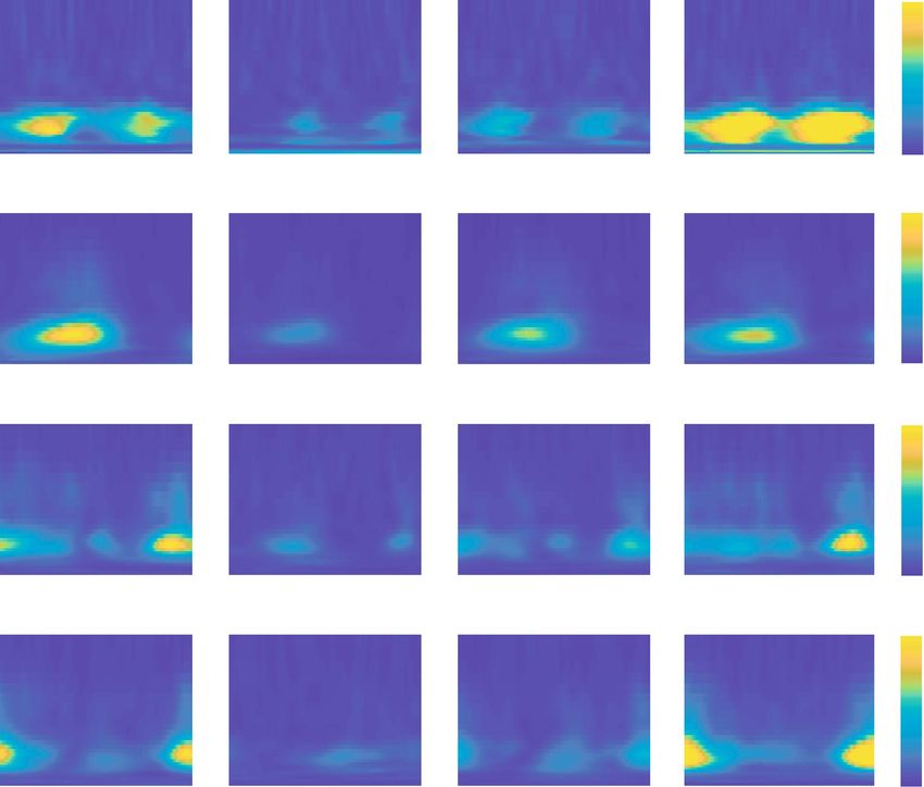
EVS‑mediolateral GRF and EVS‑EMG coherence in normal walking. We next characterized the
coupling of the electrical stimulus with both the ground reaction forces and muscle activity in normal walking
as a baseline for comparison to our manipulated conditions (Fig. 4—first column). During normal walking,
significant EVS-GRF coherence was seen in all participants over the entire gait cycle, with phase-dependent
group mean responses that peaked during single stance (Fig. 4 top row). Significant phase-dependent EVS-EMG
coupling was also prominent in the mean responses during normal walking, but muscle-specific variations were
observed: coherence peaked in mid stance in the medial gastrocnemius, just before heel strike in gluteus medius,
and at heel strike and at mid stride (though at a lower magnitude) for the erector spinae. Consistent with previ-
ous reports11,12,15, peak coherences did not align with peak EMG for any of the muscles, further confirming that
vestibular contributions do not depend purely on the excitation of the motoneuron pool. In addition, as com-
monly observed in vestibular-evoked muscles responses during standing75, the bandwidth of significant EVS-
EMG coherence spanned ~ 0–25 Hz while significant EVS-GRF coherence was observed from ~ 0–10 Hz (Fig. 4).
Stabilization demands but not dynamic stability modulate EVS‑GRF and EMG‑EVS cou-
pling. To establish the effects of stabilization demands on vestibulomotor coupling, we examined the differ-
ence in coherence between normal walking and all other conditions (Fig. 5). During externally stabilized walk-
ing, both EVS-GRF and EVS-EMG coherences decreased significantly relative to normal walking (see Figs. 4
and 5). More specifically, the reduced coupling during the stabilized condition was primarily observed during
the periods of peak coherence in normal walking (i.e. single stance for GRF and medial gastrocnemius, and
before/at heel strike for gluteus medius and erector spinae). Although the increased stride time (i.e. decreased
cadence) during stabilized walking (see Fig. 1) may have acted as a confounding factor to these c hanges11, this
effect commonly increases vestibulomotor responses in contrast to the observed decrease in coherence seen
here. During wide-base walking, EVS-GRF and EVS-EMG coherences were also significantly decreased com-
pared to normal walking (Fig. 5). The EVS-GRF coherence decreased over the majority of the gait cycle with the
most prominent changes observed during the periods of peak coherence seen in the normal condition. Further,
EVS-EMG coherence was reduced for all three muscles in wide-base walking, again with the greatest differences
observed at instants of peak coherence in normal walking (Fig. 5). Taken together, the results of stabilized and
wide-base walking show a reduction in vestibular input to the net muscle activity of the body (i.e. GRFs), which
is driven at least in part by the three muscles measured when stabilization demands (but not dynamic stability)
are decreased.
During narrow-base walking, we observed more complex changes in coupling between EVS and GRF and
between EVS and muscle activity. Figures 4 and 5 together show that EVS-GRF coherence during narrow-base
walking significantly increased compared to normal walking over the entire gait cycle. While this was matched
by an increase in EVS-EMG coherence in the erector spinae muscle, we found unchanged EVS-EMG coupling in
Scientific Reports | (2021) 11:13736 | https://doi.org/10.1038/s41598-021-93037-7 5
Vol.:(0123456789)www.nature.com/scientificreports/
100
a
reaction force [N]
Mediolateral
0
-100
gastrocnemius [-] 1.0 b
Normal
Stabilized
Medial
0.5 Wide
Narrow
0
1.0 c
medius [-]
Gluteus
0.5
0
1.0 d
spinae [-]
Erector
0.5
0
0 50 100
Gait cycle [%]
Figure 2. Ground reaction force and electromyographic amplitudes throughout the gait cycle during normal
walking (blue line), walking with external lateral stabilization (green line), wide-base walking (black line) and
narrow-base walking (red line). Mean values (lines) and standard deviation (shadowed area) of the ground
reaction force (a) and the EMG envelopes of the medial gastrocnemius (b), gluteus medius (c) and erector
spinae (d) muscles for all conditions. For each muscle, EMG signals were normalized to the maximum
amplitude across all conditions.
the gluteus medius muscle and a decrease in coherence in the medial gastrocnemius muscle. These more complex
changes in vestibular-evoked motor responses suggest that while the net output of the vestibular-evoked muscle
activity (i.e. EVS-GRF coherence) increases with increased stabilization demands, as well as the dynamic stability
(see Fig. 3), this trend is not reflected in EVS-EMG coupling of all muscles.
Scientific Reports | (2021) 11:13736 | https://doi.org/10.1038/s41598-021-93037-7 6
Vol:.(1234567890)www.nature.com/scientificreports/
Figure 3. Local divergence exponents during normal walking (blue), walking with external stabilization
(green), wide-base walking (black) and narrow-base walking (red). Mean values (bars), standard deviation
(error bar) and participants’ individual changes across conditions (lines) for the evaluated conditions.
Normal Stabilized Wide Narrow
25 0.08
Frequency [Hz]
20
reaction force
Mediolateral
0.06
15
0.04
10
0.02
5
0 0
25 0.2
Frequency [Hz]
Gastrocnemius
20 0.16
0.12
Medial
15
10 0.08
5 0.04
0 0
25 0.12
Frequency [Hz]
20
0.08
Gluteus
medius
15
10 0.04
5
0 0
25 0.12
Frequency [Hz]
20
0.08
Erector
15
spinae
10
0.04
5
0 0
0 50 100 0 50 100 0 50 100 0 50 100
Gait cycle [%] Gait cycle [%] Gait cycle [%] Gait cycle [%]
Figure 4. Coherence plots of EVS-GRF (first row) and EVS-EMG for medial gastrocnemius (second row),
gluteus medius (third row) and erector spinae (fourth row) for normal walking (first column), walking with
external stabilization (second column), wide-base walking (third column) and narrow-base walking (fourth
column). Coherence magnitude is indicated by the color bars.
Scientific Reports | (2021) 11:13736 | https://doi.org/10.1038/s41598-021-93037-7 7
Vol.:(0123456789)www.nature.com/scientificreports/
Figure 5. Differences in time–frequency coherence between the normal walking condition and walking with
external lateral stabilization (first column), wide-base walking (second column) and narrow-base walking (third
column). Color coding refers to the difference in coherence between two walking conditions (e.g. normal minus
stabilized) for the EVS-GRF coherence (first row) and EVS-EMG coherence of medial gastrocnemius (second
row), gluteus medius (third row) and erector spinae (forth row) muscles. For illustrative purposes, differences
that were not significant were plotted slightly opaque.
Discussion
We characterized how coupling of vestibular input with muscle activity and ground reaction forces modulate as
a function of the stabilization demands during locomotion. We found that as participants walked with decreased
stabilization demands through either external stabilization or wider step widths, coherence between electrical
vestibular stimulation and both muscle activity and ground reaction forces decreased compared to normal walk-
ing. These overall reductions in vestibulomotor coupling were accompanied by an increase or no change in the
stability of the gait pattern—measured as a decreased or constant local divergence exponent—during stabilized
and wide-base walking, respectively. In contrast, the increased stabilization demands of walking with narrow
steps invoked complex changes in vestibulo-muscular coupling that increased or decreased specific to each
muscle’s involvement in correcting for the imposed vestibular error. Nevertheless, these changes in vestibulo-
muscular coupling increased the collective contribution of vestibular signals to the ground reaction forces and
occurred together with a decrease in the local divergence exponent (i.e. increased gait stability). This suggests that
participants maintained a more stable gait pattern during narrow walking that was at least partially subserved
through increased use of vestibular feedback. Ultimately, these results indicate that vestibular contributions
to gait stability may be modulated with frontal plane stability, but that they more specifically depend on the
stabilization demands (i.e. control effort) required to maintain a stable gait pattern and not the stability of the
gait pattern itself.
When participants walked with external stabilization, the stability of the gait pattern increased (i.e. decreasing
LDE) while vestibular-evoked muscle and force responses decreased as compared to normal walking. Both of
these results are not entirely surprising since the control of mediolateral motion is aided by the forces generated
by the s prings19,20,25. As a result, there is a reduced reliance on vestibular signals to maintain upright locomotion
during stabilized walking. This is similar to the task dependent reductions in vestibular input observed during
Scientific Reports | (2021) 11:13736 | https://doi.org/10.1038/s41598-021-93037-7 8
Vol:.(1234567890)www.nature.com/scientificreports/
standing44,45,76–78 when participants are externally supported; stimulus-evoked responses are suppressed since
the vestibular feedback is no longer relevant to balancing the body. Our results reveal that these task depend-
ent changes in the vestibular control of standing also apply during the more dynamic task of walking. In addi-
tion, they also support the proposal that anteroposterior control of whole-body stability during locomotion is
controlled passively17. By making the body passively stable in the mediolateral direction, we saw a reduction in
vestibular-evoked responses that matched the near absence of vestibular contributions when the vestibular error
is directed in the anterior–posterior d irection16.
When participants walked with a wide base, vestibular-evoked muscle and force responses also decreased in
a manner that parallels the effects of wide stance during s tanding44,79. Walking (and standing) with a wide foot
placement increases the base of support and the passive stiffness in the frontal plane. The current results show that
the corrective contribution of vestibular signals during walking with a wide base decrease in a manner similar to
the effects seen during external stabilization. Our measure of dynamic stability, however, did not follow the same
trend. Instead, we observed a slight (albeit non-significant) increase in the local divergence exponent compared
to normal walking. This aligns with previous estimates of a constant or decreased dynamic gait stability during
wide-base walking30,34. A key difference between stabilized and wide-base walking is that in the former, increased
gait stability and upright balance is an inevitable result of the external support. Wide-base walking, on the other
hand, despite the increased base of support, still demands active stabilization, and the manner in which this
is achieved differs from normal walking. For example, push-off modulation and step-by-step foot placement
precision both decrease when walking with wide steps30. In addition, the increased moment arm of the ground
reaction forces about the center of mass generates greater fluctuations in angular plane momentum80, which in
and of themselves are destabilizing. These changes may be possible because the margins for error of balance
control are inherently increased, making wide-base walking more robust to external disturbances. The current
results therefore suggest that vestibular contributions may also decrease under walking conditions with reduced
control effort to maintain mediolateral stability. This is in line with recent findings in patients with vestibular
hypofunction who walk slower, with increased cadence and wider s teps81–85. Our results suggest that they may
adopt these changes in walking behavior to be less dependent on vestibular input.
The possibility that vestibular contributions are modulated with the control effort (i.e. stabilization demand)
of gait is further supported by our narrow-base walking results. Both the dynamic stability of gait and vestibular-
evoked ground reaction forces during narrow-base walking increased relative to normal walking. The increase
in dynamic stability (i.e. decreasing LDE) in particular is thought to be required to constrain torso motion to
margins of error within which walking with narrow steps can be maintained34. Indeed, foot placement during
narrow-base walking is more tightly coupled to center-of-mass motion30,31 and participants maintain smaller
step-to-step oscillations86. Our results show that under these highly regulated conditions, this increase in con-
trol effort may be driven, at least in part, by a net increase in vestibular input (as reflected in the ground reac-
tion forces). This overall increase in vestibular contribution to mediolateral stability, however, does not seem
to originate from a general upregulation of vestibular responses in all muscles. Instead, we found decreasing
and unchanging vestibular responses in medial gastrocnemius and gluteus medius muscles, respectively, while
vestibular responses in erector spinae muscles increased. These muscle specific changes align with the scaling
of evoked responses according to the muscle’s involvement in correcting for imposed e rrors11,26. For example,
moments generated by the ankle muscles, such as the medial gastrocnemius, strongly contribute to balance
corrections through push-off m odulation87. During narrow-base walking, however, push-off modulation in the
mediolateral direction is not a viable option since the push off force has only a minimal moment arm. Similarly,
regulation of mediolateral foot placement as provided by the gluteus m edius11,57 is rendered less effective, since
narrow-base walking constrains the foot to a restricted range. Instead, humans more commonly rely on the
modulation of the torso’s angular moment to maintain upright b alance61,86. Although the muscles around the
ankles and hips also contribute to frontal plane angular momentum during normal w alking80, the direct influence
of spinal muscles on trunk motion may make them more suited to contribute to balance during narrow walking
by producing or responding to rapid trunk tilts. A limitation to this interpretation, however, is that we cannot
rule out the influence of arm movements, which are known to influence the stability of human g ait88–90, since
we did not measure arm movements or provide specific instructions to participants to control their movement.
Nevertheless, our erector spinae results indicate that the influence of spinal muscle activity provides a key, and
perhaps primary, contribution to the net vestibular output to ground reaction forces to maintain upright balance
during narrow-base walking.
Significant coherence between the electrical stimulus and erector spinae muscle activity in all conditions also
demonstrates that the phasic contribution of this particular axial muscle to whole-body mediolateral stability
can be flexibly modulated to address varying stabilizing demands. This is similar to this muscle’s differential
response to vestibular disturbances in standing and sitting91. Others have reported, however, that erector spinae
muscles, unlike lower limb muscles, maintain a fixed response sensitivity to vestibular input throughout all
phases of w alking92. This follows a similar argument made in neck muscles where vestibulocollic reflexes are
maintained regardless of the requirement to maintain head on trunk balance93,94. By delivering square wave EVS
pulses at and slightly after (15% of the gait cycle) the heel strike, Guillaud et al. (2020) argued that the invariant
response of the muscle to the electrical stimulation at these two time points indicate a fixed vestibular sensitivity
throughout the entire gait c ycle92. The improved resolution of the time-varying techniques used here, however,
reveals that this muscle does indeed modulate its response to vestibular input throughout locomotion: EVS-
EMG coherence peaked at heel strike and dropped to near-zero at approximately 40% of the stride cycle. The
restricted time-window of the two stimulation occurrences considered (i.e. at and slightly after heel strike) in
the study from Guillaud et al., however, may have masked any changes in EVS-evoked responses. In addition,
the modulation of this muscle’s vestibular sensitivity extends across walking conditions, which nearly doubled
during narrow-base walking as compared to normal walking.
Scientific Reports | (2021) 11:13736 | https://doi.org/10.1038/s41598-021-93037-7 9
Vol.:(0123456789)www.nature.com/scientificreports/
A limitation of our study is that while vestibular-evoked responses were quantified throughout the gait cycle,
the local divergence exponent remains a mean measure of stability for the entire gait cycle. It may therefore be
possible that time-varying changes in gait stability contribute to the phase-dependent vestibular responses seen
here. While early studies using phase-dependent metrics of gait stability suggested that these measures may hold
promise95,96, a recent study from our lab doubted their use, as we found only limited correlation between phase-
dependent gait stability measures and the probability of falling in a simple dynamic walking model97. However,
if adequate phase-dependent gait stability measures become available, the relationship between phase-dependent
gait stability and phase-dependent vestibular responses may be an interesting topic of study.
In conclusion, we have shown that the muscle and whole-body responses evoked by a vestibular stimulation
differ according to the gait stabilization demands. When stability is increased by external support, the mus-
cle and whole-body responses to the vestibular stimulus are substantially reduced. During wide-base walking
vestibular-evoked muscle and force responses also decrease, though these changes in vestibular contribution
are not accompanied by increased dynamic gait stability. Conversely, narrow-base walking produced complex
muscle-specific responses that resulted in an increase in the net vestibular contribution to ground reaction forces
and increased stability of gait. Overall, our results show that although the vestibular control of gait stability may
vary with frontal plane stability, they critically depend on the stabilization demands (i.e. control effort) needed
to maintain stable walking patterns.
Received: 6 October 2020; Accepted: 4 June 2021
References
1. Bruijn, S. M., Meijer, O. G., Beek, P. J. & van Dieen, J. H. Assessing the stability of human locomotion: A review of current measures.
J. R. Soc. Interface 10, 20120999. https://doi.org/10.1098/rsif.2012.0999 (2013).
2. van Emmerik, R. E. A., Ducharme, S. W., Amado, A. C. & Hamill, J. Comparing dynamical systems concepts and techniques for
biomechanical analysis. J. Sport Health Sci. 5, 3–13. https://doi.org/10.1016/j.jshs.2016.01.013 (2016).
3. Mehdizadeh, S. The largest Lyapunov exponent of gait in young and elderly individuals: A systematic review. Gait Posture 60,
241–250. https://doi.org/10.1016/j.gaitpost.2017.12.016 (2018).
4. Misiaszek, J. Neural control of walking balance: If falling then react else continue. Exerc. Sport Sci. Rev. 34, 128–134 (2006).
5. Chien, J. H., Mukherjee, M. & Stergiou, N. Mastoid vibration affects dynamic postural control during gait. Ann. Biomed. Eng. 44,
2774–2784. https://doi.org/10.1007/s10439-016-1556-z (2016).
6. Stergiou, N. & Decker, L. M. Human movement variability, nonlinear dynamics, and pathology: Is there a connection?. Hum. Mov.
Sci. 30, 869–888. https://doi.org/10.1016/j.humov.2011.06.002 (2011).
7. Rossignol, S., Dubuc, R. & Gossard, J. P. Dynamic sensorimotor interactions in locomotion. Physiol. Rev. 86, 89–154. https://doi.
org/10.1152/physrev.00028.2005 (2006).
8. Cullen, K. E. The vestibular system: Multimodal integration and encoding of self-motion for motor control. Trends Neurosci. 35,
185–196. https://doi.org/10.1016/j.tins.2011.12.001 (2012).
9. Brandt, T. Vestibulopathic gait: You’re better off running than walking. Curr. Opin. Neurol. 13, 3–5 (2000).
10. Borel, L. et al. Walking performance of vestibular-defective patients before and after unilateral vestibular neurotomy. Behav. Brain
Res. 150, 191–200 (2004).
11. Dakin, C. J., Inglis, J. T., Chua, R. & Blouin, J. S. Muscle-specific modulation of vestibular reflexes with increased locomotor velocity
and cadence. J. Neurophysiol. 110, 86–94. https://doi.org/10.1152/jn.00843.2012 (2013).
12. Forbes, P. A. et al. Rapid limb-specific modulation of vestibular contributions to ankle muscle activity during locomotion. J. Physiol.
595, 2175–2195. https://doi.org/10.1113/JP272614 (2017).
13. Bent, L. R., Inglis, J. T., Bradford, J. & Mcfadyen, B. J. When is vestibular information important during walking?. J. Neurophysiol.
92, 1269–1275 (2004).
14. Iles, J. F., Baderin, R., Tanner, R. & Simon, A. Human standing and walking: Comparison of the effects of stimulation of the ves-
tibular system. Exp. Brain Res. 178, 151–166. https://doi.org/10.1007/s00221-006-0721-2 (2007).
15. Blouin, J. S. et al. Extracting phase-dependent human vestibular reflexes during locomotion using both time and frequency cor-
relation approaches. J. Appl. Physiol. 1985(111), 1484–1490. https://doi.org/10.1152/japplphysiol.00621.2011 (2011).
16. Tisserand, R. et al. Down regulation of vestibular balance stabilising mechanisms to enable transition between motor states. Elife
https://doi.org/10.7554/eLife.36123 (2018).
17. Bauby, C. E. & Kuo, A. D. Active control of lateral balance in human walking. J. Biomech. 33, 1433–1440 (2000).
18. McGeer, T. Passive dynamic walking. Int. J. Robot. Res. T 9, 62–82. https://doi.org/10.1177/027836499000900206 (1990).
19. Mahaki, M., Bruijn, S. M. & van Dieen, J. H. The effect of external lateral stabilization on the use of foot placement to control
mediolateral stability in walking and running. PeerJ 7, e7939. https://doi.org/10.7717/peerj.7939 (2019).
20. Donelan, J. M., Shipman, D. W., Kram, R. & Kuo, A. D. Mechanical and metabolic requirements for active lateral stabilization in
human walking. J. Biomech. 37, 827–835. https://doi.org/10.1016/j.jbiomech.2003.06.002 (2004).
21. Ortega, J. D., Fehlman, L. A. & Farley, C. T. Effects of aging and arm swing on the metabolic cost of stability in human walking. J.
Biomech. 41, 3303–3308. https://doi.org/10.1016/j.jbiomech.2008.06.039 (2008).
22. Dean, J. C., Alexander, N. B. & Kuo, A. D. The effect of lateral stabilization on walking in young and old adults. IEEE Trans. Biomed.
Eng. 54, 1919–1926 (2007).
23. Ijmker, T., Houdijk, H., Lamoth, C. J., Beek, P. J. & van der Woude, L. H. Energy cost of balance control during walking decreases
with external stabilizer stiffness independent of walking speed. J. Biomech. 46, 2109–2114. https://doi.org/10.1016/j.jbiomech.
2013.07.005 (2013).
24. Ijmker, T. et al. Can external lateral stabilization reduce the energy cost of walking in persons with a lower limb amputation?. Gait
Posture 40, 616–621. https://doi.org/10.1016/j.gaitpost.2014.07.013 (2014).
25. Bruijn, S. M., Van Dieen, J. H. & Daffertshofer, A. Beta activity in the premotor cortex is increased during stabilized as compared
to normal walking. Front. Hum. Neurosci. 9, 593. https://doi.org/10.3389/fnhum.2015.00593 (2015).
26. Forbes, P. A., Luu, B. L. & Blouin, J. S. Transformation of vestibular signals for the control of standing in humans. J. Neurosci. 36,
11510–11520. https://doi.org/10.1523/JNEUROSCI.1902-16.2016 (2016).
27. Kubinski, S. N., McQueen, C. A., Sittloh, K. A. & Dean, J. C. Walking with wider steps increases stance phase gluteus medius
activity. Gait Posture 41, 130–135. https://doi.org/10.1016/j.gaitpost.2014.09.013 (2015).
28. Aboutorabi, A., Arazpour, M. & Bahramizadeh, M. The effect of aging on gait parameters in able-bodied older subjects: A literature
review. Aging Clin. Exp. Res. 28, 393–405 (2016).
Scientific Reports | (2021) 11:13736 | https://doi.org/10.1038/s41598-021-93037-7 10
Vol:.(1234567890)www.nature.com/scientificreports/
29. Schrager, M. A., Kelly, V. E., Price, R., Ferrucci, L. & Shumway-Cook, A. The effects of age on medio-lateral stability during normal
and narrow base walking. Gait Posture 28, 466–471. https://doi.org/10.1016/j.gaitpost.2008.02.009 (2008).
30. Perry, J. A. & Srinivasan, M. Walking with wider steps changes foot placement control, increases kinematic variability and does
not improve linear stability. R. Soc. Open Sci. 4, 160627. https://doi.org/10.1098/rsos.160627 (2017).
31. Arvin, M. et al. Effects of narrow base gait on mediolateral balance control in young and older adults. J. Biomech. 49, 1264–1267.
https://doi.org/10.1016/j.jbiomech.2016.03.011 (2016).
32. Hak, L. et al. Speeding up or slowing down? Gait adaptations to preserve gait stability in response to balance perturbations. Gait
Posture 36, 260–264. https://doi.org/10.1016/j.gaitpost.2012.03.005 (2012).
33. Young, P. M. M. & Dingwell, J. B. Voluntary changes in step width and step length during human walking affect dynamic margins
of stability. Gait Posture 36, 219–224. https://doi.org/10.1016/j.gaitpost.2012.02.020 (2012).
34. Young, P. M. M. & Dingwell, J. B. Voluntarily changing step length or step width affects dynamic stability of human walking. Gait
Posture 35, 472–477. https://doi.org/10.1016/j.gaitpost.2011.11.010 (2012).
35. Murray, A. J., Croce, K., Belton, T., Akay, T. & Jessell, T. M. Balance control mediated by vestibular circuits directing lim extension
or antagonist muscle co-activation. Cell Rep. 22, 1325–1338. https://doi.org/10.1016/j.celrep.2018.01.009 (2018).
36. Goldberg, J. M., Fernandez, C. & Smith, C. E. Responses of vestibular-nerve afferents in the squirrel monkey to externally applied
galvanic currents. Brain Res. 252, 156–160 (1982).
37. Kim, J. & Curthoys, I. S. Responses of primary vestibular neurons to galvanic vestibular stimulation (GVS) in the anaesthetised
guinea pig. Brain Res. 64, 265–271. https://doi.org/10.1016/j.brainresbull.2004.07.008 (2004).
38. Kwan, A., Forbes, P. A., Mitchell, D. E., Blouin, J. S. & Cullen, K. E. Neural substrates, dynamics and thresholds of galvanic vestibular
stimulation in the behaving primate. Nat. Commun. 10, 1904. https://doi.org/10.1038/s41467-019-09738-1 (2019).
39. Peters, R. M., Rasman, B. G., Inglis, J. T. & Blouin, J. S. Gain and phase of perceived virtual rotation evoked by electrical vestibular
stimuli. J. Neurophysiol. 114, 264–273. https://doi.org/10.1152/jn.00114.2015 (2015).
40. Khosravi-Hashemi, N., Forbes, P. A., Dakin, C. J. & Blouin, J. S. Virtual signals of head rotation induce gravity-dependent infer-
ences of linear acceleration. J. Physiol. 597, 5231–5246. https://doi.org/10.1113/jp278642 (2019).
41. Fitzpatrick, R. C. & Day, B. L. Probing the human vestibular system with galvanic stimulation. J. Appl. Physiol. 1985(96), 2301–2316
(2004).
42. St George, R. J. & Fitzpatrick, R. C. The sense of self-motion, orientation and balance explored by vestibular stimulation. J. Physiol.
589, 807–813. https://doi.org/10.1113/jphysiol.2010.197665 (2011).
43. Nashner, L. M. & Wolfson, P. Influence of head position and proprioceptive cues on short latency postural reflexes evoked by
galvanic stimulation of the human labyrinth. Brain Res. 67, 255–268. https://doi.org/10.1016/0006-8993(74)90276-5 (1974).
44. Mian, O. S. & Day, B. L. Violation of the craniocentricity principle for vestibularly evoked balance responses under conditions of
anisotropic stability. J. Neurosci. 34, 7696–7703. https://doi.org/10.1523/JNEUROSCI.0733-14.2014 (2014).
45. Britton, T. C. et al. Postural electromyographic responses in the arm and leg following galvanic vestibular stimulation in man. Exp.
Brain Res. 94, 143–151. https://doi.org/10.1007/BF00230477 (1993).
46. Lund, S. & Broberg, C. Effects of different head positions on postural sway in man induced by a reproducible vestibular error
signal. Acta Physiol. Scand. 117, 307–309. https://doi.org/10.1111/j.1748-1716.1983.tb07212.x (1983).
47. Dakin, C. J., Luu, B. L., van den Doel, K., Inglis, J. T. & Blouin, J. S. Frequency-specific modulation of vestibular-evoked sway
responses in humans. J. Neurophysiol. 103, 1048–1056. https://doi.org/10.1152/jn.00881.2009 (2010).
48. Mahaki, M., Bruijn, S. M. & van Dieën, J. H. The effect of external lateral stabilization on the control of mediolateral stability in
walking and running. PeerJ https://doi.org/10.7287/peerj.preprints.27244v1 (2018).
49. Mahaki, M., IJmker, T., Houdijk, H. & Bruijn, S. M. How does external lateral stabilization constrain normal gait, apart from
improving medio-lateral gait stability? R. Soc open sci. 8, 202088. http://doi.org/10.1098/rsos.202088 (2021).
50. Matsas, A., Taylor, N. & McBurney, H. Knee joint kinematics from familiarised treadmill walking can be generalised to overground
walking in young unimpaired subjects. Gait Posture 11, 46–53. https://doi.org/10.1016/s0966-6362(99)00048-x (2000).
51. Meyer, C. et al. Familiarization with treadmill walking: How much is enough?. Sci. Rep. 9, 5232. https://doi.org/10.1038/s41598-
019-41721-0 (2019).
52. Hannan, K. B., Todd, M. K., Pearson, N. J., Forbes, P. A. & Dakin, C. J. Vestibular attenuation to random—waveform galvanic
vestibular stimulation during standing and treadmill walking. Sci. Rep. https://doi.org/10.1038/s41598-021-87485-4 (2021).
53. Cathers, I., Day, B. L. & Fitzpatrick, R. C. Otolith and canal reflexes in human standing. J. Physiol. 563, 229–234. https://doi.org/
10.1113/jphysiol.2004.079525 (2005).
54. Fitzpatrick, R. C., Butler, J. E. & Day, B. L. Resolving head rotation for human bipedalism. Curr. Biol. 16, 1509–1514. https://doi.
org/10.1016/j.cub.2006.05.063 (2006).
55. Hermens, H. J. et al. SENIAM 8: European recommendations for surface electromyography., Vol. 1 (Roessingh Research and Devel-
opment, 1999).
56. Liu, M. Q., Anderson, F. C., Schwartz, M. H. & Delp, S. L. Muscle contributions to support and progression over a range of walking
speeds. J. Biomech. 41, 3243–3252. https://doi.org/10.1016/j.jbiomech.2008.07.031 (2008).
57. Rankin, B. L., Buffo, S. K. & Dean, J. C. A neuromechanical strategy for mediolateral foot placement in walking humans. J. Neu-
rophysiol. 112, 374–383. https://doi.org/10.1152/jn.00138.2014 (2014).
58. van Leeuwen, A. M., van Dieën, J. H., Daffertshofer, A. & Bruijn, S. M. Active foot placement control ensures stable gait: Effect
of constraints on foot placement and ankle moments. PLoS ONE 15(2), e0242215. https://doi.org/10.1371/journal.pone.0242215
(2020)
59. Waters, R. L. & Morris, J. M. Electrical activity of muscles of the trunk during walking. J. Anat. 111, 191–199 (1972).
60. Thorstensson, A., Carlson, H., Zomlefer, M. R. & Nilsson, J. Lumbar back muscle activity in relation to trunk movements during
locomotion in man. Acta Physiol. Scand. 116, 13–20. https://doi.org/10.1111/j.1748-1716.1982.tb10593.x (1982).
61. Best, A. N. & Wu, A. R. Upper body and ankle strategies compensate for reduced lateral stability at very slow walking speeds. Proc.
R. Soc. B. 287, 20201685. http://doi.org/10.1098/rspb.2020.1685 (2020)
62. Roerdink, M. Online gait event detection using a large force platform embedded in a treadmill. J. Biomech. 41, 2628–2632 (2008).
63. Dingwell, J. B. & Cusumano, J. P. Nonlinear time series analysis of normal and pathological human walking. Chaos 10, 848–863.
https://doi.org/10.1063/1.1324008 (2000).
64. Dingwell, J. B. & Marin, L. C. Kinematic variability and local dynamic stability of upper body motions when walking at different
speeds. J. Biomech. 39, 444–452. https://doi.org/10.1016/j.jbiomech.2004.12.014 (2006).
65. Rosenstein, M. T., Coliins, J. J. & De Luca, C. J. A practical method for calculating largest Lyapunov exponents from small data
sets. Physica 65, 117–134 (1993).
66. Kang, H. G. & Dingwell, J. B. Dynamic stability of superior vs. inferior segments during walking in young and older adults. Gait
Posture 30, 260–263 (2009).
67. Stenum, J., Bruijn, S. M. & Jensen, B. R. The effect of walking speed on local dynamic stability is sensitive to calculation methods.
J. Biomech. 47, 3776–3779. https://doi.org/10.1016/j.jbiomech.2014.09.020 (2014).
68. Bruijn, S.M. Local Dynamic Stability. Zenodo http://doi.org/10.5281/zenodo.573285 (2017).
69. Forbes, P. A. et al. Electrical vestibular stimuli to enhance vestibulo-motor output and improve subject comfort. PLoS ONE 9,
e84385. https://doi.org/10.1371/journal.pone.0084385 (2014).
Scientific Reports | (2021) 11:13736 | https://doi.org/10.1038/s41598-021-93037-7 11
Vol.:(0123456789)You can also read