ASSESSING DIAGNOSTIC VALUE OF MICRORNAS FROM PERIPHERAL BLOOD MONONUCLEAR CELLS AND EXTRACELLULAR VESICLES IN MYALGIC ENCEPHALOMYELITIS/CHRONIC ...
←
→
Page content transcription
If your browser does not render page correctly, please read the page content below
www.nature.com/scientificreports

            OPEN           Assessing diagnostic value of
                           microRNAs from peripheral
                           blood mononuclear cells and
                           extracellular vesicles in Myalgic
                           Encephalomyelitis/Chronic Fatigue
                           Syndrome
                           Eloy Almenar-Pérez1, Leonor Sarría2, Lubov Nathanson2 & Elisa Oltra                     3*

                           Myalgic Encephalomyelitis/Chronic Fatigue Syndrome (ME/CFS) is a debilitating multisystemic
                           disease of unknown etiology, affecting thousands of individuals worldwide. Its diagnosis still relies on
                           ruling out medical problems leading to unexplained fatigue due to a complete lack of disease-specific
                           biomarkers. Our group and others have explored the potential value of microRNA profiles (miRNomes)
                           as diagnostic tools for this disease. However, heterogeneity of participants, low numbers, the variety
                           of samples assayed, and other pre-analytical variables, have hampered the identification of disease-
                           associated miRNomes. In this study, our team has evaluated, for the first time, ME/CFS miRNomes in
                           peripheral blood mononuclear cells (PBMCs) and extracellular vesicles (EVs) from severely ill patients
                           recruited at the monographic UK ME biobank to assess, using standard operating procedures (SOPs),
                           blood fractions with optimal diagnostic power for a rapid translation of a miR-based diagnostic method
                           into the clinic. Our results show that routine creatine kinase (CK) blood values, plasma EVs physical
                           characteristics (including counts, size and zeta-potential), and a limited number of differentially
                           expressed PBMC and EV miRNAs appear significantly associated with severe ME/CFS (p < 0.05). Gene
                           enrichment analysis points to epigenetic and neuroimmune dysregulated pathways, in agreement with
                           previous reports. Population validation by a cost-effective approach limited to these few potentially
                           discriminating variables is granted.

                           Myalgic Encephalomyelitis/Chronic Fatigue Syndrome (ME/CFS) is a disease characterized by long-lasting (over
                           6 months) persistent debilitating widespread fatigue, which is exacerbated by exercise (post-exertional malaise
                           or PEM), and not alleviated by rest1–5. It has been classified by the International Classification of Diseases, Tenth
                           Revision, Clinical Modification (ICD-10-CM) with code R53.82 or G93.3 if post-viral6. It affects at least 0.23
                           to 0.41% of the population worldwide7,8, although numbers can reach up to 5% depending on the diagnostic
                           criteria and geographic locations, according to a recent revision by the EUROpean Myalgic Encephalomyelitis
                           Network (EUROMENE) epidemiology working group9. The disease strikes people of all ages and within all
                           socio-economic levels, being more common in women (female:male ratios between 2:1 and 6:1)7–10. ME/CFS is
                           considered a life-long disabling disease, since only about 5% of patients return to their previous state of health11.
                               The confusion among General Health Practitioners (GPs) due to multiple clinical definitions12 and the large
                           number of comorbidities, such as fibromyalgia, IBS (irritable bowel syndrome), POTS (postural orthostatic tach-
                           ycardia syndrome), depression, etc, stigmatized the disease, delaying diagnosis and treatment of patients. This
                           highlights the urgent need of unbiased, specific methods to diagnose ME/CFS in a clinical setting.

                           1
                            Escuela de Doctorado, Universidad Católica de Valencia San Vicente Mártir, Valencia, Spain. 2Institute for Neuro
                           Immune Medicine, Dr. Kiran C. Patel College of Osteopathic Medicine, Nova Southeastern University, Fort
                           Lauderdale, Florida, USA. 3School of Medicine, Universidad Católica de Valencia San Vicente Mártir, Valencia, Spain.
                           *email: elisa.oltra@ucv.es

Scientific Reports |   (2020) 10:2064 | https://doi.org/10.1038/s41598-020-58506-5                                                            1
www.nature.com/scientificreports/ www.nature.com/scientificreports
However, despite a considerable number of studies showing significant differences at the immune and meta-
bolic levels13–16, and the report by Institute of Medicine (IOM) of the Academy of Sciences USA stressing that the
nature of ME/CFS is a physiologic/medical and not a psychiatric illness4,5, current diagnostic methods still rely
on ruling out other diseases with similar symptoms.
MicroRNAs, miRNAs or miRs are short non-coding RNAs (20–24 nt-long) with attributed gene expression
regulatory functions17,18. Their abnormal expression in disease is already quite established19–21, making them
attractive biomarker candidates in many scenarios, ME/CFS included.
Our group and others have explored the possibility to identify miRNomes or molecular miRNA profiles for
ME/CFS and comorbid diseases in the past, by analyzing PBMC (peripheral blood mononuclear cell) contents or
by evidencing their presence in body fluids (plasma, serum and cerebrospinal fluid)22–27. All body fluids typically
contain free miRNAs, miRNAs complexed with proteins, but also miRNAs packed into extracellular vesicles
(EVs). The International Society for Extracellular Vesicles or ISEV endorses “extracellular vesicle (EV) as the
generic term for particles naturally released from the cell that are delimited by a lipid bilayer and cannot replicate,
i.e. do not contain a functional nucleus”28. The EV mix is known to contain exosomes, microvesicles, microparti-
cles, ectosomes, oncosomes, apoptotic bodies, and many membranous structures with other names28.
The presence of the protective lipid bilayer in EVs constitutes a physical barrier from RNA-degrading enzymes,
possibly leading to improved replicability of assays based on RNA quantitation. In addition, some of these vesi-
cles, under the name of exosomes, are known to directionally pack miRNAs and to sustain and spread disease29–32,
and therefore may provide biomarker advantages for liquid biopsy-based diagnostic methods. As EVs in blood
may have their origin in any body cell, including the microbiome and other hosted organisms, their content
could potentially inform of dysbiosis in general, in addition to immune dysfunctions19–21,33. This last point seems
of particular importance in multisystemic diseases, such as ME/CFS, with patients presenting neuro-immune
alterations, hormonal imbalances, debilitated muscles, leaky guts, etc. Despite its potential, no study has yet been
performed to systematically evaluate particular cargo miRNAs in EVs associating with ME/CFS.
This study aimed to evaluate, for the first time, ME/CFS miRNomes in EVs from severely ill patients and
compare/contrast them with those of their PBMCs as a way to assess blood fractions with optimal diagnostic
power, for a rapid translation of a miR-based method to diagnose ME/CFS into the clinic. At the same time, the
information obtained from EV content analysis could lead to the identification of molecular targets, improving
our understanding of the cross-talk between a dysfunctional immune system and the rest of the organism in ME/
CFS pathophysiology.
Results
Population demographics. This study evaluated blood samples from a cohort of 30 participants (15
severely affected ME/CFS patients and 15 population age-matched healthy subjects), obtained from the mon-
ographic UK ME Biobank34,35. ME/CFS diagnosis was assessed with Canadian Consensus2 and/or CDC-1994
(“Fukuda”) criteria1, as detailed in methods. Included individuals were all women with an average age of 46.8 (age
range 38–53) and 45.2 (age range 18–52) years, respectively. Median ages were 48 years for the ME/CFS group
and 47 for the healthy control group (HC). Average time from disease onset was 17.5 (range 1.5–30.9) years, with
a median value of 18.4 years.
Clinical variables differences. As expected, health survey SF-3636 and General Health Questionnaire
(GHQ)37 scores, including Likert scale38 for the GHQ, scores clearly separated ME/CFS and HC groups (p < 0.05)
in all fields covered by these questionnaires, indicative of serious compromised health of ME/CFS participants
(details are shown on Table 1).
Among the 30 clinical parameters evaluated at the UK Biobank in the blood samples studied, only creatine
phosphokinase (CPK) levels showed statistically significant differences between groups (p < 0.05), with increased
levels by approximately 1.5-fold in ME/CFS patients. Although no other variable of this type showed significant
differences between groups, a tendency towards increased eosinophil counts, and lower levels for alkaline phos-
phatase and free thyroid hormone T4 levels, were noticed in ME/CFS patients. All these three variables presented
p values for group differences below 0.1 (Supplementary Table S1). Individual values for these 4 analytical blood
parameters were plotted for an improved appreciation of the differences found (Supplementary Fig. S1).
Plasma EV features differ between ME/CFS and HCs. Since the final goal of the study was to assess
the diagnostic value of miRNA profiles from PBMCs and plasma EVs in severe ME/CFS patients and since the
miRNA cargo in EVs depends on the isolated vesicle subpopulations38, we decided that characterization of the
isolated EVs to be used for RNA extraction was necessary.
Based on previous reports showing the superiority of precipitating-based methods vs other more laborious
classic methods, in terms of EV yields39, and our interest for rapidly implementing a technically simple diag-
nostic method in a clinical context, we decided that the use of a precipitating approach for EV isolation was the
most appropriate. Thus, directed by Helwa et al. results who compared ultracentrifugation and three different
commercial PEG (polyethylene glycol) precipitating methods40, in relation to the starting volume of plasma, we
chose Invitrogen’s Total Exosome Isolation Reagent (TEIR) and fixed 0.5 mls of plasma as the minimum required
volume for our project (see Methods for further details).
As shown in Fig. 1A, the EV yields obtained from equivalent volumes of plasma from ME/CFS samples were
higher than those isolated from healthy controls (HCs), although not reaching statistical significance (p > 0.05).
Interestingly, isolated EVs showed statistically significant size differences, being on average smaller in the ME/
CFS group than in the HC’s (Fig. 1B). To rule out that the observed results could derive from differences in plasma
protein content interfering with the isolation method, we proceeded to repeat the procedure, this time including
a pre-treatment with proteinase K (an optional step recommended by the vendor when using protein-rich fluids).
Scientific Reports | (2020) 10:2064 | https://doi.org/10.1038/s41598-020-58506-5 2www.nature.com/scientificreports/ www.nature.com/scientificreports
HC ME/CFS
SF-36 Test Score SD Score SD p value
Physical Functioning (PF) 56.8 2.0 21.6 3.0 1.29E-06
Role Physical (RP) 56.9 0.8 21.2 0.0 1.86E-07
Bodily Pain (BP) 56.9 5.0 32.4 12.2 3.69E-05
General Health (GH) 58.9 6.5 26.4 4.1 1.87E-14
Vitality (VT) 58.5 3.9 25.9 5.1 3.13E-06
Social Functioning (SF) 56.7 2.6 19.4 3.8 7.19E-07
Role Emotional (RE) 55.9 0.9 44.0 17.8 3.59E-03
Mental Health (MH) 55.1 2.6 45.4 11.9 1.44E-02
Physical Component Summary (PCS) 57.6 3.7 18.3 5.1 2.58E-08
Mental Health Component Summary (MCS) 55.6 2.2 43.3 12.7 4.06E-05
GHQ test Score SD Score SD p value
scale A - Somatic Symptoms 0.3 0.8 3.7 2.9 5.28E-04
scale B - Anxiety and insomnia 0.1 0.3 1.6 2.3 5.22E-03
scale C - Social dysfunction 0.0 0.0 2.9 2.9 7.96E-05
scale D - Severe depression 0.0 0.0 0.8 1.4 1.80E-02
summary 0.3 0.8 9.1 8.1 2.31E-04
GHQ test with Likert scale Score SD Score SD p value
scale A - Somatic Symptoms 2.8 1.9 12.5 5.8 1.16E-05
scale B - Anxiety and insomnia 3.1 2.1 6.5 5.4 6.66E-02
scale C - Social dysfunction 6.5 0.3 11.1 5.9 5.61E-03
scale D - Severe depression 0.0 0.3 2.7 4.2 2.10E-02
summary 12.5 3.2 32.8 19.1 3.64E-04
Table 1. SF-36 quality of life. GHQ (General Health Questionnaire) and GHQ with Likert scale results for the
cohort studied, healthy contros (HC) and ME/CFS patients (N = 15/group). Mean scores, standard deviations
(SD) and p values are shown.
By introducing this optional treatment, we could confirm that qualitative differences exist, both, in number of
vesicles and size, in plasma of ME/CFS patients with respect to HCs (p < 0.05) (Fig. 1C,D).
To further characterize the seemly diverse isolated EVs, we measured their zeta potential, a parameter reflect-
ing the surface charge of the vesicle which may be informative of membrane composition differences and indica-
tive of vesicle stability or the likelihood of these colloidal suspensions to aggregate. Although the values obtained
fit, as an overall, those reported in the literature for zeta potential of EVs isolated from human plasma (−10 to
−50 mV)41, significant differences were found between values of either group studied. In particular, ME/CFS’s
EVs presented more negative values than the EVs from the HC group, both, if they were isolated in the absence
(Fig. 2A) or presence (Fig. 2B) of proteinase K. This suggests that differences in the lipid and/or other membrane
components exist and that, as an overall, ME/CFS EVs are possibly slightly more stable than EVs from HCs.
Furthermore, when the EVs from each group were independently compared, in reference to pretreatment with
proteinase K or in its absence, significant differences were observed for both the HC (Fig. 2C) and the ME/CFS’s
group (Fig. 2D). Since in both cases the proteinase K pretreatment led to more positive results (less negative zeta
values) the results are interpreted as evidence for the role of protein factors present in plasma EVs determining
membrane electronegativity, stability in solution and, possibly, their function.
Isolated plasma EVs showed exosome features. Isolated EVs TEM (Transmission Electron
Microscope) images and Western-blot analysis show that the isolated EV fractions, from both ME/CFS and HC
plasmas, present several exosome features, including morphology (bilayered membrane “saucer shape” vesicles),
size (diameter 30–100 nm), and presence of the tetraspanins CD9 and CD81 (Fig. 3). This vesicle subset mediat-
ing long-range communication, reportedly carrying differential miRNA cargos in disease30–33, have been shown
to have potential diagnostic value in other contexts and, therefore, were of interest to our project. However, as
our final goal was to find a technically simple diagnostic strategy with independence of the isolated EV subsets by
our method of choice, no further efforts in exosomal marker detection were invested, nor additional purification
steps were performed.
RNA samples in miRNA profiling assays. RNA yields obtained from the 30 PBMC samples ranged
between 810–2800 ng, and those from EVs varied between 102–514 ng. Only PBMC RNA samples with RIN
(RNA integrity number) values above seven were considered for further analysis. The criterion was fulfilled by 28
out of 30 samples, indicating an overall acceptable quality of the PBMC samples provided by the UK ME Biobank.
Both samples showing low RIN numbers belonged to the HC group. Therefore, the final number of RNA samples
from PBMCs considered suitable for downstream analysis was of 28 (15 ME/CFS and 13 HCs) (Supplementary
Table S2).
Scientific Reports | (2020) 10:2064 | https://doi.org/10.1038/s41598-020-58506-5 3www.nature.com/scientificreports/ www.nature.com/scientificreports
Figure 1. Nanoparticle Tracking Analysis (NTA) of human plasma EVs. Quantitation (A,C) and size (B,D)
of EVs isolated from either healthy controls (HC) or ME/CFS patients, using a protocol without (A,B) or
with (C,D) proteinase K treatment (w/prot K). Analysis was performed with a NanoSight NS300, Malvern
Panalytical. Means are shown by horizontal bars, whiskers indicate standard deviations (SD).
Figure 2. Zeta potential analysis of human plasma EVs. EVs were either isolated in the absence of proteinase
K (A) or including a proteinase K pre-treatment (w/ prot K) (B), as labeled. Study groups included healthy
controls (HC) or ME/CFS patients, as indicated. Means are shown by horizontal bars, whiskers indicate
standard deviations (SD).
Scientific Reports | (2020) 10:2064 | https://doi.org/10.1038/s41598-020-58506-5 4www.nature.com/scientificreports/ www.nature.com/scientificreports
Figure 3. Characterization of human plasma EVs from healthy controls (HC) or ME/CFS patients. TEM
(Transmission Electron Microscopy) (A) (left HC, right ME/CFS) and western blot analysis with anti-CD9 (Cell
Guidance Systems, cat.EX201), anti-CD81 (Santa Cruz Biotechnology, cat. sc-23962), anti-HSP70 antibody
(Santa Cruz Biotechnology, cat. sc-7298) or anti-β-Actin antibody (Santa Cruz Biotechnology, cat. sc-130657)
(B), as primary antibodies (1:200). EVs are indicated by white triangles.
Since no quality control can be applied to total RNA obtained from EVs, and their small RNA profiles pre-
sented similar characteristics, independently of the protocol used for EV isolation (with or without proteinase K
pre-treatment), or the group analyzed (ME/CFS or HC) (Supplementary Fig. S2), all 30 RNA samples from EVs
(15 ME/CFS and 15 HCs) were included in the microRNA profiling analysis.
Differences in RNA pools between PBMCs and plasma EVs. Total small RNA content in EVs showed
similar patterns regardless of the group (ME/CFS or HCs) (Supplementary Fig. S2). No major species (size-wise
peaks) were detected on the range of 4 to >150 nts. By contrast, a broad peak in the 20–40 nt and a second nar-
row peak around 60 nts were observed in total RNA prepared from PBMCs (Supplementary Fig. S3) supporting
differential small RNA sorting into vesicles as formerly depicted by other authors29.
Upon applying recommended normalization parameters (see methods for details), only 136 miRNAs from
PBMC’s and 639 out of 798 from EV’s were used for differential expression analysis (Supplementary Dataset 1).
Next, we removed samples showing counts below background in 40% or more of the normalized miRNAs. The
loses were three (two ME/CFS samples and one from the HC group) among PBMCs and another three in EVs
(coincidentally also two ME/CFS samples and one from the HC group). This new filter reduced the total number
of analyzed samples to 13 ME/CFS and 12 HCs in PBMCs, and to 13 ME/CFS and 14 HCs in EVs. Samples elimi-
nated from analysis did not correspond to the same donors in PBMC and EV fractions indicating that the quality
of the original biobanked samples was not related to this finding.
A third filter was applied to strictly include miRNAs presenting readings in at least 75% of samples of either
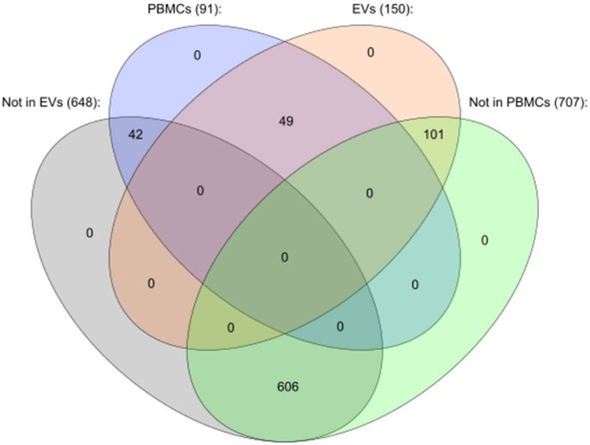
group (ME/CFS or HCs), finding only 91 miRNAs in PBMCs and 150 in EVs meeting these criteria. 49 of them
were present in both sets of samples, while 42 and 101 were exclusive of PBMCs or EVs, respectively (Fig. 4)
(Supplementary Dataset 2). These miRNAs were considered to be robustly and reproducibly expressed in PBMCs
and EVs. The restrictive filters applied lead to classify 606 miRNAs out of the 798 tested as not being highly
expressed in either PBMCs or EVs, as shown in Fig. 4.
ME/CFS PBMC miRNomes. Next, we subjected all these 241 miRNAs to differential expression (DE) anal-
ysis (as described in Methods). Since the reduced number of samples could lead to discard miRNAs, which
although not reaching statistical significance in t-student or Mann-Whitney tests, might be of relevance to the
disease, we proceeded to detect and filter potential outlier values among DE miRNAs (p < 0.1) by applying
Chauvenet’s criterion42. This method consists of detecting an acceptable band of data around the mean. Values
falling outside that band (>2-fold SD absolute values) are considered outliers.
Statistical analysis of the DE miRNAs in PBMCs, after this adjustment, showed 17 miRNAs presenting sig-
nificant differences (p < 0.05), 9 of them being overexpressed (Fig. 5 & Supplementary Table S3); and 8 under
expressed (Fig. 6 & Supplementary Table S4) in the ME/CFS group. The miRNAs showing significantly increased
values (p < 0.05) were: hsa-miR-374a-5p, hsa-miRNA-4516, hsa-miR-340–5p, hsa-miR-140-5p, hsa-miR-18a-5p,
hsa-miR-146a-5p, hsa-miR-106a-5p & hsa-miR-17-5p and hsa-miR-106b, by decreasing order of significance.
Scientific Reports | (2020) 10:2064 | https://doi.org/10.1038/s41598-020-58506-5 5www.nature.com/scientificreports/ www.nature.com/scientificreports
Figure 4 .Venn diagram showing miRNAs found in PBMCs and EVs by nanostring analysis. Numbers of
miRNAs expressed in either fraction after applying strict normalization filters are indicated. Drawn with
Cytoscape version 3.6.0 and the Venn & Euler diagram package, version 1.0.3.
Differences varied between 20 and 65% (Supplementary Table S3). While those miRNAs showing significantly
decreased values were: hsa-miR-644a, hsa-miR-451a, hsa-miR-4454 & hsa-miR-7975, hsa-miR-549a, hsa-miR-
361-3p, hsa-miR-1253 and hsa-miR-590-5p, also cited by strict order of decreasing significance. Differences
of expression in this last group varied between 13 and over 200%. In addition, hsa-miR-21-5p presented a
p-value > 0.05 and 0.05 andwww.nature.com/scientificreports/ www.nature.com/scientificreports
Figure 5 . Overexpressed miRNAs in PBMCs of ME/CFS with respect to HCs, after adjustment of outlier values
according to Chauvenet criterion, (Mann Whitney, p < 0.05). The number of samples analyzed per group for
either miRNA appears between brackets. Medians are shown by horizontal bars, whiskers indicate interquartile
ranges (IQR).
Figure 6 . Under expressed miRNAs in PBMCs of ME/CFS with respect to HCs, after adjustment of outlier
values according to Chauvenet criterion, (Mann Whitney, p < 0.05). The number of samples analyzed per
group for either miRNA appears between brackets. Medians are shown by horizontal bars, whiskers indicate
interquartile ranges (IQR).
MECP2 is subject of X-chromosome inactivation is supportive of potential environmental sex-biased effectors
for this acquired disease.
In addition, we also found MECP2 pathways among top hits in DE miRNAs overexpressed in EVs with
p < 0.05. The finding that neuronal and endocrine system pathways are among DE miRNA hits in ME/CFS EVs,
might be indicative of defects in endocrine tissue functioning, as many patients report. Neurotrophic receptor
tyrosine kinase (NTRK) signaling seems to be also impacted by significant DE miRNAs (p < 0.05) both in PBMCs
and EVs which could relate to patient symptoms such as cognitive disability and sensorial dysfunctions (altered
pain sensitivity)44.
Diagnostic value assessment of evaluated variables. Since the main goal of the study was to deter-
mine the potential diagnostic value of the variables measured, we conducted response operating characteristic
(ROC) analyses for each of the miRNAs showing significant DE between ME/CFS and HC individuals (p < 0.05),
at individual level (miRNAs shown in Fig. 5 through 7 and Supplementary Tables S3 and S4).
Scientific Reports | (2020) 10:2064 | https://doi.org/10.1038/s41598-020-58506-5 7www.nature.com/scientificreports/ www.nature.com/scientificreports
Figure 7. DE miRNAs in plasma EVs of ME/CFS and HCs, after adjustment of outlier values according to
Chauvenet criterion, (Mann Whitney, p < 0.05). Overexpressed (A–G), and under expressed (H,I) miRNAs are
shown. The number of samples analyzed per group for either miRNA appears between brackets. Medians are
shown by horizontal bars, whiskers indicate interquartile ranges (IQR).
As shown in Fig. 8, fifteen of the twenty-seven significant DE miRNAs detected in this study present values
for the area under the curve (AUC) above 0.75 indicating that they have an acceptable capacity to discriminate
blood samples of ME/CFS patients and HCs with respect to their miRNA content. The microRNAs that present
improved discrimination capacity are: hsa-miR-374a-5p (AUC: 0.876, 95% C.I:0.7139–1), hsa-miR-4516 (AUC:
0.8654, 95% C.I: 0.7158–1), hsa-miR-4454 & hsa-miR-7975 (AUC: 0.8472, 95% C.I: 0.6919–1) and hsa-miR-340
(AUC: 0.8106, 95% C.I 0.6181–1), all belonging to the PBMCs fraction, with the only exception of hsa-miR-4454
& hsa-miR-7975 which could be considered the potential top biomarker candidate in the EV fraction, with AUC
above 0.8.
Validation of DE miRNAs predicted target genes. In order to determine the impact of DE miRNAs
on PBMCs transcriptome, we designed sets of primers to quantitate the levels of representative genes within
commonly affected pathways across DE miRNAs hits (Supplementary Datasets 3 and 4), by RT-qPCR. In par-
ticular we chose: the AGO2 gene from the silencing by small RNAs category, including R-HSA-426496 and
R-HSA-426486 Reactome pathways; the MECP2 gene from MECP2 related pathways, including R-HSA-9022707,
R-HSA-9022699, R-HSA-9022692, R-HSA-8986944, R-HSA-9022534, R-HSA-9005891, R-HSA-9022538,
R-HSA-9022927, R-HSA-9022537 and R-HSA-9022702 Reactome pathways; and the NTRK1 gene from R-HSA-
187037 and R-HSA-9028731 neurotrophin signaling Reactome pathways. Figure 9 shows that AGO2 and MECP2
are under expressed, while NTRK1 levels are increased in ME/CFS PBMCs. As discussed above, the connection of
these pathways with patient symptoms suggests their contribution in the pathophysiology of ME/CFS.
Discussion
Researchers and clinicians seem to agree on the urgent need for unbiased, specific diagnostic biomarkers for ME/
CFS to expedite patient diagnosis and treatment, and to abolish disease stigmas for good45. There is also a con-
sensus on the advantages that miRNA profiles from different blood fractions, EVs included, exhibit as molecular
diagnostic candidates, and yet, a paucity of studies focusing on ME/CFS miRNA profiling is detected22,24–27.
A limitation for biomarker discovery studies in ME/CFS is that they have so far consisted of pilot studies with
low number of participants, and the present work, with an N of 30 participants, is no exception. The importance
of our design, however, relies in that, for the first time, miRNomes in PBMCs and EVs were studied in the same
exact individuals, in addition to 34 blood analytical variables and 6 additional EV features (yields, size and zeta
values, either in presence or absence of proteinase K treatment), and several health questionnaires (SF-36 &
GHQ28), leading to the most complete phenotype registry of severely affected ME/CFS individuals we are aware
of.
To prevent additional limitations hampering biomarker discovery in ME/CFS, we tried to minimize patient
heterogeneity and biases associated to pre-analytical variables by only including samples from the UK mono-
graphic ME biobank, which uses procedures complying with the NINDS (National Institute of Neurological
Disorders and Stroke) Common Data Elements (CDEs) for the study of ME/CFS46. Diagnosis criteria and stand-
ard operating procedures (SOPs) used at this facility have been briefly summarized in the methods section. For
further details, readers are referred to publications from the biobank34,35.
Blood creatine phosphokinase (CK) levels appeared significantly reduced in our ME/CFS studied cohort
(t-student, p < 0.05, Supplementary Table S1), in agreement with the recent report by Nacul et al. including eval-
uation of this variable in 56 severe ME/CFS patients47. Serum CK could therefore constitute a biomarker for
severe ME/CFS cases (96% sensitivity) albeit with low specificity (www.nature.com/scientificreports/ www.nature.com/scientificreports
Figure 8 . Calculated area under the curve (AUC) for individual DE microRNAs (p < 0.05). AUCs > 0.75 and
95% confidence interval (CI) values are shown. Blood fractions presenting differentially expressed miRNAs
(PBMCs or EVs) are indicated between brackets.
for activity maintains its significance, in addition to its potential to explain PEM in ME/CFS warrants further
exploration47.
Because technical simplicity is desirable for clinical tests, we decided that rather than pursuing the isolation
of highly purified EV subpopulations obtained through lengthy protocols, the use of optimized commercial kits
rendering high EV yields by simple-fast procedures was preferable.
It must be noted that availability of these highly valuable samples is quite restricted. This was an additional
reason for choosing TEIR reagent as the EV isolation method, as it provides highest yields from only 0.5 mls
of plasma when compared to other commercial products, or alternative isolation methods40. Therefore total
amounts of sample could be minimized. Additional authors concur with Helwa et al. that EVs isolated with
polyethylene glycol (PEG) 6000, as is the case of the commercial product TEIR, render particles of similar size to
alternative methods, including ultracentrifugation, ultrafiltration, and gel chromatography, with increased yields,
as summarized by Konoshenko et al.48.
The present study intends to pave the way in exploring blood EV features in ME/CFS, thus, opening the possi-
bility of evaluating miRNA networks affected by ME/CFS pathophysiology in the entire organism. Another main
aspect of the project was to determine whether EVs constitute a more sensitive, or otherwise improved fraction
than PBMCs, in detecting miRNA changes in ME/CFS, for diagnostic purposes. ROC analysis, however, points
towards superior diagnostic value of miRNomes from PBMCs compared to EV’s (Fig. 8). The high variability
and low miRNA copy number in EVs with respect to PBMCs (counts threshold of 17 versus 69, Supplementary
Dataset 1, PBMC & EV tabs) may, at least partly explain this finding. Surprisingly, yields, size and zeta power
values of EVs appeared as disease-discriminating parameters by the same approach (Figs. 1 & 2).
In line with findings in other pathologies, including cancer and neurodegenerative diseases49,50, we found
higher numbers of EVs in the disease state (Fig. 1A,C). Interestingly enough, a recent pilot study evaluating num-
ber and size of EVs in serum also found increased numbers in ME/CFS patients51. In addition to increased EV
yields, we also observed that the mean size of ME/CFS EVs is reduced (Fig. 1B,D), showing an average diameter
below 100 nm. This is also in agreement with the mentioned findings by Castro-Marrero et al.51. Although we
used plasma instead of serum, the parameter differences found seem to coincide, evidencing reproducibility in
more than one type of sample and across different cohorts. The question on whether one cell produces EVs of
different sizes or does size difference reflect EV production by different cells remains presently unknown.
EV size depends not only on the type of membrane phospholipids but also on the presence or absence of
particular membrane proteins52 and the difference may have functional consequences. For example, it has been
proposed that cytokines found on the EV surface may serve as bar code molecules being recognized by abundant
Scientific Reports | (2020) 10:2064 | https://doi.org/10.1038/s41598-020-58506-5 9www.nature.com/scientificreports/ www.nature.com/scientificreports
Figure 9 . Quantitation of mRNA levels in PBMCs of ME/CFS with respect to HCs. RT-qPCR amplification
of selected genes in top target pathways for DE PBMC miRNAs is shown. Expression levels were normalized to
endogenous GAPDH levels, as described in Methods. Means and standard deviations (SD) are shown (N = 10/
group, t-test, p < 0.001).
cell-specific cytokine receptors52,53. Mass spectrometry (MS) should definitively be included in follow up studies
to evaluate potential qualitative differences associating to ME/CFS.
Since the freezing/thawing process may affect the properties of the vesicles, vesicle flocculation included54, our
experiments were all based on a single thaw step. However, we cannot rule out at present vesicle collapsing in HC
as an explanation to their increased size. In fact, EVs from patients presented higher stability (within an incipient
instability range) according to their higher zeta potential absolute values (Fig. 2).
The observation that ME/CFS EV zeta values are more negative than those of the HC counterpart suggests that
the outer layer phospholipids, or the membrane proteins, or both, contain higher negative charge. Sialic acid on
membrane-bound oligosaccharides lead to increased net negative charge of secreted vesicles from tumor cells54.
It will be interesting to determine if there is an enrichment of sialic acid in ME/CFS EVs.
The fact that pretreatment with proteinase K reduces membrane electronegativity in both ME/CFS and HC
points to a contribution of proteins in the net vesicle charge. However, it must be noted that the overall charge in
ME/CFS EVs stays higher also upon proteinase K treatment in comparison to treated EVs from HCs (Fig. 2C,D),
leaving lipids and carbohydrates as main candidates to explain the observed charge differences between ME/CFS
and HCs. Unbiased distribution of lipids in vesicle leaflets and/or differences in sugar ratios are interesting possi-
bilities worth exploring as they could be indicative of different subpopulations in the disease vs the healthy state.
Despite the fact that EVs isolated with TEIR reagent include vesicles with exosomal morphology and
size-range under transmission electronic microscopy (Fig. 3A), and the presence of the exosomal markers CD9
and CD81 were evidenced by western blotting (Fig. 3B), our procedures were limited at estimating the relative
abundance of exosomes and other vesicle subgroups in general, and therefore any potential attribute of exosomes
or other vesicle subtype in ME/CFS would be merely speculative.
The total final number of miRNAs that were expressed above background seemed strikingly low, with only
91 in PBMCs and 150 in EVs (Fig. 4), out of the approximately 800 included in the nCounter Human miRNA
Panel v3 (Nanostring Technologies, GXA-MIR3-12). As the samples had passed demanding quality controls
(Supplementary Table S2), the low number of hits in PBMCs must be due to restricted blood cell-type expression,
as described by other authors55, together with the strict selective criteria imposed in this study (Supplementary
Figs. S3 and S4). Of the 27 miRNAs DE in ME/CFS (p < 0.05) (17 in PBMCs and 10 in EVs), we find, following a
database search formerly described by our group56, that hsa-miR-18a-5p can be upregulated by the anti-psychotic
drug desipramine, while hsa-miR-146a-5p and hsa-miR-150-5p expression can be induced by morphine56, raising
the possibility that the observed overexpression of these miRNAs derives from patient drug exposure and not
from the disease itself. This highlights the enormous importance of detailed medication registry when studying
molecular changes in these patients, as recommended by the ME/CFS NINDS CDEs46.
Scientific Reports | (2020) 10:2064 | https://doi.org/10.1038/s41598-020-58506-5 10www.nature.com/scientificreports/ www.nature.com/scientificreports
The fact that the number of miRNAs detected above background levels found in EVs is larger than in PBMCs
was somewhat expected since EV varied cell-source should lead to higher miRNA complexity39.
Although due to selective packaging of miRNAs, EV miRNomes could be useful to trace the cells of ori-
gin29,52, we still need to first understand the “rules” for this directional process. By contrast, some data suggests
non-selective miRNA packaging into EVs57. Interestingly Ridder et al., show, by using an elegant Cre-LoxP-based
genetic tracing system, that it is not only the cell-type, but the health status of the individual which may determine
molecule directional packaging and function of EVs. In their experiments, the authors evidenced that the transfer
of signaling RNA between immune cells and the brain, gut and additional tissues is promoted by inflammatory
injuries58.
MiRNA tissue-specific expression could provide hints regarding cell origin of plasma EVs. In this regard,
Ludwig et al., determined the relative abundance of 1997 miRNAs in 61 tissue biopsies of different organs col-
lected post-mortem from two individuals59. Based on a tissue specificity index (TSI), the authors found that the
majority of miRNAs (82.9%) fell in a middle TSI range, meaning they were neither specific for single tissues
(TSI > 0.85) nor housekeeping miRNAs (TSI < 0.5). Many different miRNAs and miRNA families’ expression,
however, were predominant in certain tissues. Moreover, miRNAs TSI values of human tissues were well corre-
lated with those of rats, supporting interspecies-conserved tissue-enriched miRNAs59. Only five miRNAs resulted
highly specific for hematopoietic cells: miR-142, miR-144, miR-150, miR-155, and miR-223. Among them only
miRNA-150 appeared DE (p < 0.05) in our EV profiling and miR-223 presented a DE tendency (0.05 > p < 0.1).
Using their web server (https://ccb-web.cs.uni-saarland.de/tissueatlas) as a search tool58 we found that EV DE
miRNAs (p < 0.05): hsa-miR-150-5p, hsa-miR-15a-5p, hsa-miR-183-5p, hsa-miR33a-5p, let-7d-5p, hsa-miR-
423-5p, hsa-miR-374a-5p and hsa-miR-130a-3p were predominantly expressed in muscle and fascia, brain and
nerves and hormonal glands (mostly thyroid), after quantile normalization. All of them correspond to tissues
affected in ME/CFS.
Although hsa-miR-4454 and hsa-miR-7875 are not found in the mentioned database, Polytarchou et al.,
reported detecting hsa-miR-4454 in serum, as a potential marker of ulcerative colitis with higher sensitivity and
specificity values than C-reactive protein60. This fits with the fact that inflammation frequently associates with
ME/CFS61. Perhaps, the large differences observed in patient EVs for this miRNA (Fig. 7A) relate to intestinal
dysbiosis in some patients. Detailed clinical history registry of participants will be key to answer this question, as
pointed by the ME/CFS CDEs46.
Only four miRNAs were found in both fractions (PBMCs and EVs). Two of them (hsa-miR-21-5p and
hsa-miR-374a-5p) followed the same trend, being overexpressed in both fractions, while the closely related
hsa-miR-4454 and hsa-miR-7975 appeared under expressed in PBMCs and overexpressed in EVs which could
be explained by directional packaging of immune cells. It seems particularly interesting that hsa-miR-4454 and
hsa-miR-7975, linked to inflammation by some authors60, appears among the top DE miRNAs with potential
diagnostic value according to ROC analysis (Fig. 8). At present, however, we can only speculate on the potential
significance of these findings. GO pathway analysis and RT-qPCR results (Fig. 9) points to potential epigenetic
and signal transduction mechanisms associating with ME/CFS, in good agreement with the differential methyla-
tion patterns formerly detected in ME/CFS patients by our group62.
Finally, although, opposite to microarrays, nanostring technology does not require validation of the results
obtained by alternate methods63, we proceeded, anyway, to test whether differential expression profiles detected
by nanostring in our samples, could be assessed by the cost-effective RT-qPCR method. As shown in Fig. 10,
both miRNAs randomly picked from the overexpressed (hsa-miR-106b) and the under expressed (hsa-miR-549a)
groups were confirmed to be statistically DE in ME/CFS PBMCs. This opens the exciting possibility for a direc-
tional low-cost screening method, including only a few miRNAs together with routine blood analytical param-
eters and some EV features, to evaluate larger ME/CFS cohorts towards population validation of the potential
biomarkers of ME/CFS detected here. Inclusion of diseased controls, such as multiple sclerosis (MS), systemic
lupus erythematosus (SLE) or other diseases presenting overlapping symptoms will be of relevance in pursuance
of ME/CFS specific biomarkers.
Methods
Samples & associated clinical data. Ethical approval of the study was granted by two independent
Research Ethics Committees: the Public Health Research Ethics Committee DGSP-CSISP, Valencia (Spain), study
number UCV_201701 and by the UCL Biobank Ethical Review Committee-Royal Free London NHS Foundation
Trust (B-ERC-RF), study number EC2017.01 before the samples were released by the UK ME Biobank. All meth-
ods were performed in accordance with relevant guidelines and regulations.
All subjects signed an informed consent before samples could be included in the UK ME Biobank34,35.
The samples included in the study consisted of PBMCs (>106 cells) and plasma from 15 severely ill female
ME/CFS patients and 15 female, age-population matched healthy subjects, obtained in EDTA blood-collection
tubes, by the monographic UK ME Biobank professionals. A total of 90 aliquots: one vial of PBMCs/participant
(total of 30 units), and two sets of 1 ml plasma vials/participant (total of 60 aliquots).
As described in the literature, patient recruitment and clinical assessment was mainly performed through the
UK National Health Service (NHS) primary and secondary health care services. Compliance with the Canadian
Consensus2 and/or CDC-1994 (“Fukuda”) criteria1 were ensured for patient recruitment. Participants exclusion
criteria were as follows: (i) taken antiviral medication or drugs known to alter immune function in the preceding
3 months (ii) had any vaccinations in the preceding 3 months; (iii) had a history of acute and chronic infectious
diseases such as hepatitis B and C, tuberculosis, HIV (but not herpes virus or other retrovirus infection); (iv)
another chronic disease such as cancer, coronary heart disease, or uncontrolled diabetes; (v) a severe mood dis-
order; (vi) been pregnant or breastfeeding in the preceding 12 months; or (vii) were morbidly obese (BMI ≥ 40).
Severely ill patients with mobility restrictions were visited at home by a research nurse while healthy subjects
Scientific Reports | (2020) 10:2064 | https://doi.org/10.1038/s41598-020-58506-5 11www.nature.com/scientificreports/ www.nature.com/scientificreports
Figure 10. RT-qPCR validation of miRNAs over (left) or underexpressed (right) in PBMCs from ME/CFS
participants, according to Nanostring analysis. Means and standard deviations (SD) are shown (N = 10/group,
t-test, p < 0.05).
were invited to a recruiting center for clinical assessment and blood sampling. SOPs were followed for participant
identification and invitation, clinical assessments, evaluation of eligibility, phlebotomy, sample transportation and
receipt, laboratory blood tests, data entry, processing, storage and sample release34,35.
All samples were provided by the UK ME Biobank together with complete health survey SF-3636, General
Health Questionnaire (GHQ)37 and Likert test scores38 plus over 30 blood analytical variables. Blood analytics
performed at the UK Biobank from aliquots taken at banking blood collection time points, allow matched corre-
lations with parameters evaluated in downstream analysis.
RNA isolation & RNA quality control. PBMC aliquots were thawed in a 37 °C bath for 1 minute and
immediately centrifuged at 300 × g for 5 min. Total RNA was isolated from PBMCs pellets (>106 cells) or isolated
EVs (500 µls) using RNAzol (Molecular Research Center) according to the manufacturer’s instructions. RNA
yields and quality were assessed using Agilent TapeStation 4200 (Agilent Technologies). PBMCs RNA samples
with an RNA Integrity number (RIN) above 7 were included in downstream analysis.
Small RNA distributions were assessed using small RNA kits and the Agilent 2100 bioanalyzer (Agilent
Technologies), following manufacturer recommendations. Agilent 2100 bioanalyzer analysis were run at the
Centro de Investigación Príncipe Felipe (CIPF) Genomic services in Valencia (Spain).
EV purification. EVs were isolated from 0.5 ml aliquots of human plasma supernatants (undergoing a sin-
gle freeze/thaw cycle), upon being centrifuged at 10,000 × g for 10 mins, with Total Exosome Isolation Reagent
(TEIR) (Invitrogen by Life Technologies, Cat. 4484450), following manufacturer’s protocol. The process was run
in parallel including a proteinase K pretreatment or skipping it for each thawed aliquot.
EV characterization. Yields and size distribution of isolated EVs were determined using Nanoparticle
Tracking Analysis (NTA), light scattering, technology with a Nanosight NS300 instrument (Malvern Panalytical)
at the Centro de Investigación Príncipe Felipe (CIPF) Genomic services in Valencia (Spain). Zeta potentials were
measured at the same facility with a DLS (Dynamic Light Scattering) Zetasizer Nano ZS (Malvern Panalytical),
using a laser wavelength of 532 nm. Capillaries used were cat. Num. DTS 1070 (Malvern Panalytical). EV samples
were diluted 1/50 (V/V) in 1X PBS buffer prior to measurements. Zeta potentials were obtained at RT and in
triplicate for each analyzed sample. The obtained 90 s videos were recorded in a level 10 camera at the setting of 25
images/s. Video analysis were performed with the NTA version 3.1 and a detection limit of 20.
TEM (transmission electronic microscopy) imaging of EVs was obtained from 1% glutaraldehyde fixed vesi-
cles. A 20 µl drop of each suspension was loaded onto a formvar/carbon-coated grid, negatively stained with 3%
(w/v) aqueous phosphotungstic acid for 1 min and observed by transmission electron microscopy (TEM) in a
Tecnai Spirit G-2 apparatus; (FEI, Eindhoven, The Netherlands) at the Centro de Investigación Príncipe Felipe
(CIPF) Genomic services in Valencia (Spain).
Western-blotting to assess exosomal markers in isolated EVs was performed using the classic Laemmli
method with some modifications. In brief, from EVs eluted in 200 µl of 1X PBS from 500 µl of human plasma,
15 µl were lysed in 5X SDS loading buffer and directly loaded into the gel after heating at 95 °C for 5 mins. Primary
antibodies used were: anti-CD9 antibody (EX201, Cell Guidance Systems) (1 mg/ml), anti-CD81 antibody (sc-
23962, Santa Cruz Biotechnology) (0.2 mg/ml), anti-HSP70 antibody (sc-7298, Santa Cruz Biotechnology)
(0.2 mg/ml) and anti-β-Actin antibody (sc-47778, Santa Cruz Biotechnology) (0.1 mg/ml), all these antibodies
were used a final dilution of 1/200. As secondary antibodies we used goat anti-mouse IgG-HRP (sc-2005, Santa
Cruz Biotechnology) (0.4 mg/ml) using a final dilution of 1/5000.
Nanostring miRNA profiling & analysis. miRNA profiling was performed in an nCounter NanoString
platform (Nanostring Technologies, Seattle, WA, USA), and the nCounter Human miRNA Panel v3 (Nanostring
Technologies, GXA-MIR3-12) which evaluates about 800 miRNAs. Each sample was analyzed by using 100 ng of
Scientific Reports | (2020) 10:2064 | https://doi.org/10.1038/s41598-020-58506-5 12www.nature.com/scientificreports/ www.nature.com/scientificreports
total RNA for hybridization (21 h at 65 °C) in addition to probe pairs consisting of a Reporter Probe, which carry
the signal on their 5′ end, and a Capture Probes, which carry biotin on their 3′ end. After hybridization, sample
cleanup and digital report counts were performed according to manufacturer’s instructions.
Raw counts were analyzed using Nanostring nSolver version 4.0 software. We calculated geometric mean
of negative ligation controls plus two standard deviations for all samples. This value served as a threshold.
Thresholds were established as 69 counts in PBMCs and 17 counts in EVs. All count values below this threshold
were excluded from normalization. After that, PBMC miRNA input levels were normalized to the geometric
mean of the top expressed100 miRNAs. EV miRNA input levels were normalized to six spiked-in controls of
non-human miRNAs.
Samples with counts above threshold established values in less than 40% of the miRNAs were eliminated.
Next, only miRNAs showing above threshold values in at least 75% of samples, in either group (ME/CFS and
HCs), were used for the differential expression analysis in the evaluated blood fraction (either PBMCs or EVs).
Statistical differences were assessed as described below.
Heatmaps were built using the Heatmapper website (http://www.heatmapper.ca/expression/), The Spearman
Range Correlation was applied to establish the distance across measurements and the Centroid link as a grouping
method64.
RT-qPCR validation. Reverse-transcription was performed with the miScript II RT kit (Qiagen, cat. 218161)
or the High-Capacity cDNA reverse Transcription kit (Applied Biosystems, cat. 4308228), using 1 µg of total RNA
according to manufacturer’s guidelines. cDNAs were used for Real time PCR using miScript SYBR Green PCR
kit (Qiagen, cat. 218073), or the PowerUP Sybr Green Master Mix (Applied Biosystems, cat. 100029283) and a
Lightcycler LC480 instrument (Roche). Standard amplification conditions were used, including a single hotstart
polymerase preactivation cycle at 94 °C for 15 min, followed by 45 amplification cycles each one consisting of
3 steps: denaturation at 95 °C for 15 s, annealing at 50–60 °C for 30 s and extension at 70 °C for 30 s. Sequences
of forward miRNA specific primers used were hsa-miR-106b-5p (5′-GTG CTG ACA GTG CAG AT-3′) and
hsa-miR-549a: (5′-TGA CAA CTA TGG ATG AGC-3′). As a reverse primer the Universal Primer (UP) provided
with the kit was used in all amplifications. U6 RNA amplification levels were used for the relative quantification of
the miRNAs amplified. For RT-qPCR of mRNAs, the following primers sets were used: AGO2 (Forward: 5′-TCG
CAC TAT CAC GTC CTC TG-3′ and Reverse: 5′-ATG GCT TCC TTC AGC ACT GT-3′); MECP2 (Forward:
5′-TGG TAA AAG CCG TCC GG-3′ and Reverse: 5′-GCC CTA ACA TCC CAG CTA-3′) and NTRK1 (Forward:
5′-TTC AAC GCT CTG GAG TCT-3′ and Reverse: 5′-CAT CAC CGT GGC TGA CT-3′). mRNA levels were
normalized to GAPDH levels obtained with primers: Forward: 5′-TGA AGG TCG GAG TCA ACG GAT-3′ and
Reverse: 5′-TTC TCA GCC TTG ACG GTG CCA-3′.
Gene enrichment & pathway analysis. For gene enrichment analysis, potential target genes of the DE
miRNAs showing statistical significance (p < 0.05) were identified with the miRDIP database65 and the Human
miRNA-gene interaction (adjacency) matrix setting. This option demands that target genes present target sites for
at least 2 of the DE miRNAs in the set66. Next, pathway analysis of the target genes was assessed in the Reactome
database67. Only target pathways with p values < 0.05 were considered potential effectors of the statistically sig-
nificant DE miRNAs.
ROC curves, statistical analysis & plotting. Response operating characteristic (ROC) for each of the
miRNAs showing significant DE between ME/CFS and HC individuals (p < 0.05) were assessed with the R pack-
age pROC version 1.13.068.
Continuous data are expressed as means ± SD, as indicated. Normal data distribution was assessed using
the Shapiro-Wilk test, assuming that p-values > 0.05, followed normal distributions. Statistical differences were
determined using t-student or Mann-Whitney tests, depending on whether the data followed or not normal
distributions, respectively. To establish group differences, two ranges of significance were applied: p < 0.05 for
statistical difference and p < 0.1 for trends or tendencies. Analysis were conducted with the R package version
3.4.2 (R Core Team, 2014)69.
Plots were drawn using the R ggplot2 environment version 2.2.170 and the GraphPad Prism 5.0 program. The
Venn diagram was drawn with Cytoscape version 3.6.0 and the Venn & Euler diagram package, version 1.0.3.
Data availability
Nanostring datasets generated during and/or analyzed during the current study are available from the NCBI Gene
Expression Omnibus (GEO) database71, (Accession Number GSE141770).
Received: 10 October 2019; Accepted: 15 January 2020;
Published: xx xx xxxx
References
1. Fukuda, K. et al. The chronic fatigue syndrome: a comprehensive approach to its definition and study. International chronic fatigue
syndrome study group. Ann. Intern. Med. 121(12), 953–959 (1994).
2. Carruthers, B. M. et al. Myalgic encephalomyelitis/chronic fatigue syndrome: clinical working case definition, diagnostic and
treatment protocols. J. Chronic Fatigue Syndrome 11, 7–115 (2003).
3. Carruthers, B. M. et al. Myalgic encephalomyelitis: international consensus criteria. J Intern Med 270(4), 327-338 (2011). Erratum
in. J Intern Med. 282(4), 353 (2017).
4. Committee on the Diagnostic Criteria for Myalgic Encephalomyelitis/Chronic Fatigue Syndrome; Board on the Health of Select
Populations; Institute of Medicine. Beyond Myalgic Encephalomyelitis/Chronic Fatigue Syndrome: Redefining an Illness.
Scientific Reports | (2020) 10:2064 | https://doi.org/10.1038/s41598-020-58506-5 13www.nature.com/scientificreports/ www.nature.com/scientificreports
Washington (DC): National Academies Press (US); Committee on the Diagnostic Criteria for Myalgic Encephalomyelitis/Chronic
Fatigue Syndrome. Available from, https://www.ncbi.nlm.nih.gov/books/NBK284899/ (2015).
5. Clayton, E. W. Beyond myalgic encephalomyelitis/chronic fatigue syndrome: a IOM report on redefining an illness. JAMA. 313(11),
1101–1102 (2015).
6. Boerma, T. et al. Revising the ICD: explaining the WHO approach. Lancet 388(10059), 2476–2477 (2016).
7. Reyes, M. et al. Prevalence and incidence of chronic fatigue syndrome in Wichita, Kansas. Arch Intern Med. 163(13), 1530–1536
(2003).
8. Jason, L. A. et al. A community-based study of chronic fatigue syndrome. Arch Intern Med. 159(18), 2129–2137 (1999).
9. Estévez-López, F. et al. European network on ME/CFS (EUROMENE). Prevalence and incidence of myalgic encephalomyelitis/
chronic fatigue syndrome in Europe-the Euro-epiME study from the european network EUROMENE: a protocol for a systematic
review. BMJ Open 8(9), e020817 (2018).
10. Holgate, S. T., Komaroff, A. L., Mangan, D. & Wessely, S. Chronic fatigue syndrome: understanding a complex illness. Nat Rev
Neurosci. 12(9), 539–544 (2011).
11. Cairns, R. & Hotopf, M. A systematic review describing the prognosis of chronic fatigue syndrome. Occup. Med. 55, 20–31 (2005).
12. Brurberg, K. G., Fonhus, M. S., Larun, L., Flottorp, S. & Malterud, K. Case definitions for chronic fatigue syndrome/myalgic
encephalomyelitis (CFS/ME): A systematic review. BMJ Open 4, e003973 (2014).
13. Scheibenbogen, C. et al. The european ME/CFS biomarker landscape project: an initiative of the european network EUROMENE. J
Transl Med. 15(1), 162 (2017).
14. Klimas, N. G., Broderick, G. & Fletcher, M. A. Biomarkers for chronic fatigue. Brain Behav Immun. 26(8), 1202–10 (2012).
15. Tomas, C. & Newton, J. Metabolic abnormalities in chronic fatigue syndrome/myalgic encephalomyelitis: a mini-review. Biochem
Soc Trans. 46(3), 547–553 (2018).
16. Tomas, C. et al. Cellular bioenergetics is impaired in patients with chronic fatigue syndrome. PLoS One 12(10), e0186802 (2017).
Erratum in. PLoS One. 13(2), e0192817 (2018).
17. Stavast, C. J. & Erkeland, S. J. The Non-Canonical Aspects of MicroRNAs: Many Roads to Gene Regulation. Cells 8(11), E146 (2019).
18. Ha, M. & Kim, V. N. Regulation of microRNA biogenesis. Nat Rev Mol Cell Biol. 15(8), 509–524 (2014).
19. Vishnoi, A. & Rani, S. MiRNA biogenesis and regulation of diseases: an overview. Methods Mol Biol. 1509, 1–10 (2017).
20. Pardini, B., Sabo, A. A., Birolo, G. & Calin, G. A. Noncoding RNAs in extracellular fluids as cancer biomarkers: the new frontier of
liquid biopsies. Cancers (Basel). 11(8), E1170 (2019).
21. Slota, J. A. & Booth, S. A. MicroRNAs in neuroinflammation: implications in disease pathogenesis, biomarker discovery and
therapeutic applications. Noncoding RNA 5(2), E35 (2019).
22. Brenu, E. W. et al. Cytotoxic lymphocyte microRNAs as prospective biomarkers for chronic fatigue syndrome/myalgic
encephalomyelitis. J. Affect Disord. 141, 261–269 (2012).
23. Cerdá-Olmedo, G., Mena-Durán, A. V., Monsalve, V. & Oltra, E. Identification of a microRNA signature for the diagnosis of
fibromyalgia. PLoS One 10(3), e0121903 (2015).
24. Brenu, E. W., Ashton, K. J., Batovska, J., Staines, D. R. & Marshall-Gradisnik, S. M. High-throughput sequencing of plasma
microRNA in chronic fatigue syndrome/myalgic encephalomyelitis. PLoS One 9, e102783 (2014).
25. Petty, R. D., McCarthy, N. E., Le Dieu, R. & Kerr, J. R. MicroRNAs hsa-miR-99b, hsa-miR-330, hsa-miR-126 and hsa-miR-30c:
potential diagnostic biomarkers in natural killer (NK) cells of patients with chronic fatigue syndrome (CFS)/myalgic
encephalomyelitis (ME). PLoS One 11, e0150904 (2016).
26. Baraniuk, J. N. & Shivapurkar, N. Exercise-induced changes in cerebrospinal fluid miRNAs in gulf war illness, chronic fatigue
syndrome and sedentary control subjects. Sci. Rep. 7, 15338 (2017). Author correction. Sci. Rep. 8, 6455 (2018).
27. Al-Rawaf, H. A., Alghadir, A. H. & Gabr, S. A. MicroRNAs as biomarkers of pain intensity in patients with chronic fatigue syndrome.
Pain Pract. 19(8), 848–860 (2019).
28. Théry, C. et al. Minimal information for studies of extracellular vesicles 2018 (MISEV2018): a position statement of the International
Society for Extracellular Vesicles and update of the MISEV2014 guidelines. J. Extracell. Vesicles. 7(1), 1535750 (2018).
29. Villarroya-Beltri, C. et al. Sumoylated hnRNPA2B1 controls the sorting of miRNAs into exosomes through binding to specific
motifs. Nat Commun. 4, 2980 (2013).
30. Wolfers, J. et al. Tumor-derived exosomes are a source of shared tumor rejection antigens for CTL cross-priming. Nat Med. 7,
297–303 (2001).
31. Melo, S. A. et al. Cancer exosomes perform cell-independent microRNA biogenesis and promote tumorigenesis. Cancer Cell 26,
707–721 (2014).
32. Zhou, W. et al. Exosomes serve as novel modes of tick-borne flavivirus transmission from arthropod to human cells and facilitates
dissemination of viral RNA and proteins to the vertebrate neuronal cells. PLoS Pathog. 14(1), e1006764 (2018).
33. Fritz, J. V. et al. Sources and functions of extracellular small RNAs in human circulation. Annu Rev Nutr. 36, 301–36 (2016).
34. Lacerda, E. M. et al. The UK ME/CFS biobank: a disease-specific biobank for advancing clinical research into myalgic
encephalomyelitis/chronic fatigue syndrome. Front Neurol. 9, 1026 (2018).
35. Lacerda, E. M. et al. The UK ME/CFS biobank for biomedical research on myalgic encephalomyelitis/chronic fatigue syndrome
(ME/CFS) and multiple sclerosis. Open J Bioresour. 4, 4 (2017).
36. McHorney, C. A., Ware, J. E. Jr. & Raczek, A. E. The MOS 36-item short-form health survey (SF-36): II. psychometric and clinical
tests of validity in measuring physical and mental health constructs. Med Care. 31(3), 247–263 (1993).
37. Jackson, C. The General Health Questionnaire. Occupational Medicine 57(1), 79–79 (2006).
38. Than, U. T. T. et al. Differential expression of keratinocyte-derived extracellular vesicle mirnas discriminate exosomes from
apoptotic bodies and microvesicles. Front Endocrinol (Lausanne). 9, 535 (2018).
39. Coumans, F. A. W. et al. Methodological guidelines to study extracellular vesicles. Circ Res. 20(10), 1632–1648 (2017).
40. Helwa, I. et al. A comparative study of serum exosome isolation using differential ultracentrifugation and three commercial reagents.
PLoS One. 12(1), e0170628 (2017).
41. Rupert, D. L. M., Claudio, V., Lässer, C. & Bally, M. Methods for the physical characterization and quantification of extracellular
vesicles in biological samples. Biochim Biophys Acta Gen Subj. 1861(1 Pt A), 3164–3179 (2017).
42. Zerbet, A. & Nikulin, M. A New Statistic for Detecting Outliers in Exponential Case. Communications in Statistics - Theory and
Methods 32(3), 573–583 (2003).
43. Leinders, M. et al. Increased cutaneous miR-let-7d expression correlates with small nerve fiber pathology in patients with
fibromyalgia syndrome. Pain. 157, 2493–2503 (2016).
44. Dhandapani, R. et al. Control of mechanical pain hypersensitivity in mice through ligand-targeted photoablationof TrkB-positive
sensory neurons. Nat Commun. 9(1), 1640 (2018).
45. Sweetman, E. et al. Current research provides insight into the biological basis and diagnostic potential for myalgic encephalomyelitis/
chronic fatigue syndrome (ME/CFS). Diagnostics (Basel). 9(3), E73 (2019).
46. NINDS. Common Data Elements. Available online, https://www.commondataelements.ninds.nih.gov/Myalgic%20
Encephalomyelitis/Chronic%20Fatigue%20SyndromeMECFS.aspx#tab=Data_Standards (accessed on 15 September 2019).
47. Nacul, L. et al. Evidence of clinical pathology abnormalities in people with myalgic encephalomyelitis/chronic fatigue syndrome
(ME/CFS) from an analytic cross-sectional study. Diagnostics (Basel) 9(2), E41 (2019).
Scientific Reports | (2020) 10:2064 | https://doi.org/10.1038/s41598-020-58506-5 14www.nature.com/scientificreports/ www.nature.com/scientificreports
48. Konoshenko, M. Y., Lekchnov, E. A., Vlassov, A. V. & Laktionov, P. P. Isolation of extracellular vesicles: general methodologies and
latest trends. Biomed Res Int. 2018, 8545347 (2018).
49. Rajendran, L. et al. Alzheimer’s disease beta-amyloid peptides are released in association with exosomes. Proc Natl Acad Sci USA
103, 11172–11177 (2006).
50. Logozzi, M. et al. High levels of exosomes expressing CD63 and caveolin-1 in plasma of melanoma patients. PLoS One. 4, e5219
(2009).
51. Castro-Marrero, J. et al. Circulating extracellular as potential biomarkers in chronic fatigue syndrome/myalgic encephalomyelitis:
an exploratory pilot study. J Extracell Vesicles. 7(1), 1453730 (2018).
52. Margolis, L. & Sadovsky, Y. The biology of extracellular vesicles: The known unknowns. PLoS Biol. 17(7), e3000363 (2019).
53. Fitzgerald, W. et al. A system of cytokines encapsulated in extracellular vesicles. Sci Rep. 8, 8973 (2018).
54. Tegegn, T. Z. et al. Characterization of procoagulant extracellular vesicles and platelet membrane disintegration in DMSO-
cryopreserved platelets. J Extracell Vesicles. 5, 30422 (2016).
55. Leidinger, P., Backes, C., Meder, B., Meese, E. & Keller, A. The human miRNA repertoire of different blood compounds. BMC
Genomics 15, 474 (2014).
56. Almenar-Pérez, E., Sánchez-Fito, T., Ovejero, T., Nathanson, L. & Oltra, E. Impact of Polypharmacy on Candidate Biomarker
miRNomes for the Diagnosis of Fibromyalgia and Myalgic Encephalomyelitis/Chronic Fatigue Syndrome: Striking Back on
Treatments. Pharmaceutics. 11(3), E126 (2019).
57. Tosar, J. P. et al. Assessment of small RNA sorting into different extracellular fractions revealed by high-throughput sequencing of
breast cell lines. Nucleic Acids Res. 43, 5601–5616 (2015).
58. Ridder, K. et al. Extracellular vesicle-mediated transfer of genetic information between the hematopoietic system and the brain in
response to inflammation. PLoS Biol. 12(6), e1001874 (2014).
59. Ludwig, N. et al. Distribution of miRNA expression across human tissues. Nucleic Acids Res. 44(8), 3865–3877 (2016).
60. Polytarchou, C. et al. Assessment of Circulating microRNAs for the diagnosis and disease activity avaluation in patients with
ulcerative colitis by using the nanostring technology. Inflamm Bowel Dis. 21(11), 2533–2539 (2015).
61. Montoya, J. G. et al. Cytokine signature associated with disease severity in chronic fatigue syndrome patients. Proc Natl Acad Sci
USA 114(34), E7150–E7158 (2017).
62. Trivedi, M. S. et al. Identification of myalgic encephalomyelitis/chronic fatigue syndrome-associated DNA methylation patterns.
PLoS One. 13(7), e0201066 (2018).
63. Foye, C. et al. Comparison of miRNA quantitation by Nanostring in serum and plasma samples. PLoS One. 12(12), e0189165 (2017).
64. Babicki, S. et al. Heatmapper: web-enabled heat mapping for all. Nucleic Acids Res. 44(W1), W147–W153 (2016).
65. Tokar, T. et al. mirDIP 4.1-integrative database of human microRNA target predictions. Nucleic Acids Res. 46(D1), D360–D370 (2018).
66. Shirdel, E. A., Xie, W., Mak, T. W. & Jurisica, I. NAViGaTing the micronome—using multiple microRNA prediction databases to
identify signalling pathway-associated microRNAs. PLoS One. 6(2), e17429 (2011).
67. Fabregat, A. et al. The Reactome pathway Knowledgebase. Nucleic Acids Research. 44(D1), D481–D487 (2016).
68. Robin, X. et al. pROC: an open-source package for R and S+ to analyze and compare ROC curves. BMC Bioinformatics. 12, 77 (2011).
69. R Core Team. R: A language and environment for statistical computing. R Foundation for Statistical Computing, Vienna, Austria.
URL, http://www.R-project.org/. (2013).
70. Wickham, H. ggplot2: Elegant Graphics for Data Analysis. Springer-Verlag New York, (2009). Wickham, H. ggplot2. Wiley
Interdisciplinary Reviews. Computational Statistics 3(2), 180–185 (2011).
71. Edgar, R., Domrachev, M. & Lash, A. E. Gene Expression Omnibus: NCBI gene expression and hybridization array data repository.
Nucleic Acids Res. 30(1), 207–210 (2002).
Acknowledgements
The study was supported by funding from the ME Association (MEA) Ramsay Research Fund, UK, and by the
2018-270-001 Fundación Universidad Católica de Valencia San Vicente Mártir, Valencia (Spain), grant award
program to E.O. Authors thank Ms. Charlotte Stephens for English language review and editing.
Author contributions
E.A.-P. isolated/characterized EVs, isolated/evaluated QC RNA & performed qRT-PCR. L.S. and L.N. isolated/
evaluated QC RNA & performed nanostring readings. E.O. designed the study and wrote the first draft of the
paper. All authors contributed to the analysis & interpretation of data in addition to reviewing the manuscript.
Competing interests
The authors declare no competing interests.
Additional information
Supplementary information is available for this paper at https://doi.org/10.1038/s41598-020-58506-5.
Correspondence and requests for materials should be addressed to E.O.
Reprints and permissions information is available at www.nature.com/reprints.
Publisher’s note Springer Nature remains neutral with regard to jurisdictional claims in published maps and
institutional affiliations.
Open Access This article is licensed under a Creative Commons Attribution 4.0 International
License, which permits use, sharing, adaptation, distribution and reproduction in any medium or
format, as long as you give appropriate credit to the original author(s) and the source, provide a link to the Cre-
ative Commons license, and indicate if changes were made. The images or other third party material in this
article are included in the article’s Creative Commons license, unless indicated otherwise in a credit line to the
material. If material is not included in the article’s Creative Commons license and your intended use is not per-
mitted by statutory regulation or exceeds the permitted use, you will need to obtain permission directly from the
copyright holder. To view a copy of this license, visit http://creativecommons.org/licenses/by/4.0/.
© The Author(s) 2020
Scientific Reports | (2020) 10:2064 | https://doi.org/10.1038/s41598-020-58506-5 15You can also read