Characterizing and classifying neuroendocrine neoplasms through microRNA sequencing and data mining
←
→
Page content transcription
If your browser does not render page correctly, please read the page content below
Published online 15 July 2020                                                                                   NAR Cancer, 2020, Vol. 2, No. 3 1
                                                                                                                     doi: 10.1093/narcan/zcaa009

Characterizing and classifying neuroendocrine
neoplasms through microRNA sequencing and
data mining
Jina Nanayakkara 1 , Kathrin Tyryshkin1 , Xiaojing Yang1 , Justin J.M. Wong 1 ,
Kaitlin Vanderbeck1 , Paula S. Ginter 2 , Theresa Scognamiglio2 , Yao-Tseng Chen2 ,
Nicole Panarelli3 , Nai-Kong Cheung4 , Frederike Dijk5 , Iddo Z. Ben-Dov 6 ,

                                                                                                                                                                  Downloaded from https://academic.oup.com/narcancer/article/2/3/zcaa009/5867117 by guest on 24 September 2020
Michelle Kang Kim7 , Simron Singh8 , Pavel Morozov9 , Klaas E.A. Max9 , Thomas Tuschl                                                                   9

and Neil Renwick 1,9,*
1
 Laboratory of Translational RNA Biology, Department of Pathology and Molecular Medicine, Queen’s University, 88
Stuart Street, Kingston, ON K7L 3N6, Canada, 2 Department of Pathology and Laboratory Medicine, Weill Cornell
Medicine, 1300 York Avenue, New York, NY 10065, USA, 3 Department of Pathology, Albert Einstein College of
Medicine, 1300 Morris Park Avenue, Bronx, NY 10461, USA, 4 Department of Pediatrics, Memorial Sloan Kettering
Cancer Center, 1275 York Avenue, New York, NY 10065, USA, 5 Department of Pathology, Amsterdam University
Medical Center, Meibergdreef 9, 1105 AZ Amsterdam, The Netherlands, 6 Department of Nephrology and
Hypertension, Hadassah-Hebrew University Medical Center, Jerusalem 91120, Israel, 7 Center for Carcinoid and
Neuroendocrine Tumors of Mount Sinai, Icahn School of Medicine at Mount Sinai, One Gustave L. Levy Place, New
York, NY 10029, USA, 8 Odette Cancer Center, Sunnybrook Health Sciences Center, Toronto, ON M4N 3M5, Canada
and 9 Laboratory of RNA Molecular Biology, The Rockefeller University, 1230 York Avenue, New York, NY 10065, USA

Received March 06, 2020; Revised May 22, 2020; Editorial Decision May 27, 2020; Accepted June 06, 2020

ABSTRACT                                                                          similarities. Using machine learning approaches, we
                                                                                  identified 17 miRNAs to discriminate 15 NEN patho-
Neuroendocrine neoplasms (NENs) are clinically di-
                                                                                  logical types and subsequently constructed a mul-
verse and incompletely characterized cancers that
                                                                                  tilayer classifier, correctly identifying 217 (98%) of
are challenging to classify. MicroRNAs (miRNAs) are
                                                                                  221 samples and overturning one histological diag-
small regulatory RNAs that can be used to classify
                                                                                  nosis. Through our research, we have identified com-
cancers. Recently, a morphology-based classifica-
                                                                                  mon and type-specific miRNA tissue markers and
tion framework for evaluating NENs from different
                                                                                  constructed an accurate miRNA-based classifier, ad-
anatomical sites was proposed by experts, with the
                                                                                  vancing our understanding of NEN diversity.
requirement of improved molecular data integration.
Here, we compiled 378 miRNA expression profiles
to examine NEN classification through comprehen-                                  INTRODUCTION
sive miRNA profiling and data mining. Following data                              Classifying neuroendocrine neoplasms (NENs) is challeng-
preprocessing, our final study cohort included 221                                ing due to tumor diversity, inconsistent terminology and
NEN and 114 non-NEN samples, representing 15 NEN                                  piecemeal molecular characterization. Currently, NENs are
pathological types and 5 site-matched non-NEN con-                                broadly divided into epithelial or non-epithelial groups
trol groups. Unsupervised hierarchical clustering of                              based on site of origin and differences in keratin and other
miRNA expression profiles clearly separated NENs                                  gene expression; each group comprises multiple patholog-
from non-NENs. Comparative analyses showed that                                   ical types (1–3). To facilitate comparisons between NENs
                                                                                  from different anatomical sites, international experts re-
miR-375 and miR-7 expression is substantially higher
                                                                                  cently proposed a common classification framework (3).
in NEN cases than non-NEN controls. Correlation                                   Here, the terms ‘category’, ‘family’, ‘type’ and ‘grade’, re-
analyses showed that NENs from diverse anatom-                                    spectively, denote predominant neuroendocrine differenti-
ical sites have convergent miRNA expression pro-                                  ation, degree of differentiation, diagnostic entity and in-
grams, likely reflecting morphological and functional                             herent biological activity. While morphological assessment

* To   whom correspondence should be addressed. Tel: +1 613 533 6411; Fax: +1 613 533 2907; Email: neil.renwick@queensu.ca

C The Author(s) 2020. Published by Oxford University Press on behalf of NAR Cancer.
This is an Open Access article distributed under the terms of the Creative Commons Attribution Non-Commercial License
(http://creativecommons.org/licenses/by-nc/4.0/), which permits non-commercial re-use, distribution, and reproduction in any medium, provided the original work
is properly cited. For commercial re-use, please contact journals.permissions@oup.com
2 NAR Cancer, 2020, Vol. 2, No. 3

and immunohistochemical staining for chromogranin A,               RNA isolation and quantitation
synaptophysin and Ki-67 proteins remain indispensable for
                                                                   Total RNA was isolated from 306 formalin-fixed paraffin-
confirming neuroendocrine differentiation and assessing tu-
                                                                   embedded tissue blocks and 72 fresh-frozen tissue sam-
mor grade, other relevant molecular findings will be inte-
                                                                   ples using the Qiagen RNeasy® Mini Kit (n = 258), TRI-
grated into this framework over time. These studies will
                                                                   zol™ Reagent (n = 68), the Ambion RecoverAll™ Total Nu-
unravel many puzzles in NEN biology, including delineat-
                                                                   cleic Acid Isolation Kit (n = 28), Amsbio RNA-Bee™ Iso-
ing the molecular differences between well-differentiated
                                                                   lation Reagent (n = 10) and Qiagen miRNeasy® Mini Kit
neuroendocrine tumors (NETs) and poorly differentiated
                                                                   (n = 5), according to the manufacturers’ instructions or as
neuroendocrine carcinomas (NECs) and finding regula-
                                                                   described (7,15–18). Total RNA concentrations were mea-
tory molecules that underpin the ‘common neuroendocrine
                                                                   sured using the Qubit™ fluorometer (n = 258), NanoDrop®
multigene program’ (3).
                                                                   ND-1000 spectrophotometer (n = 61) or Agilent 2100 Bio-

                                                                                                                                  Downloaded from https://academic.oup.com/narcancer/article/2/3/zcaa009/5867117 by guest on 24 September 2020
   MicroRNAs (miRNAs) are small (19–24 nt) regulatory
                                                                   analyzer (n = 28). RNA isolation and quantitation data
RNA molecules that can also be used to classify cancer
                                                                   were unavailable for 9 (2.4%) and 31 (8.2%) samples, respec-
(4,5). miRNAs are highly informative tissue markers be-
                                                                   tively.
cause of their abundance, cell-type and disease-stage speci-
ficity, and stability in fresh and archived materials (6,7).
These molecules also provide valuable mechanistic insights         Small RNA sequencing and sequence annotation
into cellular processes due to computationally predictable         miRNA expression profiles for all 378 samples were gener-
interactions with messenger RNAs (mRNAs) (8,9). In ad-             ated using an established small RNA sequencing approach
dition, miRNA expression profiles can be used to assess            and sequence annotation pipeline (10); spiked-in oligori-
data reliability and to prioritize mRNA targets through fur-       bonucleotide calibrator markers enabled miRNA quanti-
ther organization into miRNA cluster and sequence fam-             tation in each sample. Small RNA cDNA libraries were
ily datasets (10). To date, multiple miRNA profiling stud-         sequenced on HiSeq 2500 Illumina platforms in the Ge-
ies have been performed on single or limited combinations          nomics Resource Center, The Rockefeller University, the
of NEN pathological types using different RNA isolation,           McGill University and Génome Québec Innovation Cen-
detection and analysis methods (11). Although these dif-           ter, and the Genomics Core, Albert Einstein College of
ferences complicate interstudy comparisons, miRNAs still           Medicine. FASTQ sequence files were annotated through an
hold much promise as multi-analyte markers that better re-         automated pipeline (rnaworld.rockefeller.edu) (10), yield-
flect the ‘complexity and multidimensionality of the neo-          ing sequencing statistics and merged miRNA, miRNA clus-
plastic process’ than current mono-analyte markers (12,13).        ter and calibrator read counts. Merged miRNA refers to
Given recent advances in miRNA detection and analysis              combined counts of multicopy miRNAs from different ge-
(14), we expect that substantial biological and clinically rele-   nomic locations and miRNA clusters are transcriptional
vant insights into NEN biology will be gained through com-         units as defined (19); the term ‘miRNA’ will hereafter re-
prehensive miRNA profiling of multiple pathological types.         fer to merged miRNA data. Annotated sequencing statistics
   Through small RNA sequencing and data mining, we                for all samples are presented in Supplementary Table S2;
have generated reference miRNA expression profiles for             miRNA content was calculated using total RNA and cali-
multiple NEN pathological types and site-matched non-              brator RNA input ratio multiplied by total miRNA and cal-
NEN controls, identified candidate category- and type-             ibrator count ratio as described (7). miRNA, miRNA clus-
specific miRNAs, found evidence for constitutive and con-          ter and calibrator read counts for all samples are presented
vergent miRNA gene expression in epithelial and non-               in Supplementary Tables S3–S5, respectively.
epithelial NENs, and established a novel multilayer classi-
fier for discriminating NEN pathological types.
                                                                   Data preprocessing and filtering
MATERIALS AND METHODS                                              Data preprocessing, filtering and subsequent analyses were
                                                                   performed in MATLAB (Mathworks, Inc., Natick, MA,
Study design and clinical materials
                                                                   USA, version R2019a) as described (18). To report miRNA
Sequencing-based miRNA expression profiles from 378                abundance independent of sequencing depth, read counts
clinical samples, comprising 239 NEN cases and 139 site-           were normalized against total sequence reads annotated as
matched non-NEN controls, were used in this study. Ex-             miRNAs. Sample outliers and batch effects were identi-
pression profiles were either compiled from published stud-        fied through correlation analyses (20) of miRNA expres-
ies (7,15–18) (n = 149) or generated through small RNA se-         sion profiles and excluded from the final dataset to in-
quencing (n = 229). Diagnostic histopathology, small RNA           crease study rigor. These analyses were completed for each
cDNA library preparation and the source of each sample             NEN pathological type or site-matched non-NEN control
are presented in Supplementary Table S1. The use of de-            group prior to preprocessing of the combined sample set.
identified clinical data and banked or archived clinical ma-       Sequencing data were of sufficient quality for 221 (92%) of
terials was approved through the Research Ethics Board             239 NEN cases and 114 (82%) of 139 non-NEN controls.
at Queen’s University, the Institutional Review Boards of          Most excluded samples were individual outliers, except for
Memorial Sloan Kettering Cancer Center, The Rockefeller            10 non-NEN samples from a single sequencing run. Fol-
University and Weill Cornell Medicine, and the Medical             lowing preprocessing, all non-human miRNAs and human
Ethics Committee at the Amsterdam University Medical               miRNA STAR sequences were excluded from further anal-
Center.                                                            yses. To exclude miRNAs or miRNA clusters with low ex-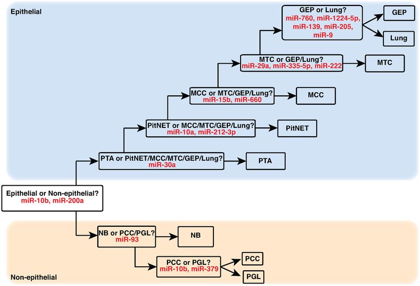
NAR Cancer, 2020, Vol. 2, No. 3 3

pression across samples, a filtering threshold was applied as     tilayer classifier for discriminating NEN pathological types
described (6); specific filtering thresholds were set as a per-   based on selected miRNAs. For each decision layer, all
centile of overall expression as indicated below.                 available algorithms (n = 23) in the MATLAB Classi-
                                                                  fication Learner App were evaluated using 5-fold cross-
Unsupervised hierarchical clustering of filtered miRNA ex-        validation. In each case, the classification model with high-
pression profiles                                                 est accuracy was a support vector machine (SVM) classi-
                                                                  fier that was used to identify the smallest subset of miRNAs
To assess sample grouping, unsupervised hierarchical clus-
                                                                  with the most discriminatory power for comparisons A–H
tering was performed using log2 transformed normalized
                                                                  above. Based on these subsets, we constructed a multilayer
read counts of miRNA and miRNA clusters from all pre-
                                                                  classifier through which miRNA profiles were first assigned
processed samples. Euclidean distance was used as the sim-
                                                                  as epithelial or non-epithelial prior to assignment to a spe-
ilarity parameter with complete agglomeration clustering

                                                                                                                                  Downloaded from https://academic.oup.com/narcancer/article/2/3/zcaa009/5867117 by guest on 24 September 2020
                                                                  cific pathological type.
applied in the heatmap.2 function of the R gplots pack-
age (www.rdocumentation.org/packages/gplots/versions/3.
0.1.1). Lowly expressed miRNAs and miRNA clusters were            Assessment of classifier performance and transferability
excluded with the filtering threshold set as the top 75%          To assess the performance and transferability of our multi-
abundant miRNA and clusters in at least one sample.               layer classifier, we used t-stochastic neighbor embedding (t-
                                                                  SNE) to visualize sample grouping patterns based on miR-
Assessment and comparative analyses of abundant miRNAs            NAs selected for classification. We also determined overall
in NEN and non-NEN samples                                        classifier accuracy, evaluated the impact of miRNA cluster
To identify candidate miRNA markers for all NENs and              member substitutions on classifier accuracy and inspected
for each NEN pathological type, we ranked miRNAs and              the expression levels of the selected miRNAs.
miRNA clusters by abundance and considered the top 1%.
These abundant miRNAs and miRNA clusters were com-                Statistical analyses
pared and correlated between NEN cases and non-NEN                Statistical analyses of clinical parameters were performed
controls, as well as between each pathological type and           using SPSS Statistics (IBM, Armonk, NY, USA, version 25)
site-matched non-NEN control group. To highlight sub-             and MATLAB. Differences in miRNA content and normal-
stantial differences in miRNA expression, only compar-            ized miRNA expression were evaluated between NEN and
isons with 20-fold or greater difference are discussed. For       non-NEN samples, and within NEN pathological types us-
single-member miRNA clusters, abundance measures ap-              ing the non-parametric Kruskal–Wallis (K–W) test (21); a
proximate the abundance of the single miRNA, and are not          two-sided P-value of 5% of samples were retained. Next, we ranked        profiles through barcoded small RNA sequencing, qual-
miRNAs and miRNA clusters that discriminate epithe-               ity controlled profiles through data preprocessing and per-
lial from non-epithelial NENs (comparison A). We subse-           formed downstream analyses using statistical and ma-
quently ranked miRNA markers that successively identified         chine learning approaches. Following data preprocessing
epithelial NENs, including parathyroid adenoma (PTA), pi-         for quality control, our final study cohort comprised 221
tuitary adenoma (PitNET), Merkel cell carcinoma (MCC),            NEN cases and 114 site-matched non-NEN controls, here-
medullary thyroid carcinoma (MTC) and lung NENs from              after termed study samples (Table 1). NEN cases comprised
gastrointestinal–pancreatic (GEP) NENs (comparisons B–            15 distinct pathological types, arising in seven anatom-
F), respectively. Lastly, we identified miRNA markers that        ical sites, including the gastrointestinal tract and pan-
discriminated neuroblastoma (NB), pheochromocytoma                creas, lung, parathyroid gland, pituitary gland, skin, thy-
(PCC) and extra-adrenal paraganglioma (PGL) from each             roid gland, and the adrenal gland and extra-adrenal sites.
other (comparisons G and H) within the non-epithelial             Site-matched non-NEN controls comprised non-diseased
group. Only the top-ranking 3% miRNAs and miRNA clus-             tissues and non-NEN cancers from five anatomical sites,
ters in these comparisons were assessed for classification be-    including the pancreas, lung, parathyroid gland, skin and
low.                                                              thyroid gland.

Construction and cross-validation of multilayer classifier        Small RNA sequencing of study samples
Scaling our existing approach to miRNA-based NEN clas-            We generated comprehensive miRNA expression profiles
sification (18,20), we constructed and cross-validated a mul-     for all samples through barcoded small RNA sequencing.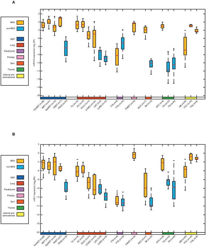
4 NAR Cancer, 2020, Vol. 2, No. 3
Table 1. Anatomical distribution and histopathological diagnoses of study miRNAs and miRNA clusters within and between sam-
samples ple sets. Abundant miRNA and miRNA cluster compo-
Number of Number of sition was similar within all NEN cases or all non-NEN
NENs samples, n (%) non-NENs samples, n (%) controls. The number of members in each miRNA cluster
is indicated in parentheses following the cluster name, e.g.
Total 221 114
Epithelial cluster-hsa-mir-98(13). Among all NEN cases, miR-375, -
Gastrointestinal 21, -143, -let-7a, -26a, -7, -let-7f and -125b and cluster-mir-
tract and pancreas 375(1), -98(13), -21(1) and -23a(6) were the most abundant
PanNET 28 (13%) PAAD 10 (9%) miRNAs and miRNA clusters, with the median relative fre-
INET 31 (14%)
AppNET 15 (7%)
quency ranging 1.5–10.6% and 3.6–10.6% of respective total
RNET 7 (3%) read counts (Supplementary Table S6). Within this group,
Downloaded from https://academic.oup.com/narcancer/article/2/3/zcaa009/5867117 by guest on 24 September 2020
Lung miR-375, -21, -143, -let-7a, -26a, -7, -let-7f, -125b and -141
TC 13 (6%) LAC 9 (8%)a and cluster-mir-98(13), -mir-375(1), -mir-7-1(3) and -mir-
AC 15 (7%) LUNG 15 (12%)a 143(2) were highly expressed in five or more pathological
SCLC 11 (5%)
LCNEC 13 (6%) types (Supplementary Table S6). In comparison, among all
Parathyroid gland non-NEN controls, miR-21, -125b, -let-7a, -143, -let-7f, -
PTA 9 (4%) PTG 15 (13%) 30a, -26a and -29a and cluster-mir-98(13), -21(1), -30a(4)
Pituitary gland and -23a(6) were the most abundant miRNA and miRNA
PitNET 10 (5%)
Skin
clusters, ranging 2.5–10% and 5.2–15.9% of respective to-
MCC 17 (8%) SK 10 (9%) tal miRNA-annotated read counts (Supplementary Table
Thyroid S7). Within this group, miR-21, -let-7a, -143, -30a, -let-
MTC 9 (4%) TG 10 (9%)b 7b and -30d and cluster-mir-98(13), -mir-21(1), -mir-23a(6)
TN 45 (39%)b and -mir-30a(4) were highly expressed in five or more non-
Non-epithelial
Adrenal gland and
NEN entities (Supplementary Table S7). Correlation analy-
extra-adrenal sites ses highlighted the similarities in abundant miRNA compo-
NB 25 (11%) sition within epithelial and non-epithelial NENs; with the
PCC 10 (5%) exception of PTA, NEN cases were less correlated with site-
PGL 8 (4%) matched non-NEN controls (Supplementary Figure S1).
a For lung NENs, neoplastic (LAC) and non-diseased (LUNG) tissue con-
trols were available. Comparative analyses of abundant miRNAs in NEN and non-
b For MTC, neoplastic (TN) and non-diseased (TG) tissue controls were
available.
NEN samples
Anatomical location and diagnostic histopathological information are pre- To better understand meaningful differences in miRNA
sented for 221 NEN cases, comprising 15 pathological types from seven
anatomical sites, and 114 site-matched non-NEN controls, comprising composition between NEN and non-NEN samples, we
seven diagnostic entities from five anatomical sites. Sample abbreviations: compared abundant miRNAs and miRNA clusters for all
AC, atypical carcinoid; AppNET, appendiceal NET; INET, ileal NET; NEN samples and for each pathological type with rele-
LCNEC, large-cell NEC; MCC, Merkel cell carcinoma; MTC, medullary vant controls. Comparative analyses indicated that miR-
thyroid carcinoma; NB, neuroblastoma; PanNET, pancreatic NET;
PCC, pheochromocytoma; PGL, paraganglioma; PitNET, pituitary ade-
375 and miR-7 were 216- and 48-fold higher in all NEN
noma; PTA, parathyroid adenoma; RNET, rectal NET; SCLC, small- cases compared to all non-NEN controls, respectively. Fold
cell lung carcinoma; TC, typical carcinoid. Non-NEN samples comprise changes ranged 59–816- and 41–69-fold higher for miR-375
lung (LUNG), lung adenocarcinoma (LAC), pancreatic adenocarcinoma and miR-7 in specific NEN pathological types compared to
(PAAD), parathyroid gland (PTG), skin (SK), thyroid gland (TG) and thy- site-matched non-NEN controls (Supplementary Table S6
roid neoplasm (TN).
and Figure 1). The only exception was observed in PTA,
which showed the lowest miR-375 and miR-7 expression of
Following sequence annotation, we obtained a median of all NENs; in fact, higher expression was observed in non-
4 386 727 (range: 53 516–40 305 4453) total small RNA neoplastic parathyroid glands. Other notable miRNA over-
reads and 258 932 (range: 1312–3 723 507) calibrator reads expression among NENs included miR-127, with 86-fold
(Supplementary Table S2). For miRNAs, we detected a higher expression in typical carcinoids (TC) compared to
median of 2 322 722 (range: 1740–34 781 174) miRNA lung non-NEN tissues (Supplementary Table S6); cluster-
sequence reads, representing a median of 63.1% total se- mir-127(8) was also 78-fold higher in TC compared to lung
quence reads; miRNA, miRNA cluster and calibrator ex- non-NEN tissues (data not shown). In addition, miR-203
pression profiles for each sample were subsequently gener- and miR-205 expression was 143- and 366-fold higher in
ated from these reads. Median miRNA content was 26.4 non-NEN skin controls than MCC, respectively (Supple-
(range: 0.0–2048.4) fmol/g total RNA (Supplementary mentary Table S7).
Table S2).
Unsupervised hierarchical clustering of filtered miRNA ex-
Abundant miRNA composition in NEN and non-NEN sam- pression profiles
ples
To assess the classificatory potential of miRNA expres-
To better understand miRNA composition in NEN and sion profiling, we first explored our data using unsuper-
non-NEN samples, we assessed and correlated abundant vised hierarchical clustering. With the exception of all PTANAR Cancer, 2020, Vol. 2, No. 3 5
Downloaded from https://academic.oup.com/narcancer/article/2/3/zcaa009/5867117 by guest on 24 September 2020
Figure 1. miR-375 and miR-7 expression in NEN and non-NEN samples. Normalized miR-375 and miR-7 expression was examined between 15 NEN
pathological types and 7 site-matched non-NEN control groups. Site-matched NEN and non-NEN groups were designated by anatomical site in the
color bar: pancreas (blue), lung (red), parathyroid (purple), skin (orange) and thyroid (green); NENs without a site-matched control were left blank. Both
miR-375 and miR-7 were higher expressed in NEN cases than non-NEN controls. With the exception of PTA, miR-375 expression was higher in NEN
pathological types than in site-matched non-NEN controls. With the exception of PTA, miR-7 was also higher in NEN pathological types compared to site-
matched non-NEN controls. Abbreviation: log2 RF, log2 normalized relative frequency. Sample abbreviations are provided in Table 1 and Supplementary
Table S1.6 NAR Cancer, 2020, Vol. 2, No. 3
Downloaded from https://academic.oup.com/narcancer/article/2/3/zcaa009/5867117 by guest on 24 September 2020
Figure 2. Unsupervised hierarchical clustering of study samples based on miRNA expression. Unsupervised hierarchical clustering using Euclidean dis-
tance and complete agglomeration clustering was performed using filtered (union of top 75% abundance) log2 normalized miRNA sequence reads for all
NEN cases (n = 221) and non-NEN controls (n = 114). Anatomical groupings comprise the following pathological types described in Table 1 and Supple-
mentary Table S1: thyroid (MTC, TG, TN), skin (MCC, SK), pituitary gland (PitNET), parathyroid gland (PTA, PTG), lung (AC, TC, SCLC, LCNEC,
LAC, LUNG), GEP (AppNET, INET, PNET, RNET), and adrenal and extra-adrenal (PCC, PGL). With noted exceptions, NEN cases and non-NEN
controls, and epithelial and non-epithelial samples, clustered distinctly and NEN pathological types preferentially clustered with each other than with
site-matched non-NEN controls.
samples and one large-cell NEC (LCNEC) sample, NEN 3% miRNAs or miRNA clusters discriminating between or
cases and non-NEN controls clustered separately (Figure within epithelial or non-epithelial NENs (Supplementary
2). In addition, epithelial samples clustered distinctly from Tables S8 and S9). These comparisons were used to con-
non-epithelial samples with the exception of one pancre- struct and assess the reliability of the multilayer classifier
atic NET (PanNET). NEN pathological types preferentially below.
clustered together rather than with site-matched non-NEN
controls. Unsupervised hierarchical clustering of filtered Construction and cross-validation of multilayer classifier
miRNA cluster expression from the same samples clustered
as above (Supplementary Figure S2). We subsequently constructed and assessed the accuracy of
a multilayer miRNA-based classifier for predicting NEN
pathological types with 5-fold cross-validation (Figure 3).
Discovery analyses for miRNA-based NEN classification
The resulting classifier consisted of eight decision layers,
Next, we identified candidate miRNA markers for NEN using the linear or cubic SVM model at each layer (Sup-
classification using an established approach comprising fea- plementary Table S10). In the first layer, miR-200a ex-
ture selection and validation (18). Using this approach, pression was significantly higher in epithelial than non-
we selected effective miRNA markers from the top-ranked epithelial NENs (K–W P-valueNAR Cancer, 2020, Vol. 2, No. 3 7
Downloaded from https://academic.oup.com/narcancer/article/2/3/zcaa009/5867117 by guest on 24 September 2020
Figure 3. Multilayer miRNA-based classifier for predicting NEN pathological types. A multilayer classifier for predicting NEN pathological types was
developed using supervised machine learning models. In the first layer, NEN miRNA profiles were classified as epithelial or non-epithelial based on miR-
10b and miR-200a expression. In subsequent layers, epithelial and non-epithelial NENs were successively identified using the selected miRNAs as indicated.
Sample abbreviations are provided in Table 1 and Supplementary Table S1.
additional prediction power (K–W P-value8 NAR Cancer, 2020, Vol. 2, No. 3
Downloaded from https://academic.oup.com/narcancer/article/2/3/zcaa009/5867117 by guest on 24 September 2020
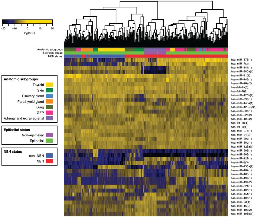
Figure 4. Scatter plot assessment of miRNAs selected for classification. Epithelial and non-epithelial NENs are effectively discriminated based on miR-
10b and miR-200a expression with one misclassification (A). Within epithelial NENs, PTA, PitNET, MCC and MTC were accurately discriminated from
the remaining NENs based on miR-30a expression (B), miR-10a and miR-212-3p expression (C), miR-15b and miR-660 expression (D), and miR-335-5p,
miR-29a and miR-222 expression (E); lung NENs and GEP NENs were discriminated based on miR-760, miR-1224-5p, miR-139, miR-205 and miR-9
expression (F, G). Within non-epithelial NENs, NB was accurately discriminated from PCC/PGL based on miR-93 expression (H), and PCC and PGL were
separated based on miR-10b and miR-379 expression (I). Similar results were generated using relevant miRNA cluster data and are not presented. Arrows
indicate misclassified samples. Abbreviation: log2 RF, log2 normalized relative frequency. Sample abbreviations are provided in Table 1 and Supplementary
Table S1.
were diffusely positive for synaptophysin, chromogranin A sis as updated clinical, pathological, biological and molecu-
and GATA3, and negative for cytokeratin (AE1/AE3 an- lar data become available. Recently, these experts proposed
tibodies). This phenotype diagnosed this tumor as a PGL, a common classification framework for evaluating NENs,
as predicted by the miRNA classifier, and not a PanNET, clarifying terminology to reduce confusion and harmoniz-
which should be cytokeratin-positive and GATA3-negative ing concepts to facilitate comparisons between pathological
(23,24). The unusual case was misidentified based on initial types (3). Although morphology-based, this framework is
histology, but was correctly diagnosed by molecular profil- designed to incorporate ‘equally solid genetic studies across
ing and miRNA-based classification. all anatomical sites (3)’ over time. Here, we generate biolog-
ical and clinical insights into NENs through miRNA-based
DISCUSSION classification.
The strength of our study stems from comparing multiple
Accurate NEN classification is essential for understanding NEN pathological types and site-matched non-NEN con-
tumor biology and guiding clinical care. NEN pathologi- trols using comprehensive miRNA detection through bar-
cal classification is modified by experts on an ongoing ba- coded small RNA cDNA library sequencing (25) and ac-NAR Cancer, 2020, Vol. 2, No. 3 9
Downloaded from https://academic.oup.com/narcancer/article/2/3/zcaa009/5867117 by guest on 24 September 2020
Figure 5. t-SNE for selected classificatory miRNAs. Sample grouping was visually assessed using miRNAs selected for multilayer classification and t-SNE
analysis. With one notable exception, samples clustered as epithelial or non-epithelial NENs and tended to group by pathological type. The exception was
a misdiagnosed PanNET later found to be a PGL on further testing. Sample abbreviations are provided in Table 1 and Supplementary Table S1.
curate sequence annotation (19). Advanced computational ences in abundant miRNA composition in NEN and non-
approaches for miRNA feature selection (20) and classi- NEN samples.
fier construction (18) further bolstered our approach. We Similarities in abundant miRNA composition between
carefully assessed data reliability through knowledge of samples provide coarse insights into cellular gene expres-
miRNA cluster composition (10), evaluated classifier per- sion programs. Within NENs, miR-375, -21, -143, -let-7a,
formance and transferability by determining overall and de- -26a, -7, -let-7f, -125b and -141 were highly expressed in
cision node level accuracy, gauged the impact of miRNA five or more pathological types; known oncogenic or tu-
cluster substitutions on accuracy and inspected the abun- mor suppressor functions for these miRNAs are reviewed
dance of selected classificatory miRNAs. Throughout the elsewhere (8,27). miR-375, the most abundant miRNA in
study, miRNA clusters measured data quality and transfer- NENs, is believed to regulate lineage-specific differentiation
ability of miRNAs as clinical markers; we then focused on (28–31), growth (32,33) and function (32,34) of neuroen-
miRNAs to build a streamlined prototype of a tool for NEN docrine cells. Correlation analyses highlighted similarities
classification. The identified miRNAs can be used as mono- in abundant miRNA composition for all NENs, including
analyte or multi-analyte markers as needed (12). epithelial or non-epithelial NENs. These findings indicate
Unsupervised hierarchical clustering of filtered miRNA that all NENs have a constitutive miRNA gene expression
expression profiles confirms existing knowledge and pro- program that likely directly or indirectly maintains the neu-
vides new knowledge of NEN grouping. With the exception roendocrine cell phenotype. Given the different cellular ori-
of all PTA and one LCNEC sample, NEN cases and non- gins of epithelial and non-epithelial NENs (35), convergent
NEN controls clustered separately. Based on these find- miRNA gene expression likely implies functional similari-
ings, we speculate that all PTA have a distinct gene expres- ties. Within non-NEN samples, miR-21, -let-7a, -143, -30a,
sion pattern linked to their indolent behavior; the LCNEC -let-7b and -30d were highly expressed in five or more non-
sample showed areas of possible squamous cell differenti- NEN entities; their cancer-related functions are reviewed
ation (data not shown) that may explain peculiar cluster- elsewhere as above. While mechanistic insights into cellu-
ing patterns. Within NENs, two major groups correspond- lar processes can be gained through predictable targeting
ing to epithelial and non-epithelial NENs are evident; in- of mRNAs by abundant miRNAs, this topic is beyond the
terestingly, one epithelial NEN clusters with non-epithelial scope of the present study (36).
NEN samples. Here, we show that these epithelial and non- Differences in abundant miRNA composition between
epithelial NENs can be discriminated through miR-200a samples can also be used to identify new and confirm
(26) and miR-10b expression, and confirm that our ep- known miRNA markers. miR-375 expression was substan-
ithelial PanNET sample is actually a non-epithelial PGL tially higher in all NEN cases compared to non-NEN con-
based on additional cytokeratin and GATA-3 immunos- trols. Where comparisons allowed, miR-375 was consis-
taining (23,24). Within non-NENs, samples group mostly tently higher in NEN pathological types compared to site-
by anatomical site of origin as expected (6). Visual inspec- matched non-NEN controls. Based on current miRNA ex-
tion of cluster diagrams indicates similarities and differ- pression tissue atlases, miR-375 is currently thought to be10 NAR Cancer, 2020, Vol. 2, No. 3
Table 2. Overall accuracy of multilayer classifier for discriminating NENs
Established diagnosis
GEP NET Lung NET MTC MCC PitNET PTA PCC PGL NB
Multilayer classifier GEP NET 80 3
designation
Lung NET 49
MTC 9
MCC 17
PitNET 10
PTA 9
PCC 10
PGL 1 8
Downloaded from https://academic.oup.com/narcancer/article/2/3/zcaa009/5867117 by guest on 24 September 2020
NB 25
Decision-level accuracy 80/81 49/52 9/9 17/17 10/10 9/9 10/10 8/8 25/25
(99%) (94%) (100%) (100%) (100%) (100%) (100%) (100%) (100%)
Overall accuracy 217/221 (98%)
Using our multilayer classifier, NEN miRNA profiles were assigned to one of nine pathological subgroups or pathological types. Cases of GEP NENs
(AppNET, INET, PNET, RNET) or lung NENs (TC, AC, SCLC, LCNEC) were not assigned to individual pathological types because we previously
developed miRNA-based classifiers for these subgroups (18) (Wong et al., in preparation). By comparing classifier designations to established histopatho-
logical diagnoses, we determined our overall classifier accuracy to be 98%. Additional measures of classifier performance were also calculated: precision
(0.98), recall (0.99) and Matthews correlation coefficient (0.98). Sample abbreviations are provided in Table 1 and Supplementary Table S1.
an endocrine gland specific marker (6,37). However, the clustering by pathological type. Given their classificatory
presence of miR-375 in enteroendocrine cells (30,38), pan- potential, we subsequently constructed and validated a mul-
creatic beta cells (32,33), thyroid C cells (39), and MCC tilayer classifier for discriminating NEN pathological types,
(7,31,40), NB (15) and SCLC cell lines (29) suggests that correctly identifying 217 (98%) of 221 samples. Three of
miR-375 is a neuroendocrine cell marker. Given the speci- the four misclassified samples occurred at the GEP NEN
ficity and distribution of miR-375 in our samples and its versus lung NEN decision node, suggesting model over-
reported abundance in seemingly disparate NEN patholog- fitting and the need for additional samples for validation.
ical types (7,18,38,41–43), we propose that miR-375 is a uni- On further testing, the fourth ‘misclassified’ sample turned
versal marker of neuroendocrine cell differentiation. miR- out to be a PGL as indicated by miRNA expression profil-
375 appears to be highly expressed in NENs, in amounts ing. We also introduced criteria for evaluating classifier per-
proportional to the number of normal neuroendocrine cells formance and transferability, including determining over-
and/or the degree of neuroendocrine differentiation within all and decision node level accuracy, assessing the impact
control tissues or tumors; neuroendocrine differentiation of of miRNA cluster substitutions on classifier accuracy and
tumors is more common than currently appreciated (44). showing the relative abundance of miRNAs selected for
More systematic studies are required to confirm this pro- classification.
posal. This study does have limitations that are commonly en-
Although less abundant than miR-375, miR-7 expres- countered in rare cancer and miRNA research. Compre-
sion was also elevated in all NEN cases compared to non- hensive clinical information is challenging to obtain, limited
NEN controls. Where comparisons allowed, miR-7 was of- sample numbers preclude hold out validation and miRNA
ten higher in NEN pathological types compared to site- content measurements can vary widely due to technical
matched non-NEN controls. Other than expression in the challenges. Nonetheless, we provide compelling evidence
pituitary gland, atlas studies provide limited information on that miRNAs are useful for NEN classification and should
miR-7 expression (6,37). However, the presence of miR-7 in be included in further multi-omic studies of these neo-
enteroendocrine cells (30), pancreatic islet cells (33,45), thy- plasms.
roid C cells (46), but not controls suggests that this miRNA Through comprehensive miRNA expression profiling, we
also has some degree of neuroendocrine specificity. Given have identified candidate universal and classificatory mark-
their specificity, some tissue profiling studies may have in- ers that may be useful as adjunct tissue markers, constructed
advertently interpreted miR-375 or miR-7 reduction in ex- a multilayer classifier for discriminating NENs and pro-
pansile cancer lesions as miRNA reduction rather than neu- vided reference profiles for hypothesis generation or inter-
roendocrine cell destruction. Although miR-127 was higher study comparisons. Our next steps involve confirming our
in TC than non-NEN controls, the significance of this dif- findings in well-annotated sample sets, evaluating miRNAs
ference is unclear. Conversely, comparisons of abundant as circulating markers and investigating upstream promoter
miRNA composition between non-NEN and NEN sam- activity and downstream targeting events.
ples identified known tissue-specific miRNA markers such
as miR-203 and miR-205 (6).
As with other cancers (4), miRNAs can be used for NEN DATA AVAILABILITY
classification. Using our feature selection algorithm, we Annotated miRNA and miRNA cluster counts (Supple-
identified 17 miRNAs to discriminate 15 NEN pathologi- mentary Tables S3 and S4) have also been deposited
cal types; t-SNE analyses using these miRNAs clearly sep- to Data Dryad (https://datadryad.org/stash/dataset/doi:10.
arated epithelial and non-epithelial NENs and suggested 5061/dryad.fn2z34tqj).NAR Cancer, 2020, Vol. 2, No. 3 11
SUPPLEMENTARY DATA 10. Farazi,T.A., Brown,M., Morozov,P., Ten Hoeve,J.J., Ben-Dov,I.Z.,
Hovestadt,V., Hafner,M., Renwick,N., Mihailovic,A., Wessels,L.F.
Supplementary Data are available at NAR Cancer Online. et al. (2012) Bioinformatic analysis of barcoded cDNA libraries for
small RNA profiling by next-generation sequencing. Methods, 58,
171–187.
ACKNOWLEDGEMENTS 11. Butz,H. and Patocs,A. (2019) MicroRNAs in endocrine tumors.
EJIFCC, 30, 146–164.
We wish to thank members of the Rockefeller University 12. Modlin,I.M., Bodei,L. and Kidd,M. (2016) Neuroendocrine tumor
Genomics Resource Core and the McGill University and biomarkers: from monoanalytes to transcripts and algorithms. Best
Pract. Res. Clin. Endocrinol. Metab., 30, 59–77.
Genome Quebec Innovation Center for outstanding service. 13. Chan,D.L., Clarke,S.J., Diakos,C.I., Roach,P.J., Bailey,D.L., Singh,S.
We wish to thank Arlene Hurley NP and the Research Fa- and Pavlakis,N. (2017) Prognostic and predictive biomarkers in
cilitation Office staff in The Rockefeller University Center neuroendocrine tumours. Crit. Rev. Oncol. Hematol., 113, 268–282.
for Clinical and Translational Science for regulatory and ad- 14. Gustafson,D., Tyryshkin,K. and Renwick,N. (2016)
Downloaded from https://academic.oup.com/narcancer/article/2/3/zcaa009/5867117 by guest on 24 September 2020
microRNA-guided diagnostics in clinical samples. Best Pract. Res.
ministrative assistance. Clin. Endocrinol. Metab., 30, 563–575.
15. Cheung,I.Y., Farazi,T.A., Ostrovnaya,I., Xu,H., Tran,H.,
Mihailovic,A., Tuschl,T. and Cheung,N.K. (2014) Deep microRNA
FUNDING sequencing reveals downregulation of miR-29a in neuroblastoma
central nervous system metastasis. Genes Chromosomes Cancer, 53,
Academic Health Sciences Center Alternative Funding 803–814.
Plan Innovation Fund; the Canada Foundation for Inno- 16. Shilo,V., Mor-Yosef Levi,I., Abel,R., Mihailovic,A., Wasserman,G.,
vation John R. Evans Leaders Fund; the Carcinoid and Naveh-Many,T. and Ben-Dov,I.Z. (2017) Let-7 and microRNA-148
regulate parathyroid hormone levels in secondary
Neuroendocrine Tumor Society Canada; the Ontario Re- hyperparathyroidism. J. Am. Soc. Nephrol., 28, 2353–2363.
search Fund––Research Infrastructure; Robertson Thera- 17. Mong,E.F., Akat,K.M., Canfield,J., Lockhart,J., VanWye,J.,
peutic Development; The Rockefeller University Center for Matar,A., Tsibris,J.C.M., Wu,J.K., Tuschl,T. and Totary-Jain,H.
Clinical and Translational Science Award, funded in part (2018) Modulation of LIN28B/let-7 signaling by propranolol
by the National Center for Advancing Translational Sci- contributes to infantile hemangioma involution. Arterioscler. Thromb.
Vasc. Biol., 38, 1321–1332.
ences, National Institutes of Health Clinical and Transla- 18. Panarelli,N., Tyryshkin,K., Wong,J.J.M., Majewski,A., Yang,X.,
tional Science Award program [UL1TR001866]; the South- Scognamiglio,T., Kim,M.K., Bogardus,K., Tuschl,T., Chen,Y.T. et al.
eastern Ontario Academic Medical Organization; the On- (2019) Evaluating gastroenteropancreatic neuroendocrine tumors
tario Institute of Cancer Research, funded by the Govern- through microRNA sequencing. Endocr. Relat. Cancer, 26, 47–57.
19. Brown,M., Suryawanshi,H., Hafner,M., Farazi,T.A. and Tuschl,T.
ment of Ontario. Funding for open access charge: Ontario (2013) Mammalian miRNA curation through next-generation
Institute of Cancer Research. sequencing. Front. Genet., 4, 145.
Conflict of Interest statement. T. Tuschl is a co-founder of 20. Ren,R., Tyryshkin,K., Graham,C.H., Koti,M. and Siemens,D.R.
Alnylam Pharmaceuticals and is on the Scientific Advisory (2017) Comprehensive immune transcriptomic analysis in bladder
Board of Regulus Therapeutics. cancer reveals subtype specific immune gene expression patterns of
prognostic relevance. Oncotarget, 8, 70982–71001.
21. Kruskal,W.H. and Wallis,W.A. (1952) Use of ranks in one-criterion
variance analysis. J. Am. Stat. Assoc., 47, 583–621.
REFERENCES 22. Spearman,C. (1906) ‘Footrule’ for measuring correlation. Br. J.
1. Wick,M.R. (2000) Neuroendocrine neoplasia. Current concepts. Am. Psychol., 2, 89–108.
J. Clin. Pathol., 113, 331–335. 23. Duan,K. and Mete,O. (2016) Algorithmic approach to
2. Kloppel,G. (2017) Neuroendocrine neoplasms: dichotomy, origin and neuroendocrine tumors in targeted biopsies: practical applications of
classifications. Visc. Med., 33, 324–330. immunohistochemical markers. Cancer Cytopathol., 124, 871–884.
3. Rindi,G., Klimstra,D.S., Abedi-Ardekani,B., Asa,S.L., Bosman,F.T., 24. Uccella,S., La Rosa,S., Volante,M. and Papotti,M. (2018)
Brambilla,E., Busam,K.J., de Krijger,R.R., Dietel,M., Immunohistochemical biomarkers of gastrointestinal, pancreatic,
El-Naggar,A.K. et al. (2018) A common classification framework for pulmonary, and thymic neuroendocrine neoplasms. Endocr. Pathol.,
neuroendocrine neoplasms: an International Agency for Research on 29, 150–168.
Cancer (IARC) and World Health Organization (WHO) expert 25. Hafner,M., Renwick,N., Farazi,T.A., Mihailovic,A., Pena,J.T. and
consensus proposal. Mod. Pathol., 31, 1770–1786. Tuschl,T. (2012) Barcoded cDNA library preparation for small RNA
4. Lu,J., Getz,G., Miska,E.A., Alvarez-Saavedra,E., Lamb,J., Peck,D., profiling by next-generation sequencing. Methods, 58, 164–170.
Sweet-Cordero,A., Ebert,B.L., Mak,R.H., Ferrando,A.A. et al. 26. Park,S.M., Gaur,A.B., Lengyel,E. and Peter,M.E. (2008) The
(2005) MicroRNA expression profiles classify human cancers. miR-200 family determines the epithelial phenotype of cancer cells by
Nature, 435, 834–838. targeting the E-cadherin repressors ZEB1 and ZEB2. Genes Dev., 22,
5. Rosenfeld,N., Aharonov,R., Meiri,E., Rosenwald,S., Spector,Y., 894–907.
Zepeniuk,M., Benjamin,H., Shabes,N., Tabak,S., Levy,A. et al. 27. Svoronos,A.A., Engelman,D.M. and Slack,F.J. (2016) OncomiR or
(2008) MicroRNAs accurately identify cancer tissue origin. Nat. tumor suppressor? The duplicity of microRNAs in cancer. Cancer
Biotechnol., 26, 462–469. Res., 76, 3666–3670.
6. Landgraf,P., Rusu,M., Sheridan,R., Sewer,A., Iovino,N., Aravin,A., 28. Kloosterman,W.P., Lagendijk,A.K., Ketting,R.F., Moulton,J.D. and
Pfeffer,S., Rice,A., Kamphorst,A.O., Landthaler,M. et al. (2007) A Plasterk,R.H. (2007) Targeted inhibition of miRNA maturation with
mammalian microRNA expression atlas based on small RNA library morpholinos reveals a role for miR-375 in pancreatic islet
sequencing. Cell, 129, 1401–1414. development. PLoS Biol., 5, e203.
7. Renwick,N., Cekan,P., Masry,P.A., McGeary,S.E., Miller,J.B., 29. Nishikawa,E., Osada,H., Okazaki,Y., Arima,C., Tomida,S.,
Hafner,M., Li,Z., Mihailovic,A., Morozov,P., Brown,M. et al. (2013) Tatematsu,Y., Taguchi,A., Shimada,Y., Yanagisawa,K., Yatabe,Y.
Multicolor microRNA FISH effectively differentiates tumor types. J. et al. (2011) miR-375 is activated by ASH1 and inhibits YAP1 in a
Clin. Invest., 123, 2694–2702. lineage-dependent manner in lung cancer. Cancer Res., 71,
8. Farazi,T.A., Hoell,J.I., Morozov,P. and Tuschl,T. (2013) MicroRNAs 6165–6173.
in human cancer. Adv. Exp. Med. Biol., 774, 1–20. 30. Knudsen,L.A., Petersen,N., Schwartz,T.W. and Egerod,K.L. (2015)
9. Bartel,D.P. (2009) MicroRNAs: target recognition and regulatory The microRNA repertoire in enteroendocrine cells: identification of
functions. Cell, 136, 215–233.12 NAR Cancer, 2020, Vol. 2, No. 3
miR-375 as a potential regulator of the enteroendocrine lineage. et al. (2018) Circulating miR-375 as a novel prognostic marker for
Endocrinology, 156, 3971–3983. metastatic medullary thyroid cancer patients. Endocr. Relat. Cancer,
31. Abraham,K.J., Zhang,X., Vidal,R., Pare,G.C., Feilotter,H.E. and 25, 217–231.
Tron,V.A. (2016) Roles for miR-375 in neuroendocrine differentiation 40. Fan,K., Ritter,C., Nghiem,P., Blom,A., Verhaegen,M.E., Dlugosz,A.,
and tumor suppression via notch pathway suppression in Merkel cell Odum,N., Woetmann,A., Tothill,R.W., Hicks,R.J. et al. (2018)
carcinoma. Am. J. Pathol., 186, 1025–1035. Circulating cell-free miR-375 as surrogate marker of tumor burden in
32. Poy,M.N., Hausser,J., Trajkovski,M., Braun,M., Collins,S., Merkel cell carcinoma. Clin. Cancer Res., 24, 5873–5882.
Rorsman,P., Zavolan,M. and Stoffel,M. (2009) miR-375 maintains 41. Hudson,J., Duncavage,E., Tamburrino,A., Salerno,P., Xi,L.,
normal pancreatic alpha- and beta-cell mass. Proc. Natl Acad. Sci. Raffeld,M., Moley,J. and Chernock,R.D. (2013) Overexpression of
U.S.A., 106, 5813–5818. miR-10a and miR-375 and downregulation of YAP1 in medullary
33. Latreille,M., Herrmanns,K., Renwick,N., Tuschl,T., Malecki,M.T., thyroid carcinoma. Exp. Mol. Pathol., 95, 62–67.
McCarthy,M.I., Owen,K.R., Rulicke,T. and Stoffel,M. (2015) 42. Galuppini,F., Bertazza,L., Barollo,S., Cavedon,E., Rugge,M.,
miR-375 gene dosage in pancreatic beta-cells: implications for Guzzardo,V., Sacchi,D., Watutantrige-Fernando,S., Vianello,F.,
regulation of beta-cell mass and biomarker development. J. Mol. Mian,C. et al. (2017) MiR-375 and YAP1 expression profiling in
Downloaded from https://academic.oup.com/narcancer/article/2/3/zcaa009/5867117 by guest on 24 September 2020
Med. (Berl.), 93, 1159–1169. medullary thyroid carcinoma and their correlation with
34. Zhang,N., Lin,J.K., Chen,J., Liu,X.F., Liu,J.L., Luo,H.S., Li,Y.Q. clinical-pathological features and outcome. Virchows Arch., 471,
and Cui,S. (2013) MicroRNA 375 mediates the signaling pathway of 651–658.
corticotropin-releasing factor (CRF) regulating 43. Miller,H.C., Frampton,A.E., Malczewska,A., Ottaviani,S.,
pro-opiomelanocortin (POMC) expression by targeting Stronach,E.A., Flora,R., Kaemmerer,D., Schwach,G., Pfragner,R.,
mitogen-activated protein kinase 8. J. Biol. Chem., 288, 10361–10373. Faiz,O. et al. (2016) MicroRNAs associated with small bowel
35. Kloppel,G. (2017) Neuroendocrine neoplasms: dichotomy, origin and neuroendocrine tumours and their metastases. Endocr. Relat. Cancer,
classifications. Visc. Med., 33, 324–330. 23, 711–726.
36. Bartel,D.P. (2018) Metazoan microRNAs. Cell, 173, 20–51. 44. La Rosa,S., Sessa,F. and Uccella,S. (2016) Mixed
37. Ludwig,N., Leidinger,P., Becker,K., Backes,C., Fehlmann,T., neuroendocrine–nonneuroendocrine neoplasms (MiNENs): unifying
Pallasch,C., Rheinheimer,S., Meder,B., Stahler,C., Meese,E. et al. the concept of a heterogeneous group of neoplasms. Endocr. Pathol.,
(2016) Distribution of miRNA expression across human tissues. 27, 284–311.
Nucleic Acids Res., 44, 3865–3877. 45. Joglekar,M.V., Joglekar,V.M. and Hardikar,A.A. (2009) Expression
38. Arvidsson,Y., Rehammar,A., Bergstrom,A., Andersson,E., of islet-specific microRNAs during human pancreatic development.
Altiparmak,G., Sward,C., Wangberg,B., Kristiansson,E. and Gene Expr. Patterns, 9, 109–113.
Nilsson,O. (2018) miRNA profiling of small intestinal 46. Santarpia,L., Calin,G.A., Adam,L., Ye,L., Fusco,A., Giunti,S.,
neuroendocrine tumors defines novel molecular subtypes and Thaller,C., Paladini,L., Zhang,X., Jimenez,C. et al. (2013) A miRNA
identifies miR-375 as a biomarker of patient survival. Mod. Pathol., signature associated with human metastatic medullary thyroid
31, 1302–1317. carcinoma. Endocr. Relat. Cancer, 20, 809–823.
39. Romeo,P., Colombo,C., Granata,R., Calareso,G., Gualeni,A.V.,
Dugo,M., De Cecco,L., Rizzetti,M.G., Zanframundo,A., Aiello,A.You can also read