SEVERE ACUTE RESPIRATORY SYNDROME CORONAVIRUS-2 (SARS-COV-2): AN UPDATE
←
→
Page content transcription
If your browser does not render page correctly, please read the page content below
Open Access Review
                                         Article                                                DOI: 10.7759/cureus.7423

                                         Severe Acute Respiratory Syndrome
                                         Coronavirus-2 (SARS-CoV-2): An Update
                                         Mahendra Pal 1 , Gemechu Berhanu 2 , Chaltu Desalegn 3 , Venkataramana Kandi 4

                                         1. Veterinary and Public Health, Narayan Consultancy on Veterinary Public Health and Microbiology,
                                         Anand, IND 2. Epidemiology and Public Health, College of Agriculture and Veterinary Medicine, Dambi
                                         Dollo University, Dambi Dollo, ETH 3. Epidemiology and Public Health, College of Agriculture and
                                         Veterinary Sciences, Ambo University, Ambo, ETH 4. Clinical Microbiology, Prathima Institute of
                                         Medical Sciences, Karimnagar, IND

                                         Corresponding author: Venkataramana Kandi, ramana20021@gmail.com

                                         Abstract
                                         Coronaviruses (CoVs) belong to the family of Coronaviridae, the order Nidovirales, and the
                                         genus Coronavirus. They are the largest group of viruses causing respiratory and
                                         gastrointestinal infections. Morphologically, CoVs are enveloped viruses containing a non-
                                         segmented positive-sense, single-stranded ribonucleic acid (RNA) viruses. CoVs are
                                         categorized into four important genera that
                                         include Alphacoronavirus, Betacoronavirus, Gammacoronavirus, and Deltacoronavirus. A novel
                                         member of human CoV that has recently emerged in Wuhan, China, is now formally named as
                                         SARS-CoV-2 (severe acute respiratory syndrome coronavirus 2). This is a unique strain of RNA
                                         viruses that have not been previously observed in humans. The virus has wide host adaptability
                                         and is capable of causing severe diseases in humans, masked palm civets, mice, dogs, cats,
                                         camels, pigs, chickens, and bats. The SARS-CoV-2 typically causes respiratory and
                                         gastrointestinal sickness in both humans and animals. It can be transmitted through aerosols
                                         and direct/indirect contact, as well as during medical cases and laboratory sample handling.
                                         Specific structural proteins, which might be found on the surface of the virus, play an
                                         important role in the pathogenesis and development of the complications. The disease is
                                         characterized by distinct medical signs and symptoms that include high fever, chills, cough,
                                         and shortness of breath or difficulty in breathing. The infected people may also present with
                                         other symptoms such as diarrhea, myalgia, fatigue, expectoration, and hemoptysis. It is
                                         important from the public health and economic point of view as it affects the growth of the
                                         country, which is majorly attributed to the restriction in the movement of the people and the
                                         cost associated with the control and prevention of the disease. Since there is no specific
                                         therapeutic intervention nor a vaccine available against the virus, supportive management and
                                         treatment with non-specific therapeutic agents (repurposed drugs) may provide relief to the
                                         patients. Some preventive strategies of the disease include blocking the routes of transmission
                                         of the infections, disinfection of instruments used during medical case handling, using
Received 03/23/2020                      personal protective equipment, proper and early diagnosis of the disease, avoiding contact with
Review began 03/24/2020
                                         the sick patients, and quarantine of the infected/exposed people.
Review ended 03/24/2020
Published 03/26/2020

© Copyright 2020
Pal et al. This is an open access        Categories: Infectious Disease, Epidemiology/Public Health
article distributed under the terms of   Keywords: coronavirus, beta coronavirus, respiratory and gastrointestinal infections, severe acute
the Creative Commons Attribution         respiratory syndrome coronavirus-2, bats, humans and animals, aerosols, quarantine, public health,
License CC-BY 4.0., which permits        sars-cov-2
unrestricted use, distribution, and
reproduction in any medium, provided
the original author and source are
credited.
                                         Introduction And Background
                                         The coronaviruses (CoVs) belong to the genus Coronavirus, the family Coronaviridae, and the

                                         How to cite this article
                                         Pal M, Berhanu G, Desalegn C, et al. (March 26, 2020) Severe Acute Respiratory Syndrome Coronavirus-
                                         2 (SARS-CoV-2): An Update. Cureus 12(3): e7423. DOI 10.7759/cureus.7423
order Nidovirales [1]. They are enveloped and have a non-segmented, single-stranded, positive-
sense ribonucleic acid (ssRNA+) as their nuclear material. On electron microscopy, these
viruses show a characteristic appearance that resembles a crown (corona in Latin means
crown) due to the presence of club-shaped surface protein projections [2-3]. The CoVs are
pleomorphic, measure between 80 and 160 nm in length, and have a small genome measuring
27-32 Kilobytes (KB) with a unique replication strategy [4].
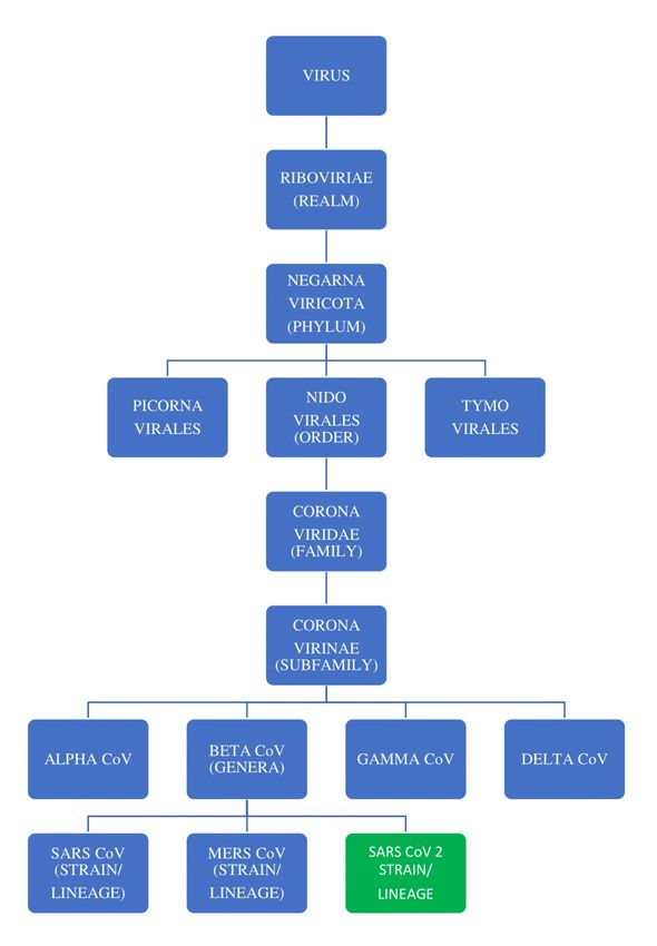
The RNA group of viruses is classified into three orders that include the order Nidovirales,
which is further classified into four families:
the Coronaviridae, Arteriviridae, Mesoniviridae, and Roniviridae. The family Coronaviridae is
further divided into two subfamilies: Coronavirinae and Torovirinae. The Coronavirinae
subfamily includes four genera of viruses (Alphacoronaviruses, Betacoronaviruses,
Gammacoronaviruses, and Deltacoronaviruses), which have been grouped primarily based on
serology and phylogenetic clustering (divisions based on the habitat/genetic
relatedness) [5]. The detailed classification along with the origin of severe acute respiratory
syndrome associated coronavirus 2 (SARS-CoV-2) is depicted in Figure 1.
2020 Pal et al. Cureus 12(3): e7423. DOI 10.7759/cureus.7423 2 of 13FIGURE 1: The classification of the RNA group of viruses and
the origin of SARS CoV-2
CoV: coronavirus; SARS CoV 2: severe acute respiratory syndrome coronavirus 2; MERS CoV,
Middle East respiratory syndrome coronavirus; RNA, ribonucleic acid
A novel member of CoV capable of infecting humans was recently identified in Wuhan, China.
This virus is now formally named as SARS-CoV-2. This new virus is noted to be a unique strain
2020 Pal et al. Cureus 12(3): e7423. DOI 10.7759/cureus.7423 3 of 13of RNA viruses that have not been previously observed to infect humans according to the
International Committee on Taxonomy of Viruses. According to the World Health Organization
(WHO), and preliminary research results, the infection with SARS-CoV-2, which was initially
called as novel coronavirus disease 2019 (nCOVID-19), could result in a human infection that
presents with signs and symptoms that include fever, dry cough, dyspnea, fatigue, and
lymphopenia. Occasionally, human infections may lead to complications such as pneumonia,
severe acute respiratory syndrome (SARS), and even death [6-7].
In this review, we attempt to update the history, genetics, epidemiology, modes of
transmission, pathogenicity, clinical features, laboratory diagnosis, public health implications,
economic impact, treatment, control, and prevention of SARS-CoV-2.
Review
History
CoVs have been described as novel respiratory tract viruses, which were observed in the
samples collected from the people who presented with signs and symptoms of respiratory tract
infection in 1962 [4]. The first cases of SARS were recognized to have emerged in mid-
November, in the year 2002 in Guangdong Province of China. According to a 2003 WHO report,
the first official report of an outbreak of peculiar pneumonia within the province had affected
305 people and caused five deaths. Around 30% of cases have been suggested to occur among
health care employees who were involved in patient care. More than one-third of the early cases
have been noted in food handlers (individuals who manage, kill, and sell animal origin food, or
those who prepare and serve food). The previous incidence of the SARS-CoV outbreak, which
started in China in the year 2002 and lasted until July 2003, had spread across the world,
affecting 24 countries that included Cambodia, Hong Kong, Singapore, Hanoi, Canada, and
others, recording 8,437 SARS cases and 813 deaths [8-10].
In December 2019, a group of patients with pneumonia have been confirmed to be infected with
a novel CoV (nCoV), which was not previously observed in humans in Wuhan, the capital city of
Hubei province of China [11-13]. The newly discovered virus, causing a similar infection (SARS-
CoV), was initially named as nCoV 2019 (January 2020) and was later called COVID-19 in
February 2020. The WHO and the China Bureau had announced the discovery of a new
coronavirus (SARS-CoV-2), which has been isolated from the patients suffering from
pneumonia. As of February 12, 2020, a total of 43,103 cases of infection and 1,018 deaths have
been recorded [14]. Thousands of human infections have been confirmed in China, along with
many exported cases throughout the globe [15].
SARS-CoV-2 was found to infect more human beings than either of its predecessors that
include the SARS-CoV and the Middle East respiratory syndrome virus (MERS) [16]. Numerous
factors have contributed to the fast spread of this virus, with the main reason being the densely
populated Wuhan, which is the capital city of China’s Hubei province, with more than
11,000,000 population. Because Wuhan is a transportation hub, there was an increased chance
of person-to-person transmission and the possibility of exporting cases to other places [14].
Genetic characteristics and virion structure
The CoVs belong to the order Nidovirales, family Coronaviridae, and the
subfamily Coronavirinae. They are genetically categorized into four important genera:
the Alphacoronavirus, Betacoronavirus, Gammacoronavirus, and Deltacoronavirus. The former
two genera typically infect mammals, whereas the latter two predominantly infect birds [9,17-
18].
2020 Pal et al. Cureus 12(3): e7423. DOI 10.7759/cureus.7423 4 of 13The whole-genome sequencing and the genetic analysis studies had revealed that SARS-CoV-2
is genetically related to SARS-CoV of the 2003 outbreak [14]. SARS-CoV-2 was also found to be
closely related to the genus Betacoronavirus and was noted to be a distinct clade in lineage B of
the subgenus Sarbecovirus, collectively with two other bat-derived SARS-CoV-like
strains [12,19]. The origin of the virus is not yet clearly understood. The latest study showed
that angiotensin-converting enzyme 2 (ACE 2), a membrane exopeptidase, is the receptor used
by SARS-CoV-2 to enter into the human cells, much like its predecessor (SARS-CoV) [20].
All CoV genomes are organized further with the replicase locus encoded in the 5' end and the
structural proteins encoded inside the 3' end of the genome. The structural proteins include
the hemagglutinin esterase (HE) (only found in some beta-CoVs), spike (S), small membrane
(E), membrane (M), nucleocapsid (N) and internal (I) protein, encoded inside the 'N' gene. The
nucleocapsid protein complexes with the genome RNA to form a helical capsid structure
observed in the viral envelope. Trimers of the spike proteins form the peplomers embedded in
the envelope, giving the virion its corona or crown-like morphology. In some CoV virions, the
HE protein forms smaller spikes at the membrane. The “M” and “E” also are transmembrane
proteins involved in virus assembly [9].
All viruses within the Nidovirales order have a very large genome, an uncommon feature for
RNA viruses, with Coronavirinae having the largest recognized RNA genomes, containing about
30 KB of genomes. Other common capabilities within the Nidovirales order encompass: (1) a
notably conserved genomic organization, with a huge replicase gene preceding structural and
accent genes; (2) expression of many nonstructural genes through ribosomal frameshifting; (3)
several unique or uncommon enzymatic activities encoded in the large replicase-transcriptase
polyprotein; and (4) expression of downstream genes with the aid of synthesis of 3′ nested sub-
genomic mRNAs. In reality, the Nidovirales order was derived from these nested 3′ mRNAs as
Nido is Latin means “nest”. The principal variations in the members of the Nidovirales are
within the variety, kind, and sizes of the structural proteins. These variations cause
considerable changes within the structure and morphology of the nucleocapsids and virions [5].
The genome consists of a 5′ cap structure together with a 3′ poly (A) tail, permitting it to behave
as an mRNA, which is ready for translation of the replicase polyproteins. The replicase gene
encoding the nonstructural proteins occupy two-thirds of the genome, approximately 20 kb,
instead of the structural and accent proteins, which make up only approximately 10 kb of the
viral genome. The 5′ end of the genome includes a leader series and untranslated region (UTR)
that consists of multiple stem-loop systems required for RNA replication and transcription.
Moreover, at the start of every structural or accent gene, there are transcriptional regulatory
sequences (TRSs) that might be required for the expression of each of those genes. The 3′ UTR
additionally consists of RNA structures required for replication and synthesis of viral RNA. The
organization of the CoV genome is 5′-leader-UTR-replicase-S (Spike)-E (Envelope)-M
(Membrane)-N (Nucleocapsid)-3′ UTR-poly (A) tail with accessory genes interspersed in the
structural genes on the 3′ end of the genome. The accent proteins are nearly completely non-
essential for the replication in tissue cultures, but some have been proven to play a crucial
role in viral pathogenesis [5,21].
CoV virions are spherical, with diameters of about 125 nm, as depicted from the available
research by employing cryo-electron tomography and cryo-electron microscopy. The maximum
distinguished characteristic features of CoVs are the club-shaped spike projections emanating
from the surface of the virion. Those spikes are a defining characteristic of the virion and
deliver them the appearance of a solar corona, prompting the name coronaviruses. In the
envelope of the virion is the nucleocapsid. CoVs have helically symmetrical nucleocapsids,
which are unusual among positive-sense RNA viruses, however, a way more common for
negative-sense RNA viruses [5,22-23].
2020 Pal et al. Cureus 12(3): e7423. DOI 10.7759/cureus.7423 5 of 13Epidemiology
There are seven CoV species recognized to infect human beings [12]. Among these, only MERS-
CoV and SARS-CoV have been able to cause severe human disease. The rest are associated with
mild respiratory ailments such as the common cold. However, they may cause serious
consequences in immunocompromised individuals. At present, the medical severity of the
nCoV-2019 (nCoV-19) is precisely unknown but life-threatening, and deaths have been
associated with the infections [24].
They have wide host adaptability and can cause severe infections in humans, birds, livestock,
masked palm civets, mice, dogs, cats, camels, pigs, chickens, and bats, wherein they typically
cause respiratory and gastrointestinal sickness [5,11,14,18]. Four human CoVs (HCoVs) (HCoV
229E, NL63, OC43, and HKU1) are endemic globally and account for 10% to 30% of upper
respiratory tract infections in adults. Ecologically, there are several types of CoVs, and the
greatest variety was noted in bats, suggesting that bats may be natural reservoirs for a lot of
these viruses. Peri-domestic mammals can also serve as intermediate hosts, facilitating
recombination, mutation, and genetic variations [1,25]. SARS-CoV-2 is currently spreading to
different countries throughout the world including European, American, Asian, and some
African countries.
Transmission
Even though the exact mechanisms of transmission are presently uncertain, human-to-human
transmission can arise, and the risk of airborne spread appears imminent [3,26]. Moreover,
SARS-CoV may be transmitted from bats to palm civets or dromedary camels and thereby spill
over into human beings [27-28]. Reintroduction into human beings from an animal reservoir,
persistent infection in previously sick people, or the laboratory strains may cause human
infections and human-to-human transmission [29].
SARS is generally transmitted through direct or indirect contact of mucous membranes (eyes,
nose, or mouth) with infectious respiratory droplets or fomites. Transmission risks increase
with period and proximity with the contacts/infected persons [30]. The time of survival of
SARS-CoV-2 within the environment is presently unknown. Recent research showed that
SARS-CoV can live up to two weeks after drying and five days at temperatures of 22-25°C and
40-50% relative humidity, with a gradual decline in the viability of the virus thereafter. The
viability of SARS-CoV was found to decrease after 24 hours at 38°C and 80-90% relative
humidity [31]. The virus may remain viable on distinct surfaces for 48 hours at 20°C and 40%
relative humidity, even though viability reduced to 8 hours at 30°C and 80% relative humidity
conditions [3,32]. This confirms the fact that at low temperature, low humidity conditions favor
the virus survival in the environment.
Based on the evidence of a rapidly increasing incidence of infections and the possibility of
transmission by asymptomatic carriers, SARS-CoV-2 can be transmitted effectively among
humans and exhibits high potential for a pandemic [6,33-36]. Additionally, the advancement
and convenience of global travel could further facilitate the worldwide spread of SARS-CoV-
2 [34,37]. The possibility of feco-oral transmission of SARS-CoV-2 has public health
implications, especially in areas with poor sanitation [38].
Pathogenesis
CoVs demonstrate versatile host ranges and tissue tropism [14]. The preliminary attachment of
the virion to the host cell is initiated by interactions between the “S” protein and its
receptor [9]. The sites of the receptor-binding domain (RBD) in the S1 vicinity of a CoV’s “S”
protein vary with the virus strain. Some have the RBD at the N-terminus of S1 (MHV) and the
2020 Pal et al. Cureus 12(3): e7423. DOI 10.7759/cureus.7423 6 of 13others (SARS-CoV) have the RBD at the C-terminus of S1, as noted by the results of the recent
research studies [5,39-40].
CoV proteins, structural, enzymatic, and accent proteins play key roles in the pathogenesis of
the CoV disease (COVID). Structural proteins, in addition to their role in the maintenance of
virion shape and morphogenesis, also contribute to the viral spread in vivo and antagonizing
host cellular and immune responses. Nonstructural proteins include the small accent proteins
that are not at all conserved among mouse hepatitis virus (MHV) and SARS-CoVs and the 16
conserved proteins encoded in the replicase locus, a lot of which have an enzymatic role in RNA
metabolism or protein processing, which further assists the viruses in antagonizing hosts
immune responses during infections [9,14].
Clinical signs
CoVs naturally cause illnesses in mammals and birds that include enteritis in cows and pigs and
respiratory diseases in chickens. They may also be responsible for potentially lethal respiratory
tract infections (acute and chronic) in humans [5,9]. From the available preliminary clinical
data, SARS can develop in COVID in stages, consisting of acute constitutional signs and
symptoms, acute viral pneumonitis, acute lung damage, or even acute respiratory distress
syndrome, evolving over one to two weeks. The preliminary infection could be followed by a
hyperactive immune reaction, which seems to underlie the severe manifestations of SARS [41].
The incubation period of COVID may average between two to seven days (range of one to two
weeks). Clinical manifestation is characterized by systemic symptoms such as high fever, chills,
cough, shortness of breath or difficulty in breathing, diarrhea, myalgia or fatigue,
expectoration, and hemoptysis. In severe forms, the patients may develop pneumonia, and the
case fatality rates may vary considerably. Serious complications such as heart failure,
respiratory failure, and liver failure most likely occur in elderly patients [8,10,13].
Respiratory failure is the most important problem of COVID; at least half of the patients
(mostly the elderly people) require supplemental oxygen during the intense phase, whereas
around 20% of patients progress to acute respiratory distress syndrome requiring invasive
mechanical ventilator support. In contrast, the severity is usually mild in infected young
children [42]. Deaths may take place as early as day 4 and as overdue as 108 days after the onset
of symptoms. Virus shedding from the respiratory tract was found to peak around day 10 and
later declined. Virus excretion from the gastrointestinal tract was also noted. Immunoglobulin
G (IgG) antibodies had been detected 10-15 days after the onset of symptoms, and the patient’s
improvement correlated with the decrease in virus load. The severity of the disease was found
to correlate with increasing age, with increased mortality (up to 50%) in patients over 60 years
of age [9].
Generally, early clinical signs of COVID can be similar to other seasonal viral respiratory
illnesses, thereby limiting the ability of the physicians to suspect the disease at its early stages.
Respiratory symptoms frequently increase from two to seven days after the onset of infection
and usually include a non-productive cough and dyspnea. More severe respiratory symptoms
along with rhinorrhea and sore throat may arise, which are unusual. Patients with positive
laboratory tests for SARS-CoV may show advanced radiographic changes of lung indicating
pneumonia after 7-10 days of infection. Many CoV infected patients (70-90%) were noted to
develop lymphopenia [6].
Laboratory diagnosis
Laboratory tests, which are currently available for the diagnosis of SARS-CoV-2 in various
human clinical specimens (Sputum, throat swab, nasal secretions, feces, blood/serum/plasma),
2020 Pal et al. Cureus 12(3): e7423. DOI 10.7759/cureus.7423 7 of 13include real-time polymerase chain reaction (RT-PCR), viral cultural techniques for the isolation
of virus from clinical specimens, immunological tests for the detection of antibodies and
antigens such as enzyme-linked immunosorbent assay, indirect fluorescent antibody technique,
rapid immunochromatographic tests, and immunofluorescence techniques [7]. Other tests that
may assist in the diagnosis may include the flow-cytometry analysis for CD4+ and CD8+ T cell
counts, chest radiography (pneumonia), complete blood picture (to demonstrate lymphopenia),
and serum biochemistry (serum protein and others) [9-10,13-14,41,43-44].
The 10 genome sequences of SARS-CoV-2 obtained from the nine patients have been noted to
be extremely identical and exhibiting more than 99.98% sequence similarity. Notably, SARS-
CoV-2 was closely related (with 88% similarity) to two bat-origin SARS-like CoVs (Bat-SL-CoV),
Bat-SL-CoVZC45 and Bat-SL-CoVZXC21, which were collected in 2018 from Zhoushan, eastern
China. Interestingly, SARS-CoV-2 was found to be non-identical with SARS-CoV (about 79%
similarity) and MERS-CoV (about 50% similarity). Phylogenetic analysis revealed that the
nCoV-19 fell within the subgenus Sarbecovirus of the genus Betacoronavirus, with a relatively
long branch length to its closest relatives (Bat-SL-CoVZC45 and Bat-SL-CoVZXC21) and was
genetically distinct from SARS-CoV and MERS-CoV. Notably, homology modeling revealed that
SARS-CoV-19 had a similar RBD structure to that of SARS-CoV despite amino acid variations at
some key residues [11].
Electron microscopy, virus isolation, cloning, and sequencing studies have demonstrated that
an nCoV was the etiologic agent of SARS [9]. An in-depth annotation of the newly discovered
CoV (SARS-CoV-2) genome has revealed differences with SARS or SARS-like CoVs. A systematic
comparison study identified 380 amino acid substitutions between these CoVs, which may have
caused functional and pathogenic divergences of SARS-CoV-2 [28].
Public health importance
While most CoVs cause a mild common cold-like condition in human beings (children and
adults), the emergence of the agents such as SARS and SARS-associated CoVs under the
subgenus Betacoronavirus highlight the nature of adaptability and genetic variations of CoVs
and their potential to cause significant/serious human illnesses. Because of their novel nature
and the unavailability of specific anti-viral agents and a vaccine, isolation and quarantine of
exposed/infected persons appear to be of increased significance to control and prevent the
spread of the virus among the general population [9,45].
SARS-CoVs have the necessary potential to cause community and nosocomial transmission and
result in severe morbidity and mortality [18]. They also contribute to zoonotic infections, which
may result in epidemics and represent a huge threat to public health. Previous research had
suggested that CoV infection in pregnant women may result in poor obstetric consequences,
which include maternal morbidity and mortality [46-48]. As noted earlier in this review, people
older than 60 years of age and those with co-morbid conditions may suffer from serious/life-
threatening COVID.
Economic impact
The outbreak of SARS-CoV-2 substantially affects the economic system of an individual,
society, and the country as a whole. It affects transportation within the country and throughout
the globe. Outbreaks result in financial losses associated with tourism, trade, and recreational
activities [42]. They may also result in gross domestic product increase. The economic loss with
SARS-CoV-2 may also be attributed to the loss of life of animals from the sickness and cost of
treatment for both animals and humans during the outbreak [14]. Additionally, the disease has
moral or psychological, legal, and political impacts throughout the world. Many countries are
currently following restricted entries of foreigners. Such hindrances greatly affect the economic
2020 Pal et al. Cureus 12(3): e7423. DOI 10.7759/cureus.7423 8 of 13development of the country due to no foreign exchange.
Treatment
At present, there is no single specific anti-viral therapy available against COVID, and the
treatment is mostly supportive. The cases of 2019 nCoV (SARS-CoV-2) infection have been
continuously increasing throughout the world ever since its outbreak in China. Currently,
SARS-CoV-2 M protein has been used as a target, which may be inhibited by the already
available and approved drugs. For this reason, the safety profile of these FDA-approved drugs is
carefully documented and the efficacy of the selected few can be quickly examined against the
novel virus (drug repurposing). Previous studies have demonstrated the efficacy of the available
drugs against SARS-CoV-2 “P” and “L” proteins and their potential to inhibit the catalytic area
and inactivate the virus [11,14].
Anti-viral treatment with interferon-alpha inhalation (50 μg two times daily), lopinavir and
ritonavir (anti-retroviral drugs) (400 mg twice each day and 100 mg twice every day,
respectively), and arbidol (200 mg two times every day) is recommended. Patients obtained
treatment with a corticosteroid (40-80 mg/day) and gamma globulin (15-20 g/day) for three to
five days while their resting respiratory rate became greater than 30 per minute, or oxygen
saturation was under 93% without oxygen, or multiple pulmonary lobes showed more than 50%
progression of sickness in 48 hours on imaging. Patients additionally may be treated with
probiotics to relieve gastrointestinal illness. Quinolones and higher generation cephalosporins
(oral and intravenous) can be administered if fever lasted for more than seven days or C-
reactive protein levels were 30 mg/L or more (normal range: 0-8 mg/L). After the course of
treatment, the patients infected with SARS-CoV-2 can be discharged from the hospital only
after two negative results of the samples collected with 24-hour interval using an RT-PCR [13].
Other drugs like chloroquine (anti-malaria drug), hydroxychloroquine (used to treat rheumatoid
arthritis, Lupus), azithromycin (anti-bacterial drug), and favipiravir are all either drugs
recommended for other causes (repurposed) or drugs that are under clinical trials to treat
serious infections caused by SARS-CoV-2.
Protease inhibitors (lopinavir/ritonavir) in combination with ribavirin may be used for anti-
viral therapy in the early phase, and nelfinavir was found to be a promising alternative. The role
of interferon and systemic corticosteroid therapy in preventing immune-mediated lung injury
requires further investigation. Besides, other anti-viral treatments, RNA interference,
monoclonal antibody, synthetic peptides, and vaccines are being developed [42]. Corticosteroids
can be used to limit excessive lung damage due to an inflammatory response, and a high flow of
oxygen supplementation and mechanical ventilation can be used in cases of respiratory
failure [41]. Tracheostomy may be performed in patients who require prolonged mechanical
ventilation and longer intensive care unit (ICU) stay. Strict adherence to infection control
guidelines is mandatory while performing tracheostomy in the ICU or operating rooms, as well
as during subsequent changes of the tracheostomy tube. Care should be taken during the
treatment procedures to reduce complications and the chances of transmission [30]. The use of
high-dose pulse methylprednisolone during the clinical course of a SARS outbreak was
associated with clinical improvement, but randomized controlled trials are needed to ascertain
its efficacy [49].
Control and preventive measures
The SARS-CoV disease may precipitate nosocomial transmission, and, therefore, it is important
to enhance ordinary infection control measures in healthcare settings. Healthcare facilities
must additionally ensure “respiratory hygiene/cough etiquette” strategy to restrict the
nosocomial transmission of respiratory pathogens including SARS-CoV-2. To contain the
spread of respiratory secretions, all people with signs and symptoms of respiratory infection,
2020 Pal et al. Cureus 12(3): e7423. DOI 10.7759/cureus.7423 9 of 13irrespective of presumed cause, have to be advised to cover the nose and mouth while coughing
or sneezing. People must be advised to use tissues to contain respiratory secretions and get rid
of them within the nearest waste disposal container. They have to also be sensitized about hand
hygiene after contact with respiratory secretions and contaminated items and substances.
Healthcare centers have to ensure the availability of materials (tissues and no-contact
receptacles for used tissue disposal, provide conveniently placed dispensers for alcohol-based
hand sanitizer, and provide cleaning soap and disposable towels for handwashing) for adhering
to respiratory hygiene/cough etiquette in waiting areas for patients and site visitors [44].
Because of the potential survival of the virus in the environment for several days, the premises
and areas doubtlessly contaminated with SARS-CoV-2 need to be wiped clean earlier than their
re-use, with disinfectants containing antimicrobial agents recognized to be effective against
CoVs. Even though there is a lack of particular evidence for their effectiveness against SARS-
CoV-2, cleansing with water and household detergents and the use of common disinfectant
products ought to be sufficient for general precautionary cleaning. Many antimicrobial agents
have been examined against different CoVs. Several active substances, e.g., sodium
hypochlorite (the household bleach) and ethanol, are extensively available in non-healthcare
and non-laboratory settings [3].
A previous research study had proposed three prevention strategies for SARS [44]. These
include the primary prevention strategies that deal with stopping transmission of infection to
increase personal protection for individuals; secondary prevention focused on detecting the
SARS infection as early as possible and referring suspected individuals to the quarantine or
emergency room in the nearby medical center if needed; and tertiary prevention is focused on
restoration and rehabilitation. The major strategies at the tertiary level of prevention include
providing training and education in hospital and community facilities to maximize the use of
preventive strategies.
Early recognition of the disease, rapid diagnosis, isolation, and stringent infection control
measures are the keys to control this highly contagious disease. Isolation facilities, strict
droplet and contact precautions (hand hygiene, gown, gloves, masks, eye protection), contact
tracing, and quarantine/isolation of close contacts are all important measures in controlling the
spread of the infection in the hospital and the community [42].
Effective staff education in infection control, personal protection equipment (PPE),
disinfecting the environment, controlling patient transport, the accuracy and timeliness of the
reporting and dissemination of data relating to SARS are important issues affecting public
perception, thereby removing the fear and limiting the spread of disease [30]. Since SARS and
any other novel infectious disease poses a great challenge to the healthcare community with
medical, social, political, legal, and economic implications, all countries have to be prepared at
different levels of the pandemic to deal with the threat [50]. Because there is no effective
therapy or vaccine, the best measures to control is identification of the source of infection,
early diagnosis, reporting, isolation, supportive treatments, and timely publishing epidemic
information to avoid unnecessary panic.
Conclusions
SARS-CoV-2 is a viral disease that is caused by an nCoV. Currently, it is one of the global issues
ever since it had first emerged and caused the outbreak in China. It is now spreading to
different countries of the world. The disease can be transmitted from person to person through
aerosol droplets, direct and indirect contact, and handling clinical cases by the medical
practitioner, as well as in the laboratory setting. Also, it can be transmitted from bats to
humans, which confirms its zoonotic importance. COVID can present various clinical signs that
include high fever, chills, cough, shortness of breath or difficulty in breathing, diarrhea,
2020 Pal et al. Cureus 12(3): e7423. DOI 10.7759/cureus.7423 10 of 13myalgia or fatigue, expectoration, and hemoptysis. It can be diagnosed by clinical findings and
laboratory tests including serology, viral isolation, and molecular techniques. COVID has great
public health and economic impact. Since the disease has no specific treatment, proper
measures should be taken to control and prevent the spread.
As a future recommendation for the prevention of SARS-CoV-2, each country of the world
should give attention to the diagnosis and prevention of the disease and have quarantine
facilities where the suspected persons can be kept in isolation until the confirmation of the
disease or otherwise, and all healthcare centers should have personal protective equipment
during the diagnosis and identification of the disease. The governments of the respective
countries of the world should give attention to the prevention of the disease by promoting or
amending the laws concerning prevention strategies to combat the disease. The scientists,
medical workers, and pharmaceutical organizations should work hard to prepare a vaccine for
prevention and control and to discover a specific drug for the treatment of the disease. Most
importantly, timely disease surveillance and preventive measures should be implemented all
over the world to fight the disease globally.
Additional Information
Disclosures
Conflicts of interest: In compliance with the ICMJE uniform disclosure form, all authors
declare the following: Payment/services info: All authors have declared that no financial
support was received from any organization for the submitted work. Financial relationships:
All authors have declared that they have no financial relationships at present or within the
previous three years with any organizations that might have an interest in the submitted work.
Other relationships: All authors have declared that there are no other relationships or
activities that could appear to have influenced the submitted work.
References
1. Paules CI, Marston HD, Fauci AS: Coronavirus infections-more than just the common cold .
JAMA. 2020, 323:707-708. 10.1001/jama.2020.0757
2. Ksiazek TG, Erdman D, Goldsmith CS, et al.: A novel coronavirus associated with severe acute
respiratory syndrome. N Engl J Med. 2003, 348:1953-1966. 10.1056/NEJMoa030781
3. ECDC technical report: Interim guidance for environmental cleaning in non-healthcare
facilities exposed to SARS-CoV-2. (2020). Accessed: March 15, 2020:
https://www.ecdc.europa.eu/sites/default/files/documents/coronavirus-SARS-CoV-2-
guidance-environmental-cleaning-non-h....
4. Sahin AR, Erdogan A, Agaoglu PM, et al.: 2019 novel coronavirus (COVID-19) outbreak: a
review of the current literature. EJMO. 2020, 4:1-7. 10.14744/ejmo.2020.12220
5. Fehr AR, Perlman S: Coronaviruses: an overview of their replication and pathogenesis .
Methods Mol Biol. 2015, 1282:1-23. 10.1007/978-1-4939-2438-7_1
6. Huang C, Wang Y, Li X, et al.: Clinical features of patients infected with 2019 novel
coronavirus in Wuhan, China. Lancet. 2020, 395:497-506. 10.1016/S0140-6736(20)30183-5
7. Wang D, Hu B, Hu C, et al.: Clinical characteristics of 138 hospitalized patients with 2019
novel coronavirus-infected pneumonia in Wuhan, China. JAMA. 2020, 323:1061-1069.
10.1001/jama.2020.1585
8. Cumulative number of reported probable cases of SARS . (2003). Accessed: March 15, 2020:
https://www.who.int/csr/sars/country/2003_07_11/en/.
9. Weiss SR, Leibowitz JL: Coronavirus pathogenesis . Adv Virus Res. 2011, 81:85-164.
10.1016/B978-0-12-385885-6.00009-2
10. Pal M: Severe acute respiratory syndrome: a newly recognized viral zoonosis of public health
concern. Acta Scientific Microbiology. 2018, 1:1.
11. Lu R, Zhao X, Li J, et al.: Genomic characterisation and epidemiology of 2019 novel
coronavirus: implications for virus origins and receptor binding. Lancet. 2020, 395:565-574.
2020 Pal et al. Cureus 12(3): e7423. DOI 10.7759/cureus.7423 11 of 1310.1016/S0140-6736(20)30251-8
12. Coronavirus disease (COVID-19) pandemic . (2020). Accessed: March 20, 2020:
https://www.who.int/emergencies/diseases/novel-coronavirus-2019.
13. Xu XW, Wu XX, Jiang XG, et al.: Clinical findings in a group of patients infected with the 2019
novel coronavirus (SARS-Cov-2) outside of Wuhan, China: retrospective case series. BMJ.
2020, 368:m606. 10.1136/bmj.m606
14. Chen Y, Liu Q, Guo D: Emerging coronaviruses: genome structure, replication, and
pathogenesis. J Med Virol. 2020, 92:418-423. 10.1002/jmv.25681
15. Situation summary. (2020). Accessed: March 20, 2020:
https://www.cdc.gov/coronavirus/2019-ncov/cases-updates/summary.html.
16. Guarner J: Three emerging coronaviruses in two decades: the story of SARS, MERS, and now
COVID-19. Am J Clin Pathol. 2020, 153:420-421. 10.1093/ajcp/aqaa029
17. Li F: Structure, function, and evolution of coronavirus spike proteins . Annu Rev Virol. 2016,
3:237-261. 10.1146/annurev-virology-110615-042301
18. Schwartz DA, Graham AL: Potential maternal and infant outcomes from (Wuhan) coronavirus
2019-nCoV infecting pregnant women: lessons from SARS, MERS, and other human
coronavirus infections. Viruses. 2020, 12:pii: E194. Accessed: March 26, 2020:
10.3390/v12020194
19. Zhu N, Zhang D, Wang W, et al.: A novel coronavirus from patients with pneumonia in China,
2019. N Engl J Med. 2020, 382:727-733. 10.1056/NEJMoa2001017
20. Zhou P, Yang XL, Wang XG, et al.: A pneumonia outbreak associated with a new coronavirus
of probable bat origin. Nature. 2020, 579:270-273. 10.1038/s41586-020-2012-7
21. Zhao J, Zhao J, Perlman S: T cell responses are required for protection from clinical disease
and for virus clearance in severe acute respiratory syndrome coronavirus-infected mice. J
Virol. 2010, 84:9318-9325. 10.1128/JVI.01049-10
22. Neuman BW, Adair BD, Yoshioka C, et al.: Supramolecular architecture of severe acute
respiratory syndrome coronavirus revealed by electron cryomicroscopy. J Virol. 2006, 80:7918-
7928. 10.1128/JVI.00645-06
23. Bárcena M, Oostergetel GT, Bartelink W, et al.: Cryo-electron tomography of mouse hepatitis
virus: Insights into the structure of the coronavirion. Proc Natl Acad Sci U S A. 2009, 106:582-
587. 10.1073/pnas.0805270106
24. COVID-19. (2020). Accessed: March 20, 2020:
http://www.centerforhealthsecurity.org/resources/COVID-19/index.html.
25. de Wit E, van Doremalen N, Falzarano D, Munster VJ: SARS and MERS: recent insights into
emerging coronaviruses. Nat Rev Microbiol. 2016, 14:523-534. 10.1038/nrmicro.2016.81
26. Wax RS, Christian MD: Practical recommendations for critical care and anesthesiology teams
caring for novel coronavirus (2019-nCoV) patients. Can J Anaesth. 2020, 10.1007/s12630-020-
01591-x
27. Cui J, Li F, Shi ZL: Origin and evolution of pathogenic coronaviruses . Nat Rev Microbiol. 2019,
17:181-192. 10.1038/s41579-018-0118-9
28. Wu A, Peng Y, Huang B, et al.: Genome composition and divergence of the novel coronavirus
(2019-nCoV) originating in China. Cell Host Microbe. 2020, 27:325-328.
10.1016/j.chom.2020.02.001
29. Poon LL, Guan Y, Nicholls JM, Yuen KY, Peiris JS: The aetiology, origins, and diagnosis of
severe acute respiratory syndrome. Lancet Infect Dis. 2004, 4:663-671. 10.1016/S1473-
3099(04)01172-7
30. Lau AC, Yam LY, So LK: Management of critically ill patients with severe acute respiratory
syndrome (SARS). Int J Med Sci. 2004, 1:1-10. Accessed: March 26, 2020: 10.7150/ijms.1.1
31. Chan KH, Peiris JS, Lam SY, Poon LL, Yuen KY, Seto WH: The effects of temperature and
relative humidity on the viability of the SARS coronavirus. Adv Virol. 2011, 2011:734690.
10.1155/2011/734690
32. van Doremalen N, Bushmaker T, Munster VJ: Stability of Middle East respiratory syndrome
coronavirus (MERS-CoV) under different environmental conditions. Euro Surveill. 2013,
18:pii: 20590. 10.2807/1560-7917.es2013.18.38.20590
33. Zhao S, Lin Q, Ran J, et al.: Preliminary estimation of the basic reproduction number of novel
coronavirus (2019-nCoV) in China, from 2019 to 2020: A data-driven analysis in the early
phase of the outbreak. Int J Infect Dis. 2020, 92:214-217. 10.1016/j.ijid.2020.01.050
34. Biscayart C, Angeleri P, Lloveras S, Chaves TDSS, Schlagenhauf P, Rodríguez-Morales AJ: The
2020 Pal et al. Cureus 12(3): e7423. DOI 10.7759/cureus.7423 12 of 13next big threat to global health? 2019 novel coronavirus (2019-nCoV): What advice can we
give to travellers? - Interim recommendations January 2020, from the Latin-American society
for Travel Medicine (SLAMVI). Travel Med Infect Dis. 2020, 33:101567.
10.1016/j.tmaid.2020.101567
35. Carlos WG, Dela Cruz CS, Cao B, Pasnick S, Jamil S: Novel Wuhan (2019-nCoV) coronavirus .
Am J Respir Crit Care Med. 2020, 201:P7-P8. 10.1164/rccm.2014P7
36. Munster VJ, Koopmans M, van Doremalen N, van Riel D, de Wit E: A novel coronavirus
emerging in China - key questions for impact assessment. N Engl J Med. 2020, 382:692-694.
10.1056/NEJMp2000929
37. Lai CC, Shih TP, Ko WC, Tang HJ, Hsueh PR: Severe acute respiratory syndrome coronavirus 2
(SARS-CoV-2) and coronavirus disease-2019 (COVID-19): the epidemic and the challenges.
Int J Antimicrob Agents. 2020, 55:105924. 10.1016/j.ijantimicag.2020.105924
38. Geller C, Varbanov M, Duval RE: Human coronaviruses: insights into environmental
resistance and its influence on the development of new antiseptic strategies. Viruses. 2012,
4:3044-3068. Accessed: March 26, 2020: 10.3390/v4113044
39. Cheng PK, Wong DA, Tong LK, et al.: Viral shedding patterns of coronavirus in patients with
probable severe acute respiratory syndrome. Lancet. 2004, 363:1699-700. 10.1016/S0140-
6736(04)16255-7
40. Frieman MB, Chen J, Morrison TE, et al.: SARS-CoV pathogenesis is regulated by a STAT1
dependent but a type I, II and III interferon receptor independent mechanism. PLoS Pathog.
2010, 6:e1000849. 10.1371/journal.ppat.1000849
41. Nie QH, Luo XD, Hui WL: Advances in clinical diagnosis and treatment of severe acute
respiratory syndrome. World J Gastroenterol. 2003, 9:1139-1143. 10.3748/wjg.v9.i6.1139
42. Beutels P, Jia N, Zhou QY, Smith R, Cao WC, de Vlas SJ: The economic impact of SARS in
Beijing, China. Trop Med Int Health. 2009, 14:85-91. 10.1111/j.1365-3156.2008.02210.x
43. Guan M, Chan KH, Peiris JS: Evaluation and validation of an enzyme-linked immunosorbent
assay and an immunochromatographic test for serological diagnosis of severe acute
respiratory syndrome. Clin Diagn Lab Immunol. 2004, 11:699-703. 10.1128/CDLI.11.4.699-
703.2004
44. Chan PK, To WK, Ng KC, et al.: Laboratory diagnosis of SARS. Emerg Infect Dis. 2004, 10:825-
831. 10.3201/eid1005.030682
45. Liu HE: Severe acute respiratory syndrome (SARS) prevention in Taiwan . J Sch Nurs. 2004,
20:76-80. 10.1177/10598405040200020401
46. Hui DSC, Zumla A: Severe acute respiratory syndrome: historical, epidemiologic, and clinical
features. Infect Dis Clin North Am. 2019, 33:869-889. 10.1016/j.idc.2019.07.001
47. Song Z, Xu Y, Bao L, et al.: From SARS to MERS, thrusting coronaviruses into the spotlight .
Viruses. 2019, 11:pii: E59. Accessed: March 26, 2020: 10.3390/v11010059
48. Perlman S: Another decade, another coronavirus. N Engl J Med. 2020, 382:760-762.
10.1056/NEJMe2001126
49. Sung JJ, Wu A, Joynt GM, et al.: Severe acute respiratory syndrome: report of treatment and
outcome after a major outbreak. Thorax. 2004, 59:414-420. 10.1136/thx.2003.014076
50. Abdullah AS, Tomlinson B, Cockram CS, Thomas GN: Lessons from the severe acute
respiratory syndrome outbreak in Hong Kong. Emerg Infect Dis. 2003, 9:1042-1045.
10.3201/eid0909.030366
2020 Pal et al. Cureus 12(3): e7423. DOI 10.7759/cureus.7423 13 of 13You can also read