ATAZANAVIR INHIBITS SARS-COV-2 REPLICATION AND PRO-INFLAMMATORY CYTOKINE PRODUCTION - ARCA
←
→
Page content transcription
If your browser does not render page correctly, please read the page content below
bioRxiv preprint doi: https://doi.org/10.1101/2020.04.04.020925. The copyright holder for this preprint (which was not peer-reviewed) is the
                              author/funder. It is made available under a CC-BY-NC-ND 4.0 International license.

             Atazanavir inhibits SARS-CoV-2 replication and pro-inflammatory cytokine
             production

             Natalia Fintelman-Rodrigues1,8#, Carolina Q. Sacramento1,8#, Carlyle Ribeiro Lima8,#,
             Franklin Souza da Silva2,8, André C. Ferreira1,3,8, Mayara Mattos1,8, Caroline S. de
             Freitas1,8, Vinicius Cardoso Soares1, Suelen da Silva Gomes Dias1, Jairo R. Temerozo4,
             Milene Miranda5, Aline R. Matos5, Fernando A. Bozza6,7, Nicolas Carels8, Carlos
             Roberto Alves2, Marilda M. Siqueira5, Patrícia T. Bozza1, Thiago Moreno L. Souza1,8,*
             # - These authors contributed equally to this work
             1 – Laboratório de Imunofarmacologia, Instituto Oswaldo Cruz (IOC), Fundação
             Oswaldo Cruz (Fiocruz), Rio de Janeiro, RJ, Brazil.
             2 – Laboratório de Biologia Molecular e Doenças Endêmicas, IOC, Fiocruz, Rio de
             Janeiro, RJ, Brazil.
             3 – Universidade Iguaçu, Nova Iguaçu, RJ, Brazil.
             4 – Laboratório de Pesquisas em Timo, IOC, Fiocruz, Rio de Janeiro, RJ, Brazil.
             5 – Laboratório de Vírus Respiratório e do Sarampo, IOC, Fiocruz, Rio de Janeiro, RJ,
             Brazil.
             6 – Instituto Nacional de Infectologia Evandro Chagas, Fiocruz, Rio de Janeiro, RJ,
             Brazil
             7 – Instituto D’or de Pesquisa e Ensino, Rio de Janeiro, RJ, Brazil
             8 - National Institute for Science and Technology on Innovation in Diseases of
             Neglected Populations (INCT/IDNP), Center for Technological Development in Health
             (CDTS), Fiocruz, Rio de Janeiro, RJ, Brazil.

             *Correspondence footnote:
             Thiago Moreno L. Souza, PhD

             ***********************************

             Fundação Oswaldo Cruz (Fiocruz)
             Centro de Desenvolvimento Tecnológico em Saúde (CDTS)
             Instituto Oswaldo Cruz (IOC)
             Pavilhão Osório de Almeida, sala 16
             Av. Brasil 4365, Manguinhos, Rio de Janeiro - RJ, Brasil, CEP 21060340
             Tel.: +55 21 2562-1311
             Email: tmoreno@cdts.fiocruz.br

                                                                     1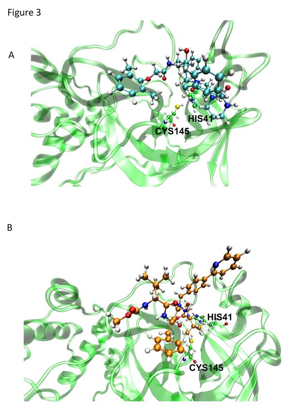
bioRxiv preprint doi: https://doi.org/10.1101/2020.04.04.020925. The copyright holder for this preprint (which was not peer-reviewed) is the
                              author/funder. It is made available under a CC-BY-NC-ND 4.0 International license.

             Abstract

             Severe acute respiratory syndrome coronavirus 2 (SARS-CoV-2) is already responsible
             for far more deaths than previous pathogenic coronaviruses (CoVs) from 2002 and
             2012. The identification of clinically approved drugs to be repurposed to combat 2019
             CoV disease (COVID-19) would allow the rapid implementation of potentially life-
             saving procedures. The major protease (Mpro) of SARS-CoV-2 is considered a
             promising target, based on previous results from related CoVs with lopinavir (LPV), an
             HIV protease inhibitor. However, limited evidence exists for other clinically approved
             antiretroviral protease inhibitors, such as atazanavir (ATV). ATV is of high interest
             because of its bioavailability within the respiratory tract. Our results show that ATV
             could dock in the active site of SARS-CoV-2 Mpro, with greater strength than LPV.
             ATV blocked Mpro activity. We confirmed that ATV inhibits SARS-CoV-2 replication,
             alone or in combination with ritonavir (RTV) in Vero cells, human pulmonary epithelial
             cell line and primary monocytes, impairing virus-induced enhancement of IL-6 and
             TNF-α levels. Together, our data strongly suggest that ATV and ATV/RTV should be
             considered among the candidate repurposed drugs undergoing clinical trials in the fight
             against COVID-19.

                                                                     2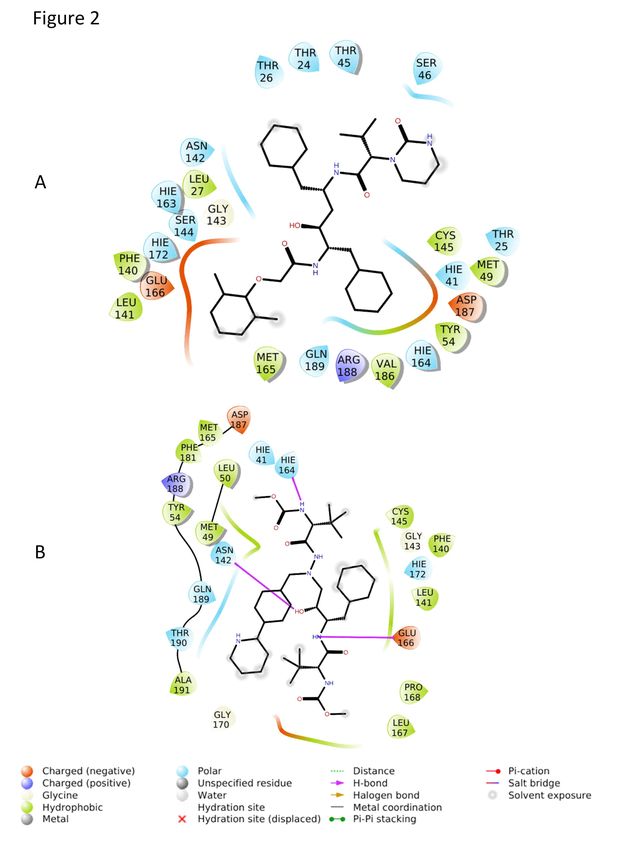
bioRxiv preprint doi: https://doi.org/10.1101/2020.04.04.020925. The copyright holder for this preprint (which was not peer-reviewed) is the
                              author/funder. It is made available under a CC-BY-NC-ND 4.0 International license.

             1) Introduction

                      Coronaviruses (CoVs) are single-stranded positive sense RNA viruses with large
             enveloped nucleocapsids that are able to infect a range of hosts including both animals
             and humans1. Although a number of human CoV are known to circulate seasonally, two
             highly pathogenic variants emerged in the 21st century that cause life-threatening
             infection, the severe acute respiratory syndrome (SARS-CoV) and middle-east
             respiratory syndrome (MERS-CoV)2. At the end of 2019, a novel variant of SARS-CoV
             (SARS-CoV-2) appeared in the citizens of the City of Wuhan, China that is believed to
             have spilled over to humans from animal reservoirs, most likely bats and/or pangolins3.
             The novel 2019 CoV is phylogenetically closer to SARS-CoV (from the 2002 outbreak)
                                                                      2,3
             than MERS-CoV (from 2012 outbreak)                          . Both SARS- and MERS-CoV raised
             international public health concerns with rates of mortality of 10 and 35%,
             respectively4,5. Soon after its discovery, the contemporary SARS-CoV-2 became a
             pandemic threat, with the number of confirmed infections ramping up globally6. To
             date, SARS-CoV-2 is responsible for 10 times more deaths than the total sum from
             SARS- and MERS-CoV, with more causalities daily that are continue to scale up6.

                      Currently, the most effective response to the SARS-CoV-2 pandemic has been
             self-quarantining and social distancing to avoid contact between infected and uninfected
             individuals that can flatten the virus dissemination curve, which aim to reduce the
             burden on medical resources to prevent loss of service for those with the highest need.
             While these social actions can disrupt virus transmission rates, they are not expected to
             reduce the absolute number of infected individuals. Furthermore, these strategies are
             also provoking a severe reduction in global economic activity7. To effectively combat
             the impact of SARS-CoV-2 on infected individuals, and society as a whole, it is
             essential to identify antiviral drugs for immediate use, as well as develop new drugs and
             a vaccine for long-term solutions to the disease associated with SARS-CoV-2 (COVID-
             19) .

             Repurposing of clinically approved drugs is the fastest pathway towards an effective
             response to a pandemic outbreak8. Some of the most promising antiviral candidates
             against SARS-CoV-2 have been under investigation since the outbreak of SARS-CoV
             in 2002. Building on this continuous investigation, an unprecedented effort to run a
             global clinical trial, called SOLIDARITY, is ongoing under the auspicious of the World
             Health Organization (WHO) and the United Nations (UN)9. This mega trial has been
                                                                     3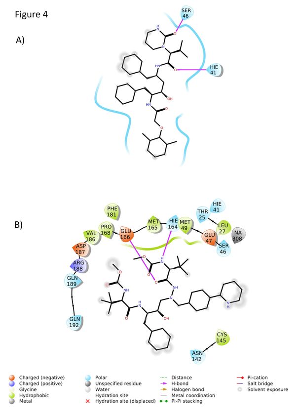
bioRxiv preprint doi: https://doi.org/10.1101/2020.04.04.020925. The copyright holder for this preprint (which was not peer-reviewed) is the
                              author/funder. It is made available under a CC-BY-NC-ND 4.0 International license.

             putting forward lopinavir (LPV)/ritonavir (RTV), in combination or not with interferon-
             β (IFN-β), chloroquine (CQ) and remdesivir to treat COVID-199. LPV, RTV and
             remdesivir target viral enzymes, while the actions of CQ and IFN-β target host cells.

                      The most successful antiviral drugs often directly target viral enzymes10. For
             CoVs, its major protease (Mpro) has been a promissing drug target for almost two
             decades, starting with early studies on 2002 SARS-CoV that showed this enzyme to be
             inhibited by LPV/RTV, inhibitors of HIV protease11. Mpro is required during the CoV
             replication cycle to process viral polyprotein12. Highly pathogenic CoVs contain two
             open reading frames, ORF1a and ORF1b, that are translated by host ribosomes into
             their two respective viral polyproteins, pp1a and pp1ab. ORF1a encodes two cysteine
             proteases, the papain-like protease (PLpro) and Mpro. While PLpro cuts the polyprotein
             at three sites, Mpro is responsible for cleavage at 11 another locations that, together,
             produce the 16 nonstructural proteins.

                      In a combined therapy of LPV with RTV, LPV is included as the principle
             antiviral compound and RTV as an inhibitor cytochrome p45013. Although RTV can
             also display weak anti-protease activity, at current therapeutic dosages its activity
             enhances the plasmatic concentration of the main antiviral compound by its ability to
             block drug metabolism. However, in an open-label clinical trial using LPV/RTV against
             COVID-19, their combination showed a limited benefit for treated patients14. In the
             early 2000s, another contemporary antiretroviral protease inhibitor, atazanavir (ATV),
             replaced LPV/RTV due to fewer side effects for the patients15,16. Contemporarily, in
             silico evidence suggested that other HIV protease inhibitors would target SARS-CoV-2
             Mpro better than LPV or RTV, that included ATV17. Importantly, ATV has been
             described to reach the lungs after intravenous administration18,19. Moreover, a proposed
             secondary use of ATV to treat pulmonary fibrosis suggested that this drug could
             functionally reach the lungs19.

                      The seriousness of COVID19 and the need for an immediate intervention, along
             with this series of observations with LPV, RTV and ATV, motivated us to evaluate the
             susceptibility of SARS-CoV-2 to ATV. Since ATV is available as a clinical treatment
             alone or in combination with RTV, both therapies were studied here, which for the first
             time describes that SARS-CoV-2 Mpro is a target for ATV. Further, ATV alone or
             withRTV could inhibit viral replication in cell culture models of infection that also
             prevented the release of a cytokine storm-associated mediators. Our timely data
                                                                     4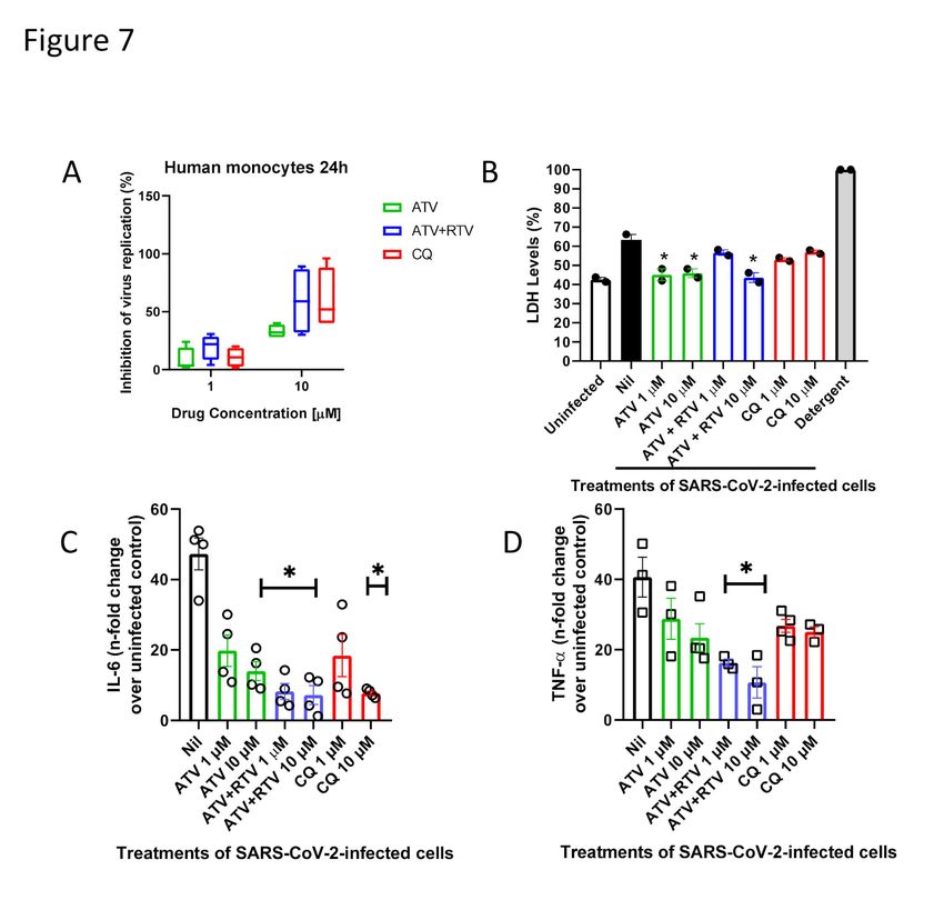
bioRxiv preprint doi: https://doi.org/10.1101/2020.04.04.020925. The copyright holder for this preprint (which was not peer-reviewed) is the
                              author/funder. It is made available under a CC-BY-NC-ND 4.0 International license.

             highlights an additional therapeutic approach against COVID-19 that should be
             considered for clinical trials.

             2) Results

             2.1) ATV docks into SARS-CoV-2 Mpro more spontaneously and stably than LPV

                        The targeting of the enzyme Mpro from SARS-CoV-2 by both ATV and LPV
             was evaluated by molecular modeling using a representative structure (PDB:6LU7). As
             shown in Figure 1, ATV occupied the S1* and S1 regions, whereas LPV occupied S1*
             and S2 regions with calculated free energy scores for LPV and ATV of -59.87 and -
             65.49 Kcal/mol, respectively. The more spontaneous binding of ATV, suggested by its
             lower energy score, may be related to its projected ability to form hydrogens bonds with
             the amino acid residues Asn142, His164, and Glu166 in Mpro, whereas the binding of
             LPV depends on hydrophobic interactions (Figure 2).

                        A molecular dynamic analysis revealed that the root-mean-square deviation
             (RMSD) for the SARS-CoV-2 Mpro backbone presented different conformations in
             complex with ATV or LPV (Figure S1). LPV was initially at a 3.8 Å distance from the
             catalytic residue Cys145 (Figure S2A and S3A), which after conformational changes
             extended to a distance equivalent to 7.17 Å (Figure 3A and 4A) that is projected to most
             likely limit the extent of its antiviral inhibition. Another critical residue, His41, was
             satisfactorily at a distance of 2.89 Å from bound LPV (Figure 3A and 4A). While ATV
             did not interact with His41 or Cys145 (Figure S2B and S3B), its position remained
             stable within the active site independent of conformational changes displayed by the
             enzyme (Figure 3B and 4B). The steric occupation of the cleft in the enzymatic active
             site by ATV, which block the residues of the catalytic amino acids, can be explained by
             its stronger interactions with Mpro, compared to LPV, through multiple hydrogen bonds
             during stationary docking and molecular dynamics (Tables S1-S3).

             2.2) ATV inhibits SARS-CoV-2 Mpro enzymatic activity

                      Next, we evaluated whether ATV could inhibit SARS-CoV-2 Mpro activity by
             partially purifying the enzyme in cellular fractions obtained from SARS-CoV-2-infected
             cells and performing zymographic profiles. To assure that the proteinase profiles were
             not dependent on cellular enzymes, similar fractions of mock-infected cells were also

                                                                     5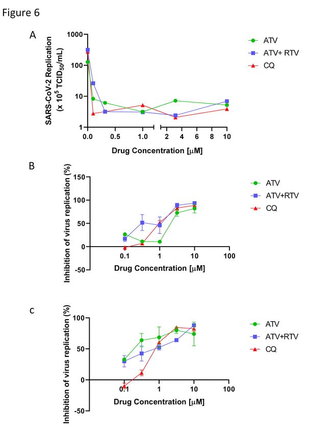
bioRxiv preprint doi: https://doi.org/10.1101/2020.04.04.020925. The copyright holder for this preprint (which was not peer-reviewed) is the
                              author/funder. It is made available under a CC-BY-NC-ND 4.0 International license.

             prepared for comparison. The results from cysteine proteinase zymographic profiles in
             gelatinolytic gels reveled a cellular related band of approximately 70 kDa under both
             conditions (Figure 5, lanes Nil). This activity was blocked by the drug E-64, an epoxide
             that acts as an irreversible inhibitor of cysteine proteases (Figure 5, lanes E-64). In the
             infected cells, a region of activity was observed between 31 and 38 kDa that was not
             present in the mock fraction. This zone of molecular weight is consistent with expected
             size of SARS-CoV-2 Mpro as was the inhibition of activity in this region by exposure
             of the gels to 10 μM of ATV, which did not affect the cellular cysteine proteinase at 70
             kDa (Figure 5, lanes ATV). Further confirmation of the presence and activity of SARS-
             CoV-2 Mpro in fractions from infected cells was obtained by treatment with RTV,
             which inhibited activity in the molecular range of 31-38 kDa without a change in the 70
             kDa region (Figure 5, lanes RTV). These data are consistent with predictions from the
             molecular modeling and dynamic analyses that suggested that ATV could bind and
             target the enzymatic activity of the Mpro encode by the novel 2019 CoV.

             2.3) SARS-CoV-2 is susceptible to ATV in different cell types
                      We extended our investigation to the inhibition of SARS-CoV-2 replication by
             ATV using a range of different cellular systems. Vero cells are a well-known model
             system that produce high virus titers and display visual cytopathic effects to viral
             infections. ATV alone, or in combination with RTV, inhibited infectious virus
             production and SARS-CoV RNA levels in Vero cells (Figure 6A and B, respectively).
             CQ was used as a positive control because of its inclusion in the SOLIDARITY trial
             due to its encouraging pre-clinical and clinical results against SARS-CoV-2 replication
             and COVID-19, respectively20,21. ATV/RTV was the most potent therapy tested; with an
             EC50 of 0.5 ± 0.08 µM. ATV alone and CQ’s potencies were 2.0 ± 0.12 µM and 1.0 ±
             0.07 µM, respectively. SARS-CoV-2 susceptibility to CQ is consistent with recent
             reports in the literature20, validating our analysis. The ATV/RTV, ATV, and CQ
             cytotoxicity values, CC50, were 280 ± 3 µM, 312 ± 8 µM and 259 ± 5 µM, respectively.
             Our results indicate that the selectivity index (SI, which represents the ratio between the
             CC50 and EC50 values) for ATV/RTV, ATV and CQ were 560, 156 and 259,
             respectively, which shows that ATV/RTV has a high therapeutic potential that was
             greater than CQ.
                  Since the results regarding the pharmacologic activity of ATV and ATV/RTV
             against SARS-CoV-2 replication in Vero cells were promising, we next investigated

                                                                     6
bioRxiv preprint doi: https://doi.org/10.1101/2020.04.04.020925. The copyright holder for this preprint (which was not peer-reviewed) is the
author/funder. It is made available under a CC-BY-NC-ND 4.0 International license.
whether the proposed drug therapies could inhibit virus replication in a human epithelial
pulmonary cell line (A549). ATV alone showed a nearly 10-fold increase in potency for
inhibiting SARS-CoV-2 replication in A549 (Figure 6C) compared to Vero cells (Figure
6B). ATV/RTV and CQ were similarly potent in inhibiting virus replication in both cell
types (Figure 6B and C). ATV/RTV, ATV and CQ EC50 values to inhibit SARS-CoV-2
replication in A549 cells were 0.60 ± 0.05 µM, 0.22 ± 0.02 µM and 0.89 ± 0.02 µM,
respectively. In vitro results confirmed the rational that SARS-CoV-2 would be
susceptible to ATV that included cells derived from the respiratory tract.
2.4) ATV prevents cell death and pro-inflammatory cytokine production in SARS-
CoV-2-infected monocytes.
Recent reports on the COVID-19 outbreak have implicated that an increase in the
levels of lactate dehydrogenase (LDH) and interleukin 6 (IL-6) is associated with
mortality22. Viral infection in the respiratory tract often trigger the migration of blood
monocytes to orchestrate the transition from innate to adaptive immune responses23. For
these reasons, ATV and ATV/RTV were tested at suboptimal (1 μM) or optimal (10
μM) doses in a SARS-CoV-2-infection model utilizing human primary monocytes.
ATV/RTV and CQ were similarly efficient to inhibit viral replication in the human
monocytes (Figure 7A). Virus infection increased cellular mortality by 75%, which was
prevented by ATV, at both doses tested, and by ATV/RTV, at 10 μM (Figure 7B). As a
control, detergent treatment completely destroyed all cells (Figure 7B). Moreover, we
observed that infections by SARS-CoV-2 triggered the expected increase in the IL-6
levels in the culture supernatant, which ranged from 20- to 60-fold depending on the
cell donor (Figure 7C, open circles in nil-treated cells). The virus-induced enhancement
of IL-6 levels were significantly prevented by treatment with ATV at 10 µM,
ATV/RTV at both 1 and 10 µM and CQ at 10 µM (Figure 7C). Another biomarker of
uncontrolled pro-inflammatory cytokine response, TNF-α, was up-regulated 40-fold
during virus infection (Figure 7D). Only the combination of ATV/RTV could
significantly prevent the induction of TNF-α release (Figure 7D). Altogether, our results
confirm that ATV and ATV/RTV should not be ignored as an additional therapeutic
option against COVID-19.
3) Discussion
7bioRxiv preprint doi: https://doi.org/10.1101/2020.04.04.020925. The copyright holder for this preprint (which was not peer-reviewed) is the
author/funder. It is made available under a CC-BY-NC-ND 4.0 International license.
In these two decades of the 21st century, the human vulnerability to emerging viral
diseases has been notable24. The emergence of infectious disease highlights the
undeniable fact that existing countermeasures are inefficient to prevent virus spill over
and diseases outbreak. Preclinical data on the susceptibility of an emerging virus to
clinically approved drugs can allow for the rapid mobilization of resources towards
clinical trials8. This approach proved feasible for combating the Zika, yellow fever and
chikungunya outbreaks experienced in Brazil over the past 5 years, when our group
demonstrated that sofosbuvir, a blockbuster drug against hepatitis C, could represent a
compassionate countermeasure against these diseases25–29.
Currently, the rate of SARS-CoV-2 dissemination has become one of the most
rapidly evolving pandemics known in modern times with the number of cases and
deaths doubling every week and the peak of the pandemic has yet to arrive6. The
existence of several ongoing clinical trials against COVID-19 reinforces the suggestion
that drug repurposing represents the fastest approach to identify therapies to emerging
infectious disease8. The WHO/UN, under the auspicious of the SOLIDARITY trial,
have highlighted the most promising anti-CoV drugs, such as LPV/RTV with or without
interferon-β, CQ and remdesivir30. Here, we provide preclinical evidence that another
HIV protease inhibitor, ATV, can inhibit the activity of a critical protease of SARS-
CoV-2, Mpro, and that this inhibition can extend to a disruption of viral replication as
well as the release of cytokine storm-associated mediators associated with viral
infection. The results suggest that the performance of ATV could be better than LPV
and strongly support that inclusion of ATV-based therapies in clinical trials for COVID-
19 either alone, in combination with RTV or both.
Kaletra® is an LPV/RTV formulation from Abbot Laboratories that was approved
by Food and Drug Administration (FDA) in 2000 and was evaluated for use in the
treatment during the SARS-CoV outbreak in 2002 and again for MERS-CoV. Its
continued evaluation with the SARS-CoV-2 outbreak is a logical choice due to the
conservation of the Mpro among these highly pathogenic viruses31,32. Nevertheless,
information on the susceptibility of SARS-CoV-2 to other antiviral protease inhibitors
that have been approved since 2003 has been scarce.
ATV was approved in 2003, and become a wider prescribed drug among HIV-
infected individuals, than LPV, including for critically ill patients16. ATV shows a safer
profile than LPV in both short- and long-tem therapeutic regimens15,33. ATV has a
8bioRxiv preprint doi: https://doi.org/10.1101/2020.04.04.020925. The copyright holder for this preprint (which was not peer-reviewed) is the
author/funder. It is made available under a CC-BY-NC-ND 4.0 International license.
documented bioavailability to reach the respiratory tract18,34, which lead to its proposed
use against pulmonary fibrosis19. However, it is not currently under consideration for
clinical trials against COVID-19.
The potencies of LPV and LPV/RTV against CoV are from 10 to 8 μM,
respectively32. Based on our data, ATV and ATV/RTV are at least 10 times more
potent. The ATV and ATV/RTV in vitro potencies are comparable to other small
molecule inhibitors of the SARS-CoV-2, such as remdesivir and CQ20. The improved
potency of ATV, in comparison to LPV, may be at least in part due to its multiple
hydrogen bond driven interactions within the Mpro active site. Other investigators have
also recognized a wider range of interactions of ATV and Mpro compared to LPV17,35,
although none provided functional evidence through phenotypic assays as presented
here. Neither ATV nor LPV displayed any interactions with the catalytic dyad of
Cys145 and His41 at the start of the molecular dynamic simulations. However,
important interactions were observed at its end, such as LPV-His41 and ATV-Glu166.
Glu166 is one of the residues that promotes the opeing of Mpro for its substrate to
interact with the active site36,37.
LPV/RTV was the first line of defense for early patients with COVID-1931. In
patients with severe COVID-19, the open-labeled clinical trial with LPV/RTV revealed
that treated patients had 5% less deaths and better clinical improvement then controls14.
Throughout the course of this study, LPV/RTV-treated patients continued to shed
SARS-CoV-2 at the same magnitude and duration of the control group14, limiting the
enthusiasm on the part of the medical and scientific community for this therapeutic
option. In this context, ATV and/or ATV/RTV should not be ignored in the treatment of
this novel respiratory disease. Indeed, our results demonstrate that the potency of ATV
and ATV/RTV potency against SARS-CoV-2 in A549 cells is likely to be consistent
with their bioavailability in the lungs in experimental models18,34.
Highly pathogenic respiratory viruses, such as influenza A virus, have been
associated with a cytokine storm that describes an uncontrolled pro-inflammatory
cytokine response38,39. Cytokine storms also seem to be highly relevant for pathogenic
human CoVs40. Contemporary investigations on SARS-CoV-2 strongly suggest the
involvement of cytokine storm with disease severity22. COVID-19 mortality is
associated with enhanced IL-6 levels and consistent cell death, as measured by LDH
9bioRxiv preprint doi: https://doi.org/10.1101/2020.04.04.020925. The copyright holder for this preprint (which was not peer-reviewed) is the
author/funder. It is made available under a CC-BY-NC-ND 4.0 International license.
release22. We showed that ATV and ATV/RTV decreased IL-6 release in SARS-CoV-2-
infected human primary monocytes. Moreover, we also included in our analysis TNF-α,
another hallmark of inflammation during respiratory virus infections22,41. Our results
reveled that cellular mortality and cytokine storm-associated mediators were reduced
after treatment with the repurposed antiretroviral drugs used in this study.
Among the most promising anti-SARS-CoV-2 drugs, CQ, IFN-β and LPV displayed
a higher toxic profile than ATV. Moreover, ATV and ATV/RTV have in vitro antiviral
potencies comparable to CQ and remdesivir, which were superior to LPV/RTV. In
summary, our study highlights a new option among clinically approved drugs that
should be considered in ongoing clinical trials for an effective treatment for COVID-19.
Material and Methods
4.1. Reagents.
The antiviral ATV, ATV/RTV and CQ were received as donations from Instituto de
Tecnologia de Fármacos (Farmanguinhos, Fiocruz). ATV/RTV was prepared in the
proportion of 3:1 as the pharmaceutical pills are composed of 300 mg ATV and 100 mg
RTV daily. ELISA assays were purchased from R&D Bioscience. All small molecule
inhibitors were dissolved in 100% dimethylsulfoxide (DMSO) and subsequently diluted
at least 104-fold in culture or reaction medium before each assay. The final DMSO
concentrations showed no cytotoxicity. The materials for cell culture were purchased
from Thermo Scientific Life Sciences (Grand Island, NY), unless otherwise mentioned.
Triton X-100 (TX-100), 3-[(3-Cholamidopropyl)dimethylammonio]-1-
propanesulfonate hydrate (CHAPS), 1,2,3-Propanetriol (glycerol), bovine serum
albumin (BSA), Phosphate-buffered saline (PBS), N-benzyloxycarbonyl-l-phenylalanyl-
l-arginine 7-amino-4-methylcoumarin (Z-FR-AMC; ε= 1.78 × 10⁴ M−1 cm−1),
dithiothreitol (DTT) and trans-epoxysuccinyl-l-leucylamido(4-guanidino)butane (E-64)
were purchased from Sigma Aldrich Chemical Co. (St. Louis, MO, USA). HiTrap Q FF
anion exchange chromatography column (HiTrap Q FF) was purchase from GE
Healthcare Life Sciences. Micro-bicinchoninic acid (BCA) protein assay kit was
purchased from Pierce Chemical Co. (Appleton, WI). All other reagents were of
analytical grade or better.
4.2. Cells and Virus
10bioRxiv preprint doi: https://doi.org/10.1101/2020.04.04.020925. The copyright holder for this preprint (which was not peer-reviewed) is the
author/funder. It is made available under a CC-BY-NC-ND 4.0 International license.
African green monkey kidney (Vero, subtype E6) and A549 (human lung epithelial
cells) cells were cultured in high glucose DMEM with 10% fetal bovine serum (FBS;
HyClone, Logan, Utah), 100 U/mL penicillin and 100 μg/mL streptomycin (Pen/Strep;
ThermoFisher) at 37 °C in a humidified atmosphere with 5% CO2.
Human primary monocytes were obtained after 3 h of plastic adherence of
peripheral blood mononuclear cells (PBMCs). PBMCs were isolated from healthy
donors by density gradient centrifugation (Ficoll-Paque, GE Healthcare). PBMCs (2.0 x
106 cells) were plated onto 48-well plates (NalgeNunc) in RPMI-1640 without serum for
2 to 4 h. Non-adherent cells were removed and the remaining monocytes were
maintained in DMEM with 5% human serum (HS; Millipore) and
penicillin/streptomycin. The purity of human monocytes was above 95%, as determined
by flow cytometric analysis (FACScan; Becton Dickinson) using anti-CD3 (BD
Biosciences) and anti-CD16 (Southern Biotech) monoclonal antibodies.
SARS-CoV-2 was prepared in Vero E6 cells from an isolate contained on a
nasopharyngeal swab obtained from a confirmed case in Rio de Janeiro, Brazil. Viral
experiments were performed after a single passage in a cell culture in a 150 cm2 flasks
with DMEM plus 2% FBS. Observations for cytopathic effects were performed daily
and peaked 4 to 5 days after infection. All procedures related to virus culture were
handled in a biosafety level 3 (BSL3) multiuser facility according to WHO guidelines.
Virus titers were determined as the tissue culture infectious dose at 50% (TCID50/mL).
Virus stocks were kept in - 80 °C ultralow freezers.
The virus strain was sequenced to confirm the virus identity and its complete
genome is publicly deposited (https://nextstrain.org/ncov: Brazil/RJ-314/2020 or
GISAID EPI ISL #414045).
4.3. Cytotoxicity assay
Monolayers of 1.5 x 10⁴ Vero cells in 96-well plates were treated for 3 days with
various concentrations (semi-log dilutions from 1000 to 10 µM) of ATV, ATV/RTV or
CQ. Then, 5 mg/ml 2,3-bis-(2-methoxy-4-nitro-5-sulfophenyl)-2H-tetrazolium-5-
carboxanilide (XTT) in DMEM was added to the cells in the presence of 0.01% of N-
methyl dibenzopyrazine methyl sulfate (PMS). After incubating for 4 h at 37 °C, the
plates were measured in a spectrophotometer at 492 nm and 620 nm. The 50% cytotoxic
11bioRxiv preprint doi: https://doi.org/10.1101/2020.04.04.020925. The copyright holder for this preprint (which was not peer-reviewed) is the
author/funder. It is made available under a CC-BY-NC-ND 4.0 International license.
concentration (CC50) was calculated by a non-linear regression analysis of the dose–
response curves.
4.4. Yield-reduction assay
Cells were infected with a multiplicity of infection (MOI) of 0.01. Vero or A549
cells were infected at densities of 5 x 10⁵ cells/well. Human primary monocytes were
infected at density of 2-8 x 10⁵ cells/well, depending on the endogenous characteristic
of the cell donor. Infections were performed in 48-well plates for 2h at 37 °C. The cells
were washed, and various concentrations of compounds were added to DMEM with 2%
FBS. After 48h, supernatants were collected and harvested virus was quantified by real
time RT-PCR and infectious titers by TCID50/mL. A variable slope non-linear
regression analysis of the dose-response curves was performed to calculate the
concentration at which each drug inhibited the virus production by 50% (EC50).
4.5. Virus titration
Monolayers of Vero cells (2 x 10⁴ cell/well) in 96-well plates were infected with a
log-based dilution of supernatants containing SARS-CoV-2 for 1h at 37°C. Cells were
washed, fresh medium added with 2% FBS and 3 to 5 days post infection the cytopathic
effect was scored in at least 10 replicates per dilution by independent readers. The
reader was blind with respect to source of the supernatant. A Reed and Muench scoring
method was employed to determine TCID50/mL42.
4.6. Molecular detection of virus RNA levels.
The total RNA from a culture was extracted using QIAamp Viral RNA (Qiagen®),
according to manufacturer’s instructions. Quantitative RT-PCR was performed using
QuantiTect Probe RT-PCR Kit (Quiagen®) in an ABI PRISM 7500 Sequence Detection
System (Applied Biosystems). Amplifications were carried out in 25 µL reaction
mixtures containing 2× reaction mix buffer, 50 µM of each primer, 10 µM of probe, and
5 µL of RNA template. Primers, probes, and cycling conditions recommended by the
Centers for Disease Control and Prevention (CDC) protocol were used to detect the
SARS-CoV-243. The standard curve method was employed for virus quantification. For
reference to the cell amounts used, the housekeeping gene RNAse P was amplified. The
Ct values for this target were compared to those obtained to different cell amounts, 107
to 102, for calibration.
12bioRxiv preprint doi: https://doi.org/10.1101/2020.04.04.020925. The copyright holder for this preprint (which was not peer-reviewed) is the
author/funder. It is made available under a CC-BY-NC-ND 4.0 International license.
4.7. Measurements Inflammatory Mediators and cell death marker
The levels of TNF-α, IL-6 and LDH were quantified in the monocyte supernatants
from infected and uninfected cells. ELISA for TNF-α and IL-6 required 100 µL of
supernatants to be exposed to capture antibody in 96-well plates. After a 2h incubation
period at room temperature (RT), the detection antibody was added. Plates were
incubated for another 2h at RT. Streptavidin-HRP and its substrate were added,
incubated for 20 minutes and the optical density was determined using a microplate
reader set to 450 nm.
Extracellular lactate dehydrogenase (LDH) was quantified using Doles® kit
according to manufacturer’s` instructions. Supernatant was centrifuged at 5,000 rpm for
1 minute, to remove cellular debris. A total of 25 µL of supernatant was placed into 96-
well plates and incubated with 5 µL of ferric alum and 100 µL of LDH substrate for 3
minutes at 37 °C. Nicotinamide adenine dinucleotide (NAD, oxidized form) was added
followed by the addition of a stabilizing solution. After a 10 min incubation, plates were
measured in a spectrophotometer at 492 nm.
4.8. Molecular docking
ATV (PubChem CID: 148192) and LPV (PubChem CID: 92727) were used as
inhibitors of the SARS-CoV-2 Mpro. ATV and LPV were prepared using the
Generalized Amber Force Field (GAFF) and their charges were obtained using the
AM1-BCC loading scheme 44,45.
Molecular docking experiments were performed with DOCK 6.946 for identifying
the binding site of the Mpro. SARS-CoV-2 Mpro structure was obtained from Protein
Data Bank (RCSB PDB, http://www.rcsb.org), under the accession code #6LU7 47. The
active site region was identified by using a complexed peptide (N-[(5-methylisoxazol-3-
yl)carbonyl]alanyl-l-valyl-n~1~-((1r,2z)-4-(benzyloxy)-4-oxo-1-{[(3r)-2-oxopyrrolidin-
3-yl]methyl}but-2-enyl)-l-leucinamide) as a guide. The creation of the DOCK 6.9 input
files for docking was performed using Chimera 1.1448.
The docking of ligands was performed in a box of 10 Å edges with its mass center
matching that of the complexed peptide. Each scan produced 20 conformations for each
ligand with the best score being used for molecular dynamics simulations.
4.9. Molecular dynamics
13bioRxiv preprint doi: https://doi.org/10.1101/2020.04.04.020925. The copyright holder for this preprint (which was not peer-reviewed) is the
author/funder. It is made available under a CC-BY-NC-ND 4.0 International license.
Since the tertiary structure (3D) of the SARS-CoV-2 Mpro is a homodimer, we
focused the molecular dynamics only one chain, henceforward chain A. Molecular
dynamics calculations were performed using NAMD 2.949 and Charmm27* force field50
at pH 7, i.e., with deprotonated Glu and Asp, protonated Arg and Lys, and neutral His
with a protonated Nε atom. This all-atom force field has been able to fold properly
many soluble proteins51–53. The soluble proteins were centered in a cubic box of TIP3P
water molecules54; the box extended 1.2 nm outside the protein on its four lateral sides,
and the appropriate numbers of Na+ and Cl- ions were added to ensure system
neutralization. The electrostatic interactions were calculated using the Particle Mesh
Ewald method and a cutoff of 1.2 nm55. The same cutoff of 1.2 nm was used for the Van
der Waals interactions. The non-bonded pair lists were updated every 10 fs. In what
follows, the analysis is based on MD simulation of 100 ns at 310 K.
4.10. Protein extraction
Protein extracts containing SARS-CoV-2 Mpro activity were obtained from
Vero cell monolayers at 25 cm2 flasks that were infected for 1h with an MOI of 0.1 at
37 °C and 5% CO2. After 1 or 2 days of infection, the supernatant was harvested and
monolayers were washed 3 times with in sterile cold PBS (pH 7.2). Next, cells were
suspended into 1 mL of lysis buffer (100 mM Tris-HCl (pH 8.0), 150 mM NaCl, 10%
glycerol and 0.6% Triton X-100) and kept at 4 °C. The soluble protein fraction was
isolated as the supernatant after centrifugation (100,000 x g, 30 min, 4 °C) and stored at
-20°C until further use. The protein concentrations of the samples were determined
using the BCA protein assay kit.
4.11. Zymographic assays
Proteinases were assayed after electrophoresis on 10% SDS-PAGE with 0.1%
copolymerized gelatin56. Briefly, the gels were loaded per slot with 12 μg of soluble
proteins dissolved in Laemmli’s buffer, and following electrophoresis at a constant
voltage of 200 V at 4°C, they were soaked for 1 h at 25 °C in washing buffer (0.1 mM
sodium acetate buffer (pH 5.5) containing 2.5% TX-100). Proteinase activity was
detected by incubating (16 h at 37 °C) the gels in reaction buffer (0.1 mM sodium
acetate buffer pH 5.5 containing 1.0 mM DTT), in the presence and absence of same
concentration of 10 µM of E-64, ATV, RTV or the ATV/RTV combination. Hydrolysis
of gelatin was visualized by staining the gels with amido black 0.2%57.
14bioRxiv preprint doi: https://doi.org/10.1101/2020.04.04.020925. The copyright holder for this preprint (which was not peer-reviewed) is the
author/funder. It is made available under a CC-BY-NC-ND 4.0 International license.
4.12. Statistical analysis
The assays were performed blinded by one professional, codified and then read
by another professional. All experiments were carried out at least three independent
times, including a minimum of two technical replicates in each assay. The dose-
response curves used to calculate EC50 and CC50 values were generated by variable
slope plot from Prism GraphPad software 8.0. The equations to fit the best curve were
generated based on R2 values ≥ 0.9. Student’s T-test was used to access statistically
significant P valuesbioRxiv preprint doi: https://doi.org/10.1101/2020.04.04.020925. The copyright holder for this preprint (which was not peer-reviewed) is the
author/funder. It is made available under a CC-BY-NC-ND 4.0 International license.
The authors declare no competing financial interests.
References
1. Masters, P. S. The molecular biology of coronaviruses. Adv. Virus Res. 66, 193–292
(2006).
2. Cui, J., Li, F. & Shi, Z.-L. Origin and evolution of pathogenic coronaviruses. Nat.
Rev. Microbiol. 17, 181–192 (2019).
3. Lam, T. T.-Y. et al. Identifying SARS-CoV-2 related coronaviruses in Malayan
pangolins. Nature (2020) doi:10.1038/s41586-020-2169-0.
4. WHO | Middle East respiratory syndrome coronavirus (MERS-CoV). WHO
http://www.who.int/emergencies/mers-cov/en/ (2020).
5. WHO | Severe Acute Respiratory Syndrome (SARS). WHO
https://www.who.int/csr/sars/en/ (2020).
6. Dong, E., Du, H. & Gardner, L. An interactive web-based dashboard to track
COVID-19 in real time. Lancet Infect. Dis. 0, (2020).
7. Romer, P. & Garber, A. M. Opinion | Will Our Economy Die From Coronavirus?
The New York Times (2020).
8. Harrison, C. Coronavirus puts drug repurposing on the fast track. Nat. Biotechnol.
(2020) doi:10.1038/d41587-020-00003-1.
9. Organization, W. H. WHO R&D Blueprint: informal consultation on prioritization of
candidate therapeutic agents for use in novel coronavirus 2019 infection, Geneva,
Switzerland, 24 January 2020. (2020).
10. De Clercq, E. & Li, G. Approved Antiviral Drugs over the Past 50 Years. Clin
Microbiol Rev 29, 695–747 (2016).
11. Wu, C.-Y. et al. Small molecules targeting severe acute respiratory syndrome
human coronavirus. Proc. Natl. Acad. Sci. U. S. A. 101, 10012–10017 (2004).
12. Fehr, A. R. & Perlman, S. Coronaviruses: an overview of their replication and
pathogenesis. Methods Mol. Biol. Clifton NJ 1282, 1–23 (2015).
13. Gong, Y. et al. Pharmacokinetics and pharmacodynamics of cytochrome P450
inhibitors for HIV treatment. Expert Opin. Drug Metab. Toxicol. 15, 417–427 (2019).
14. Cao, B. et al. A Trial of Lopinavir-Ritonavir in Adults Hospitalized with Severe
Covid-19. N. Engl. J. Med. (2020) doi:10.1056/NEJMoa2001282.
16bioRxiv preprint doi: https://doi.org/10.1101/2020.04.04.020925. The copyright holder for this preprint (which was not peer-reviewed) is the
author/funder. It is made available under a CC-BY-NC-ND 4.0 International license.
15. Stanley, T. L. et al. Effects of Switching from Lopinavir/ritonavir to
Atazanavir/ritonavir on Muscle Glucose Uptake and Visceral Fat in HIV Infected
Patients. AIDS Lond. Engl. 23, 1349–1357 (2009).
16. Gibert, C. L. Treatment Guidelines for the Use of Antiretroviral Agents in HIV-
Infected Adults and Adolescents: An Update. Fed. Pract. 33, 31S-36S (2016).
17. Dayer, M. R. Old Drugs for Newly Emerging Viral Disease, COVID-19:
Bioinformatic Prospective. 16.
18. Gautam, N. et al. Preclinical pharmacokinetics and tissue distribution of long-
acting nanoformulated antiretroviral therapy. Antimicrob. Agents Chemother. 57,
3110–3120 (2013).
19. Song, S. et al. Protective Effect of Atazanavir Sulphate Against Pulmonary
Fibrosis In Vivo and In Vitro. Basic Clin. Pharmacol. Toxicol. 122, 199–207 (2018).
20. Wang, M. et al. Remdesivir and chloroquine effectively inhibit the recently
emerged novel coronavirus (2019-nCoV) in vitro. Cell Res. 30, 269–271 (2020).
21. Gautret, P. et al. Hydroxychloroquine and azithromycin as a treatment of
COVID-19: results of an open-label non-randomized clinical trial. Int. J. Antimicrob.
Agents 105949 (2020) doi:10.1016/j.ijantimicag.2020.105949.
22. Zhou, F. et al. Clinical course and risk factors for mortality of adult inpatients
with COVID-19 in Wuhan, China: a retrospective cohort study. The Lancet 395,
1054–1062 (2020).
23. Newton, A. H., Cardani, A. & Braciale, T. J. The host immune response in
respiratory virus infection: balancing virus clearance and immunopathology. Semin.
Immunopathol. 38, 471–482 (2016).
24. Solomon, T., Baylis, M. & Brown, D. Zika virus and neurological disease-
approaches to the unknown. Lancet Infect Dis 16, 402–4 (2016).
25. de Freitas, C. S. et al. Yellow fever virus is susceptible to sofosbuvir both in
vitro and in vivo. PLoS Negl Trop Dis 13, e0007072 (2019).
26. Ferreira, A. C. et al. Beyond members of the Flaviviridae family, sofosbuvir also
inhibits chikungunya virus replication. Antimicrob Agents Chemother (2018)
doi:10.1128/aac.01389-18.
27. Ferreira, A. C. et al. Sofosbuvir protects Zika virus-infected mice from
mortality, preventing short- and long-term sequelae. Sci. Rep. 7, 9409 (2017).
28. Sacramento, C. Q. et al. The clinically approved antiviral drug sofosbuvir
inhibits Zika virus replication. Sci Rep 7, 40920 (2017).
17bioRxiv preprint doi: https://doi.org/10.1101/2020.04.04.020925. The copyright holder for this preprint (which was not peer-reviewed) is the
author/funder. It is made available under a CC-BY-NC-ND 4.0 International license.
29. Figueiredo-Mello, C. et al. Efficacy of sofosbuvir as treatment for yellow fever:
protocol for a randomised controlled trial in Brazil (SOFFA study). BMJ Open 9,
e027207 (2019).
30. UN health chief announces global ‘solidarity trial’ to jumpstart search for
COVID-19 treatment. UN News https://news.un.org/en/story/2020/03/1059722
(2020).
31. Jin, Y.-H. et al. A rapid advice guideline for the diagnosis and treatment of 2019
novel coronavirus (2019-nCoV) infected pneumonia (standard version). Mil. Med.
Res. 7, 4 (2020).
32. Sheahan, T. P. et al. Comparative therapeutic efficacy of remdesivir and
combination lopinavir, ritonavir, and interferon beta against MERS-CoV. Nat.
Commun. 11, 1–14 (2020).
33. Lv, Z., Chu, Y. & Wang, Y. HIV protease inhibitors: a review of molecular
selectivity and toxicity. HIVAIDS Auckl. NZ 7, 95–104 (2015).
34. Huang, J. et al. UPLC-MS/MS quantification of nanoformulated ritonavir,
indinavir, atazanavir, and efavirenz in mouse serum and tissues. J. Chromatogr. B
Analyt. Technol. Biomed. Life. Sci. 879, 2332–2338 (2011).
35. Beck, B. R., Shin, B., Choi, Y., Park, S. & Kang, K. Predicting commercially
available antiviral drugs that may act on the novel coronavirus (2019-nCoV), Wuhan,
China through a drug-target interaction deep learning model. bioRxiv
2020.01.31.929547 (2020) doi:10.1101/2020.01.31.929547.
36. Yang, H. et al. The crystal structures of severe acute respiratory syndrome virus
main protease and its complex with an inhibitor. Proc. Natl. Acad. Sci. U. S. A. 100,
13190–13195 (2003).
37. Macchiagodena, M., Pagliai, M. & Procacci, P. Inhibition of the Main Protease
3CL-pro of the Coronavirus Disease 19 via Structure-Based Ligand Design and
Molecular Modeling. (2020).
38. Gao, R. et al. Cytokine and chemokine profiles in lung tissues from fatal cases
of 2009 pandemic influenza A (H1N1): role of the host immune response in
pathogenesis. Am J Pathol 183, 1258–68 (2013).
39. Peschke, T., Bender, A., Nain, M. & Gemsa, D. Role of macrophage cytokines
in influenza A virus infections. Immunobiology 189, 340–55 (1993).
18bioRxiv preprint doi: https://doi.org/10.1101/2020.04.04.020925. The copyright holder for this preprint (which was not peer-reviewed) is the
author/funder. It is made available under a CC-BY-NC-ND 4.0 International license.
40. Channappanavar, R. & Perlman, S. Pathogenic human coronavirus infections:
causes and consequences of cytokine storm and immunopathology. Semin.
Immunopathol. 39, 529–539 (2017).
41. Monteerarat, Y. et al. Induction of TNF-alpha in human macrophages by avian
and human influenza viruses. Arch Virol 155, 1273–9 (2010).
42. Reed, H., L. J. ,. &. Muench. A simple method of estimating fifty percent
endpoints. Am J Hyg. (1938).
43. CDC. Coronavirus Disease 2019 (COVID-19). Centers for Disease Control and
Prevention https://www.cdc.gov/coronavirus/2019-ncov/lab/rt-pcr-panel-primer-
probes.html (2020).
44. Jakalian, A., Jack, D. B. & Bayly, C. I. Fast, efficient generation of high-quality
atomic charges. AM1-BCC model: II. Parameterization and validation. J. Comput.
Chem. 23, 1623–1641 (2002).
45. Wang, J., Wolf, R. M., Caldwell, J. W., Kollman, P. A. & Case, D. A.
Development and testing of a general amber force field. J. Comput. Chem. 25, 1157–
1174 (2004).
46. Allen, W. J. et al. DOCK 6: Impact of new features and current docking
performance. J. Comput. Chem. 36, 1132–1156 (2015).
47. Jin, Z. et al. Structure-based drug design, virtual screening and high-throughput
screening rapidly identify antiviral leads targeting COVID-19. bioRxiv
2020.02.26.964882 (2020) doi:10.1101/2020.02.26.964882.
48. Pettersen, E. F. et al. UCSF Chimera--a visualization system for exploratory
research and analysis. J. Comput. Chem. 25, 1605–1612 (2004).
49. Phillips, J. C. et al. Scalable molecular dynamics with NAMD. J. Comput.
Chem. 26, 1781–1802 (2005).
50. MacKerell, A. D., Banavali, N. & Foloppe, N. Development and current status
of the CHARMM force field for nucleic acids. Biopolymers 56, 257–265 (2000).
51. Zhang, T., Nguyen, P. H., Nasica-Labouze, J., Mu, Y. & Derreumaux, P.
Folding Atomistic Proteins in Explicit Solvent Using Simulated Tempering. J. Phys.
Chem. B 119, 6941–6951 (2015).
52. Hoang Viet, M., Derreumaux, P. & Nguyen, P. H. Communication: Multiple
atomistic force fields in a single enhanced sampling simulation. J. Chem. Phys. 143,
021101 (2015).
19bioRxiv preprint doi: https://doi.org/10.1101/2020.04.04.020925. The copyright holder for this preprint (which was not peer-reviewed) is the
author/funder. It is made available under a CC-BY-NC-ND 4.0 International license.
53. Lindorff-Larsen, K., Piana, S., Dror, R. O. & Shaw, D. E. How fast-folding
proteins fold. Science 334, 517–520 (2011).
54. Jorgensen, W. L., Chandrasekhar, J., Madura, J. D., Impey, R. W. & Klein, M.
L. Comparison of simple potential functions for simulating liquid water. (1998)
doi:1.445869.
55. Darden, T., York, D. & Pedersen, L. Particle mesh Ewald: An N⋅log(N) method
for Ewald sums in large systems. J. Chem. Phys. 98, 10089–10092 (1993).
56. Heussen, C. & Dowdle, E. B. Electrophoretic analysis of plasminogen activators
in polyacrylamide gels containing sodium dodecyl sulfate and copolymerized
substrates. Anal. Biochem. 102, 196–202 (1980).
57. Alves, C. R., Marzochi, M. C. & Giovanni-de-Simone, S. Heterogeneity of
cysteine proteinases in Leishmania braziliensis and Leishmania major. Braz. J. Med.
Biol. Res. Rev. Bras. Pesqui. Medicas E Biol. 26, 167–171 (1993).
20bioRxiv preprint doi: https://doi.org/10.1101/2020.04.04.020925. The copyright holder for this preprint (which was not peer-reviewed) is the
author/funder. It is made available under a CC-BY-NC-ND 4.0 International license.
Legend for the Figures
Figure 1. The active site of SARS-CoV-2 Mpro in the absence and presence of the
inhibitors. A representative structure of Mpro (PDB:6LU7) was color coded to show
the electrostatic potential of residues in the active site for negative (blue) and positive
(red) charges. Panel A, the cavities of ligand interaction designated S1*, S1 and S2 in
the absence of inhibitors. Panel B, placement of LPV (cyan) docked in the S1* and S2
regions of the active site. Panel C, placement of ATV (orange) docked in the S1* and
S1 regions of the active site.
Figure 2. Binding profile of antiretroviral drugs onto SARS-CoV-2 Mpro. Two-
dimensional (2D) representations of the interactions of LPV (A) and ATV (B) in the
Mpro active site based on a molecular docking analysis. Two hydrogen bonds are
predicted between ATV and Mpro.
Figure 3. Final positions of ATV and LPV on Mpro at the end of a molecular
dynamic simulation. Representative images of the molecular dynamics after 100 ns of
simulation. LPV (A) and ATV (B) are positioned in the Mpro active site at the end of
100 ns simulation.
Figure 4. Position profile of ATV and LPV during molecular dynamics. Two-
dimensional (2D) representation of the interactions of LPV (A) and ATV (B) in the
Mpro active site at the end of 100 ns molecular dynamic simulation.
Figure 5. Inhibition of proteinase activity through an analysis of gelatinolytic
activity. Vero cells were mock treated or infected with SARS-CoV-2 at an MOI of 0.1
for 48h before lysis and preparation of a cellular fraction. Fractions containing 12 µg of
total protein separated by electrophoresis followed by cutting the gels into their
individual lanes that were incubated in 10 mM sodium acetate buffer (pH 5.5) in the
absence (Nil) or presence of 10 µM of E-64, ATV or RTV. Gelatinolytic bands
indicative of enzymatic activity were revealed by negative staining with amide black
solution. Molecular mass markers are indicated (kDa).
Figure 6. The antiviral activity of ATV and ATV/RTV against SARS-CoV-2. Vero
(A and B) or A549 (C) cells were infected with SARS-CoV-2 at the MOI of 0.01 and
exposed to indicated concentrations of the drugs. After 2 days, the viral replication in
the culture supernatant was measured by TCID50/mL (A) or RT-PCR (B and C). The
data represent means ± SEM of three independent experiments.
Figure 7. ATV and ATV/RTV impairs SARS-CoV-2 replication, cell death and
cytokine storm in human primary monocytes. Human primary monocytes were
infected at the indicated MOI of 0.01 and treated with indicated concentration of the
compounds. After 24h, virus replication (A) and LDH release (B) as well as the levels
of IL-6 (C) and TNF-α (D) were measured in the culture supernatant. The data represent
means ± SEM of experiments with cells from at least three healthy donors. Differences
with P < 0.05 are indicates (*), when compared to untreated cells (nil).
21You can also read