Iron Ladies - How Desiccated Asexual Rotifer Adineta vaga Deal With X-Rays and Heavy Ions? - DLR
←
→
Page content transcription
If your browser does not render page correctly, please read the page content below
ORIGINAL RESEARCH
                                                                                                                                                 published: 31 July 2020
                                                                                                                                         doi: 10.3389/fmicb.2020.01792

                                             Iron Ladies – How Desiccated
                                             Asexual Rotifer Adineta vaga Deal
                                             With X-Rays and Heavy Ions?
                                             Boris Hespeels 1,2* , Sébastien Penninckx 3† , Valérie Cornet 2† , Lucie Bruneau 1 ,
                                             Cécile Bopp 3 , Véronique Baumlé 1,2 , Baptiste Redivo 2 , Anne-Catherine Heuskin 3 ,
                                             Ralf Moeller 4,5 , Akira Fujimori 6 , Stephane Lucas 3 and Karine Van Doninck 1,2
                                             1
                                               Research Unit in Environmental and Evolutionary Biology (URBE), Laboratory of Evolutionary Genetics and Ecology (LEGE),
                                             NAmur Research Institute for Life Sciences (NARILIS), University of Namur, Namur, Belgium, 2 Research Unit
                                             in Environmental and Evolutionary Biology (URBE), Institute of Life, Earth & Environment (ILEE), University of Namur, Namur,
                                             Belgium, 3 Laboratory of Analysis by Nuclear Reaction (LARN), NAmur Research Institute for Life Sciences (NARILIS),
                                             University of Namur, Namur, Belgium, 4 Space Microbiology Research Group, Radiation Biology Department, Institute
                                             of Aerospace Medicine, German Aerospace Center (DLR), Cologne, Germany, 5 Department of Natural Sciences, University
                            Edited by:       of Applied Sciences Bonn-Rhein-Sieg (BRSU), Rheinbach, Germany, 6 Department of Basic Medical Sciences for Radiation
                        Rakesh Mogul,        Damages, National Institute of Radiological Sciences (NIRS), Chiba, Japan
California State Polytechnic University,
                Pomona, United States
                        Reviewed by:
                                             Space exposure experiments from the last 15 years have unexpectedly shown that
                      Tim Barraclough,       several terrestrial organisms, including some multi-cellular species, are able to survive in
University of Oxford, United Kingdom
                                             open space without protection. The robustness of bdelloid rotifers suggests that these
                  Fernando Rodriguez,
 Marine Biological Laboratory (MBL),         tiny creatures can possibly be added to the still restricted list of animals that can deal
                         United States       with the exposure to harsh condition of space. Bdelloids are one of the smallest animals
                Dinakar Challabathula,
Central University of Tamil Nadu, India
                                             on Earth. Living all over the world, mostly in semi-terrestrial environments, they appear
                  *Correspondence:
                                             to be extremely stress tolerant. Their desiccation tolerance at any stage of their life cycle
                      Boris Hespeels         is known to confer tolerance to a variety of stresses including high doses of radiation
           boris.hespeels@unamur.be
                                             and freezing. In addition, they constitute a major scandal in evolutionary biology due
     † These   authors have contributed
                                             to the putative absence of sexual reproduction for at least 60 million years. Adineta
                    equally to this work
                                             vaga, with its unique characteristics and a draft genome available, was selected by ESA
                   Specialty section:        (European Space Agency) as a model system to study extreme resistance of organisms
         This article was submitted to
                Extreme Microbiology,
                                             exposed to space environment. In this manuscript, we documented the resistance of
               a section of the journal      desiccated A. vaga individuals exposed to increasing doses of X-ray, protons and Fe
             Frontiers in Microbiology       ions. Consequences of exposure to different sources of radiation were investigated in
         Received: 25 February 2020          regard to the cellular type including somatic (survival assay) and germinal cells (fertility
            Accepted: 09 July 2020
            Published: 31 July 2020          assay). Then, the capacity of A. vaga individuals to repair DNA DSB induced by different
                              Citation:      source of radiation was investigated. Bdelloid rotifers represent a promising model in
             Hespeels B, Penninckx S,        order to investigate damage induced by high or low LET radiation. The possibility of
        Cornet V, Bruneau L, Bopp C,
    Baumlé V, Redivo B, Heuskin A-C,
                                             exposure both on hydrated or desiccated specimens may help to decipher contribution
    Moeller R, Fujimori A, Lucas S and       of direct and indirect radiation damage on biological processes. Results achieved
   Van Doninck K (2020) Iron Ladies –        through this study consolidate our knowledge about the radioresistance of A. vaga
      How Desiccated Asexual Rotifer
       Adineta vaga Deal With X-Rays         and improve our capacity to compare extreme resistance against radiation among living
                      and Heavy Ions?        organisms including metazoan.
            Front. Microbiol. 11:1792.
      doi: 10.3389/fmicb.2020.01792          Keywords: bdelloid rotifer, LET, DNA damage, extremophile, astrobiology, panspermia

Frontiers in Microbiology | www.frontiersin.org                                     1                                                July 2020 | Volume 11 | Article 1792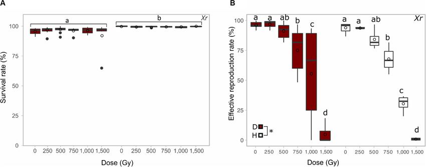
Hespeels et al.                                                                                           Resistance A. vaga Individuals Exposed to IR

INTRODUCTION                                                              semi-terrestrial habitats and in particular limno-terrestrial
                                                                          environments such as mosses and lichens (Melone and
The exposition of cells to ionizing radiations (IR) generates a           Fontaneto, 2005). Their desiccation tolerance at any stage of
succession of physical, chemical and biological processes that            their life cycle allows bdelloid rotifers to thrive in semi-terrestrial
differ in time, spatial and energy-scale leading to a cellular            environments but also confers tolerance to a variety of stresses
response complicated to predict (Joiner and van der Kogel, 2018).         including freezing, deep vacuum, UV and high doses of ionizing
The ionization density in particle tracks is usually characterized        radiation (Ricci et al., 2005; Gladyshev and Meselson, 2008;
by a physical parameter called Linear Energy Transfer (LET)               Fischer et al., 2013; Hespeels et al., 2014; Jönsson and Wojcik,
describing the average energy (in keV) given up by a charged              2017; Ricci, 2017).
particle traversing a distance of 1 µm. High-Z charged particles             Nevertheless, an overview of the radiation tolerance among
(such as Fe ions) are high LET particles that will produce                bdelloids is lacking and only few radiation studies have been
dense ionizations along their path while photons (like X-rays)            published. No lethal dose is currently available in the literature.
which produce sparse ionizations are considered as low LET                In 2008, Gladyshev and Meselson demonstrated that two bdelloid
IR (Joiner and van der Kogel, 2018; Hagiwara et al., 2019).               rotifer species maintained hydrated, Adineta vaga and Philodina
Although a same dose of radiation leads to the same amount                roseola, are extraordinary resistant to ionizing radiation (IR),
of ionizations, the differences in their spatial distribution is          surviving up to 1,200 Gy of gamma radiation with fecundity
translated into different types of damages at biological level. It        and fertility showing a dose response (Gladyshev and Meselson,
was demonstrated that sparse ionizations originating from low             2008). These doses were contrasting the Lethal Dose 50 (i.e., dose
LET radiations mainly induce DNA base oxidation and single                required to kill 50% of the irradiated population) of mammalian
strand breaks (Lehnert, 2007). Contrastingly, complex DNA                 cells ranging from 2 to 6 Gy after X-ray exposure (Kohn and
damages including clustered double strand breaks (DSBs) are               Kallman, 1957; Mole, 1984). In 2014, Hespeels et al. demonstrated
preferentially created following high-LET exposition resulting in         that the rehydration process of desiccated A. vaga individuals was
a poor survival of irradiated organisms (Semenenko and Stewart,           not affected by doses up to 800 Gy of protons 1,7 MeV. In 2017,
2004). Moreover, an increasing amount of studies highlighted              Jönsson and Wojcik highlighted that desiccated bdelloid Mniobia
that the complexity of damages induced by high LET particles              russeola is able to tolerate exposure to iron ions up to at least
was associated to lower repair kinetic leading to an increase             2,000 Gy with no apparent impact in term of survival and egg
in chromosomal aberrations and cancer incidence (Loucas and               production (Jönsson and Wojcik, 2017).
Cornforth, 2013; Penninckx et al., 2019). Although the health                By analogy with the desiccation- and radiation resistant
risks from exposure to terrestrial radiations (electrons and              bacterium D. radiodurans (Mattimore and Battista, 1996), it has
photons) are well known, the ones associated to heavy ions                been postulated that the extraordinary radiation resistance of
particles (HZE) remain poorly understood since they are only              bdelloid rotifers reflects an adaptation to survive desiccation and
naturally present in space. Therefore, the lack of data regarding         a high capacity to deal with numerous DNA DSBs (Gladyshev
whole-body response following HZE exposure and solutions                  and Meselson, 2008; Gladyshev and Arkhipova, 2010). Indeed,
to counteract their deleterious effects were identified as major          high radiation doses are known to induce numerous cellular
showstoppers for future long-duration space missions (White               damages, through the formation of reactive oxygen species
and Averner, 2001; Cucinotta and Durante, 2006). Within the               (ROS) and inducing DNA DSBs (Riley, 1994). We previously
animals, bdelloid rotifers are known to survive high doses of high        demonstrated that A. vaga accumulate DNA double strand
and low LET ionizing radiation (Gladyshev and Meselson, 2008;             breaks (DSBs) with time spent in dehydrated state. Upon
Hespeels et al., 2014; Jönsson and Wojcik, 2017), making them a           rehydration, rehydrated animals were able to reassemble large
new, suitable model system for space research.                            fragments of DNA supporting the presence of active DNA repair
   Bdelloid rotifers are among the smallest animals on Earth,             mechanisms. Even when the genome was entirely shattered into
with most species being less than 1 mm in size and containing             small DNA fragments by exposure to 800 Gy of protons 1.7 MeV
∼1,000 cells. Despite their small size, these eutelic (i.e., a            radiations, A. vaga individuals were able to efficiently recover
fixed number of cells at maturity) metazoans have a complete              from desiccation and to repair the DNA DSBs (Hespeels et al.,
nervous, muscular, digestive and excretory system (Ricci and              2014). Krisko et al. (2012) demonstrated that A. vaga is far more
Melone, 2000). One remarkable feature of bdelloid rotifers,               resistant to IR-induced protein carbonylation than is the much
being even more unusual among animals, is the absence of                  more radiosensitive nematode Caenorhabditis elegans. Therefore,
males, vestigial male structures or hermaphrodites in any of              like bacteria Deinococcus radiodurans, bdelloid rotifers appear to
the populations studied within the 460 described morphospecies            survive such extreme conditions because of efficient antioxidants
(Segers, 2007). Bdelloid rotifers are females apparently cloning          and DNA repair mechanisms (Flot et al., 2013; Latta et al.,
themselves since millions of years. While the exact nature of their       2019). However, the exact biological processes of these resistance
parthenogenetic reproductive mode remains unknown, bdelloid               mechanisms remain largely unknown.
rotifers are considered an “evolutionary scandal” because they               Space exposure experiments from the last 15 years have
persist without sexual reproduction (Maynard Smith, 1986).                unexpectedly shown that several terrestrial organisms, including
   Living all over the world, mostly in semi-terrestrial                  some multi-cellular species, are able to survive in open space
environments, they appear to be extremely stress tolerant.                without protection. In the absence of protective effects from
Around 90% of bdelloid species were described to live in                  the atmosphere and the magnetic field, organisms are exposed

Frontiers in Microbiology | www.frontiersin.org                       2                                           July 2020 | Volume 11 | Article 1792
Hespeels et al. Resistance A. vaga Individuals Exposed to IR
to a mixture of radiations and accelerated particles. More Meselson at Harvard University (Flot et al., 2013). The cultures
specifically, biological samples in space are expected to be were maintained hydrated in Petri dishes supplemented with
exposed to galactic cosmic rays (GCR) and to radiation bursts natural spring water (Spa), in thermostatic chambers at 21◦ C,
generated by solar eruption (Simpson, 1983; Wilson, 1991). and fed with sterilized lettuce juice (Lactuca sativa). Bacterial
GCR, originating from extra solar events like supernova, are proliferation and dirt accumulation were avoided by changing the
composed of 83% protons, 12% alpha particles and 1.5% water minimum twice a month.
high atomic number and energy particle (HZE) including Fe
(Ferrari and Szuszkiewicz, 2009). Despite reduced occurrence X-Ray Exposure
in terms of particle number, Fe ions driven by their huge Desiccated and hydrated A. vaga individuals were irradiated
LET are the primary component of GCR in term of energy using an X-ray irradiator PXi X-RAD 225 XL, with a dose rate
deposited on the target (Leuko and Rettberg, 2017; Moeller of ∼7.8 Gy/min (19 mA, 225 kV, no filter) available at LARN
et al., 2017). In addition, the collision of GCR with matter (UNamur-Belgium). Irradiation time was calculated from this
may result in the generation of low LET gamma and X-ray dose rate to obtain the different doses. All samples, including
radiation (Dartnell, 2011; Leuko and Rettberg, 2017). The control samples, were put on a refrigerated water bag to mitigate
robustness of bdelloid rotifers toward IR suggests that these heating from X-ray. For each dose, a minimum of three replicates
tiny creatures can possibly be added to the still restricted list of were recorded from at least two independent exposure campaigns
animals used for astrobiology experiments, including exposure to (except for 5,000 and 7,500 Gy where 3 replicates per dose
simulated deep space environment. Bdelloid rotifers can indeed were obtained during one exposure campaign). The exposure of
contribute significantly to our understanding of living in extreme hydrated animals was limited to 1,500 Gy. For hydrated samples,
environments. The bdelloid species A. vaga, whose genome a total of 2,000 to 5,000 living and healthy animals stored in
was sequenced (Flot et al., 2013), got recently selected for an 2 mL of Spa water supplemented by 100 µL of sterile lettuce
astrobiology experiment aimed at identifying and quantifying juice were exposed to X-ray in refrigerated multi-well plates.
biological damage suffered by bdelloids when exposed to space As animals were metabolically active during irradiation and
environment. Through this project, bdelloid rotifers will be because maximum dose rate was limited, it was decided to expose
exposed to a combination of full-spectrum electromagnetic hydrated A. vaga individuals to a maximum dose of 1,500 Gy. For
radiation from the Sun, cosmic particle radiations, vacuum, wide desiccated A. vaga samples individuals were dried for 1 day (see
temperature fluctuations and microgravity. In the preparation desiccation protocol below) before irradiation to X-rays doses
of this experiment, the present manuscript aims to determine ranging from 100 to 7,500 Gy.
the limits and consequences of bdelloid rotifer extreme
resistance to radiation. 4 MeV Proton Exposure
In this study, the resistance of A. vaga versus multiple sources Only desiccated A. vaga were irradiated with a homogenous
of radiation was investigated. The biological response of these broad proton beam defocused over 1 cm2 area, produced by a
animals to high doses of low and high LET was evaluated, 2 MV Tandem accelerator (High Voltage Engineering Europa).
respectively, using X-ray, protons 4 MeV and Fe 0.5 GeV A thorough description of the experimental set-up and the
particles. The high resistance of A. vaga individuals to radiation irradiation procedure is given in Wéra et al. (2011). Briefly,
prone them as a good model to study the prevalence of direct 4 MeV pristine proton beam was extracted in air through a 1 µm
and indirect damages coming from high doses of ionizing silicon nitride window. The linear energy transfer (LET) at the
radiation (>100 Gy). Here, hydrated and desiccated A. vaga sample location was computed using SRIM software (Ziegler
individuals were exposed to increasing dose of X-ray, and only et al., 2010), using two stacked layers representing the setup:
desiccated A. vaga to proton and Fe ions. For each radiation 1 µm Si3 N4 exit window and a small 3 mm air gap between
dose, we evaluated the survival and the capacity of irradiated the exit window and the sample dish. The energy of transmitted
animals to produce viable offspring. These data were confronted particles was averaged and the LET was obtained for this average
with the amount of DNA damage, evaluated by pulsed field proton energy. The irradiation chambers were placed vertically
gel electrophoresis (PFGE), and with the capacity of irradiated on a sample holder fixed at the end of the beamline. Rotifer
animals to repair, at least in somatic cells, DNA breaks induced samples were thus positioned perpendicularly to the incoming
by X-ray, proton and Fe radiation. Results achieved through this beam. Homogeneity was achieved by defocusing the beam and
study consolidate our knowledge about the radioresistance of checked using a passivated implanted planar silicon detector
bdelloid rotifer A. vaga and improve our capacity to compare moved along x and y directions. Dose rate was assessed every
extreme resistance against low and high LET radiation among millimeter in a 1 cm2 surface with errors less than 5% in the
living organisms including metazoan. cell sample region. Dataset generated for this study was acquired
through 6 campaigns of irradiation. Between campaigns, the
dose rate ranged from 42 to 134 Gy min−1 . The particle flux
MATERIALS AND METHODS was accordingly adjusted using the classic broad beam formula
(Turner, 2007) as shown below (Eq. 1):
Bdelloid Rotifer Cultures
Experiments were performed using isogenic A. vaga clones 1.6 10−9 LET ∅
issued from a single individual from the laboratory of Matthew Ḋ = (1)
ρ
Frontiers in Microbiology | www.frontiersin.org 3 July 2020 | Volume 11 | Article 1792Hespeels et al. Resistance A. vaga Individuals Exposed to IR
Here the density ρ is taken as 1 g cm−3 and 8 is the proton beam contracted individuals. All living and dead animals were collected
flux in part cm−2 s−1 . For each dose, a minimum of 3 replicates separately. Then, the survival rate was estimated manually
were recorded from at least 2 independent campaigns. by counting living and dead individuals under the binocular.
The desiccated A. vaga individuals were exposed to 4 MeV When the number of individuals was higher than ∼750, the
proton, with a LET of 10 keV/µm, ranging from 100 to 10,000 Gy. number of individuals was extrapolated by counting 6 times
the number of living/dead animals observed in 2 µL taken
from a homogenously mixed culture in a final volume of 2–
Fe 500 MeV/n Exposure 5 mL. Data are available in Supplementary Data S1. In the
Only desiccated A. vaga individuals were also irradiated with
absence of intermediary exposure doses between 5,000 and
iron ions (Fe 500 MeV/n, with a LET of 200 keV/µm and
7,500 Gy, it was not possible to compute an accurate LD50 for
a dose rate of 10 Gy/min). Desiccated A. vaga individuals
each radiation type.
were stored in 0.2 mL PCR tubes and sent to the Heavy
Ion Medical Accelerator in Chiba (HIMAC) facility at the
National Institute of Radiological Sciences (NIRS) in Chiba Fertility Assays
(Japan). Two campaigns of irradiations were performed The reproductive capacity was defined as the ability for each
in January 2018 and June 2018, respectively. During the A. vaga individual to lay eggs and to develop clonal populations
first campaign, samples were exposed to 250, 500, 1,000, after being desiccated and potentially irradiated. We tested
and 2,000 Gy. In the second campaign, desiccated A. vaga the fertility of 1 day desiccated and irradiated individuals by
rotifers were exposed to 1,000, 2,000, and 3,000 Gy Fe randomly selecting and isolating a minimum of 60 successfully
ions. For each campaign a set of 4 replicates per dose was rehydrated individuals per condition; each isolated female was
prepared. In parallel, two sets of controls were prepared for deposited in a well of a 12-well petri plate. Each well was filled
each exposure campaign. One set consisted of desiccated with 2 mL of Spa water and 50 µL of sterilized lettuce juice.
A. vaga individuals that were sent to HIMAC facility and After 30 days, wells were observed under a binocular stereoscope
back to Belgium without irradiation (Control transport). The checking for: (1) the presence of a population (minimum 2 adults
second set consisted of desiccated A. vaga individuals stored and 1 egg per well), (2) the presence of only eggs that did not
dry at controlled temperature (15◦ C) in LEGE laboratory hatch (+eventually the single adult from the start defined as a
(UNamur) for the whole duration of the exposure experiment sterile individual), and (3) the presence of only dead individual
(from sample preparation, exposure to HIMAC, and travel (s). When the number of survivors after irradiation was below
back to Belgium). For each campaign, it was impossible to 60 individuals, the number of isolated individuals lacking to
rehydrate the whole dataset of desiccated samples on the arrive at this total was automatically associated with the category
same day due to the workload and analysis required post dead individual (s). This correction avoids the overweighting of
rehydration. As a consequence, samples were rehydrated certain values following the drastic reduction of survivors at dose
randomly after their return to Belgium. Therefore, samples equal or higher than 5,000 Gy (X-ray and proton only). Data are
from first campaign were desiccated for a minimum available in Supplementary Data S1. Based on these data, it was
and a maximum duration of 30 to 37 days, respectively. possible to evaluate the minimal dose required to sterilize 50% of
For the second campaign, the total desiccation ranged the irradiated population. A similar approach to the evaluation of
between 39 and 48 days. a standard LD50 was applied here to define the Sterilizing Dose
50 (SD50 ). All curve fittings were performed with the OriginLab R
software (MA, United States). A dose-response equation was used
Desiccation and Survival Rate to fit fertility data according to the following equation (Eq. 2):
For desiccation experiments, we used the protocol previously
described in Hespeels et al. (2014). Briefly, healthy cultures A2 − A1
Sterilization = A1 + (2)
were shortly washed in filtered Spa water. Individuals were 1 + 10(logx0 − x)p
collected using quick vortex and transferred to a 50 mL
Falcon tube for centrifugation. The pellet of concentrated Where A1 is the bottom asymptote (i.e., representing
A. vaga individuals was resuspended in 1.25 mL of Spa water the fertility of sterilized animals), A2 is the top
and placed in the center of Petri dishes containing 30 mL asymptote (i.e., representing the fertility of control
of 3% LMP agarose (Invitrogen). Plates with approximately animals), logx0 correspond to SD50 and p is the hill
15,000–45,000 A. vaga individuals are placed in a climatic slope.
chamber (WEKK 0028) for desiccation following the optimized
desiccation protocol: (1) linear decrease of relative humidity from Statistical Analysis
70 to 55% for 17 h (temperature: 23◦ C), (2) linear decrease Data were expressed as means ± standard deviations (SD).
of relative humidity from 55 to 41% for 1 h (temperature: Statistical analyses were performed using Rstudio software
23◦ C), and (3) maintenance of 41% RH and 23◦ C during version 3.6.3 (RStudio Team, 2015) with packages car
the whole exposure period. After desiccation, dried bdelloid version 3.0–7 (Fox and Weisberg, 2019) and multcomp
individuals were rehydrated using 15 mL Spa water and stored
R
version 1.4–13 (Hothorn et al., 2008). Effects of radiation
at 21◦ C for 48 h. Bdelloids were considered alive when they dose were analyzed using a Global Linear Model (using a
had fully recovered motility or when the mastax moved in Gaussian family). Post hoc comparisons at a 5% significant
Frontiers in Microbiology | www.frontiersin.org 4 July 2020 | Volume 11 | Article 1792Hespeels et al. Resistance A. vaga Individuals Exposed to IR
level were performed with General Linear Hypotheses the plot profile tool (ImageJ) calculated on straight lines of
(Tukey’s test) and multiple comparisons for GLM. This identical length.
approach allowed not restricting the statistical analysis
to the comparison of each condition versus the control
condition (i.e., non-irradiated samples). Multiple comparisons
RESULTS
between each dose ensure to discriminate significant
differences between all conditions. Group characterized
letters indicate the significant differences between groups:
Evaluation of the Survival Rate of A. vaga
a significant difference (P-value < 0.05) between two Individuals Exposed to X-Ray, PROTONS
conditions is observed when these conditions do not share and Fe Sources
any letter. Desiccated A. vaga individuals were exposed to increasing doses
of ionizing radiation. The maximal doses achieved were about
Genomic DNA Integrity 3,000 Gy for Fe radiation, 7,500 Gy for X-ray and up to 10,000 Gy
The genomic integrity, i.e., the accumulation of DNA for protons 4 MeV. The survival rate of desiccated, irradiated
DSBs induced by desiccation or radiation, and the DNA individuals was investigated 2 days post rehydration.
repair kinetics were screened using Pulsed Field Gel For the proton exposure assay, unirradiated rotifers presented
Electrophoresis (PFGE) according to a protocol adapted a survival rate of 94.8% (SD ± 3.5%). After exposure to protons
from Hespeels et al. (2014). The genomic integrity was 4 MeV, no significant differences were reported for samples
checked on hydrated and desiccated samples after their exposed from 0 to 1,000 Gy (Figure 1A. group a). A significant
exposure to increasing dose of radiation. In order to screen decrease, in comparison with unirradiated samples, was reported
the potential DNA repair process occurring post irradiation after exposure to 2,500 Gy and more (Figure 1A. group b and c).
(and post rehydration for desiccated samples), the genomic Then, the survival rate of desiccated A. vaga individuals decreased
integrity was evaluated after 2, 30, 8, 24, 48, and 168 h of to 19,1% (SD ± 34.1%) at 5,000 Gy, with a large variation
rehydration by PFGE. observed between replicates. Finally, the clonal population of
For each time point, A. vaga individuals were isolated in desiccated A. vaga individuals were dead when exposed to
1,300 µL of EDTA 50 mM pH 8 and Tris HCl 10 mM 7,500 Gy proton or more.
pH 8 and stored at −80◦ C until their analysis. Plugs were In comparison, X-ray exposed desiccated A. vaga do not
prepared by mixing a volume of 31 µL of EDTA 50 mM show any significant decrease of survival rate until 2,500 Gy
pH 8 and Tris HCl 10 mM pH 8 containing approx. (Figure 1B. group a). Unirradiated samples were characterized by
1,000 individuals and 19 µL of melted CleanCut agarose a survival rate of 95,6% (SD ± 2.5%). The survival rate at 5,000 Gy
2% (Bio-Rad, Hercules, CA, United States). After 15 min was 82,7% (SD ± 11.4%) and most animals (>99%) died after
of polymerization at 4◦ C, plugs were individually transferred exposure to a dose of 7,500 Gy X-ray (see Figure 1B. group b and
into 500 µL of digestion buffer 100 mM EDTA 50 mM Tris c, respectively).
pH 8, supplemented with 1 mg/mL proteinase K (Thermo Both for proton and X-ray exposure, few animals remained
Fisher Scientific, Waltham, MA, United States) and 3.3% (Hespeels et al. Resistance A. vaga Individuals Exposed to IR
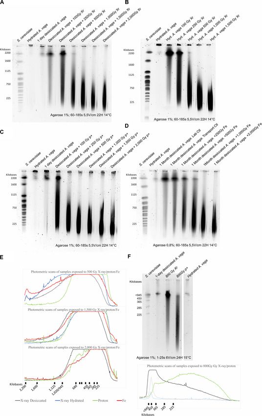
FIGURE 1 | Survival rate of desiccated A. vaga individuals exposed to increasing doses of (A) protons (p+) (B) X-ray (Xr) (C) Fe. Survival rate of desiccated,
irradiated A. vaga individuals was evaluated 48 h post rehydration. For Fe exposure, the dose of 0 Gy corresponded to laboratory control and “0T” corresponded to
unirradiated, transported samples. X-ray and proton exposed samples were desiccated for a period of 24 h. Fe samples, irradiated in Japan, were stored approx.
1 month in desiccated state. A minimum of 3 replicates per dose was achieved. Data were visualized as boxplot (# = average; – = median). Letters indicate the
significant differences between groups (P-value < 0.05).
FIGURE 2 | Comparison between the survival rate (A) and the reproductive capacity (B) of desiccated (red) and hydrated (white) A. vaga individuals exposed to
X-ray. Survival rate was evaluated 2 days post rehydration or post radiation and evaluated on minimum 3 replicates. The reproductive capacity was evaluated with a
minimum of 3 replicates per dose (see section “Materials and Methods”). For each replicate, reproduction was evaluated by direct observation under binocular
30 days after rehydration/radiation. Effective reproduction was validated when at least 2 adults and 1 egg per well were observed. Statistical analysis included
comparison between and within each group of desiccated and hydrated individuals. Statistical analysis of survival data highlights the significant effect of
hydrated/desiccated status. The reproductive capacity was characterized by independent effect of doses and hydrated/desiccated status. Data were visualized as
boxplot (# = average; – = median). Letters or “*” indicate the significant differences between groups (P-value < 0.05).
desiccated animals (Figure 2A group a). No sign of abnormal For proton radiation, no significant decrease of fertility was
behavior was reported 2 days post irradiation. reported up to 250 Gy (Figure 3A group a). However, the fertility
started to decrease significantly at 500 Gy where a big variability
between replicates was observed (Figure 3A group b). Above
Comparing the Reproductive Capacity of 500 Gy, most A. vaga individuals were sterilized by proton 4 MeV
A. vaga Individuals Exposed to X-Ray, radiation (Figure 3A group c).
Proton and Fe Sources For X-ray radiation on desiccated samples, no significant
The reproductive capacity of irradiated A. vaga was evaluated by decrease of fertility was reported at doses ranging from 0 to
checking their capacity to lay eggs and to restart a new population 750 Gy. However, a significant drop was reported at 1,000 Gy
of clonal A. vaga within a month post rehydration and isolation. where a higher variability between replicates was observed. Then,
Frontiers in Microbiology | www.frontiersin.org 6 July 2020 | Volume 11 | Article 1792Hespeels et al. Resistance A. vaga Individuals Exposed to IR
FIGURE 3 | Reproductive capacity of desiccated A. vaga individuals exposed to (A) protons (P +) (B) X-ray (Xr) (C) Fe. The reproductive capacity was calculated
with a minimum of 3 replicates per dose. For each replicate, 60 individuals were randomly isolated and individually placed in multi-well plates. Reproduction was
evaluated by direct observation under binocular 30 days after rehydration/radiation. Effective reproduction was validated when at least 2 adults and 1 egg per well
were observed. Data were visualized as boxplot (# = average; – = median). Letters indicate the significant differences between groups (P-value < 0.05).
a significant decrease of fertility was reported from 1,500 Gy, between factors dose and hydrated/desiccated status of samples
and most A. vaga individuals were sterilized at X-ray doses of when looking at the effective reproduction rate data. As
2,000 Gy and higher when desiccated (Figure 3B group c). suggested by SD50 a significant difference between hydrated and
For Fe exposure, no significant difference in reproductive desiccated samples was reported: independently of the dose,
capacity between lab and transport control was reported. the reproduction rate was significantly higher in desiccated
Moreover, no decrease of the fertility capacity was reported up to samples than hydrated specimens. However, no significant
250 Gy of Fe radiation (Figure 3C group a). A significant decrease difference was reported when data collected from hydrated
of fertility was reported at 500 Gy (Figure 3C group b) and a and desiccated samples were compared for each tested dose
complete sterilization of samples was reported at 1,000 Gy and (Figure 2B). At 1,000 Gy the fertility rate of desiccated
higher (Figure 3C group c). samples was characterized by a high variability (55.5 ± 40.9%)
Based on collected data, it was possible to evaluate the in comparison with hydrated samples (30.4 ± 7.5%). 1%
minimal dose required to sterilize 50% of the irradiated A. vaga (SD ± 1%) of the exposed hydrated A. vaga were able to
population. The median dose to sterilize a population [Sterilizing restart a population after exposure to 1,500 Gy X-ray in
Dose 50 (SD50 )] were 1,035 ± 20 Gy, 453 ± 23 Gy and contrast to desiccated samples where 5% (SD ± 8%) of
460 ± 1 Gy for X-ray, proton and iron radiation, respectively (see animals were still able to produce offspring after radiation.
Supplementary Figure S1). These differences were not statistically significant (Figure 2B).
The failure of irradiated A. vaga individuals to restart a new Nevertheless, a significant increase of animals unable to lay
population was not automatically linked with the non-ability eggs or with premature deaths were reported in hydrated
of animals to produce eggs. Up to 2,500 Gy we noticed an samples exposed to doses equal or higher to 1,000 Gy in
increase of animals laying eggs that were unable to perform comparison with desiccated, irradiated samples (Supplementary
a complete embryonic development independently of radiation Figure S3b group c and d).
type (see Supplementary Figure S2). Multiple cellular divisions
were reported in these sterile eggs that failed to reach a complete
embryonic development (binocular observation of DAPI stained
Loss of Genomic Integrity in Desiccated
eggs, B. Hespeels and M. Terwagne, personal communication). and Irradiated A. vaga Individuals
Higher doses were characterized by an increased proportion Exposed to Increasing Dose of Radiation
of animals unable to lay eggs or dying within a month (see To investigate how different types of radiation can affect the
Supplementary Figure S2). genomic integrity of A. vaga genome, the loss of genomic
The effect of the hydration state (desiccated or hydrated) integrity induced by the accumulation of DNA DSBs was
on irradiated animals was investigated through X-ray using assessed using PFGE on 1,000 A. vaga individuals. As observed
doses ranging from 0 to 1,500 Gy. A higher impact of previously for A. vaga species (Hespeels et al., 2014), the
radiation on hydrated samples versus desiccated samples genomic integrity of hydrated or 1 day desiccated bdelloid
was highlighted by the comparison of both SD50 . Those individuals was characterized by large DNA fragments remaining
were evaluated to 1,125 ± 88 Gy and 889 ± 12 Gy for in the gel plug with a very weak signal around 2200 kbp
samples exposed desiccated or hydrated, respectively (see (Figures 4A–C lanes 2 and 3; Figure 4B lane 2). At a
Supplementary Figure S1b). Statistical analysis (see section dose of 100 Gy, loss of genomic integrity was observed
“Materials and Methods”) reveal the absence of interaction through the visualization of a smear ranging between 225
Frontiers in Microbiology | www.frontiersin.org 7 July 2020 | Volume 11 | Article 1792Hespeels et al. Resistance A. vaga Individuals Exposed to IR
to 2,200 kbp. Size of DNA fragments was negatively linked DISCUSSION
with the increase of the radiation exposure. After a dose of
1,000 Gy and higher, the genomes of A. vaga individuals This research highlights that lethal doses for desiccated A. vaga
were fragmented in DNA pieces mostly ranging between individuals range between 5,000 to 7,500 Gy using low and high
225 and 1000 kbp (Figures 4A–D). Interestingly, for a LET radiation (for X-ray and Proton, respectively, see Figure 1).
similar dose, it was not possible to observe any significant At dose of 5,000 Gy, the survival rate was higher for animals
variation in term of DNA fragmentation between X-ray, exposed to X-ray (82.7% SD ± 11.4) than to proton radiation
Proton and Iron radiation (see photometric scans Figure 4E). (19.1% SD ± 34.1). This is in congruence with the idea that
Moreover, when comparing hydrated and desiccated individuals high LET radiation has a higher relative biological effectiveness
exposed to similar dose of X-ray, no difference was reported than low LET radiation (Goodhead, 1999). A similar trend was
(Figures 4A,B,E). reported for A. vaga samples exposed to high energy Fe particles
For samples exposed to Fe radiation, desiccated and despite the lack of exposure to doses higher than 3,000 Gy (see
unirradiated control presented a slight smear of DNA fragments Figure 1). Noticeably, doses associated with drop of survival
ranging from small to high molecular weight. These controls or fertility rates, in both X-ray and proton radiation exposure,
were exposed to 1 month of dry storage and transported from were characterized by a higher level of heterogeneity between
Belgium to Chiba irradiation location and vice versa. These data replicates. We hypothesize that this variability is linked to the
are congruent with previous results suggesting that DNA DSB uncertainty of the delivered dose. Although this is low (of
are accumulating with time in dried A. vaga even in absence of the order of 1 to 2% of the delivered dose depending on the
irradiation. As a consequence, the number of DNA DSBs may irradiation system used), it can become non-negligible in view of
be overestimated in the dataset of samples exposed to increasing the total doses delivered in this study. Moreover, the dose received
dose of Fe radiation. by animals is a Gaussian centered around the average reported
Second, the genomic integrity of desiccated A. vaga dose (Poisson statistic). This is a characteristic specific to the use
individuals exposed to X-ray and proton was investigated of broad beam radiation system and is a well-known source of
with a resolution window of DNA fragments ranging between variability in radiobiology. Finally, other uncontrolled biological
15 to 242 kbp (Figure 4F). No smear was observed in hydrated factors, such as the average age of the exposed population between
and 1-day desiccated A. vaga individuals. However, both samples each assay, may contribute to the variability observed.
were characterized by a band of size >945 kbp. In contrast to The radio-resistance of A. vaga individuals was differentially
previous results, different patterns between 1-day desiccated, interpreted, in term of magnitude, when focusing on the survival
irradiated A. vaga individuals exposed to 800 Gy of X-ray and rate or on their capacity to produce viable offspring. In our
proton were reported. The X-ray sample was characterized study, for X-ray for example, a factor of approximately 3
by intense signal for DNA fragments >600 kbp followed by a was reported between doses required to sterilize a population
smear of decreasing intensity for DNA fragments 600 kbp fragments and a smear of higher intensity and 2,000 Gy X-ray while it is between 5,000 and 7,500 Gy
than X-ray < 600 kbp. Similar patterns, highlighting progressive to kill the entire population). At minimal doses required to
loss of genomic integrity, were reported when comparing sterilize A. vaga individuals, no decrease of survival rate is
1-day desiccated samples irradiated at 500 and 1,000 Gy (see observed in comparison with controls (see Figures 1, 3).
Supplementary Figure S4). Animals were sterilized by doses of 750, 2,000 and 1,000 Gy
for Protons, X-ray and Fe radiation, respectively (Figure 3).
The median sterilizing doses (SD50 ) for the A. vaga clone were
Partial Restoration of Genomic Integrity 1,035 ± 20 Gy, 453 ± 23 Gy and 461 ± 1 Gy for X-ray,
After Exposure to Radiation protons and iron, respectively (see Supplementary Figure S1).
The potential repair of DNA fragmentation induced by 800 Gy Due to the lack of intermediary dose for Fe particles between
of X-ray and proton radiation in the somatic cells of (re- 500 and 1,000 Gy, no significant difference was reported between
)hydrated animals were assessed through a kinetic from 0 to Protons and Fe. Nevertheless, data confirmed a clear difference
1-week post exposure (Figures 5A–C). Following the rehydration between low and high LET radiation at the reproductive
or irradiation arrest (for hydrated animals), the genomic level. A 2.3 factor was reported when comparing SD50 of
integrity was characterized by a smear of small DNA fragments X-ray versus protons and Fe particles. Indeed, it would have
(Hespeels et al. Resistance A. vaga Individuals Exposed to IR FIGURE 4 | Loss of genomic integrity in A. vaga individuals exposed to increasing doses of X-ray (A,B), Protons 4 MeV (C) and Fe 1 GeV (D); Comparative analysis of photometric scans from samples exposed to 500; 1,500 and 2,000 Gy (E); Comparative analysis of 1-day desiccated A. vaga individuals exposed to 800 Gy of X-ray and protons 4 MeV using short fragments run parameters (15–291 kbp) (F). For X-ray exposure, rotifers were irradiated after 1 day of desiccation (A) or irradiated in hydrated state (B). Rotifers were desiccated 1 day before exposure to Protons 4 MeV (C). However, samples exposed to Fe were desiccated 1 month (D). The first lane on the pulsed-field gel electrophoresis corresponds to the karyotype of Saccharomyces cerevisiae. Second lanes correspond to the control (1,000 hydrated A.vaga individuals). Third lanes of (A,C) correspond to DNA fragmentation induced by 1-day desiccation on 1,000 individuals. Other lanes correspond to 1,000 desiccated (A,C,D) or hydrated individuals (B) after exposition to 100, 250, 500, 1,000, 1,500 and 2,000 Gy (in exception of (B) where maximum dose was 1.500 Gy). For the comparative analysis focusing on short DNA fragments dose of 800 Gy of respectively X-ray and protons was used (Lane 3 and 4). Hydrated control was in lane 5. 1-day desiccated A. vaga individuals was at lane 2. The run parameters are documented under each gel. Photometric scans were generated using ImageJ (see “Materials and Methods” section). Top flat lines from photometric scans correspond to saturated levels. Frontiers in Microbiology | www.frontiersin.org 9 July 2020 | Volume 11 | Article 1792
Hespeels et al.                                                                                                             Resistance A. vaga Individuals Exposed to IR

  FIGURE 5 | Repair kinetic of A. vaga individuals on hydrated or desiccated samples with exposure to 800 Gy of X-ray [(A) 1-day desiccated samples (B) hydrated
  animals] and protons [(C) 1-day desiccated samples]. The first lane on the pulsed-field gel electrophoresis correspond to the karyotype of Saccharomyces
  cerevisiae. Second lanes correspond to the control (1,000 hydrated individuals). Third and fourth lanes correspond, respectively, to hydrated or desiccated bdelloids
  not submitted and submitted to 800 Gy radiation. Other lanes correspond to 1,000 desiccated individuals after 2, 30, 8, 24, 48, and 168 h of rehydration/post
  exposure. The run parameters are documented under each gel.

gamma radiation in both hydrated (LD50/48 h 5,000 ± 1,900 Gy)                           through survival or fecundity rate assays. The survival rate
and anhydrobiotic (LD50/48 h 4,400 ± 500 Gy) states, but an                             in those metazoans is related to non-dividing somatic cells,
irradiation dose >1,000 Gy makes them sterile (Horikawa et al.,                         while fertility data is linked to germinal cells that will ensure
2006). Similarly, the tardigrade Hypsibius dujardini exposed to                         cellular multiplication during embryonic development and as
gamma radiation presented a LD50/48 h for survival estimated at                         consequence are more sensitive to damages induced by radiation
∼ 4,200 Gy, and doses higher than 100 Gy reduced drastically                            (Pagani et al., 1993; Beltran-Pardo et al., 2015). Nevertheless,
production and hatchability of laid eggs (Beltran-Pardo et al.,                         when compared to other metazoans, the tested bdelloid rotifer
2015). These observations reported for tardigrades and bdelloids,                       and tardigrade species show a very high resistance to ionizing
both eutelic, may be attributed to the nature of the cells evaluated                    radiation (for examples see Krisko et al., 2012).

Frontiers in Microbiology | www.frontiersin.org                                    10                                                July 2020 | Volume 11 | Article 1792
Hespeels et al.                                                                                           Resistance A. vaga Individuals Exposed to IR

   For future comparative analysis between radiation sensitive             occur also in germline cells, remain to be investigated at genomic
and radioresistant organisms, it may be more relevant to                   scale on offspring originated from irradiated individuals. The
focus on fertility rate. Moreover, in term of evolution, there             genome of rotifer A. vaga was previously reported to carry
is no advantage to survive to extreme dose of radiation if                 an enriched complement of DNA repair genes compared to
no offspring is produced. As expected, the exposure to high                other invertebrates models (Flot et al., 2013; Hecox-Lea and
doses of ionizing radiation was visualized through PFGE as                 Mark Welch, 2018). Genes of both homology-directed and
an increased level of DNA fragmentation in function of the                 non-homologous end joining (NHEJ) repair pathways are
dose (Figure 4). An attempt was made here to discriminate                  present in numerous copies, some of which likely acquired
DNA fragmentation induced by low and high LET, as well                     by horizontal transfers (Hecox-Lea and Mark Welch, 2018).
as the contribution of indirect damages by exposing hydrated               Functional genetics are, however, required to determine the exact
A. vaga individuals to X-ray. No significant difference was                nature of their DNA repair process.
reported between radiation source for comparable dose when                    It is well known that ionizing radiation damages on biological
focusing on fragment sizes ranging between 225 and 1,600 kbp.              samples can be generated by two paths of action. The first
Similarly, it was not possible to detect significant differences in        one, called “direct damage” literally means either the incident
DNA fragmentation between samples exposed in hydrated or                   radiation itself or secondary particles ionize the target and
desiccated state (Figure 4). Nevertheless, absence of differences          generate breaks in the molecules. The second one, called “indirect
may be due to the lack of resolution inherent to PFGE                      action” refers to the generation of effectors such as reactive
focusing on high molecular weight DNA fragments. The                       oxygen species (ROS), most frequently generated through water
resolution range of 225 kbp to 1,600 kbp is hypothesized                   radiolysis, due to the incident radiation that, in turn, will generate
to miss clustered damages induced by high LET radiation                    the damages to cell compounds (Dartnell, 2011). In this line of
that may result in short DNA fragments. A comparative                      thought, the contribution of direct and indirect damages remains
analysis was performed in order to check the genomic integrity             to be investigated at high doses of radiation. The capacity of
pattern of fragments ranging from 15 to 240 kbp. Using                     bdelloid rotifers to be exposed to ionizing radiation in hydrated
this resolution window, it was possible to observe different               and desiccated state constitutes an opportunity to evaluate
DNA migration patterns between X-ray and proton irradiated                 biological effectiveness played by direct and indirect damages.
A. vaga individuals (see Figure 4F). The increased smear of                Here, the impact of X-ray on desiccated and hydrated A. vaga
short size DNA fragments reported after proton exposure, in                was investigated. Within a radiation range of 0 to 1,500 Gy,
comparison to X-ray irradiated samples, may be attributed to               no significant effects on the survival rate of irradiated A. vaga
the clustered damages generated by proton exposure resulting               were reported 48 h post-exposition and eventual rehydration (see
in the higher relative biological effectiveness of proton radiation        Figure 2A). Nevertheless, SD50 of hydrated individuals appeared
vs X-rays. However, these data need to be validated by a                   to be lower than the one calculated for desiccated individuals.
systematic approach testing the impact of different particles,             These data may suggest an increased level of damages due
with increasing LET and other resolution range. Indeed, fine               to the presence of liquid water (see Figure 2B). However, no
quantification of short DNA fragments induced by radiation                 significant difference was found when compared fertility rate of
is still challenging (Höglund et al., 2000; Alloni et al., 2013;           both conditions. In contrast, a higher proportion of dead animals,
Barbieri et al., 2019).                                                    unable to produce eggs was reported 30 days post exposure
   The loss of genomic integrity induced by low or high doses              among A. vaga individuals exposed in hydrated state starting
of radiation do not prevent the recovery process of A. vaga,               1,000 Gy (see Supplementary Figure S3b, group “d”). This latest
neither in hydrated nor in desiccated state (Figure 4). Despite            result may be interpreted as a harmful impact of X-ray on
the dramatic loss of genomic integrity, A. vaga individuals                hydrated animals that appeared to die prematurely in comparison
were able to repair, at least partially, DNA damage induced                with desiccated and irradiated samples. Indeed, the survival rate
by 800 Gy of X-ray or proton radiation in the somatic cells                was only recorded 2 days post irradiation or rehydration and
of (re-)hydrated animals (Figure 5). Indeed the 1,000 A. vaga              provides, as consequence, an evaluation of it at a short term.
individuals of the PFGE analysis contain only few germline                 Investigation at different time points should be implemented
cells per individual (900). DNA repair kinetics suggest that most                  somatic cells of desiccated bdelloid individuals are less affected
DNA damage induced by radiation are repaired within 24 h                   by radiation in comparison with hydrated ones. According to
independently of the radiation source or on the reproductive               the literature, 60 to 70% of damages induced in vitro cells
capacity (see Figures 2, 3, 5). At first glance, these results             by X-ray exposure originate from indirect damages (Komatsu,
suggest that the DNA repair kinetic taking place in somatic                2019). However, the comparison of SD50 from animals exposed
cells of irradiated A. vaga individuals appeared to be insensitive         hydrated (SD50 = 889 ± 12 Gy) or desiccated (1,125 ± 88 Gy)
to the complexity of damage induced by low vs. high LET                    suggested a contribution of only 21% of indirect damages
radiation exposures. The exact mechanism of DNA repair                     on A. vaga fecundity rate (see Supplementary Figure S1b).
and the potential divergence in mechanism taking place in                  The radioresistance of A. vaga was previously attributed to
somatic or germline cells remain mostly unknown. Moreover,                 the presence of an unusually effective system of anti-oxidant
consequences of such massive DNA fragmentation, expected to                protection, protecting also the DNA repair proteins required to

Frontiers in Microbiology | www.frontiersin.org                       11                                          July 2020 | Volume 11 | Article 1792
Hespeels et al.                                                                                                   Resistance A. vaga Individuals Exposed to IR

repair the fragmented genome (Krisko et al., 2012). The reduced             FUNDING
contribution of indirect damage observed here in hydrated,
exposed samples may be linked to the efficiency of the antioxidant          The authors were supported by the European Space Agency
protection system present in A. vaga. Complementary studies                 (ESA) and the Belgian Federal Science Policy Office (BELSPO)
must be addressed to document the biological response of                    in the framework of the PRODEX Programme. Fe radiation was
living organisms to low and high LET radiation as well as the               part of STARLIFE astrobiology inter-comparison experiments at
contribution of direct and indirect damage using desiccated and             HIMAC (led by RM). STARLIFE was supported by the MEXT
hydrated individuals.                                                       Grant-in-Aid for Scientific Research on Innovative Areas “Living
                                                                            in Space” (Grant Numbers: 15H05935 and 15K21745). RM was
                                                                            supported by the DLR grant FuE-Projekt “ISS LIFE” (Programm
CONCLUSION                                                                  RF-FuW, TP 475). SP was funded by the Walloon Region
                                                                            (PROTHERWAL, grant n◦ 7289).
In conclusion, the robustness of bdelloid rotifers against multiple
stresses coupled with their desiccation tolerance at any stage
of their life cycle prone them to the short list of animals                 ACKNOWLEDGMENTS
that can be used as model systems to study how life can
be adapted and could evolve in extra-terrestrial environments               R. Coos, R. Tonneau, and T. Tabarrant are strongly acknowledged
(Jönsson et al., 2008; Vukich et al., 2012; Rabbow et al., 2015).           for their assistance at any time during X-rays and proton
As a reference, this study focused on the bdelloid species                  exposure sessions. The authors also thank the European
A. vaga. It remains unknown if A. vaga, isolated 40 years                   Space Agency (ESA) and the Belgian Federal Science Policy
ago from the wild and continuously maintained hydrated in                   Office (BELSPO) for their support in the framework of
laboratory condition, may reflect the overall limit of bdelloid             the PRODEX Program.
radioresistance. Moreover, it is not excluded that this species
presents a lower radioresistant phenotype in comparison with
species living in extreme environments and Mars analog sites like           SUPPLEMENTARY MATERIAL
Antarctic or Atacama Desert (Robeson et al., 2009; Iakovenko
et al., 2015). Further studies are ongoing to decipher the impact of        The Supplementary Material for this article can be found
the environment of bdelloid species to their desiccation/radiation          online at: https://www.frontiersin.org/articles/10.3389/fmicb.
tolerance. Finally, the exposure of biological organisms in                 2020.01792/full#supplementary-material
laboratory remains a simple view of the complex radiation                   FIGURE S1 | Evaluation of Sterilizing Dose 50 (SD50 , Gy) for A. vaga individuals
flux found in space environment (Moeller et al., 2017) and                  exposed to X-ray (a,b), protons (c) and Fe (d), overview (e). Each dose was
experiments ongoing on ISS may reveal more insights into                    tested in min 3 replicates. For each replicate, 60 individuals were randomly
the resistance to cosmic radiation and space environment.                   isolated and individually placed in multiwell plates. Reproduction was evaluated by
                                                                            direct observation under binocular 30 days after irradiation and rehydration. All
Nevertheless, the combination of different type of low and
                                                                            curve fittings were performed with the OriginLab R software (MA, United States)
high LET radiations, exposure to UV radiation, variation of                 (see section “Materials and Methods”). Two SD50 for desiccated A. vaga species
temperature may significantly affect the biological effectiveness           irradiated with X-ray were calculated. First SD50 included all data from 0 to
of unprotected samples ongoing lithopanspermia or “living” on               7,500 Gy X-ray. The second SD50 was calculated by limiting the dose range from
martian-like atmosphere.                                                    0 to 1,500 Gy X-ray. This last value was used for comparison with SD50 of
                                                                            hydrated specimens exposed to X-ray similarly calculated with dose
                                                                            from 0 to 1,500 Gy.

                                                                            FIGURE S2 | Production of sterile eggs or premature dead in desiccated A. vaga
DATA AVAILABILITY STATEMENT                                                 individuals exposed to (a) protons (P +) (b) X-ray (Xr) c. Fe. For each replicate, 60
                                                                            individuals were randomly isolated and individually placed in multi-well plates.
All datasets generated for this study are included in the                   Reproduction was evaluated by direct observation under binocular 30 days after
article/Supplementary Material.                                             irradiation and rehydration. Samples were characterized as “Sterile egg
                                                                            production” when one A. vaga individual and minimum one sterile egg was
                                                                            reported. If animals died within a month or were not able to lay eggs, it was
                                                                            annotated as “Dead or no egg production.” When the number of survivors was
AUTHOR CONTRIBUTIONS                                                        below 60 individuals, the number of isolated individuals lacking to arrive at this
                                                                            total was automatically associated with the category “Dead or no egg production.”
BH, LB, CB, and VB maintained the rotifer cultures. BH and LB               This correction avoids the overweighting of certain values following the drastic
performed all the experiments with help of CB and VB. Exposures             reduction of survivors at dose equal or higher than 5,000 Gy (X-ray and proton
to radiation were performed by BH, VB, CB, and LB under the                 only). Data were visualized as boxplot (# = average; – = median). Letters indicate
                                                                            the significant differences between groups (P-value < 0.05; see section
supervision of A-CH, SL, RM, and AF. The results were analyzed              “Materials and Methods”).
by BH, VC, BR, and SP. The manuscript was written by BH.
The manuscript was revised by KV, A-CH, SL, VC, RM, AF, and                 FIGURE S3 | Production of sterile eggs (a) or premature dead (b) in A. vaga
                                                                            individuals exposed to X-ray in desiccated (Red) or hydrated (white) state. Each
SP. The project was designed and supervised by BH and KV.                   dose was tested in min 3 replicates. For each replicate, 60 individuals were
The acquisition of funding was done by BH and KV. All authors               randomly isolated and individually placed in multiwell plates. Reproduction was
contributed to the article and approved the submitted version.              evaluated by direct observation under binocular 30 days after irradiation and

Frontiers in Microbiology | www.frontiersin.org                        12                                                  July 2020 | Volume 11 | Article 1792
Hespeels et al. Resistance A. vaga Individuals Exposed to IR
rehydration. Sample were characterized as “Sterile eggs production” when one fragments run parameters (15–291 kbp). Lanes 1 and 5 on the pulsed-field gel
A. vaga individuals and min one sterile eggs was reported. If animals died within a electrophoresis corresponds to the karyotype of Saccharomyces cerevisiae.
month or was not able to lay egg, it was annotated as “Dead or no eggs Lanes 2 and 6 correspond to the control (1,000 hydrated A. vaga individuals).
production.” Statistical analysis of the sterile eggs production highlighted only an Lanes 3 and 7 correspond to 1-day desiccated A. vaga individuals exposed,
effect of dose. Related to premature dead data, an interaction effect between respectively, to 500 and 1,000 Gy of X-ray radiation. Lanes 4 and 8 correspond to
dose factor and hydrated/desiccated status was highlighted. Data were visualized 1-day desiccated A. vaga individuals exposed, respectively, to 500 and 1,000 Gy
as boxplot (# = average; – = median). Letters indicate the significant differences of protons 4 MeV radiation. The run parameters are documented under the gel.
between groups (P-value < 0.05; see section “Materials and Methods”). Samples used for Supplementary Figure 4, were irradiated and prepared
independently from samples used for Figure 4.
FIGURE S4 | Genomic integrity of 1-day desiccated A. vaga individuals exposed
to 500 and 1,000 Gy with X-rays and 4 MeV Protons, respectively, using short DATA S1 | Survival and fertility data.
REFERENCES Horikawa, D. D., Sakashita, T., Katagiri, C., Watanabe, M., Kikawada, T., Nakahara,
Y., et al. (2006). Radiation tolerance in the tardigrade Milnesium tardigradum.
Alloni, D., Campa, A., Friedland, W., Mariotti, L., and Ottolenghi, A. (2013). Int. J. Radiat. Biol. 82, 843–848. doi: 10.1080/09553000600972956
Integration of Monte Carlo Simulations with PFGE Experimental Data Yields Hothorn, T., Bretz, F., and Westfall, P. (2008). Simultaneous inference in
Constant RBE of 2.3 for DNA Double-Strand Break Induction by Nitrogen Ions general parametric models. Biom. J. 50, 346–363. doi: 10.1002/bimj.20081
between 125 and 225 keV/µm LET. Radiat. Res. 179, 690–697. doi: 10.1667/ 0425
r3043.1 Iakovenko, N. S., Smykla, J., Convey, P., Kasparova, E., Kozeretska, I. A.,
Barbieri, S., Babini, G., Morini, J., Friedland, W., Buonanno, M., Grilj, V., et al. Trokhymets, V., et al. (2015). Antarctic bdelloid rotifers: diversity, endemism
(2019). Predicting DNA damage foci and their experimental readout with 2D and evolution. Hydrobiologia 761, 5–43. doi: 10.1007/s10750-015-2463-2
microscopy: a unified approach applied to photon and neutron exposures. Sci. Joiner, M. C., and van der Kogel, A. J. (2018). Basic Clinical Radiobiology. Available
Rep. 9:14019. doi: 10.1038/s41598-019-50408-5 online at: https://books.google.be/books?id=pnxqDwAAQBAJ (accessed May
Beltran-Pardo, E., Jonsson, K. I., Harms-Ringdahl, M., Haghdoost, S., and Wojcik, 27, 2020).
A. (2015). Tolerance to gamma radiation in the tardigrade Hypsibius dujardini Jönsson, K. I., Rabbow, E., Schill, R. O., Harms-Ringdahl, M., and Rettberg, P.
from embryo to adult correlate inversely with cellular proliferation. PLoS One (2008). Tardigrades survive exposure to space in low Earth orbit. Curr. Biol.
10:e0133658. doi: 10.1371/journal.pone.0133658 18, 729–731. doi: 10.1016/j.cub.2008.06.048
Cucinotta, F. A., and Durante, M. (2006). Cancer risk from exposure to galactic Jönsson, K. I., and Wojcik, A. (2017). Tolerance to X-rays and heavy ions (Fe, He)
cosmic rays: implications for space exploration by human beings. Lancet Oncol. in the tardigrade Richtersius coronifer and the bdelloid rotifer Mniobia russeola.
7, 431–435. doi: 10.1016/S1470-2045(06)70695-7 Astrobiology 17, 163–167. doi: 10.1089/ast.2015.1462
Dartnell, L. R. (2011). Ionizing radiation and life. Astrobiology 11, 551–582. doi: Kohn, H. I., and Kallman, R. F. (1957). The influence of strain on acute X-Ray
10.1089/ast.2010.0528 lethality in the mouse: II. Recovery rate studies. Radiat. Res. 6, 329–338. doi:
Ferrari, F., and Szuszkiewicz, E. (2009). Cosmic rays: a review for astrobiologists. 10.2307/3570613
Astrobiology 9, 413–436. doi: 10.1089/ast.2007.0205 Komatsu, K. (2019). Contemporary Radiobiology. Available online at: https://books.
Fischer, C., Ahlrichs, W. H., Buma, A. G. J., van de Poll, W. H., and Bininda- google.be/books?id=CRu7DwAAQBAJ (accessed May 27, 2020).
Emonds, O. R. P. (2013). How does the “ancient” asexual Philodina roseola Krisko, A., Leroy, M., Radman, M., and Meselson, M. (2012). Extreme anti-oxidant
(Rotifera: Bdelloidea) handle potential UVB-induced mutations? J. Exp. Biol. protection against ionizing radiation in bdelloid rotifers. Proc. Natl. Acad. Sci.
216(Pt 16), 3090–3095. doi: 10.1242/jeb.087064 U.S.A. 109, 2354–2357. doi: 10.1073/pnas.1119762109
Flot, J.-F., Hespeels, B., Li, X., Noel, B., Arkhipova, I., Danchin, E. G. J., et al. (2013). Latta, L. C., Tucker, K. N., and Haney, R. A. (2019). The relationship between
Genomic evidence for ameiotic evolution in the bdelloid rotifer Adineta vaga. oxidative stress, reproduction, and survival in a bdelloid rotifer. BMC Ecol. 19:7.
Nature 500, 453–457. doi: 10.1038/nature12326 doi: 10.1186/s12898-019-0223-2
Fox, J., and Weisberg, S. (2019). An {R} Companion to Applied Regression (Third). Lehnert, S. (2007). Biomolecular Action of Ionizing Radiation. Boca Raton, FL: CRC
Available online at: https://socialsciences.mcmaster.ca/jfox/Books/Companion/ Press.
(accessed June 1, 2020). Leuko, S., and Rettberg, P. (2017). The effects of HZE particles, γ and X-ray
Gladyshev, E., and Meselson, M. (2008). Extreme resistance of bdelloid rotifers to radiation on the survival and genetic integrity of Halobacterium salinarum
ionizing radiation. Proc. Natl. Acad. Sci. U.S.A. 105, 5139–5144. doi: 10.1073/ NRC-1, Halococcus hamelinensis, and Halococcus morrhuae. Astrobiology 17,
pnas.0800966105 110–117. doi: 10.1089/ast.2015.1458
Gladyshev, E. A., and Arkhipova, I. R. (2010). Genome structure of bdelloid Loucas, B. D., and Cornforth, M. N. (2013). The LET dependence of unrepaired
rotifers: Shaped by asexuality or desiccation? J. Heredity 101(Suppl. 1), 85–93. chromosome damage in human cells: A break too far? Radiat. Res. 179, 393–405.
doi: 10.1093/jhered/esq008 doi: 10.1667/RR3159.2
Goodhead, D. T. (1999). Mechanislns for the Biological Effectiveness of Mattimore, V., and Battista, J. R. (1996). Radioresistance of Deinococcus
High-LET Radiations. J. Radiat. Res. 40(Suppl.), 1–13. doi: 10.1269/jrr. radiodurans: functions necessary to survive ionizing radiation are also necessary
40.s1 to survive prolonged desiccation. J. Bacteriol. 178, 633–637. doi: 10.1128/jb.178.
Hagiwara, Y., Oike, T., Niimi, A., Yamauchi, M., Sato, H., Limsirichaikul, S., et al. 3.633-637.1996
(2019). Clustered DNA double-strand break formation and the repair pathway Maynard Smith, J. (1986). Contemplating life without sex. Nature 324, 300–301.
following heavy-ion irradiation. J. Radiat. Res. 60, 69–79. doi: 10.1093/jrr/ doi: 10.1038/324300a0
rry096 Melone, G., and Fontaneto, D. (2005). Trophi structure in bdelloid rotifers.
Hecox-Lea, B. J., and Mark Welch, D. B. (2018). Evolutionary diversity and novelty Hydrobiologia 546, 197–202. doi: 10.1007/s10750-005-4197-z
of DNA repair genes in asexual Bdelloid rotifers. BMC Evol. Biol. 18:177. doi: Moeller, R., Raguse, M., Leuko, S., Berger, T., Hellweg, C. E., Fujimori, A., et al.
10.1186/s12862-018-1288-9 (2017). STARLIFE -An international campaign to study the role of galactic
Hespeels, B., Knapen, M., Hanot-Mambres, D., Heuskin, A.-C., Pineux, F., Lucas, cosmic radiation in astrobiological model systems. Astrobiology 17, 101–109.
S., et al. (2014). Gateway to genetic exchange? DNA double-strand breaks in doi: 10.1089/ast.2016.1571
the bdelloid rotifer Adineta vaga submitted to desiccation. J. Evol. Biol. 27, Mole, R. H. (1984). The LD50 for uniform low LET irradiation of man. Br. J. Radiol.
1334–1345. doi: 10.1111/jeb.12326 57, 355–369. doi: 10.1259/0007-1285-57-677-355
Höglund, E., Blomquist, E., Carlsson, J., and Stenerlöw, B. (2000). DNA damage Pagani, M., Ricci, C., and Redi, C. A. (1993). Oogenesis in Macrotrachela
induced by radiation of different linear energy transfer: Initial fragmentation. quadricornifera (Rotifera, Bdelloidea). Hydrobiologia 255-256, 225–230. doi:
Int. J. Radiat. Biol. 76, 539–547. doi: 10.1080/095530000138556 10.1007/BF00025843
Frontiers in Microbiology | www.frontiersin.org 13 July 2020 | Volume 11 | Article 1792You can also read