Complement associated microvascular injury and thrombosis in the pathogenesis of severe COVID-19 infection: A report of five cases
←
→
Page content transcription
If your browser does not render page correctly, please read the page content below
Complement associated microvascular injury
and thrombosis in the pathogenesis of severe
COVID-19 infection: A report of five cases

CYNTHIA MAGRO, J. JUSTIN MULVEY, DAVID BERLIN, GERARD NUOVO, STEVEN SALVATORE,
JOANNA HARP, AMELIA BAXTER-STOLTZFUS, and JEFFREY LAURENCE
NEW YORK, NEW YORK; POWELL, OHIO; AND NEW YORK, NEW YORKX X

                                    Acute respiratory failure and a systemic coagulopathy are critical aspects of the
                                    morbidity and mortality characterizing infection with severe acute respiratory dis-
                                    tress syndrome-associated coronavirus-2, the etiologic agent of Coronavirus dis-
                                    ease 2019 (COVID-19). We examined skin and lung tissues from 5 patients with
                                    severe COVID-19 characterized by respiratory failure (n= 5) and purpuric skin rash
                                    (n = 3). COVID-19 pneumonitis was predominantly a pauci-inflammatory septal
                                    capillary injury with significant septal capillary mural and luminal fibrin deposition
                                    and permeation of the interalveolar septa by neutrophils. No viral cytopathic
                                    changes were observed and the diffuse alveolar damage (DAD) with hyaline mem-
                                    branes, inflammation, and type II pneumocyte hyperplasia, hallmarks of classic
                                    acute respiratory distress syndrome, were not prominent. These pulmonary findings
                                    were accompanied by significant deposits of terminal complement components
                                    C5b-9 (membrane attack complex), C4d, and mannose binding lectin (MBL)-asso-
                                    ciated serine protease (MASP)2, in the microvasculature, consistent with sustained,
                                    systemic activation of the complement pathways. The purpuric skin lesions similarly
                                    showed a pauci-inflammatory thrombogenic vasculopathy, with deposition of C5b-
                                    9 and C4d in both grossly involved and normally-appearing skin. In addition, there
                                    was co-localization of COVID-19 spike glycoproteins with C4d and C5b-9 in the
                                    interalveolar septa and the cutaneous microvasculature of 2 cases examined. In
                                    conclusion, at least a subset of sustained, severe COVID-19 may define a type of
                                    catastrophic microvascular injury syndrome mediated by activation of comple-
                                    ment pathways and an associated procoagulant state. It provides a foundation for
                                    further exploration of the pathophysiologic importance of complement in COVID-
                                    19, and could suggest targets for specific intervention. (Translational Research 2020;
                                    220:1 13)

From the Department of Pathology and Laboratory Medicine, Weill Cornell Medicine, New York, New York; Department of Laboratory Medicine,
Memorial Sloan-Kettering Cancer Center, New York, New York; Department of Medicine, Division of Pulmonary and Critical Care Medicine,
Weill Cornell Medicine, New York, New York; The Ohio State University Comprehensive Cancer Center, Columbus Ohio and Discovery Life Sci-
ences, Powell, Ohio; Department of Dermatology, Weill Cornell Medicine; Department of Medicine, Division of Hematology and Medical Oncol-
ogy, Weill Cornell Medicine, New York, New York.
Submitted for PublicationApril 9, 2020; accepted for publication April 9, 2020.
Reprint requests: Jeffrey Laurence, Weill Cornell Medicine, 1300 York Avenue, New York, NY 10065.   E-mail address: jlaurenc@med.cornell.edu.
1931-5244/$ - see front matter
Ó 2020 Elsevier Inc. All rights reserved.
https://doi.org/10.1016/j.trsl.2020.04.007

                                                                                                                                                1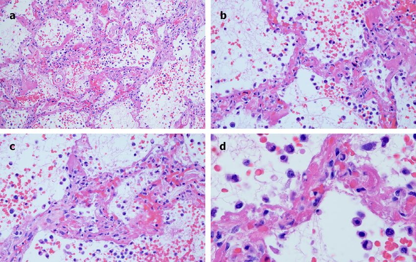
Translational Research
2    Magro et al                                                                                           June 2020

                                                            of acute respiratory distress syndrome (ARDS).7 Pre-
    AT A GLANCE COMMENTARY                                  liminary pathology studies of COVID-19 patients dem-
                                                            onstrated diffuse alveolar damage (DAD) with edema,
    Background                                              hyaline membranes, and inflammation, followed by
    The respiratory distress syndrome accompanying          type II pneumocyte hyperplasia, features characteristic
    a subset of severe Coronavirus disease 2019             of typical ARDS.8,9 But many patients with COVID-
    (COVID-19) may be distinct from classic acute           19-related severe respiratory distress have a delayed
    respiratory distress syndrome. There is relatively      onset of respiratory distress,10 then manifest relatively
    well-preserved lung mechanics despite the sever-        well-preserved lung mechanics, despite the severity of
    ity of hypoxemia, characterized by high respira-        hypoxemia, characterized by high respiratory compli-
    tory compliance and high shunt fraction, and            ance and high shunt fraction, and prolonged require-
    increasing recognition of systemic features of a        ment for mechanical ventilation.10,11 Therefore,
    hypercoaguable state in this disease. Therefore,        significant aspects of the pathology of COVID-19
    the pathology and pathophysiology of COVID-19           might be expected to differ from classic ARDS.
    might differ from that of typical acute respiratory        We examined lung and cutaneous tissues from
    distress syndrome. We sought to define the role of      5 patients with SARS-CoV-2 infection and severe
    complement activation and microvascular throm-          respiratory failure, 3 of whom also had features con-
    bosis in cases of persistent, severe COVID-19.          sistent with a systemic procoagulant state, including
                                                            retiform purpura or livedo racemosa—prominent
    Translational Significance                              dermatologic signs of a generalized microvascular
                                                            thrombotic      disorder—and       markedly     elevated
    A pattern of tissue damage consistent with com-
                                                            d-dimers. Histologic and immunohistochemistry
    plement-mediated microvascular injury was noted
                                                            studies defined a pattern of cutaneous and pulmonary
    in the lung and/or skin of 5 individuals with severe
                                                            pathology involving microvascular injury and throm-
    COVID-19. Our demonstration of the striking
                                                            bosis, consistent with activation of the alternative
    deposition of C5b-9, C4d, and MASP2 in the
                                                            pathway (AP) and lectin pathway (LP) of comple-
    microvasculature of 2 organ systems is consistent
                                                            ment. Co-localization of SARS-CoV-2-specific spike
    with profound and generalized activation of both
    alternative and lectin-based pathways. It provides      glycoproteins with complement components in the
                                                            lung and skin was also documented. Our studies sug-
    a foundation for further exploration of the patho-
                                                            gest that at least a subset of severe COVID-19 infec-
    physiologic importance of complement in
                                                            tion involves a catastrophic, complement-mediated
    COVID-19, and could suggest targets for specific
                                                            thrombotic microvascular injury syndrome with sus-
    intervention.
                                                            tained activation of the AP and LP cascades. Poten-
                                                            tial mechanisms of complement activation, including
                                                            involvement of positive feedback loops with the
INTRODUCTION                                                coagulation system, are discussed in the context of
   The severe acute respiratory distress syndrome-asso-     hypothesis-generating studies providing the founda-
ciated coronavirus-2 (SARS-CoV-2), etiologic agent of       tion for potential therapeutic intervention.
Coronavirus disease 2019 (COVID-19), was initially
identified in Wuhan, Hubei, China in December 2019.1
It was documented to be pandemic by the World
Health Organization in early March 2020,2 and by            METHODS
early April there were over 1.5 million cases world-           Patient population. All 5 patients were selected for
wide, with over 90,000 deaths.3 Organ dysfunction,          pathologic studies based on a respiratory tract sample
particularly progressive respiratory failure and a gener-   positive for SARS-CoV-2 in a reverse transcriptase-
alized coagulopathy, are associated with the highest        polymerase chain reaction assay, as tested by a desig-
mortality.1,4,5                                             nated diagnostic laboratory. They represent the first 2
   It was soon recognized that SARS-CoV-2 is but one        patients succumbing to COVID-19 and undergoing
of a large pool of prepandemic SARS-like bat corona-        autopsy that were available to us, and the first 3
viruses which replicate in primary human airway epi-        infected individuals for whom a dermatologic consult
thelial cells.6 These include the etiologic agents of the   had been requested to evaluate an extensive skin
original SARS-CoV and Middle East respiratory syn-          rash. These 5 cases were assembled over a period
drome (MERS)-CoV, for which mortality is also linked        of 2 weeks. A respiratory pathogen PCR panel was
to severe respiratory failure, with pathologic evidence     used to test for other potential pulmonary viral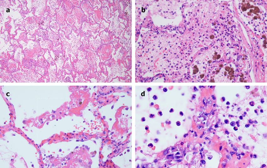
Translational Research
Volume 220                                                                                             Magro et al   3

pathogens, along with standard bacterial and fungal          demonstrated bilateral airspace opacities most promi-
respiratory cultures, and these tests were unreveal-         nent in the peri-hilar distribution. After discussion with
ing. Cases 3 and 4 received hydroxychloroquine               his family, he was placed on comfort measures and
and/or azithromycin, as noted, with dosing as:               died a few hours after presentation. A limited autopsy
hydroxychloroquine, 600 mg every 12 hours for 1              was performed. Grossly, the lungs had a congested and
day then 400 mg every 12 hours for 4 days; azithro-          hemorrhagic appearance. Light microscopic examina-
mycin: 500 mg daily for 5 days.                              tion revealed a severe organizing hemorrhagic pneu-
   Microscopic and immunohistologic studies. Routine         monitis (Fig 1A), including significant fibrin
light microscopy and immunohistochemical (IHC)               deposition within septal capillary lumens and walls
assessment for the deposition of C5b-9 (membrane             accompanied by endothelial cell necrosis, consistent
attack complex, MAC), C3d, and C4d via a diamino-            with a thrombotic necrotizing capillary injury syn-
benzidene (DAB) technique was conducted. Identifica-         drome (Fig 1B). The pattern of septal capillary injury
tion of C5b-9, C3d, or C4d within any epithelial             ranged from a pauci-inflammatory pattern (Fig 1C) to
basement membrane zone, elastic fibers, or the elastic       one characterized by permeation of the interalveolar
lamina of vessels was considered nonspecific staining.       septa by neutrophils amidst the damaged capillaries,
IHC was performed using C3d (Cell Marque, Rocklin            along with intra-alveolar neutrophils (Fig 1D). No viral
CA, 403A-78), C4d (Alpco, Salem NH, BI-RC4d),                cytopathic changes were observed. Hyaline membranes
C5b-9 (Agilent, Santa Clara CA, M077701-5), and              reflective of DAD were not observed. In addition, and
MASP2 (Sigma, St. Louis, MO, HPA029313) antibod-             in contrast to preliminary reports of COVID-19 lung
ies on paraffin embedded sections using a modified           pathology,8,9 type II pneumocyte hyperplasia was not
Leica protocol. Heat mediated antigen retrieval with         appreciated (Fig 1A D). The brunt of the lung injury
Tris-EDTA buffer (pH = 9, epitope retrieval solution 2)      was restricted to septal capillaries, without pneumocyte
was performed for 20 minutes, followed by incubation         involvement.
with each antibody for 15 minutes. Details have been            Extensive C4d deposition localized to the interalveo-
described by one of the authors, including documenta-        lar septal capillaries was then demonstrated (Fig 2A
tion of the involvement of many of these complement          and B). C5b-9 deposition was also seen, showing a
components in pathologic processes linked to comple-         similar pattern of septal capillary localization as for
ment activation in both dermal and lung tissues.12 15        C4d, although reduced in intensity (Fig 2C). Granular
Staining for SARS-CoV-2 spike (catalogue #3525) and          deposition of C3d was also noted; although minimal in
envelope (catalogue #3531) proteins (Prosci, Poway,          comparison to the C5b-9 and C4d deposits, it expressed
CA) was optimized with cells from nasopharyngeal             a similar pattern of septal capillary localization (Fig
preparations obtained from individuals known to be           2D). MASP2 staining was attempted but, due to high
positive or negative for the virus before being              background staining in this case, was deemed techni-
employed in the current studies. It involves use of          cally unsatisfactory.
horseradish peroxidase conjugate (ENZO, catalogue               In order to explore possible generalized complement
#ADI-950-113-0100) and an HRP-conjugated compact             activation in this patient, a sample of clinically-appear-
polymer system, with DAB as chromogen. Hematoxy-             ing normal skin was also found to have significant vas-
lin and eosin counterstain was used and mounted with         cular deposits of C5b-9 within dermal capillaries (not
Leica Micromount.                                            shown).
                                                                Case 2. A 73-year-old male with a history of smok-
                                                             ing, obesity, and prediabetes developed respiratory dis-
                                                             tress and was evaluated in the ED. He was febrile,
RESULTS                                                      tachypneic, severely hypoxemic, and required emer-
   Case 1. A 62-year-old male with a history of coro-        gent endotracheal intubation. He had a serum creati-
nary artery disease, diabetes mellitus, heart failure with   nine of 2.4. Chest x-ray showed bilateral airspace
preserved ejection fraction, prior treatment for hepatitis   opacities. Sedatives and neuromuscular blockade were
C virus infection, and end-stage renal disease on inter-     used to enforce a lung protective low-stretch strategy.
mittent hemodialysis was evaluated in an emergency           His course was complicated by atrial fibrillation, shock,
department (ED), in extremis and obtunded. He had            and progressive renal failure. His INR and PTT were
severe hypoxemia and a blood pressure of 180/100.            within normal limits. On the 4th day of mechanical
His international normalized ratio (INR) was mildly          ventilation the patient developed thrombocytopenia
elevated at 1.2, with a partial thromboplastic time          (platelets 148 £ 109/L) and severe hypercapnia, with a
(PTT) within the normal range (23.0 37.0), and nor-          dramatic increase in the estimated pulmonary dead
mal platelet count (178 £ 109/L). Chest x-ray                space fraction, and expired the following day. A
Translational Research
4 Magro et al June 2020
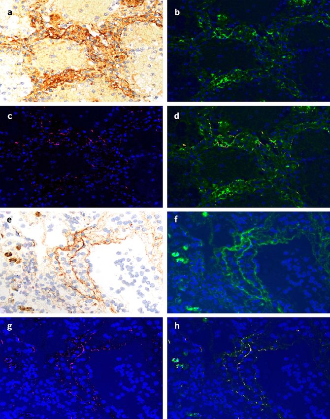
Fig 1. Microscopic features of pulmonary autopsy samples from Case 1. A, Significant fibrin deposition within
the interalveolar septa and alveolar spaces, accompanied by marked hemorrhage and hemosiderin deposition.
(Hematoxylin and eosin stain, 200£). B, Prominent destructive septal capillary injury is apparent, with fibrinoid
necrosis of the capillaries accompanied by evidence of vascular compromise, with hemorrhage and fibrin and
hemosiderin deposition within alveolar spaces. (Hematoxylin and eosin stain, 400£). C, Septal capillary injury
is a pauci-inflammatory response. (Hematoxylin and eosin, 1000£). D, The septal capillary injury includes an
interstitial and intra-alveolar accumulation of neutrophils. (Hematoxylin and eosin, 400£).
Fig 2. Immunohistochemistry analysis of pulmonary autopsy samples from Case 1. A, Extensive C4d deposi-
tion is seen throughout the lung parenchyma, with striking septal capillary localization. (Diaminobenzidene
stain, 200£). B, Higher power magnification documents a clear localization of C4d within septal capillaries.
(Diaminobenzidene, 1000£). C, A similar septal capillary distribution for C5b-9 deposition is observed,
although it is less pronounced than that observed for C4d. (Diaminobenzidene, 1000£). D, A similar septal cap-
illary distribution of C3d staining is observed, although it is also less pronounced than was observed for C4d.
(Diaminobenzidene, 1000£).
Translational Research
Volume 220                                                                                                                      Magro et al   5

limited autopsy demonstrated a pattern of lung injury                       tracheal soft tissues, while C4d was localized to
that mirrored Case 1. Grossly, the lungs had a hemor-                       interalveolar septa in regions of microvascular injury
rhagic and congested appearance. Microscopically, the                       (Fig 4C and D). MASP2 staining demonstrated gran-
lungs showed extensive hemorrhagic pneumonitis. The                         ular and punctate staining localized to the interalveo-
interalveolar septa were congested, with luminal and                        lar septa (Fig 4E).
mural fibrin deposition within septal capillaries. Focal                       Case 3. A 32-year-old male with a medical history of
intra-alveolar collections of neutrophils and monocytes                     obesity-associated sleep apnea and anabolic steroid
were seen. Viral cytopathic changes were not appreci-                       use, currently taking testosterone, presented with a 1
ated. Due to vascular compromise, there was concomi-                        week history of fever and cough. He became progres-
tant striking red cell extravasation found within the                       sively more dyspenic with fevers to 40˚C, ultimately
alveolar spaces, along with intra-alveolar fibrin deposi-                   becoming ventilator dependent from acute respiratory
tion. Unlike Case 1, there was some focal hyaline                           failure. Chest x-ray showed bilateral airspace opacities.
membrane formation and type II pneumocyte hyperpla-                         He had an elevated d-dimer of 1024 ng/ml (normal
sia in areas of hemorrhagic pneumonitis (Fig 3A D).                         range 0 229) on presentation, which peaked at
However, unlike Case 1, this patient had been on venti-                     2090 ng/ml on hospital day 19, and a persistently ele-
lator support for 1 week, which can itself lead to some                     vated INR of 1.6 1.9, but a normal PTT and platelet
DAD, and yet, as in Case 1, the dominant pattern was                        count. Serum complement levels for CH50 (177 CAE
septal capillary injury, not DAD.                                           Units, normal range 60 144), C4 (42.6 mg/dL, normal
   On IHC there was prominent deposition of C5b-9                           range 12 36), and C3 (178 mg/dL, normal range
within the microvasculature of the interalveolar                            90 180) were elevated. Over his continuing 3-plus
septa as well as in larger caliber vessels of the lung                      weeks on ventilator support he completed courses of
parenchyma (Fig 4A and B). Of interest, C5b-9 sep-                          hydroxychloroquine and azithromycin, followed by the
tal deposition was not limited to areas of diseased                         experimental anti-CoV agent remdesivir (5 mg/kg i.v.
lung, but was also seen in normal-appearing lung and                        daily for 10 days).

              Fig 3. Microscopic features of pulmonary autopsy samples from Case 2. A, Extensive hemorrhagic pneumonitis
              was seen, with red cell extravasation and fibrin in alveolar spaces and luminal and mural fibrin deposition within
              septal capillaries. (Hematoxylin and eosin, 200£). B, The septa exhibit a pauci-cellular pattern of capillary
              injury as evidenced by significant fibrin deposition, with thrombi seen in capillaries. There is red cell extravasa-
              tion in the alveolar spaces along with collections of neutrophils and monocytes. (Hematoxylin and eosin,
              400£). C, There is slight widening of the septa by a few inflammatory cells, predominantly neutrophils. There
              is evidence of capillary injury characterized by fibrin deposition in the lumens and walls with red cell extravasa-
              tion within the septa and adjacent alveolar space. Type II pneumocyte hyperplasia and viral cytopathic effects
              are not discernible. (Hematoxylin and eosin, 400£). D, Higher power examination further illuminates capillary
              wall disruption accompanied by fibrin deposition and red cell extravasation, with neutrophils in the septa and
              within the alveolar spaces. (Hematoxylin and eosin, 1000£).
Translational Research
6   Magro et al                                                                                                                  June 2020

              Fig 4. Immunohistochemistry analysis of pulmonary autopsy samples from Case 2. A, There was striking depo-
              sition of C5b-9 within the microvasculature of the interalveolar septa. (Diaminobenzidene, 200£). B, Higher
              power magnification again shows localization of C5b-9 within the septa, including C5b-9 deposits in areas of
              normal appearing lung, suggestive of systemic complement activation. (Diaminobenizdene, 1000£). C, C4d
              deposition was largely localized to the interalveolar septa in areas of microvascular injury. (Diaminobenzidene
              400£). D, A higher power image demonstrates the extensive degree of C4d deposition within the septa. (Diami-
              nobenzidene, 1000£). E, MASP2 staining showed granular and punctate deposits localized to the interalveolar
              septa.

   After only 4 days on ventilator support, retiform pur-                 extensive necrosis of the epidermis and adnexal struc-
pura with extensive surrounding inflammation was                          tures, including the eccrine coil. There was a signifi-
noted on his buttocks (Fig 5A). Skin biopsy showed a                      cant degree of interstitial and perivascular neutrophilia
striking thrombogenic vasculopathy accompanied by                         with prominent leukocytoclasia (Fig 5B). IHC showed

              Fig 5. Clinical, microscopic, and immunhohistochemical analyses of Case 3. A, Striking retiform purpura with
              surrounding inflammation was noted on the buttocks. B, Skin biopsy showed an extensive pattern of pauci-
              inflammatory vascular thrombosis with endothelial cell injury. (Hematoxylin and eosin, 400£).
              C, Prominent deposits of C5b-9 are seen within the microvasculature. (Diaminobenzidene, 400£).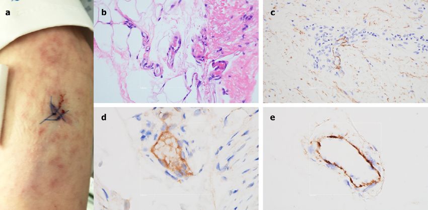
Translational Research
Volume 220                                                                                                                 Magro et al   7

striking and extensive deposition of C5b-9 within the                     with complete infarction of the area supplied by the left
microvasculature (Fig 5C).                                                middle cerebral artery.
   Case 4. A 66-year-old female, with no significant past                    Case 5. A 40-year-old female, with no significant
medical history, was brought to the ED after 9 days of                    past medical history, presented to the ED after 2 weeks
fever, cough, diarrhea, and chest pain. She was hypox-                    of dry cough, fever, myalgias, diarrhea, and progres-
emic, with diffuse bilateral patchy airspace opacities,                   sive dyspnea. She had been diagnosed 1 week earlier,
without effusions, on chest x-ray. She was admitted and                   at an outside hospital, with COVID-19. Echocardio-
treated with hydroxychloroquine and prophylactic anti-                    gram revealed severely reduced left ventricular
coagulation with enoxaparin. Three days later she                         function. Patchy bilateral airspace opacities were noted
became confused, increasingly hypoxemic with rising                       and she was soon intubated for respiratory failure and
serum creatinines, and was intubated. Renal replacement                   shock. D-dimer was elevated at 1187 ng/ml, with a nor-
was initiated. On hospital day 10, thrombocytopenia                       mal platelet count and PTT, but an elevated INR of 1.4.
(platelets 128 £ 109/L) and a markedly elevated d-dimer                   Mildly purpuric reticulated eruptions on her chest, legs
of 7030 ng/ml, but normal INR and PTT, were noted.                        and arms, consistent with livedo racemosa, were noted
The next day dusky purpuric patches appeared on her                       (Fig 7A), and a skin biopsy performed. There was a
palms and soles bilaterally (Fig 6A). A skin biopsy of                    modest perivascular lymphocytic infiltrate in the super-
one lesion showed superficial vascular ectasia and an                     ficial dermis along with deeper seated small thrombi
occlusive arterial thrombus within the deeper reticular                   within rare venules of the deep dermis, in the absence
dermis in the absence of inflammation (Fig 6B). Exten-                    of a clear vasculitis (Fig 7B). Significant vascular
sive vascular deposits of C5b-9 (Fig 6C), C3d, and C4d                    deposits of C5b-9 (Fig 7C) and C4d (Fig 7D) were
(Fig 6D) were observed throughout the dermis, with                        observed. As for Case 4, a biopsy of normal deltoid
marked deposition in an occluded artery. A biopsy of                      skin showed microvascular deposits of C5b-9 through-
normal-appearing deltoid skin also showed conspicuous                     out the dermis (Fig 7E).
microvascular deposits of C5b-9 (Fig 6E). Sedative infu-                     Co-localization of SARS-CoV-2 envelope proteins with
sions were discontinued that day, unmasking a comatose                    complement components in dermal and pulmonary
state. Computerized tomographic imaging of the head                       microvessels of 2 COVID-19 patients. SARS-CoV-2
                                                                                                                     spike
revealed multifocal supra- and infratentorial infarctions,                and envelope proteins strongly localized to the

              Fig 6. Clinical, microscopic, and immunhohistochemical analyses of Case 4. A, Prominent livedo rashes on the
              palmar and plantar aspects of the hands and feet, respectively, were noted. B, Skin biopsy demonstrated an
              occlusive arterial thrombus within deeper dermis. (Hematoxylin and eosin, 200£). C, Extensive endothelial and
              subendothelial deposits of C5b-9 are observed within the thrombosed artery. (Diaminobenzidene, 400£). D, A
              similar striking pattern of endothelial and subendothelial C4d deposition is noted within the artery. (Diamino-
              benzidene, 400£). E, A biopsy of normal-appearing deltoid skin showing conspicuous microvascular deposits
              of C5b-9. (Diaminobenzidene, 400£).
Translational Research
8   Magro et al                                                                                                              June 2020

              Fig 7. Clinical, microscopic, and immunhohistochemical analyses of Case 5. A, A lacey livedoid rash on the
              lower extremities was noted. B, A skin biopsy revealed a few deep-seated venules at the dermal-subcuticular
              interface containing small fibrin thrombi. (Hematoxylin and eosin, 400£). C, There are significant vascular
              deposits of C5b-9 within the dermis. (Diaminobenzidene, 400£). D, Vascular deposits of C4d were also
              observed within the dermal microvasculature. (Diaminobenzidene, 400£). E, A biopsy of normal-appearing del-
              toid skin shows microvascular deposits of C5b-9. (Diaminobenzidene, 400£).

respiratory epithelia and the interalveolar septa in tis-              in the skin of 3 cases with retiform and purpuric
sue from Case 1. No signal was found in control lung                   lesions, with C5b-9 and C4d deposition in samples
samples (data not shown). Using NUANCE software,                       taken from both cutaneous lesions and normal-appear-
by which the fluorescent signal tagging CoV proteins                   ing skin.
appears red and the DAB chromogen tagging C4d                             Our histologic findings are consistent with emerging
appears green, a merged image shows a strong yellow                    observations suggesting that COVID-19 has clinical
signal indicative of septal capillary C4d and SARS-                    features distinct from typical ARDS. That is, COVID-
CoV-2 co-localization (Fig 8A D). A similar analysis                   19-related severe respiratory distress can be manifest
in which the DAB chromogen was used to tag C5b-9                       by relatively well-preserved lung mechanics, despite
also gave a merged image with a strong yellow signal,                  the severity of hypoxemia, characterized by high respi-
indicative of septal vascular C5b-9 co-deposition with                 ratory compliance, high shunt fraction, and prolonged
SARS-CoV-2 protein (Fig 8E H). In terms of the                         requirement for mechanical ventilation.10,11 The
skin, co-localization of C4d with SARS-CoV-2 glyco-                    pathology in these cases might therefore be expected to
protein was we demonstrate for Case 3 (Fig 9A and B).                  differ from the diffuse alveolar damage and hyaline
                                                                       membrane formation which are hallmarks of typical
                                                                       ARDS. Albeit preliminary pathology studies of lungs
                                                                       from COVID-19 cases described DAD with edema,
DISCUSSION                                                             hyaline membranes, and inflammation, followed by
   Using pulmonary and cutaneous biopsy and autopsy                    type II pneumocyte hyperplasia, features characteristic
samples from 5 individuals with severe COVID-19, we                    of typical ARDS,8,9 the pulmonary abnormalities in
document that at least some SARS-CoV-2-infected                        our patients appear largely restricted to the alveolar
patients who become critically ill suffer a generalized                capillaries, that is, more of a thrombotic microvascular
thrombotic microvascular injury. Such pathology                        injury with few signs of viral cytopathic or fibroproli-
involves at least the lung and skin, and appears medi-                 ferative changes. An increase in the dead space fraction
ated by intense complement activation. Specifically,                   might be anticipated with this type of pathology, i.e.,
we found striking septal capillary injury accompanied                  respiratory failure accompanied by greater lung com-
by extensive deposits of the terminal complement com-                  pliance and less pulmonary consolidation than is char-
plex C5b-9 as well as C4d and MASP2 in the lungs of 2                  acteristic of typical ARDS. Indeed, the histologic
cases examined, and a similar pattern of pauci-inflam-                 pattern of pauci-cellular terminal lung parenchymal
matory complement mediated microthrombotic disease                     injury with septal capillary damage we document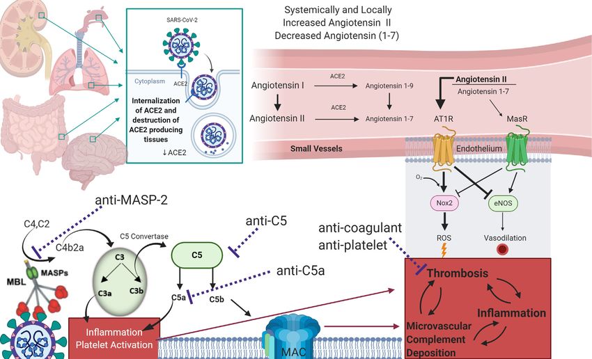
Translational Research
Volume 220                                                                                                                    Magro et al   9

              Fig 8. Demonstration of co-localization of complement components with SARS-CoV2 spike glycoprotein in the
              lung of Case 1. A, Striking deposition of C4d within the interalveolar septa of the lung was first demonstrated by
              DAB staining. Using NUANCE software the C4d image appears green (B) while the SARS-CoV2 spike protein
              appears red (C). D, A merged image shows a significant degree of C4d and SARS-CoV2 co-localization, as
              revealed by intense yellow staining. E H, A similar pattern was observed using an anti-C5b-9 reagent whose
              image appears green, with a significant degree of C5b-9 and SARS-CoV2 co-localization, as revealed by intense
              yellow staining.

resembles images captured in early case reports in the                     deposition, might be anticipated,17 as observed in our
Chinese language literature of severe COVID-19                             cases. It is also consistent with the very high d-dimer
pneumonitis.16                                                             levels found in the 3 cases in which it was assessed.
   We now show that this pathologic pattern, atypical                      Vascular deposition of C5b-9 is a key feature of many
for classic ARDS, is accompanied by extensive deposi-                      microthrombotic syndromes, regardless of the particu-
tion of complement components within the lung septal                       lar syndromic complex, including catastrophic anti-
microvasculature. With such extensive complement                           phospholipid antibody syndrome, atypical hemolytic
involvement, membrane attack complex-mediated                              uremic syndrome, purpura fulminans and severe multi-
microvascular endothelial cell injury and subsequent                       organ malignant atrophic papulosis, and they may
activation of the clotting pathway, leading to fibrin                      respond to anticomplement therapies.18 23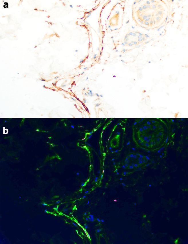
Translational Research
10    Magro et al                                                                                                       June 2020

                                                                        in limited series of COVID-19 patients from China26
                                                                        or the United States,27 although there are insufficient
                                                                        numbers to assess its correlation with severity of dis-
                                                                        ease. We have observed an increase in peripheral blood
                                                                        neutrophil vacuolization and granule content in our
                                                                        patients, consistent with an activated state, even if
                                                                        absolute numbers are not elevated.
                                                                           Systemic activation of complement was reflected by
                                                                        elevated complement levels in the sera of SARS-CoV-
                                                                        infected wild-type mice. We measured complement
                                                                        levels in only one of our cases, Case 3. They were only
                                                                        moderately elevated, perhaps reflecting consumption in
                                                                        tissues, with reciprocal depression of circulating levels.
                                                                        Indeed, we found deposition of AP and LP components
                                                                        in normal-appearing skin in 3 cases, not only in biop-
                                                                        sies of retiform cutaneous lesions from those patients.
                                                                        Retiform purpura reflects the cutaneous manifestation
                                                                        of an occlusive microthrombotic process.28 In sum-
                                                                        mary, while complement does not appear to play a
                                                                        major role in controlling CoV replication, it may have
                                                                        a critical role in its pathogenicity.
                                                                           There are several intriguing questions that remain to
                                                                        be addressed as additional cases are investigated. For
                                                                        example, one might have anticipated thrombocytopenia
Fig 9. Demonstration of co-localization of C4d and SARS-CoV2            in the context of a systemic microvascular thrombosis
spike glycoprotein in the skin of Case 3. A, The skin biopsy was        in our cases. Although mild thrombocytopenia
stained for C4d showing significant vascular localization (DAB          (100 150 £ 109/L) has been recorded in 20% of
stain). B, Using NUANCE software C4d is highlighted green while
COVID-19 spike protein shows a red staining pattern; a yellow signal
                                                                        COVID-19 patients,26 such values were seen in only 2
is discernible indicative of co-localization of C4d and viral protein   of our 5 patients, nor were platelets a prominent com-
within the microvasculature.                                            ponent of the fibrin microthrombi seen histologically.
                                                                        It is possible that infection-related thrombocytosis was
   A preclinical model of SARS infection emphasizes                     mitigated by the development of severe vasculopathy.
the potential role of complement in the parenchymal                     However, there is also precedent for such platelet val-
lung injury of coronavirus infection.7 Mouse-adapted                    ues in an atypical hemolytic-uremic syndrome
SARS-CoV MA15 infection of C3 / mice led to sig-                        (aHUS)-type of microangiopathy. Platelet counts may
nificantly less weight loss and respiratory dysfunction                 be in the normal range despite severe thrombotic dis-
than seen in wild-type mice, and this occurred despite                  ease.29 Second, there appears to have been a delay of
equivalent viral loads in the lung.7 Another feature                    5 9 days in most, if not all, of our patients between
reflective of a prominence of complement-mediated                       the onset of typical respiratory symptoms, including a
pathology rather than simply inflammation or vasculitis                 nonproductive cough, myalgias, fatigue, fever, and
in our COVID-19 patients was the extent of septal and                   mild dyspnea, and a catastrophic change in respiratory
intra-alveolar neutrophilia. It was present, but pauci-                 status. This may reflect an evolution of immune pro-
inflammatory. SARS-CoV-infected wild-type mice                          cesses—antiviral IgM levels would expect to emerge at
also had higher levels of neutrophils in the lung than                  this point—or perhaps the fact that the complement
their C3 / counterparts.7 Tissue neutrophilia may be                    cascade is a threshold pathway. When activated to a
attributable to the neutrophil chemoattractant proper-                  great extent it may exceed the capacity of complement
ties of complement. Both neutrophils and complement                     regulatory proteins, both soluble and normally present
are key sentinels of innate immunity and also modulate                  in abundance on the microvasculature.29 That raises
thrombogenic pathways, the latter thought related to                    the issue of why only a subset of SARS-CoV2-infected
C5a receptor/tissue factor cross-talk mediated by neu-                  patients develops such severe disease with features
trophils.24 Neutrophilia in human SARS-CoV patients                     atypical for ARDS. The age range was wide among our
is associated with a poor outcome and could be an                       cases, as was the degree of pre-existing immune sup-
index of the extent of complement activation.7,25                       pression. But there are a variety of complement regula-
Peripheral neutrophilia is not a significant observation                tory factor polymorphisms or mutations21 as well as
Translational Research
Volume 220 Magro et al 11
coagulation pathway mutations which could promote to activation of the AP and LP with SARS-CoV-2 spike
susceptibility to enhanced complement activation and glycoproteins are consistent with this hypothesis.
thrombosis in a given individual that should be investi- There may also be pathways apart from virus spike
gated. engagement by which LP and AP are activated. First,
Our results, particularly when viewed in the context MBL and ficolins bind to altered glycan structures
of murine models of SARS-CoV infection, suggest the present on injured cells, enabling complex formation
hypothesis that intervention with inhibitors of the alter- with MASP2.36 In addition, SARS-Cov1 and SARS-
native or lectin complement pathways, or both, may be CoV use Angiotensin Converting Enzyme (ACE2) as
relevant to severe COVID-19 in humans. Multiple an entry point to cells.6 Angiotensin I and angiotensin
studies have documented a role for the AP in microvas- II have been associated with inflammation, oxidative
cular injury with C5b-9 deposition,13,30,31 but the lectin stress, and fibrosis, and ACE2 is involved in their deac-
pathway has not been similarly investigated. Support tivation.37 If overwhelming coronavirus infection, with
for the involvement of the LP in COVID-19 comes binding to ACE2 on epithelial targets not only in the
from the discovery that MBL binds to the SARS-CoV lung but in other tissues expressing these proteins,
spike glycoprotein.32 A complex of MBL with MASP2 including the kidney, intestines, and brain, were to
is the first step in LP activation, and part of a positive interfere with ACE2 activity, the resulting increases in
feedback loop leading to sustained AP activation, angiotensin II could lead to reactive oxygen species
with inflammation and concurrent activation of the formation and interference with antioxidant and vaso-
coagulation cascade.33,34 Although documented experi- dilatory signals such as NOX2 and eNOS, with further
mentally for SARS-CoV, this binding is speculative complement activation. This has been demonstrated in
in terms of SARS-CoV-2, as certain sites on its glyco- a rat model of angiotensin II overexpression linked to
protein spikes are mutated compared to SARS-CoV.35 other disorders.38 The potential loss of auto-vasocon-
However, glycosylation sites for high-mannose structures striction and regulation of lung blood flow through
with the potential to similarly engage MBL, and thus acti- injured vascular segments would also lead to increased
vate MASP2, have been identified for SARS-CoV-2.35 shunting and severe hypoxemia. Fig 10 summarizes
Our data showing co-localization of products linked the potential mechanisms of SARS-CoV2 associated
Fig 10. Model for AP and LP complement activation by SARS-CoV2, and its interaction with coagulation cascades.Translational Research
12 Magro et al June 2020
complement activation, and its possible interaction in Wuhan, China. JAMA 2020. https://doi.org/10.1001/
with coagulation pathways. jama.2020.1585.
5. Tang N, Li D, Wang X, Sun Z. Abnormal coagulation parame-
Given these data, use of agents that block LP activa-
ters are associated with poor prognosis in patients with novel
tion such as narsoplimab (OMS721), a human mono- coronavirus pneumonia. J Thromb Haemost 2020. https://doi.
clonal antibody against MASP2 which recently org/10.1111/jth.14768.
received Breakthrough Therapy Designation for 6. Phan T. Novel coronavirus: from discovery to clinical diagnos-
hematopoietic stem cell transplant-linked thrombotic tics. Infect Genet Evol 2020;79:104211. https://doi.org/10.1016/
microangiopathies,39 or eculizumab, a humanized anti- j.meegid.2020.104211.
7. Gralinski LE, Sheahan TP, Morrison TE, et al. Complement acti-
C5 monoclonal antibody FDA-approved for aHUS, vation contributes to severe acute respiratory syndrome corona-
might be considered on a case-by-case basis in severe virus pathogenesis. mBio. 2018;9:e01753–18.
COVID-19. Investigating critical biomarkers consis- 8. Xu Z, Shi L, Wang Y, et al. Pathologic findings of COVID-19
tent with complement-mediated microvascular injury associated with acute respiratory distress syndrome. Lancet
and thrombosis in COVID-19 patients would be impor- Respiratory Med 2020;8:420–2.
9. Zhang H, Zhou P, Wei Y, et al. Histopathologic changes and
tant. A panel to study might include: d-dimers; factor SARS-Cov-2 immunostaining in the lung of a patient with
VIII, fibrinogen, and other coagulation factors; anti- COVID-19. Ann Intern Med 2020. https://doi.org/10.7326/M20-
phospholipid antibodies; C-reactive protein; pro- 0533.
inflammatory cytokines, particularly IL-1 and IL-6; cir- 10. Zhou F, Yu T, Du R, et al. Clinical course and risk factors
culating complement proteins including C3, C4, C5b-9, for mortality of adult inpatients with COVID-19 in Wuhan,
China: a retrospective cohort study. Lancet 2020;395:
and Bb (the latter remaining in circulation longer than 1054–62.
other components, as a marker of AP activation); and 11. Gattinoni L, Coppola S, Cressoni M, Busana M, Chiumello D.
tissue biopsy, the skin being most readily accessible. Covid-19 does not lead to a “Typical” acute respiratory distress
This could enable establishment of criteria for clinical syndrome. Am J Respir Crit Care Med 2020. https://doi.org/
trials with anti-complement and/or anticoagulants, and 10.1164/rccm.202003-0817LE.
12. Magro CM, Poe JC, Kim C, et al. Degos disease: a C5b-9/inter-
perhaps open the possibility for earlier intervention feron-a-mediated endotheliopathy syndrome. Am J Clin Pathol
than at the end-stages of severe COVID-19. 2011;135:599–610.
13. Magro CM, Momtahen S, Mulvey JJ, Yassin AH, Kaplan RB,
Laurence JC. The role of the skin biopsy in the diagnosis of atyp-
ical hemolytic uremic syndrome. Am J Dermatopathol
ACKNOWLEDGMENTS 2015;37:349–59.
The authors are aware of the journal’s authorship 14. Magro CM, Pope Harman A, Klinger D, et al. Use of C4d as a
statement and only Dr. Laurence has a potential con- diagnostic adjunct in lung allograft biopsies. Am J Transplant
flict of interest. He has received an unrestricted edu- 2003;3:1143–54.
15. Magro CM, Deng A, Pope-Harman A, et al. Humorally mediated
cational grant and consulting fees from Omeros, Inc.
posttransplantation septal capillary injury syndrome as a com-
He has recent grants and consulting fees in the recent mon form of pulmonary allograft rejection: a hypothesis. Trans-
past from Alexion, Inc. He is also Senior Scientist plantation 2002;74:1273–80.
for Programs at amfAR, The Foundation for AIDS 16. Yao XH, Li TY, He ZC, et al. A pathological report of three
Research, and has funding from the Angelo Donghia COVID-19 cases by minimally invasive autopsies. Zhonghua
bing li xue za zhi = Chinese J Pathol 2020. https://doi.org/
Foundation.
10.3760/cma.j.cn112151-20200312-00193.
The expert technical assistance of Bing He, Weill 17. Chaturvedi S, Braunstein EM, Yuan X, et al. Complement activ-
Cornell Medicine, Department of Pathology and Labo- ity and complement regulatory gene mutations are associated
ratory Medicine, Translational Research Program, is with thrombosis in APS and CAPS. Blood 2020. https://doi.org/
gratefully acknowledged. 10.1182/blood.2019003863.
18. Magro CM, Poe JC, Kim C, et al. Degos disease: a C5b-9/inter-
Conflicts of Interest: All authors have read the jour- feron-a-mediated endotheliopathy syndrome. Am J Clin Pathol
nal’s policy on disclosure of potential conflicts of inter- 2011. https://doi.org/10.1309/AJCP66QIMFARLZKI.
est and have none to declare. 19. Ruffatti A, Calligaro A, Lacognata CS, et al. Insights into the
pathogenesis of catastrophic antiphospholipid syndrome. A case
REFERENCES report of relapsing catastrophic antiphospholipid syndrome and
review of the literature on ischemic colitis. Clin Rheumatol
1. Zhu N, Zhang D, Wang W, et al. A novel coronavirus from 2019. https://doi.org/10.1007/s10067-019-04888-5.
patients with pneumonia in China. 2019. N Engl J Med 2020. 20. Manrique-Caballero CL, Peerapornratana S, Formeck C, Del
https://doi.org/10.1056/NEJMoa2001017. Rio-Pertuz G, Gomez Danies H, Kellum JA. Typical and atypi-
2. Cucinotta D, Vanelli M. WHO declares COVID-19 a Pandemic. cal hemolytic uremic syndrome in the critically Ill. Crit Care
Acta Biomed 2020. https://doi.org/10.23750/abm.v91i1.93973. Clin 2020. https://doi.org/10.1016/j.ccc.2019.11.004.
3. cdc.gov/coronavirus/2019.ncov 21. Fremeaux-Bacchi V, Fakhouri F, Garnier A, et al. Genetics and
4. Wang D, Hu B, Hu C, et al. Clinical characteristics of 138 hospi- outcome of atypical hemolytic uremic syndrome: a nationwide
talized patients with 2019 novel coronavirus-infected pneumoniaTranslational Research
Volume 220 Magro et al 13
French series comparing children and adults. Clin J Am Soc 32. Zhou Y, Lu K, Pfefferle S, et al. A single asparagine-linked gly-
Nephrol 2013;8:554–62. cosylation site of the severe acute respiratory syndrome corona-
22. Magro CM, Wang X, Garrett-Bakelman F, Laurence J, Shapiro virus spike glycoprotein facilitates inhibition by mannose-
LS, Desancho MT. The effects of Eculizumab on the pathology binding lectin through multiple mechanisms. J Virol 2010;84:
of malignant atrophic papulosis. Orphanet J Rare Dis 2013. 8753–64.
https://doi.org/10.1186/1750-1172-8-185. 33. Beltrame MH, Catarino SJ, Goeldner I, et al. The lectin pathway
23. Perera TB, Murphy-Lavoie HM. Purpura fulminans. StatPearls of complement and rheumatic heart disease. Front Ped
Publishing; 2020. Available at: http://www.ncbi.nlm.nih.gov/ 2015;2:1–14.
pubmed/30422460 Accessed March 28, 2020. 34. Krarup A, Wallis R, Presanis JS, Gal P, Sim RB. Simultaneous
24. Rits K, Doumas M, Mastellos D, et al. Anovel C5a receptor-tis- activation of complement and coagulation by MBL-associated
sue factor cross-talk in neutrophils links innate immunity to serine protease 2. PLoS One 2007;7:e623.
coagulation pathways. J Immunol 2006;177:4794–802. 35. Walls AC, Park Y-J, Tortorici MA, Wall A, McGuire AT, Veesler
25. Yen Y-T, Liao F, Hsiao C-H, Kao C-L, Chen Y-C, Wu-Hsieh D. Structure, function, and antigenicity of the SARS-CoV-2 spike
BA. Modeling the early events of severe acute respiratory syn- glycoprotein. Cell 2020. https://doi.org/10.1016/j.cell.2020.02.058.
drome coronavirus infection in vitro. J Virol 2006;80:2684–93. 36. Dodo J, Kocsis A, Gal P. Be on target: strategies of targeting
26. Fan BE, Chong VCL, Chan SSW, et al. Hematologic parameters alternative and lectin pathway components in complement-medi-
in patients with COVID-19 infection. Am J Hematol 2020. ated diseases. Front Immunol 2018;9:1–22.
https://doi.org/10.1002/ajh.25774. 37. Srivastava P, Badhwar S, Chandran DS, Jaryal AK, Jyotsna
27. Bhatraju PK, Ghassemieh BJ, Nichols M, et al. Covid-19 in criti- VP, Deepak KK. Imbalance between Angiotensin II - Angio-
cally ill patients in the Seattle region case series. N Engl J Med tensin (1-7) system is associated with vascular endothelial
2020. https://doi.org/10.1056/NEJMoa2004500. dysfunction and inflammation in type 2 diabetes with newly
28. Wysong A, Venkatesan P. An approach to the patient with reti- diagnosed hypertension. Diabetes Metab Syndr 2019;13:
form purpura. Dermatologic Ther 2011;24:151–72. 2061–8.
29. Laurence J, Haller H, Mannucci PM, Nangaku M, Praga M, de Cor- 38. Shagdarsuren E, Wellner M, Braesen JH, et al. Complement acti-
doba SR. Atypical hemolytic uremic syndrome (aHUS): essential vation in angiotensin II-induced organ damage. Circ Res
aspects of an accurate diagnosis. Clin Adv Hematol Oncol 2016;14 2005;97:716–24.
(11S1):1–15. 39. Rambaldi A, Khaled S, Smith M, et al. Improved survival fol-
30. Prufer F, Scheiring J, Sautter S, et al. Terminal complement lowing OMS721 treatment of hematopoietic stem cell trans-
complex (C5b-9) in children with recurrent hemolytic uremic plant-associated thrombotic microangiopathy (HCT-TMA).
syndrome. Sem Thromb Hemost 2006;32:121–7. Stockholm, Sweden: Eur Hematol Assoc; 2018 June
31. Noris M, Galbusera M, Gastoldi S, et al. Dynamics of comple- 14 17Abst PF724.
ment activation in aHUS and how to monitor eculizumab ther-
apy. Blood 2014;124:1715–26.You can also read