MiRNA profiles of canine cutaneous mast cell tumours with early nodal metastasis and evaluation as potential biomarkers - Nature
←
→
Page content transcription
If your browser does not render page correctly, please read the page content below
www.nature.com/scientificreports
OPEN miRNA profiles of canine cutaneous
mast cell tumours with early
nodal metastasis and evaluation
as potential biomarkers
Valentina Zamarian1, Roberta Ferrari1, Damiano Stefanello1, Fabrizio Ceciliani1,
Valeria Grieco1, Giulietta Minozzi1, Lavinia Elena Chiti1, Maddalena Arigoni2,
Raffaele Calogero2 & Cristina Lecchi1*
Cutaneous mast cell tumours (MCTs) are common skin neoplasms in dogs. MicroRNAs (miRNAs) are
post-transcriptional regulators involved in several cellular processes, and they can function as tumour
promoters or suppressors. However, the role of miRNAs in canine MCTs has not yet been elucidated.
Thus, the current study aimed to characterize miRNA profiles and to assess their value as biomarkers
for MCTs. miRNA expression profiles were assessed in formalin-fixed, paraffin-embedded samples
by next-generation sequencing. Ten samples were MCT tissues, and 7 were healthy adjacent tissues.
Nine dysregulated miRNAs (DE-miRNAs) were then validated using RT-qPCR in a larger group of MCT
samples, allowing the calculation of ROC curves and performance of multiple factor analysis (MFA).
Pathway enrichment analysis was performed to investigate miRNA biological functions. The results
showed that the expression of 63 miRNAs (18 up- and 45 downregulated) was significantly affected
in MCTs. Five DE-miRNAs, namely, miR-21-5p, miR-92a-3p, miR-338, miR-379 and miR-885, were
validated by RT-qPCR. The diagnostic accuracy of a panel of 3 DE-miRNAs—miR-21, miR-379 and miR-
885—exhibited increased efficiency in discriminating animals with MCTs (AUC = 0.9854) and animals
with lymph node metastasis (AUC = 0.8923). Multiple factor analysis revealed clusters based on nodal
metastasis. Gene Ontology and KEGG analyses confirmed that the DE-miRNAs were involved in cell
proliferation, survival and metastasis pathways. In conclusion, the present study demonstrated that
the miRNA expression profile is changed in the MCT microenvironment, suggesting the involvement
of the altered miRNAs in the epigenetic regulation of MCTs and identifying miR-21, miR-379 and miR-
885 as promising biomarkers.
Cutaneous mast cell tumours (MCTs) are a common skin neoplasm in dogs, accounting for up to 21% of all
skin tumours1. Mast cell tumours originate from uncontrolled proliferation of neoplastic mast cells in cutaneous
and subcutaneous tissues and usually occur as single tumours but sometimes as multiple t umours2. The clinical
phenotype of MCTs ranges from relatively benign to highly malignant tumours that can spread to the local lymph
nodes, liver and spleen3,4. Their diagnosis and prognosis are usually based on tumour grading as determined
by the Patnaik5 and Kiupel4,6 systems, and tumour staging, including evaluation of draining lymph nodes7.
In addition. the proliferation rate (Ki-67), c-kit proto-oncogene mutations and KIT expression are significant
prognostic factors7,8. The pathogenesis and aetiology of MCTs are poorly understood, and the main causes that
lead to MCT carcinogenesis remain e lusive9.
Canine MCTs have been recently investigated at the molecular level using next-generation RNA sequencing10,
and two distinct tumour subtypes have been identified with differential expression of 71 genes involved in cell
proliferation processes. However, no information about the epigenetic regulation of MCT development, such as
regulation mediated by microRNAs, is available.
MicroRNAs (miRNAs) are small non-coding RNAs that are involved in post-transcriptional regulation and
thus affect virtually every cellular process. MiRNAs regulate mRNA translation, leading to modulation of protein
expression. MiRNA dysregulation occurs during the pathogenesis of several diseases, including cancer, and leads
1
Dipartimento di Medicina Veterinaria, Università degli Studi di Milano, Milan, Italy. 2Molecular Biotechnology
Center, Department of Biotechnology and Health Sciences, Università di Torino, 10126 Turin, Italy. *email:
cristina.lecchi@unimi.it
Scientific Reports | (2020) 10:18918 | https://doi.org/10.1038/s41598-020-75877-x 1
Vol.:(0123456789)www.nature.com/scientificreports/
Grade
Breed Sex Age (years) Tumor location Size (cm) Patnaik Kiupel Lymph node s tatuse
American Staffordshire
1 Male 6 Trunk 1 II Low HN1
Terriera,d
2 Bernese Female 4 Limb 2.5 II Low HN2
3 Boxer Male 8 Limb 2.2 II Low HN2
4 Dachshund Female 9 Trunk 0.8 II Low HN0
5 Dogoa,b Male 6 Limb 2 II Low HN2
6 English settera,b Female 6 Trunk 3 II Low HN2
7 English settera,c,d Female 11 Trunk 5 II Low HN3
8 Italian pointera,b Male 5.5 Trunk 4 II Low HN1
9 Jack Russella,b Male 13 Head 5 II High HN2
10 Labrador Male 1 Head 1 I Low HN0
11 Labrador d Male 10 Scrotum 2 II Low HN0
12 Labrador Male 9 Trunk 0.6 II Low HN2
13 Labrador Female 6 Trunk 3.5 II Low HN2
14 Mixed breeda,b,c Female 11 Trunk 3 II Low HN0
15 Mixed breeda,b Female 6 Trunk 4 II Low HN2
16 Mixed breeda,b Male 11 Limb 3 III High HN2
17 Mixed breeda,c,d Female 8 Limb 3 III High –
18 Mixed breedc Male 12 Neck 7 II Low HN3
19 Pug Male 3.5 Head 1 II Low HN2
20 Tosa inu Male 4 Trunk 3 II Low HN2
21 Weimaraner Male 7 trunk 2 II Low HN2
Table 1. Summary of clinical and histopathological data. a MCT samples sequenced using NGS. b Healthy
margins sequenced using NGS. c $amples in which miRNAs selected for the RT-qPCR validation step were not
detected. d Samples for which healthy margins were not collected. e Classification system proposed by Weishaar
et al.7. HN histological node, NGS next-generation sequencing.
to disruption of pathways that play a role in cancer initiation and progression11. MiRNAs can act as oncomiRs,
targeting tumour suppressor genes and promoting tumour progression, or as tumour suppressors, although
overall miRNA dysregulation is a hallmark of c ancer12–14. The role of miRNAs in canine MCT is still unknown.
The present study aims to close this gap by assessing the miRNA expression profile of canine MCTs using next-
generation sequencing, investigating whether miRNAs are dysregulated in the MCT microenvironment, and
identifying links between miRNAs and their target genes and relevant biological processes.
Results
Determination of miRNA profiles and identification of DE‑miRNAs in healthy margins versus
MCTs. After RNA extraction, small RNA libraries were generated, pooled, concentrated and size selected on
a non-denaturing acrylamide gel (≈ 141 bp bands). After elution from the gel, the size-selected libraries were
quantified and sequenced on a NextSeq 500 sequencer (Illumina). The resulting number of reads per sample
varied from 53,000 to 29,000,000. Eight MCT samples with insufficiently high mapping rates were excluded from
further analysis. For two dogs (numbers 8 and 15 in Table 1), the results for the tumours and matched healthy
adjacent margins were reported.
A count table was used to identify differentially expressed miRNAs via DESeq2 analysis15. The DESeq thresh-
old was set by discarding low-expression miRNAs having an average count of 2 or less. This analysis revealed the
expression of 246 and 116 Canis familiaris (cfa) miRNAs in healthy margins and MCTs, respectively.
The expression profiles of sequenced samples were used to carry out cluster analysis. Samples were grouped
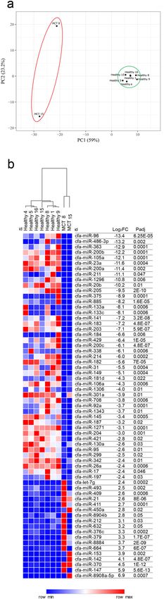
into two clusters: healthy tissues and MCTs (Fig. 1a).
To rank the most differentially expressed miRNAs (DE-miRNAs) between healthy and MCT samples, the
results of differential expression analysis performed with DESeq2 were further filtered a more stringent cut-off
criteria of an adjusted P value of 0.01 and an absolute log2FC of 2.4. This filtering allowed the identification of
sixty-three miRNAs whose abundance differed significantly between MCT and healthy samples, demonstrating
that 45 miRNAs were downregulated with a log2FC of between − 2.4 and − 13.4 and 18 miRNAs were upregulated
with a log2FC of between 2.4 and 6.9 (Fig. 1b).
Quantification of DE‑miRNAs in healthy versus MCT samples by RT‑qPCR. To validate the NGS
results and measure the abundances of DE-miRNAs in MCTs, RT-qPCR was performed on both the sequenced
samples and a separate group of 11 MCTs and related healthy adjacent (normal) tissue samples. To validate the
sequencing results, 9 DE-miRNAs—miR-370, miR-379, miR-92a, miR-21, miR-26a, miR-342, miR-885, miR-
375 and miR-338—were selected based on the fold change and read count values. MiR-122, miR-128 and miR-
Scientific Reports | (2020) 10:18918 | https://doi.org/10.1038/s41598-020-75877-x 2
Vol:.(1234567890)www.nature.com/scientificreports/
Figure 1. NGS results. (a) Principal component analysis (PCA) of sequenced samples. Two-dimensional PCA
was used to determine whether MCTs (red circle) could be differentiated from healthy (green circle) samples.
(b) Identification of DE-miRNAs between MCTs and healthy samples. Heat map and table displaying the fold
change and Padj of DE-miRNAs.
Scientific Reports | (2020) 10:18918 | https://doi.org/10.1038/s41598-020-75877-x 3
Vol.:(0123456789)www.nature.com/scientificreports/
101 were quantified as controls for normalization. The artificial spike-in cel-miR-39 was used as the internal
control. MiRNAs selected for the validation step were detected in almost all samples, except for sample numbers
7, 14, 17 and 18 (Table 1). The results are presented in Fig. 2. The RT-qPCR results confirmed that five miR-
NAs were differentially regulated in healthy adjacent margin tissues versus MCTs. In detail, miR-21 (P = 0.004,
log2FCMCT/Healthy = 2.84) and miR-379 (P = 0.0005, log2FCMCT/Healthy = 2.61) were upregulated, while miR-885
(P = 0.008, log2FCMCT/Healthy = − 2.53), miR-338 (P = 0.025, log2FCMCT/Healthy = − 0.86) and miR-92a (P = 0.021,
log2FCMCT/Healthy = − 0.78) were downregulated in MCT samples compared to healthy margin samples. Con-
versely, miR-26a, miR-342, miR-370 and miR-375 did not exhibit statistically significant differences between
the groups.
Diagnostic value of DE‑miRNAs in dogs with MCT. Receiver operating characteristic (ROC) analysis
was used to assess the diagnostic value of DE-miRNAs as biomarkers to further discriminate between MCT and
healthy adjacent tissue. To confirm the diagnostic efficacy of each miRNA, the associated area under the curve
(AUC) was calculated. Table 2 shows a summary of the diagnostic performance of each DE-miRNA and shows
combinations of three DE-miRNAs. The AUC was fair for miR-92a (AUC = 0.7427) and miR-338 (AUC = 0.7339)
and excellent for miR-21 (AUC = 0.9825), miR-379 (AUC = 0.9211) and miR-885 (AUC = 0.9181) (Fig. 3).
Discriminant analysis was used to further investigate the potential for improving diagnostic performance
by analysing multiple DE-miRNAs. Statistical analysis was performed to examine the weighted average rela-
tive quantification (RQ) values of the miRNAs with an AUC of > 0.9 (miR-21, miR-379 and miR-885) (Fig. 4).
The median expression levels were 0.0301 (range 0.0069–0.9334) and 0.99998 (range 0.3485–1) in healthy
margin and MCT samples, respectively (Fig. 4a). The predicted probability of being able to discriminate a
sample as positive based on the logit model [logit = 1/(1 + exp (− (− 4.92611–1.31822 × expression level of miR-
885 + 0.40746 × expression level of miR-379 + 0.86787 expression level of miR-21)))] was used to construct the
ROC curve (Fig. 4b). The AUC for the panel of these three DE-miRNAs was 0.9854 (95% CI 0.9854–0.9854)
with a cut-off value of 0.1654, and a sensitivity of 100% and a specificity of 94.4%.
Potential of the miRNA panel for the detection of nodal metastases. Excised lymph nodes were
categorized in accordance with the Weishaar classification system for nodal metastases (2014)7, and the potential
of the three-miRNA panel to discriminate patients with and without lymph node involvement was evaluated.
Two groups, namely, HN0/1, including non-metastatic and pre-metastatic samples, and HN2, including early
metastatic samples, were included for further analysis (Table 1). The weighted average relative quantification
(RQ) values of the miRNA panel (miR-21, miR-379 and miR-885) were calculated (Fig. 5). The median expres-
sion levels were 0.3179 (range 0.0071–0.8858) and 0.9424 (range 0.3741–1) in the HN0/1 and HN2 groups,
respectively (Fig. 5a). The predicted probability of being able to discriminate a sample as metastatic based on the
logit model [logit = 1/(1 + exp (−(− 1.58980–7.91569 × expression level of miR-885 + 0.13130 × expression level of
miR-379 + 0.05084 expression level of miR-21)))] was used to construct the ROC curve (Fig. 5b). The AUC for
the panel of these three DE-miRNAs was 0.8923 (95% CI 0.759–1.000) with a cut-off value of 0.5528, a sensitiv-
ity of 92.3% and a specificity of 80%. MFA identifies individuals with similar profiles who are close to each other
on the factor map. Collectively, the components F1 and F2 explained 68.53% of the total variance in the data
(Fig. 5c). The first component (F1) explained 42.76% of the variance and separated the HN0/1 group from the
HN2 group according to lymph node involvement. The second component (F2) explained 25.77%, discriminat-
ing non-metastatic HN0 samples (samples 4 and 10) in the upper right panel from high-grade early metastatic
HN2 samples (samples 16 and 9) in the lower-left panel (Supplementary Table S1).
Gene Ontology and pathway enrichment analysis of miRNAs. The MiRWalk 3.0 and DAVID data-
bases were searched to retrieve the candidate target genes of DE-miRNAs and to perform mRNA enrichment
analysis, respectively. Of the predicted mRNA targets of downregulated miRNAs, 196 were in the 3′UTR, 45
were in the 5′UTR and 171 were in the CDS. Of the predicted mRNA targets of upregulated miRNAs, 16 were in
the 3′UTR, 3 were in the 5′UTR and 11 were in the CDS. The list of candidate target genes is provided in Table 3.
Gene Ontology (GO) analysis was performed using DAVID for three categories: biological process (BP),
cellular component (CC) and molecular function (MF). An overview of the top 10 terms significantly enriched
with target genes for each of the above GO categories is presented in Fig. 6. The enriched GO BP terms mainly
included regulation of transcription from RNA polymerase II promoter and protein ubiquitination involved in
ubiquitin-dependent protein catabolic process; the CC terms were related to the cytoplasm, nucleus and nucleo-
plasm, while the MF terms focused on transcription factor activity and sequence-specific DNA binding. KEGG
pathway analysis was performed on candidate targets of DE-miRNAs. Figure 7 shows the top 10 significantly
enriched KEGG pathways, with PI3K-Akt signalling pathway, small cell lung cancer, viral carcinogenesis and
microRNAs in cancer at the top of the list.
Discussion
The role of miRNAs in canine MCT has not yet been elucidated. The current study aimed to characterize the
miRNA profile of canine MCTs using FFPE samples. A multi-step approach was adopted: the pilot part of the
study profiled miRNAs in MCTs and healthy adjacent margins via next-generation sequencing. In the second
step, the DE-miRNAs were validated by performing RT-qPCR on the samples selected for sequencing and on a
separate independent group of FFPE samples. Our results showed that the expression of 63 miRNAs, of which
18 were upregulated and 45 were downregulated, was significantly affected in MCTs. Nine DE-miRNAs were
then validated in a larger group by RT-qPCR, demonstrating that five of these DE-miRNAs, namely, miR-21-5p,
miR-92a-3p, miR-338, miR-379 and miR-885, were effectively modulated. The diagnostic accuracy of three
Scientific Reports | (2020) 10:18918 | https://doi.org/10.1038/s41598-020-75877-x 4
Vol:.(1234567890)www.nature.com/scientificreports/
Figure 2. Box plots of DE-miRNAs in MCTs compared with healthy margins. Significance was accepted at
P < 0.05 (*), P < 0.01 (**) and P < 0.001 (***). The black lines inside the boxes denote the medians. The whiskers
indicate variability outside the upper and lower quartiles.
Scientific Reports | (2020) 10:18918 | https://doi.org/10.1038/s41598-020-75877-x 5
Vol.:(0123456789)www.nature.com/scientificreports/
miRNA AUC 95% CI P-value Cut-off Sensitivity 1-Specificity
miR-885 0.9181 0.8276–1.000 < 0.0001 0.0357 0.8889 0.9474
Downregulated miR-92a 0.7427 0.5925–0.8929 = 0.0015 0.814 0.7222 0.6842
miR-338 0.7339 0.5827–0.8851 = 0.0024 1.7878 0.6111 0.7895
miR-21 0.9825 0.9825–0.9825 < 0.0001 1.6250 0.9444 0.9474
Upregulated
miR-379 0.9211 0.8328–1.000 < 0.0001 11.5688 1.000 0.7895
W-AV* miR-379 + miR-21 + miR-885 0.9854 0.9854–0.9854 < 0.0001 0.1654 1.000 0.9444
W-AV-HN** miR-379 + miR-21 + miR-885 0.8923 0.759–1.000 < 0.0001 0.5528 0.9231 0.8000
Table 2. The area under the curve (AUC), sensitivity and specificity values of DE-miRNAs. *W-AV =
weighted average relative quantification of miR-379 + miR-21 + miR-885 in healthy versus MCT samples.
**W-AV-HN = weighted average relative quantification of miR-379 + miR-21 + miR-885 in HN0/1 versus HN2
samples.
Figure 3. Receiver operating characteristic (ROC) curve analysis of DE-miRNAs. (a) AUC of miR-885; (b)
AUC of miR-92a; (c) AUC of miR-338; (d) AUC of miR-21; (e) AUC of miR-379. AUCarea under the curve, CI
confidence interval.
DE-miRNAs—miR-21, miR-379 and miR-885—was excellent, and the AUC of their combination increased to
0.9854 with 100% sensitivity and 94.4% specificity. Due to their limited nucleotide length, miRNAs have shown
higher stability than DNA and mRNA in sample types such as FFPE t issues16. However, the preparation of
miRNA NGS libraries from FFPE samples is particularly challenging because of the intersample heterogeneity
of the RNA q uality17. In the present investigation, the library preparations were performed in parallel to avoid
the batch effect, but mappable miRNA reads were produced from only 2 of 10 tumours. Moreover, mast cells,
which release their granules into the tumour m ass9, may also affect library preparation.
Mast cells are crucial players in allergies, immune responses, angiogenesis and the maintenance of tissue
function and integrity18, also promoting tissue r epair19. Furthermore, mast cells modulate the tumour micro-
environment by performing a two-pronged role: they perform a pro-neoplastic role by releasing mitogenic and
pro-angiogenic factors such as histamine, IL-10, TNF, FGF2, VEGF, IL-18 and M MP20, that promote immune
suppression, proliferation and angiogenesis; and they perform an anti-neoplastic role by inhibiting cell growth
and motility and promoting antitumour inflammatory reactions and apoptosis21. Few studies have investigated
the dysregulation of miRNAs in MCTs in dogs. Using real-time PCR-based TaqMan Low-Density miRNA Arrays,
Fenger and colleagues22 demonstrated that the expression level of miR-9 was increased in high-grade canine
Scientific Reports | (2020) 10:18918 | https://doi.org/10.1038/s41598-020-75877-x 6
Vol:.(1234567890)www.nature.com/scientificreports/
Figure 4. The average expression of the DE-miRNAs with AUC > 0.9, including miR-379, miR-21 and miR-885.
(a) The weighted average relative quantification (RQ) values of DE-miRNAs in healthy versus MCT samples (a)
and ROC curve analysis performed using the logit model, for healthy versus MCT samples (b). AUC, area under
the curve; CI, confidence interval. The black lines denote the medians. **P < 0.001; ***P < 0.0001.
Figure 5. The average expression of the DE-miRNAs with AUC > 0.9, including miR-379, miR-21 and miR-885.
(a) The weighted average relative quantification (RQ) values of DE-miRNAs in HN0/1 versus HN2 samples; (b)
ROC curve analysis performed using the logit model for HN0/1 versus HN2 samples; (c) individual map for
Multiple Factor Analysis (MFA): each sample name represents the barycentre of the two positions according to
the dataset coloured according to lymph node involvement: HN0/1 (blue) and HN2 (green). AUCarea under
the curve, CI confidence interval. The black lines denote the medians. **P < 0.001; ***P < 0.0001.
MCTs, promoting an invasive phenotype. Furthermore, circulating miRNA-126 resulted in exacerbation of
non-epithelial neoplasms, including M CT23.
The DE-miRNAs identified herein have been related to neoplasms in humans and, in some cases, in dogs.
Of the five miRNAs that were found to be differentially regulated, two, namely, miR-21 and miR-379 were
upregulated, whereas three, namely, miR-92a-3p, miR-338, and miR-885, were downregulated. MiR-21, which
was found to be upregulated in the present study, has been widely investigated in cancer, and its upregulation
has been associated with cell proliferation, invasion, apoptosis and drug resistance24,25. MiR-21 is frequently
overexpressed in human cancers, including breast cancer, lung cancer, pancreatic cancer, ovarian cancer, glioma,
liver neoplasms, gastric cancer, colorectal cancer and kidney c ancer26, and in canine oral m
elanoma27, hepatocel-
lular carcinoma28 and malignant mammary tumours29. In humans, overexpression of miR-21 has been related
to downregulation of tumour suppressor genes, including programmed cell death 4 (PDCD4), matrix metallo-
proteinases (MMPs), phosphatase and tensin homolog (PTEN), reversion inducing cysteine-rich protein (RECK),
and phosphoinositide 3-kinase (PI3K)26. MiR-379, which was also upregulated, is an onco-suppressor miRNA.
MiR-379 negatively regulates cell proliferation, migration and invasion in several human cancers, including
nasopharyngeal carcinoma30, cervical c arcinoma31, gastric c ancer32 and bladder cancer33, by targeting tumor
protein D52 (TPD52), V-crk avian sarcoma virus CT10 oncogene homolog-like (CRKL), focal adhesion kinase
(FAK) and mouse double minute 2 (MDM2).
Our results demonstrated that miR-92a, miR-338 and miR-885 were downregulated in canine MCTs. MiR-
92a belongs to the miR-17-92a cluster, which is dysregulated in many different types of human t umours34. The
mechanisms by which miR-92a promotes tumorigenesis include augmenting tumour proliferation, inhibiting
tumour apoptosis, and enhancing tumour invasion and m etastasis35 by targeting PTEN in oesophageal squamous
cell cancer36 and Dickkopf-related protein 3 (DKK3) in osteosarcoma37. These features identify miR-92a as an
Scientific Reports | (2020) 10:18918 | https://doi.org/10.1038/s41598-020-75877-x 7
Vol.:(0123456789)www.nature.com/scientificreports/
Target genes of downregulated miRNAs
FOS, POF1B, TP63, PTGES2, CCDC113, LPIN1, IL6ST, GTF2A1, HDAC2, MED19, TET2,
PBLD, ZBTB7B, KIF1B, PPP1R3D, CNNM4, MED29, PAFAH1B1, RASAL2, ABL2, PAWR,
TMCC3, SMARCA5, KLHL3, CDK5R1, TIA1, PDIK1L, FKBP1A, MACROD2, RAB14, ATG9A,
SCN1A, FAM168A, NOL4, HEXIM1, C16orf87, SH3PXD2A, TSFM, ARPC1B, MSL2, RIC1,
NFIA, GNG12, ZZZ3, DUSP16, SERINC5, LAMC1, IPO9, HNF4G, YRDC, RPH3A, PPM1B,
AAK1, LSM14A, CNOT6, SOX6, ZADH2, NOL4L, NFIB, MAP2K4, NEDD4L, TMEM50B,
SLC30A7, LIN54, GNAQ, PALM2, SYNDIG1, KIF3B, JPH2, LPP, PCGF3, RGS3, DAB2IP, TSC1,
COL5A1, ATXN1, MARCH4, PAX3, TRIP12, EPC2, MAP3K20, SESTD1, ITGAV, TPBGL, LDL-
RAD4, TECPR2, TRAF3, ACTC1, UBE2Z, OTUD3, LUZP1, MTMR9, RNF157, SLC9A7, TAF1,
3′UTR ATRX, USP28, GRAMD1B, SRPRA, C21orf91, AFF3, KCNC4, ANP32E, SEC31B, CCDC186,
PLEKHA1, PITPNM2, LRCH1, AXL, PPP1R37, CDC42BPA, ZBTB18, GRHL1, PHLPP2,
ZFHX3, RASSF3, DYRK2, STRN3, STYX, PPP1R9A, SMURF1, PIK3CB, TFDP2, DCX,
TMEM255A, MBNL3, PCDH11Y, DESI1, SHOX, PTPRD, DENND4B, KCNN3, NF2, PLXDC2,
GPR158, PIK3R3, EVI5, ERGIC2, RAB3C, AGGF1, BTG2, ELK4, ARID1B, FAM20C, SNX13,
ATG14, KCNK10, FOXN3, ATXN3, PCMTD1, SGK3, UBE2W, MMP16, CCNE2, MTDH,
NIPAL1, CXCL5, G3BP2, WDFY3, TCF21, LIN28A, GAS2L3, EFR3B, KCNK3, YIPF4, SOCS5,
SERTAD2, CEP41, CREB3L2, ELOVL6, SETD7, DCLK2, SH3D19, PPARGC1B, CAMK2A,
CPEB4, RNF141, EIF4G2, CHST1, ARRDC3, MAN2A1, FNIP1, SLX4, GLYR1, SNN, ATXN7,
ZBTB20, SPOCK2, DNAJB12, CCSER2, GID4, PIP5K1C, SCUBE3, CD2AP, TRAM2, PHF3
MYLIP, PCMTD1, RRBP1, ELOVL6, E2F3, GAA, AURKA, ANP32E, CCDC186, PAX9,
DYNLT3, DENND4B, WASL, SETD5, FAM135A, XKR7, GAN, CNOT2, RABGAP1L, ZNF287,
5′UTR IDH1, NR4A3, TBC1D19, PLEKHB2, TGIF1, ZNF532, CPEB1, KCNA1, SPHK2, CBFA2T3,
COL1A2, TBL1XR1, AIFM1, PTPRD, KLF6, RAB3C, KCNK10, PAPOLA, NAV3, FOSL2, ATL3,
SH3D19, SCRG1, WWC1, SNX2
FOS, DAB2IP, HIPK1, MYLIP, GRAMD1B, MAP2K4, MTF1, PTAR1, RRBP1, NOL4L, TSC1,
HIVEP1, ATXN1, IKZF2, TRIP12, SSFA2, VPS4B, TECPR2, GOLGA8A, GOLGA8B, NSF,
XYLT2, ELOA, ADAM10, GAA, RBL2, ARFGEF2, SYNJ1, BCL2L11, MCL1, SH3PXD2A,
PDZD8, GOLGA3, IPO5, PHLPP2, ITGA5, ANKIB1, FNDC3B, MPP1, RAD21, ZNF17,
ZNF776, MYH9, GPBP1L1, CD69, SCAF11, ATP2B4, DSTYK, TULP4, CUX1, CREB3L2,
KIAA1109, RBM27, SETD5, TRIM36, TMF1, VPS52, ZBTB34, MBD2, UBR1, NPTN, CNOT2,
TTLL7, XPR1, FKBP14, TMCC3, TCHP, PDCD6IP, LCOR, USP7, FRMD3, GNL1, RAB30,
WNK4, SS18L1, CELSR2, MYT1L, COX4I1, TSFM, CACNA2D1, PVALB, NFIA, ADGRA2,
CDS PPM1B, FBXW7, B4GALT7, RASA1, ZBTB20, SLC35G1, KIAA1024, USF2, TAGAP, STX17,
SEL1L3, TBC1D19, ANGPTL2, ADAM23, GIGYF2, LRP1B, SLC4A10, MAP3K20, ZNF385B,
ARHGEF17, EPG5, HERC2, RYR3, EPHA8, CSMD1, ATP6V1B2, CHMP7, CPEB1, NRK,
FRMPD3, DOK5, TACC2, SBNO1, ITM2B, SPTBN4, HNRNPU, HAS3, WWP2, SCN8A,
GDF11, DTX2, PPP1R9A, PIK3CB, PEX5L, VWA5B2, TENM1, CSMD3, EFR3A, PTPRD,
TTC28, NOTCH1, P3H3, EPS8, PDZD2, NIPBL, RAB3C, DCAF6, SNX13, PLEKHG3, BSN,
ZC2HC1A, RBM47, REST, WDFY3, PTPRK, TCF21, GAS2L3, ADCY3, SRPK2, CTTNBP2,
NRF1, NFIX, RHPN2, NPNT, OTUD4, ADAM19, CNTN4, OXSR1, ASB7, CLEC16A, FOXP1,
ROBO2, BTLA, FSTL1, MYO18A, C2CD4C, PHF3, COL19A1
Target genes of upregulated miRNAs
RECK, NCAPG, PAN3, KLHL42, GID4, CCL1, CD59, SLC20A1, PPP1R3B, NEGR1, THRB,
3′UTR
PCDH17, FIGN, HTR2C, FAM126B, ETNK1
5′UTR KAT6A, ZBTB26, TNRC6B
CDS EPHA4, ADNP, TNRC6B, ATF7IP, FBXO11, NR2C2, PTPN14, SPRY4, KLF3, CASKIN1, ROBO2
Table 3. Candidate target genes retrieved from the miRWalk 3.0 database.
onco-miRNA. However, onco-suppressor activities of the miR-17-92a cluster have also been reported, including
anti-proliferative and senescence effects in bladder cancer c ells38 and in p
rostate39 and g astric40 cancers by target-
ing, among other pathways, the NOTCH/EP4 pathway. Similarly, the role of miR-338 is controversial, as it has
been associated with both pro- and antitumour roles. MiR-338 targets oncogenes such as RAB32 and EYA2, and
its downregulation in cancer is also linked to overexpression of epidermal growth factor receptor (EGFR)41,42 and
MET transcriptional regulator (MACC1)43. MiR-338 plays a tumour-promoting role in melanoma that is linked to
tumour growth and metastasis44. MiR-338 is also involved in hypoxia-induced epithelial-to-mesenchymal transi-
tion by targeting HIF-1α45. MiR-885 is a tumour suppressor miRNA that interferes with cell proliferation and
migration by targeting SOCS in colorectal cancer46 and the Wnt/β-catenin pathway in hepatocellular carcinoma47.
Gene Ontology and KEGG pathway analysis suggested that DE-miRNAs have an impact on transcription
activities, cell cycle progression and cell survival and, in general, on several pathways involved in cancer develop-
ment. This hypothesis is supported by gene expression analysis of canine cutaneous M CTs10,48. Gene expression
profiling of metastatic and non-metastatic MCTs using an array approach identified differentially expressed
genes involved in apoptosis, cell cycle arrest and loss of cell polarity and adhesion48. Comparison between these
genes and the genes potentially modulated by DE-miRNAs identified in the present study showed that seven
genes (Fos Proto-Oncogene or FOS, Histone Deacetylase or HDAC, Striatin or STRN, Neurofibromin 2 or NF2,
Phosphoinositide-3-Kinase Regulatory Subunit 3 or PIK3R3, Rho Guanine Nucleotide Exchange Factor or ARHGEF,
C-Type Lectin Domain Containing or CLEC) are potentially modulated by downregulation of miR-92a, miR-338
and miR-885. Conversely, Sprouty RTK Signaling Antagonist or SPRY can be modulated by upregulation of miR-
21 and miR-379. Transcriptome analysis comparing low- and high-risk canine MCTs using next-generation
RNA sequencing identified 71 differentially expressed genes associated with cell proliferation and the extracel-
lular matrix10. Comparison between these genes and the candidate target genes identified in the present study
showed that eight genes belonging to the kelch-like (KLHL), collagen (COL), matrix metallopeptidase (MMP),
multi-domain (WW, PDZ, FERM) containing protein (FRMPD), C-type lectin domain (CLEC) and suppressor
of cytokine signaling (SOCS) families may be downregulated. Conversely, genes belonging to the KLHL and
Scientific Reports | (2020) 10:18918 | https://doi.org/10.1038/s41598-020-75877-x 8
Vol:.(1234567890)www.nature.com/scientificreports/
Figure 6. Gene Ontology (GO) enrichment analysis of terms potentially regulated by DE-miRNAs. The target
genes were annotated by DAVID in three categories: biological process, cellular component and molecular
function. The top 10 significantly enriched terms are shown.
5-Hydroxytryptamine Receptor (HTR) families may be upregulated DE-miRNAs. Previous results obtained via
two different strategies, an array-based approach and RNA-seq, support our hypothesis that the dysregulation
of miRNAs identified in this study may influence the expression of genes involved in cell proliferation, survival
and tumour s pread10.
Although the prognostic role of both the Patnaik5 and K iupel4 grading systems in canine MCTs is widely
etastases3–5,9,49. Nodal
accepted, histological grading alone cannot accurately predict the risk of local and distant m
metastases have been reported in 20–49% of cutaneous MCTs at first presentation, and identification of lymph
node involvement is crucial for accurate tumour staging and prognosis3,49,50. Recently, a novel classification
system for the evaluation of nodal metastasis in canine MCTs has been proposed and correlated with the clini-
cal outcome, providing evidence that dogs diagnosed with early metastatic/overt metastatic (HN2-HN3) nodes
have a shorter life expectancy7. In our study, a three-miRNA signature (miR-379-miR-21-miR-885) accurately
discriminated between healthy adjacent tissue and MCT tissue (AUC = 0.9854) and identified patients with early
nodal metastases (AUC = 0.8923). Since the number of enrolled patients did not allow us to perform discrimi-
nant analysis of parameters such as survival time and progression-free interval, the present results provide a
background to investigate new biomarkers of MCT outcome in different matrices, including blood, to support
clinical decision making.
In conclusion, the present study demonstrated that the expression levels of miR-21, miR-379, miR-92a,
miR-885 and miR-338 in the tumour microenvironment are changed compared to those in healthy adjacent
tissues and differ in dogs with early nodal metastases compared to those without nodal involvement, suggesting
that these miRNAs may epigenetically modulate genes involved in MCT progression and metastasis. Our study
provides insights into the emerging roles of miRNAs in veterinary oncology, although more efforts are required
to establish the role and molecular targets of the investigated DE-miRNAs. Since the sample size influences the
clinical sensitivity and specificity of the test, further studies are necessary to confirm the diagnostic value of
miRNAs by increasing the number of patients.
Scientific Reports | (2020) 10:18918 | https://doi.org/10.1038/s41598-020-75877-x 9
Vol.:(0123456789)www.nature.com/scientificreports/
Figure 7. Pathway enrichment analysis for genes potentially regulated by DE-miRNAs. Genes regulated by
DE-miRNAs were retrieved and analysed for enrichment in KEGG pathways using DAVID. The P value was −
log10 transformed. The top 10 enriched KEGG pathways are reported.
Materials and methods
Inclusion criteria and sample collection. Thirty-seven formalin-fixed, paraffin-embedded (FFPE) sam-
ples, including 21 MCT samples and 16 healthy adjacent tissue samples (dermal tissue at the excision mar-
gins), were selected from the archives of the Department of Veterinary Medicine of the Università degli Studi
di Milano. Samples were collected from client-owned dogs that underwent veterinary consultation and sur-
gery during routine oncological management of canine mast cell tumour. All experimental procedures were
reviewed and approved by the Ethics Committee of the University of Milano (approval number 118/19). Patients
were recruited after the owner provided written informed consent. All experiments were performed following
the relevant guidelines and regulations. Samples were trimmed and processed according to currently recom-
mended guidelines51 (Table 1). Cutaneous MCTs at first presentation without distant m etastasis52 and sentinel/
regional lymph nodes were surgically removed and histologically classified and graded4,5. In addition, neoplas-
53
tic involvement of sentinel lymph nodes was categorized as previously described7,52.
For all samples, after bright field microscopy observation of the haematoxylin–eosin-stained slide, the cor-
responding paraffin block was penetrated using a biopsy punch with a plunger (Miltex) to collect a portion of
the tumour (21 MCTs) or a portion of the healthy dermal connective tissue (16 margins); the latter samples were
used as controls. For MCT samples, areas of necrosis, haemorrhage or inflammation were avoided, if present.
miRNA extraction and next‑generation sequencing (NGS). MiRNAs were extracted using an
miRNeasy FFPE kit (Qiagen, Cat. No. 217504) following the manufacturer’s instructions. The RNA quality and
quantity were verified according to MIQE guidelines54. The RNA concentration was determined in a Qubit 2.0
fluorometer with a Qubit microRNA Assay Kit (Invitrogen, Cat. No. Q32880). A pilot NGS study was performed
on 10 MCTs and 7 healthy adjacent tissue samples (Table 1). Small RNA transcripts were converted into bar-
coded cDNA libraries. Library preparation was performed as previously r eported55 using an NEBNext Multiplex
Small RNA Library Prep Set (Cat. No. NEB#E7560) for Illumina, and sequencing was performed in a NextSeq
500 sequencer (Illumina Inc., USA).
Computational analysis. The output of the NextSeq500 Illumina sequencer was demultiplexed using
bcl2fastq Illumina software embedded in the docker4seq p ackage56. miRNA expression quantification was per-
formed using the workflow and implementation previously d escribed57. In brief, after adapter trimming with
cutadapt58, sequences were mapped using SHRIMP59 to Canis familiaris precursor miRNAs available in miR-
Base 22.0-March 2018 (https://www.mirbase.org/). Using G enomicsRanges60, an R script, was used to identify
the number of reads on precursor miRNAs mapping to the expected location on mature miRNAs. The detected
counts were organized in a table including all analysed samples. For visualization purposes, only CPM (counts
per million reads) values were used. Differential expression analysis was conducted using the DESeq2 Biocon-
Scientific Reports | (2020) 10:18918 | https://doi.org/10.1038/s41598-020-75877-x 10
Vol:.(1234567890)www.nature.com/scientificreports/
ductor package15 implemented in the docker4seq package. Differential expression analysis was performed using
the abovementioned count table comparing the tumour and control groups using an adjusted P value of ≤ 0.05
and an absolute log2 fold change (log2FC) of ≥ 1 as the threshold criteria.
miRNA quantification by RT‑qPCR. Small RNAs were extracted using an miRNeasy kit for FFPE sam-
ples (Qiagen, Cat. No. 217504). The Caenorhabditis elegans miRNA cel-miR-39 (25 fmol final concentration)
(Qiagen, Cat. No. 219610) was used as a synthetic spike-in control due to its lack of sequence homology to
canine miRNAs. RNA extraction was then carried out according to the manufacturer’s instructions.
To obtain cDNA, reverse transcription was performed using a TaqMan Advanced miRNA cDNA Synthesis
Kit (Cat. No. A28007, Applied Biosystems) following the manufacturer’s instructions.
Quantitative real-time PCR (RT-qPCR) was performed to validate the sequencing results following the MIQE
guidelines54. The selected DE-miRNAs included miR-370-3p (assay ID 478326_mir), miR-379-5p (assay ID
478077_mir), miR-92a-3p (assay ID 477827_mir), miR-21-5p (assay ID rno481342_mir), miR-26a-5p (assay
ID mmu481013_mir), miR-342-3p (assay ID 478043_mir), miR-885-5p (assay ID 478207_mir), miR-375-3p
(assay ID 481141_mir), and miR-338-3p (assay ID rno480884_mir). The endogenous controls were selected from
sequencing data based on microRNA that did not show significant differences, with a log2 fold change equal to
zero and the lowest standard error, and included miR-122-5p (assay ID rno480899_mir), miR-128-3p (assay ID
mmu480912_mir) and miR-101 (custom probe SO_66039417_6871885).
Quantitation was performed in a scaled down reaction volume (15 µl) in a CFX Connect Real-Time PCR
Detection System (Bio-Rad) using 7.5 μl of 2X TaqMan Fast Advanced Master Mix (catalogue number 4444557),
0.75 μl of miRNA-specific TaqMan Advance assay reagent (20X), 1 μl of cDNA and water to make up the remain-
ing volume. The thermal cycling profile was as follows: 50 °C for 2 min, 95 °C for 3 min and 40 cycles at 95 °C
for 15 s and 60 °C for 40 s. No-RT controls and no-template controls were included. The geometric mean of the
reference miRNA abundance was used for normalization. Relative quantification of target miRNAs was carried
out after sample normalization using the geometric mean of the reference miRNA abundance in Bio-Rad CFX
Maestro Software using the 2 -ΔΔCq method.
miRNA target prioritization. The target genes of DE-miRNAs were retrieved using MiRWalk 3.061, which
includes 3 miRNA target prediction programs ( miRDB62, miRTarBase63 and T argetScan64). Analysis was per-
formed on the entire gene sequence (including the 5′UTR, CDS, and 3′UTR). The list of target genes predicted
by at least two of the three tools was included in further analysis, mRNA functional enrichment analysis was
performed using the DAVID bioinformatic resource65,66, and biological pathways in the KEGG database67 were
examined for enrichment.
Statistical analysis. Statistical analysis was performed using XLStat software for Windows (Addinsoft,
New York, USA). Data were tested for normality using the Shapiro–Wilk test; as the data were not normally
distributed, the nonparametric Wilcoxon signed-rank test for paired samples was applied. Quantitative (miRNA
quantification and tumour size) and qualitative (lymph node HN c lassification7) variables were used for ordi-
nation analysis using the ‘Multiple Factor Analysis’ (MFA) function. Receiver operating characteristic (ROC)
analysis was performed as previously reported to determine the diagnostic accuracyy68. Statistical significance
was accepted at a P value of ≤ 0.05.
Data availability
The data that support the findings of this study are available from the corresponding author upon reasonable
request.
Received: 28 April 2020; Accepted: 9 October 2020
References
1. Welle, M. M., Bley, C. R., Howard, J. & Rüfenacht, S. Canine mast cell tumours: A review of the pathogenesis, clinical features,
pathology and treatment. Vet. Dermatol. 19, 321–339 (2008).
2. Mullins, M. N. et al. Evaluation of prognostic factors associated with outcome in dogs with multiple cutaneous mast cell tumors
treated with surgery with and without adjuvant treatment: 54 cases (1998–2004). J. Am. Vet. Med. Assoc. 228, 91–95 (2006).
3. Stefanello, D. et al. Comparison of 2- and 3-category histologic grading systems for predicting the presence of metastasis at the
time of initial evaluation in dogs with cutaneous mast cell tumors: 386 cases (2009–2014). J. Am. Vet. Med. Assoc. 246, 765–769
(2015).
4. Kiupel, M. et al. Proposal of a 2-tier histologic grading system for canine cutaneous mast cell tumors to more accurately predict
biological behavior. Vet. Pathol. 48, 147–155 (2011).
5. Patnaik, A. K., Ehler, W. J. & MacEwen, E. G. Canine cutaneous mast cell tumor: Morphologic grading and survival time in 83
dogs. Vet. Pathol. 21, 469–474 (1984).
6. Kiupel, M. & Camus, M. Diagnosis and prognosis of canine cutaneous mast cell tumors. Vet. Clin. N. Am. Small. Anim. Pract. 49,
819–836 (2019).
7. Weishaar, K. M., Thamm, D. H., Worley, D. R. & Kamstock, D. A. Correlation of nodal mast cells with clinical outcome in dogs
with mast cell tumour and a proposed classification system for the evaluation of node metastasis. J. Comp. Pathol. 151, 329–338
(2014).
8. Sledge, D. G., Webster, J. & Kiupel, M. Canine cutaneous mast cell tumors: A combined clinical and pathologic approach to diag-
nosis, prognosis, and treatment selection. Vet. J. 215, 43–54 (2016).
9. Blackwood, L. et al. European consensus document on mast cell tumours in dogs and cats. Vet. Comp. Oncol. 10, e1–e29 (2012).
10. Pulz, L. H. et al. Identification of two molecular subtypes in canine mast cell tumours through gene expression profiling. PLoS One
14, e0217343 (2019).
Scientific Reports | (2020) 10:18918 | https://doi.org/10.1038/s41598-020-75877-x 11
Vol.:(0123456789)www.nature.com/scientificreports/
11. Bracken, C. P., Scott, H. S. & Goodall, G. J. A network-biology perspective of microRNA function and dysfunction in cancer. Nat.
Rev. Genet. 17, 719–732 (2016).
12. Di Leva, G., Garofalo, M. & Croce, C. M. miRNA in cancer. Annu. Rev. Pathol. 9, 287–314 (2014).
13. Rupaimoole, R., Calin, G. A., Lopez-Berestein, G. & Sood, A. K. MiRNA deregulation in cancer cells and the tumor microenviron-
ment. Cancer Discov. 6, 235–246 (2016).
14. Lu, J. et al. MicroRNA expression profiles classify human cancers. Nature 435, 834–838 (2005).
15. Love, M. I., Huber, W. & Anders, S. Moderated estimation of fold change and dispersion for RNA-seq data with DESeq2. Genome
Biol. 15, 550 (2014).
16. Peiró-Chova, L. et al. High stability of microRNAs in tissue samples of compromised quality. Virch. Arch. 463, 765–774 (2013).
17. Jones, W. et al. Deleterious effects of formalin-fixation and delays to fixation on RNA and miRNA-Seq profiles. Sci. Rep. 9, 6980
(2019).
18. Maurer, M. et al. What is the physiological function of mast cells?. Exp. Dermatol. 12, 886–910 (2003).
19. Noli, C. & Miolo, A. The mast cell in wound healing. Vet. Dermatol. 12, 303–313 (2001).
20. da Silva, E. Z., Jamur, M. C. & Oliver, C. Mast cell function: A new vision of an old cell. J. Histochem. Cytochem. 62, 698–738 (2014).
21. Dyduch, G., Kaczmarczyk, K. & Okoń, K. Mast cells and cancer: Enemies or allies?. Pol. J. Pathol. 63, 1–7 (2012).
22. Fenger, J. M. et al. Overexpression of miR-9 in mast cells is associated with invasive behavior and spontaneous metastasis. BMC
Cancer 14, 84 (2014).
23. Heishima, K. et al. Circulating microRNA-214 and -126 as potential biomarkers for canine neoplastic disease. Sci. Rep. 7, 2301
(2017).
24. Feng, Y. H. & Tsao, C. J. Emerging role of microRNA-21 in cancer. Biomed. Rep. 5, 395–402 (2016).
25. Grimaldi, A. et al. Non-coding RNAs as a new dawn in tumor diagnosis. Semin. Cell Dev. Biol. 78, 37–50 (2018).
26. Javanmardi, S., Aghamaali, M. R., Abolmaali, S. S., Mohammadi, S. & Tamaddon, A. M. miR-21, an oncogenic target miRNA for
cancer therapy: Molecular mechanisms and recent advancements in chemo and radio-resistance. Curr. Gene Ther. 16, 375–389
(2017).
27. Ushio, N. et al. Identification of dysregulated microRNAs in canine malignant melanoma. Oncol. Lett. 17, 1080–1088 (2019).
28. Lai, Y. C. et al. Aberrant expression of microRNAs and the miR-1/MET pathway in canine hepatocellular carcinoma. Vet. Comp.
Oncol. 16, 288–296 (2018).
29. Boggs, R. M., Wright, Z. M., Stickney, M. J., Porter, W. W. & Murphy, K. E. MicroRNA expression in canine mammary cancer.
Mamm. Genome 19, 561–569 (2008).
30. Zhao, X. & Chu, J. Microrna-379 suppresses cell proliferation, migration and invasion in nasopharyngeal carcinoma by targeting
tumor protein D52. Exp. Ther. Med. 16, 1232–1240 (2018).
31. Shi, X. et al. MicroRNA-379 suppresses cervical cancer cell proliferation and invasion by directly targeting V-crk avian sarcoma
virus CT10 oncogene homolog-like (CRKL). Oncol. Res. 26, 987–996 (2018).
32. Xu, M., Qin, S., Cao, F., Ding, S. & Li, M. MicroRNA-379 inhibits metastasis and epithelial-mesenchymal transition via targeting
FAK/AKT signaling in gastric cancer. Int. J. Oncol. 51, 867–876 (2017).
33. Wu, D. et al. MicroRNA-379-5p plays a tumor-suppressive role in human bladder cancer growth and metastasis by directly target-
ing MDM2. Oncol. Rep. 37, 3502–3508 (2017).
34. Li, M. et al. miR-92a family and their target genes in tumorigenesis and metastasis. Exp. Cell Res. 323, 1–6 (2014).
35. Zhang, X., Li, Y., Qi, P. & Ma, Z. Biology of MiR-17-92 cluster and its progress in lung cancer. Int. J. Med. Sci. 15, 1443–1448 (2018).
36. Li, X., Guo, S., Min, L., Guo, Q. & Zhang, S. miR-92a-3p promotes the proliferation, migration and invasion of esophageal squamous
cell cancer by regulating PTEN. Int. J. Mol. Med. 44, 973–981 (2019).
37. Yu, H. et al. MiR-92a modulates proliferation, apoptosis, migration, and invasion of osteosarcoma cell lines by targeting Dickkopf-
related protein 3. Biosci. Rep. 39, 20 (2019).
38. Tao, J. et al. microRNA-18a, a member of the oncogenic miR-17-92 cluster, targets Dicer and suppresses cell proliferation in blad-
der cancer T24 cells. Mol. Med. Rep. 5, 167–172 (2012).
39. Ottman, R., Levy, J., Grizzle, W. E. & Chakrabarti, R. The other face of miR-17-92a cluster, exhibiting tumor suppressor effects in
prostate cancer. Oncotarget 7, 73739–73753 (2016).
40. Shin, V. Y. et al. MiR-92 suppresses proliferation and induces apoptosis by targeting EP4/Notch1 axis in gastric cancer. Oncotarget
9, 24209–24220 (2018).
41. Wang, Y. & Qin, H. miR-338-3p targets RAB23 and suppresses tumorigenicity of prostate cancer cells. Am. J. Cancer Res. 8,
2564–2574 (2018).
42. Liang, Y. et al. The EGFR/miR-338-3p/EYA2 axis controls breast tumor growth and lung metastasis. Cell Death Dis. 8, e2928 (2017).
43. Zhang, C., Li, H., Wang, J., Zhang, J. & Hou, X. MicroRNA-338-3p suppresses cell proliferation, migration and invasion in human
malignant melanoma by targeting MACC1. Exp. Ther. Med. 18, 997–1004 (2019).
44. Long, J., Luo, J. & Yin, X. MiR-338-5p promotes the growth and metastasis of malignant melanoma cells via targeting CD82.
Biomed. Pharmacother. 102, 1195–1202 (2018).
45. Shan, Y. et al. MicroRNA-338 inhibits migration and proliferation by targeting hypoxia-induced factor 1α in nasopharyngeal
carcinoma. Oncol. Rep. 34, 1943–1952 (2015).
46. Su, M., Qin, B., Liu, F., Chen, Y. & Zhang, R. Mir-885-5p upregulation promotes colorectal cancer cell proliferation and migration
by targeting suppressor of cytokine signaling. Oncol. Lett. 16, 65–72 (2018).
47. Zhang, Z. et al. miR-885-5p suppresses hepatocellular carcinoma metastasis and inhibits Wnt/β-catenin signaling pathway. Onco-
target 7, 75038–75051 (2016).
48. Blacklock, K. B. et al. Identification of molecular genetic contributants to canine cutaneous mast cell tumour metastasis by global
gene expression analysis. PLoS One 13, e0208026 (2018).
49. Ferrari, R. et al. The impact of extirpation of non-palpable/normal-sized regional lymph nodes on staging of canine cutaneous
mast cell tumours: A multicentric retrospective study. Vet. Comp. Oncol. 16, 505–510 (2018).
50. Marconato, L. et al. Therapeutic impact of regional lymphadenectomy in canine stage II cutaneous mast cell tumours. Vet. Comp.
Oncol. 16, 580–589 (2018).
51. Kamstock, D. A. et al. Recommended guidelines for submission, trimming, margin evaluation, and reporting of tumor biopsy
specimens in veterinary surgical pathology. Vet. Pathol. 48, 19–31 (2011).
52. Stefanello, D. et al. Ultrasound-guided cytology of spleen and liver: A prognostic tool in canine cutaneous mast cell tumor. J. Vet.
Intern. Med. 23, 1051–1057 (2009).
53. Thompson, J. J. et al. Canine subcutaneous mast cell tumors: Cellular proliferation and KIT expression as prognostic indices. Vet.
Pathol. 48, 169–181 (2011).
54. Bustin, S. A. et al. The MIQE guidelines: Minimum information for publication of quantitative real-time PCR experiments. Clin.
Chem. 55, 611–622 (2009).
55. Pardini, B. et al. microRNA profiles in urine by next-generation sequencing can stratify bladder cancer subtypes. Oncotarget 9,
20658–20669 (2018).
56. Beccuti, M. et al. SeqBox: RNAseq/ChIPseq reproducible analysis on a consumer game computer. Bioinformatics 34, 871–872
(2018).
Scientific Reports | (2020) 10:18918 | https://doi.org/10.1038/s41598-020-75877-x 12
Vol:.(1234567890)www.nature.com/scientificreports/
57. Cordero, F., Beccuti, M., Arigoni, M., Donatelli, S. & Calogero, R. A. Optimizing a massive parallel sequencing workflow for
quantitative miRNA expression analysis. PLoS One 7, e31630 (2012).
58. Martin, M. Cutadapt removes adapter sequences from high-throughput sequencing reads. EMBnet.journal 17, 10 (2011).
59. Rumble, S. M. et al. SHRiMP: Accurate mapping of short color-space reads. PLoS Comput. Biol. 5, e1000386 (2009).
60. Lawrence, M. et al. Software for computing and annotating genomic ranges. PLoS Comput. Biol. 9, e1003118 (2013).
61. Sticht, C., De La Torre, C., Parveen, A. & Gretz, N. Mirwalk: An online resource for prediction of microRNA binding sites.
PLoS One 13, e0206239 (2018).
62. Wong, N. & Wang, X. miRDB: An online resource for microRNA target prediction and functional annotations. Nucleic Acids Res.
43, D146–D152 (2015).
63. Hsu, S. D. et al. MiRTarBase: A database curates experimentally validated microRNA-target interactions. Nucleic Acids Res. 39,
D163-169 (2011).
64. Agarwal, V., Bell, G. W., Nam, J. W. & Bartel, D. P. Predicting effective microRNA target sites in mammalian mRNAs. Elife 4, 20
(2015).
65. Huang, D. W., Sherman, B. T. & Lempicki, R. A. Bioinformatics enrichment tools: Paths toward the comprehensive functional
analysis of large gene lists. Nucleic Acids Res. 37, 1–13 (2009).
66. Huang, D. W., Sherman, B. T. & Lempicki, R. A. Systematic and integrative analysis of large gene lists using DAVID bioinformatics
resources. Nat. Protoc. 4, 44–57 (2009).
67. Kanehisa, M., Goto, S., Sato, Y., Furumichi, M. & Tanabe, M. KEGG for integration and interpretation of large-scale molecular
data sets. Nucleic Acids Res. 40, D109-114 (2012).
68. Lecchi, C. et al. Characterization of circulating miRNA signature in water buffaloes (Bubalus bubalis) during Brucella abortus
infection and evaluation as potential biomarkers for non-invasive diagnosis in vaginal fluid. Sci. Rep. 9, 1945 (2019).
Acknowledgements
The funding for this research was provided by Linea 2-2018, awarded by Università degli Studi di Milano, and by
Fondo per il Finanziamento delle Attività Base di Ricerca—FFABR-Lecchi, awarded by Ministero dell’Istruzione,
dell’Università e della Ricerca.
Author contributions
C.L. and V.Z. designed the study. F.C. and C.L. guided the performance of the experiments. D.S, R.F. and L.E.C.
enrolled patients and surgically removed the tumours. V.G. performed histological classification and assessed
the grading of tumours. V.Z. and M.A. performed the laboratory experiments. R.C. performed the bioinformatic
data analysis. C.L., V.Z. and G.M. performed the statistical analysis. C.L., F.C. and D.S. provided the funding. C.L.
and V.Z. wrote the main manuscript. All authors critically read and approved the manuscript.
Competing interests
The authors declare no competing interests.
Additional information
Supplementary information is available for this paper at https://doi.org/10.1038/s41598-020-75877-x.
Correspondence and requests for materials should be addressed to C.L.
Reprints and permissions information is available at www.nature.com/reprints.
Publisher’s note Springer Nature remains neutral with regard to jurisdictional claims in published maps and
institutional affiliations.
Open Access This article is licensed under a Creative Commons Attribution 4.0 International
License, which permits use, sharing, adaptation, distribution and reproduction in any medium or
format, as long as you give appropriate credit to the original author(s) and the source, provide a link to the
Creative Commons licence, and indicate if changes were made. The images or other third party material in this
article are included in the article’s Creative Commons licence, unless indicated otherwise in a credit line to the
material. If material is not included in the article’s Creative Commons licence and your intended use is not
permitted by statutory regulation or exceeds the permitted use, you will need to obtain permission directly from
the copyright holder. To view a copy of this licence, visit http://creativecommons.org/licenses/by/4.0/.
© The Author(s) 2020
Scientific Reports | (2020) 10:18918 | https://doi.org/10.1038/s41598-020-75877-x 13
Vol.:(0123456789)You can also read