Neurokinin 3 Receptor Antagonism Ameliorates Key Metabolic Features in a Hyperandrogenic PCOS Mouse Model
←
→
Page content transcription
If your browser does not render page correctly, please read the page content below
Endocrinology, 2021, Vol. 162, No. 5, 1–15
                                                                                            doi:10.1210/endocr/bqab020
                                                                                                       Research Article

Research Article

Neurokinin 3 Receptor Antagonism Ameliorates

                                                                                                                                                                 Downloaded from https://academic.oup.com/endo/article/162/5/bqab020/6125280 by Sydney College of Arts user on 09 April 2021
Key Metabolic Features in a Hyperandrogenic
PCOS Mouse Model
Irene E. Sucquart,1 Ruchi Nagarkar,1 Melissa C.                                                                           Edwards,1
Valentina Rodriguez Paris,1 Ali Aflatounian,1 Michael J.                                                                  Bertoldo,1
Rebecca E. Campbell,2 Robert B. Gilchrist,1 Denovan                                                                       P. Begg,3
David J. Handelsman,4 Vasantha Padmanabhan,5 Richard A.                                                                  Anderson,6
and Kirsty A. Walters1
1
 Fertility and Research Centre, School of Women’s & Children’s Health, University of New South Wales,
Sydney, NSW 2052, Australia; 2Centre of Neuroendocrinology and Department of Physiology, School
of Biomedical Sciences, University of Otago, Dunedin 9054, New Zealand; 3Department of Behavioural
Neuroscience, School of Psychology, University of New South Wales, Sydney, NSW 2052, Australia;
4
 Andrology Laboratory, ANZAC Research Institute, University of Sydney, Concord Hospital, NSW 2139,
Australia; 5Department of Pediatrics, University of Michigan, Ann Arbor, MI 48109, USA; and 6Medical
Research Council Centre for Reproductive Health, University of Edinburgh, Edinburgh EH16 4TJ, UK
ORCiD numbers: 0000-0002-7592-5855 (I. E. Sucquart); 0000-0002-0880-6661 (V. Rodriguez Paris); 0000-0002-9004-6920
(A. Aflatounian); 0000-0002-9471-501X (M. J. Bertoldo); 0000-0002-0309-532X (R. E. Campbell); 0000-0003-1611-7142 (R. B.
Gilchrist); 0000-0002-4551-2697 (D. P. Begg); 0000-0002-4200-7476 (D. J. Handelsman); 0000-0002-8443-7212 (V. Padmanabhan);
0000-0002-7495-518X (R. A. Anderson); 0000-0002-1504-9734 (K. A. Walters).

Abbreviations: AR, androgen receptor; ARC, arcuate nucleus; DHT, dihydrotestosterone; ER, estrogen receptor; GnRH,
gonadotropin-releasing hormone; GTT, glucose tolerance test; LH, luteinizing hormone; KNDy, kisspeptin-/neurokinin B-/
dynorphin; NK3R, neurokinin 3 receptor; NKB, neurokinin B; PCOM, polycystic ovary morphology; PCOS, polycystic ovary
syndrome; RER, respiratory exchange ratio; RT-PCR, reverse transcription polymerase chain reaction; SEM, standard error
of the mean; SNS, sympathetic nervous system
Received: 3 December 2020; Editorial Decision: 25 January 2021; First Published Online: 1 February 2021; Corrected and
Typeset: 18 March 2021.

Abstract
Polycystic ovary syndrome (PCOS) is a prevalent endocrine condition characterized by
a range of endocrine, reproductive, and metabolic abnormalities. At present, manage-
ment of women with PCOS is suboptimal as treatment is only symptomatic. Clinical and
experimental advances in our understanding of PCOS etiology support a pivotal role
for androgen neuroendocrine actions in PCOS pathogenesis. Hyperandrogenism is a
key PCOS trait and androgen actions play a role in regulating the kisspeptin-/neurokinin
B-/dynorphin (KNDy) system. This study aimed to investigate if targeted antagonism of
neurokinin B signaling through the neurokinin 3 receptor (NK3R) would reverse PCOS
traits in a dihydrotestosterone (DHT)-induced mouse model of PCOS. After 3 months,
DHT exposure induced key reproductive PCOS traits of cycle irregularity and ovulatory

ISSN Online 1945-7170
© The Author(s) 2021. Published by Oxford University Press on behalf of the Endocrine Society. All rights reserved.
For permissions, please e-mail: journals.permissions@oup.com
                                                                                                                             https://academic.oup.com/endo   1
2 Endocrinology, 2021, Vol. 162, No. 5
dysfunction, and PCOS-like metabolic traits including increased body weight; white and
brown fat pad weights; fasting serum triglyceride and glucose levels, and blood glu-
cose incremental area under the curve. Treatment with a NK3R antagonist (MLE4901) did
not impact the observed reproductive defects. In contrast, following NK3R antagonist
treatment, PCOS-like females displayed decreased total body weight, adiposity, and adi-
pocyte hypertrophy, but increased respiratory exchange ratio, suggesting NK3R antag-
onism altered the metabolic status of the PCOS-like females. NK3R antagonism did not
improve circulating serum triglyceride or fasted glucose levels. Collectively, these find-
ings demonstrate that NK3R antagonism may be beneficial in the treatment of adverse
Downloaded from https://academic.oup.com/endo/article/162/5/bqab020/6125280 by Sydney College of Arts user on 09 April 2021
metabolic features associated with PCOS and support neuroendocrine targeting in the
development of novel therapeutic strategies for PCOS.
Key Words: hyperandrogenism, polycystic ovary syndrome (PCOS), animal model, neuroendocrine
Polycystic ovary syndrome (PCOS) is a heterogenous endo- from developing the majority of reproductive and meta-
crine condition that affects up to 20% of women of re- bolic PCOS-like traits (24, 25). This highlights the brain
productive age (1-3). Women with PCOS can suffer from as a key site at the core of PCOS pathogenesis and neuro-
a wide range of ill-health traits as PCOS is associated endocrine AR-mediated pathways as potential targets for
with adverse reproductive, endocrine, metabolic, and psy- the development of novel therapeutic strategies.
chological features (4, 5). Using the Rotterdam criteria, The key neuroendocrine aberration in women with
a woman is diagnosed with PCOS if she exhibits 2 of 3 PCOS is increased luteinizing hormone (LH) pulse fre-
features (clinical and/or biochemical hyperandrogenism, quency driven by an increase in activity of gonadotropin-
oligo-ovulation or anovulation, and polycystic ovary releasing hormone (GnRH) neurons in the hypothalamus
morphology [PCOM]) on ultrasound after exclusion of all (4). GnRH neuron activity and the pattern of pulsatile
other differential diagnoses (5). PCOS is associated with GnRH secretion are highly dependent upon homeostatic
obesity, metabolic syndrome, hyperinsulinemia, insulin re- feedback from gonadal steroid hormone signaling in the
sistance, hepatic steatosis, and dyslipidemia, and heightens brain. While GnRH neurons express estrogen receptor (ER)
the risk of type 2 diabetes and cardiovascular disease (4, β, they do not express AR, ERα, or progesterone receptors
6-8). Despite the high prevalence and significant health im- (26). Hence, steroid-mediated negative feedback regula-
pact of PCOS, there is no specifically approved treatment tion is largely facilitated through the neuronal network
for PCOS (9) and current treatment strategies for PCOS are that lies upstream to the GnRH neurons. The kisspeptin-/
suboptimal as they rely on treatment of symptoms (5). At neurokinin B-/dynorphin-expressing “KNDy” neurons
present, mechanism-based treatments remain unavailable form a network that play a critical role in mediating go-
as the etiology of PCOS remains unclear. Hence, there is a nadal steroid hormone feedback to GnRH neurons and
real need for ongoing research to define the causative fac- control episodic GnRH/LH release (27-31). It is proposed
tors driving PCOS pathogenesis. that the colocalized KNDy peptides, in neurons of the ar-
Hyperandrogenism represents a major feature of PCOS cuate nucleus (ARC), work in concert to regulate GnRH/
(5, 10). Numerous findings support a causative role for LH pulse dynamics. Neurokinin B (NKB) activates local
androgen excess acting via the androgen receptor (AR) in KNDy neurons through reciprocal connections within the
driving the pathogenesis of PCOS (11, 12). Exposure of ARC, promoting kisspeptin release and subsequent GnRH
nonhuman primates (13), sheep (14-16), and rodents (17) neuron activation and peptide secretion, while dynorphin
to androgen excess reliably induces the development of a plays a role in pulse termination (32). NKB can act through
range of key PCOS-like traits that closely resemble features several tachykinin receptors (NK1R-NK3R); however,
of human PCOS. Blockade of androgen actions by the AR NK3R has the highest affinity for NKB (33). In humans,
antagonist flutamide restores menstrual regularity and ovu- loss of function mutation in NK3R results in pubertal delay
lation in some women with PCOS (18), and rectifies repro- (34) and specific antagonism of NK3R decreases LH pulse
ductive and/or metabolic traits in PCOS mouse and sheep frequency in women (35). KNDy neurons express AR (36)
models (19-22). Genetically modified mice that exhibit and AR-mediated actions have been shown to regulate the
haplo- or complete AR insufficiency are protected against KNDy system (26). Moreover, KNDy neurons in ARC of
the development of PCOS traits (23, 24). Importantly, in- rodents, sheep, and pigs appear to be targets for metabolic
activation of AR solely in the brain protects female mice hormones such as leptin and insulin like growth factor 1Endocrinology, 2021, Vol. 162, No. 5 3
(37-42), indicating that they may be involved in integrating silastic implant (inner diameter, 1.47 mm; outer diameter,
metabolism with reproduction. 1.95 mm; Dow Corning, Midland, MI; catalog no. 508-
Clinical studies have reported increased kisspeptin 006) containing ~10 mg of DHT, or an empty implant as
signaling in some (43-45), but not all women with PCOS (46, a control. Silastic implants were made in-house and pro-
47). Similarly, KNDy expression and circuitry are reported vide steady-state DHT release for at least 6 months (54).
to be altered in some, but not all, PCOS animal models (48- At the time of tissue collection all implants were removed
51). The letrozole-treated hyperandrogenic mouse PCOS and checked to ensure they still had DHT powder in them
model displays a small increase in kisspeptin receptor (which they did) and had not ruptured or leaked.
mRNA in the rostral forebrain region containing GnRH
Downloaded from https://academic.oup.com/endo/article/162/5/bqab020/6125280 by Sydney College of Arts user on 09 April 2021
neurons (52). Similarly, rats prenatally exposed to dihydro-
testosterone (DHT) display a small increase in the number Experimental Design
of kisspeptin- and neurokinin B–immunoreactive neurons PCOS-like mice were exposed to chronic DHT excess for 11
in the ARC (48). While kisspeptin expression remained weeks, to allow full development of a range of reproductive
unchanged, a reduction in dynorphin and neurokinin B and metabolic traits (Fig. 1). Control and PCOS-like mice
immunoreactivity (50), and a decrease in innervation to (generated as above) were injected daily intraperitoneal
GnRH neurons and total synaptic input to KNDy neurons for 4 weeks with either a NK3R antagonist (NK3Ra,
in the arcuate nucleus have also been reported in pre- MLE4901, Millendo Therapeutics Inc.) or vehicle (0.5%
natally testosterone-treated ewes (51). Importantly, the (w/v) hydroxypropylmethylcellulose, 0.1% (v/v) Tween 80
KNDy signaling system has been highlighted as a potential in milli-Q water). In PCOS-like mice, the DHT implant re-
attractive therapeutic target in PCOS, as a recent clinical mained in place for the entire duration of the experiment.
study revealed beneficial effects of NK3R antagonism in MLE4901 was given at a dose of 25 mg/kg/day, based on
women with PCOS, manifested as reduced LH pulse fre- in house data from Astra Zeneca/Millendo Therapeutics
quency and reduced serum LH and testosterone levels (35). Inc., where a dose-related effect on estrous cycles was ob-
Recent clinical findings support targeting the neuro- served from 20 mg/kg/day in rats. Study groups comprised
endocrine pathophysiology of PCOS as a potential new Control + vehicle (n = 8), Control + NK3Ra (n = 7), DHT +
approach to treat PCOS (35, 47). In the present study we vehicle (n = 8), DHT + NK3Ra (n = 8). Precollection assess-
tested the hypothesis that NK3R antagonism can ameli- ments of estrous cycles and glucose tolerance tests (GTTs)
orate the reproductive and metabolic PCOS traits in a were performed between 13 and 15 weeks after the initi-
DHT-induced PCOS mouse model. ation exposure to DHT. Female mice were euthanized, and
tissues harvested at the end of the 4-week period of NK3Ra
or vehicle administration (Fig. 1).
Materials and Methods
Mouse Housing and Experimental Procedures Assessment of Estrous Cyclicity
Female mice were purchased from Australian BioResources, Two weeks following the start of NK3Ra treatment, va-
New South Wales, and were randomly allocated to the ginal epithelial cell smears were taken daily for 14 consecu-
different treatment groups. Mice were maintained under tive days to determine estrous cycle stage (53). An estrous
standard housing conditions (ad libitum access to food and cycle was defined as complete when a mouse exhibited all 4
water in a temperature- and humidity-controlled, 12-hour stages of the estrous cycle in the following order: proestrus,
light/dark environment) at the Biological Resources Centre estrus, metestrus, and diestrus.
Facility, University of New South Wales [UNSW], Sydney.
Subcutaneous implantation surgeries were performed under
isoflurane inhalation anesthesia. All procedures were ap- Ovarian Follicle and Corpora Lutea Enumeration
proved by the UNSW Animal Care and Ethics Committee Ovaries were collected and weighed from every animal.
within National Health and Medical Research Council Dissected ovaries were weighed, fixed in 4% (w/v)
guidelines for animal experimentation. paraformaldehyde overnight at 4°C and stored in 70%
ethanol before histological processing. An ovary was
histologically examined from 4 randomly selected ani-
Generation of a PCOS-like Mouse Model mals per treatment group. Ovaries were processed through
The PCOS-like mouse model was generated as previ- graded alcohols and embedded in glycol methacrylate resin
ously described (53). Peripubertal (4-5 week old) female (Technovit 7100; Heraeus Kulzer). Serial sections of the
mice (C57BL/6J) were implanted with either a 1-cm whole ovary (20 μm) were stained with periodic acid Schiff4 Endocrinology, 2021, Vol. 162, No. 5
Downloaded from https://academic.oup.com/endo/article/162/5/bqab020/6125280 by Sydney College of Arts user on 09 April 2021
Figure 1. Experimental design. For this study, polycystic ovary syndrome was induced in female mice by subcutaneous insertion of a dihydrotes-
tosterone (DHT) implant in a peripubertal mice for 11 weeks. Control mice were implanted with a blank (empty) pellet. Mice were administered daily
intraperitoneal for 4 weeks with NK3R antagonist (NK3Ra, MLE4901) or vehicle. Estrous cycling and glucose tolerance test were performed between
2 and 4 weeks after the initiation NK3Ra treatment, before collection of serum and tissues following 4 full weeks of NK3Ra administration.
and counterstained with hematoxylin. Growing antral fol- microscope (DP70). Adipocyte area was quantified using
licle populations were quantified in every serial section ImageJ version 1.51 software (NIH), as previously described
with small antral follicles defined as consisting of an oocyte (53). All parametrial fat pads were blindly analyzed.
surrounded with more than 5 layers of cuboidal granulosa
cells, and/or 1 or 2 small areas of follicular fluid, while large
antral follicles contained a single large antral cavity, as pre- Metabolic Cage Study
viously described (53, 55, 56). Corpora lutea were iden- In an additional experiment, Control + vehicle (n = 4),
tified based on morphological properties consistent with Control + NK3Ra (n = 4), DHT + vehicle (n = 4), and DHT +
luteinized follicles and by being visible throughout several NK3Ra (n = 5) mice were monitored individually in metabolic
serial sections. All ovaries were blindly analyzed and to chambers (Columbus Laboratory Animal Monitoring System
avoid repetitive counting, each follicle was only counted in [CLAMS/Oxymax]), as described previously (57). During
the section where the oocyte’s nucleolus was visible. the third week of NK3Ra/Vehicle treatment, each mouse
was assessed for lean mass and body fat, using ECHO mag-
netic resonance imaging, before being housed individually in
Body and Fat Pads Weight Assessment and the CLAMS system and maintained under standard housing
Adipocyte Morphometry conditions as described above. Mice were acclimatized for 24
Total body weight and dissected white inguinal (representing hours and data were collected for the following 24 hours. All
subcutaneous fat depot), parametrial, mesenteric, and retro- the outputs were normalized against the lean mass reading
peritoneal (all representing visceral fat depots) and brown fat taken from the ECHO magnetic resonance imaging.
pads were weighed. Parametrial fat pads were fixed in 4%
(w/v) paraformaldehyde overnight at 4°C and stored in 70%
ethanol before histological processing. Fixed parametrial Adiponectin and Leptin Measures
fat pads were embedded in paraffin, sectioned at 8 µm and A Quantikine enzyme-linked immunosorbent assay kit
stained with hematoxylin and eosin. To assess adipocyte from R&D Systems (catalog no. MRP300) was used
cell size, 5 images from 3 distinct sections of parametrial fat to determine serum concentrations of total full-length
pad (at least 160 µm apart) were taken using an Olympus mouse adiponectin. The mouse adiponectin detectionEndocrinology, 2021, Vol. 162, No. 5 5
limit was 0.003 ng/mL and the intra- and interassay co- Cholesterol and Triglyceride Assays
efficients of variation were both ≤10%. A mouse Leptin Serum levels of total cholesterol and triglycerides were
enzyme-linked immunosorbent assay kit from Chrystal obtained by enzymatic assay using commercial kits obtained
Chem (catalog no. 90030) was as used to determine from Wako (Cholesterol E kit, catalog no. 439–17501; and
serum concentrations of mouse leptin. The minimum Triglyceride E kit, catalog no. 432–40201).
detectable dose of mouse leptin was 200 pg/mL and
intra- and interassay coefficients of variations were both
≤10%. Fasting Blood Glucose and Glucose
Tolerance Tests
Downloaded from https://academic.oup.com/endo/article/162/5/bqab020/6125280 by Sydney College of Arts user on 09 April 2021
GTTs were performed within the third week of NK3Ra treat-
RNA Extraction and Quantitative reverse
ment, as previously reported (53). Mice were fasted for 6
transcription polymerase chain reaction
hours before a baseline blood glucose reading, followed by
Parametrial fat tissue was thawed on ice and solubilized an intraperitoneal injection of glucose at 2 g/kg body weight.
in Qiazol lysis reagent (Qiagen, Hilden, Germany) using a Blood samples were obtained from a tail prick at 15-, 30-,
homogenizer. The RNeasy Lipid Tissue Mini Kit (#74804, 60-, and 90-minute periods after glucose injection and blood
Qiagen) was used according to the manufacturer’s instruc- glucose was measured with glucose strips and an Accu-Chek
tions to extract total RNA. RNA was eluted in 30 μL glucometer (Roche).
of RNase-free water and the concentration was deter-
mined using a Nanodrop spectrophotometer (ND-1000).
An equal amount of total RNA (1 μg) from each sample Statistical Analysis
was reverse transcribed using SuperScript®III Reverse Statistical analysis was performed using Prism 7 soft-
Transcriptase (#18080085; Life Technologies, Scoresby, ware (GraphPad Software). Datasets were subjected to the
Victoria, Australia). Following the manufacturer’s protocol, D’Augusto & Pearson normality omnibus test. All groups
RNA was mixed with random primers (1 μL) and dNTPs passed this test and statistical differences were determined by
(1 μL) followed by a 5-minute incubation period at 65°C. 2-way analysis of variance (to assess the effect of DHT ex-
To this mix, the following was added: 5× first strand buffer posure to induce PCOS, NK3R antagonist (NK3Ra) treatment
(4 μL), 0.1 M dithiothreitol (1 μL), and enzyme (1 μL) and DHT exposure X NK3Ra treatment interaction), with a
on ice. The samples were then incubated for 10 minutes post hoc test using Fisher’s least significant difference multiple
at 25°C, 60 minutes at 50°C, and 15 minutes at 70°C. comparison test. P < .05 was considered statistically significant.
Primers (1 μL forward and reverse; primer sequences are
listed in Table 1) and cDNA (3 μL) were then added to
20 μL of total reaction volume with SYBR Green (10 μL). Results
The reverse transcription quantitative polymerase chain re-
action (RT-qPCR) was performed on QuantStudio 12 Flex NK3R Antagonism Did Not Ameliorate
(Applied Biosystems, Foster City, California). Thermal cyc- Reproductive PCOS-like Features in
ling conditions were as follows: 50°C for 2 minutes, then A Peripubertal DHT-induced PCOS
95°C for 10 minutes for denaturing, then 40 cycles at 95°C Mouse Model
and 60°C for 15 and 60 seconds, respectively, for annealing All control females cycled and displayed ~2.5 cycles in a 2-week
and extension, followed by 95°C to 60°C ramp for melt period (Fig. 2A-C). In contrast, the key PCOS feature of in-
curve analysis. Expression levels of genes of interest were frequent cycles (5) was observed in the DHT-induced PCOS
normalized to the geometric mean of β-actin and RPL19. mouse model, as peripubertal DHT exposure had a significant
Relative gene expression methods were calculated using main effect on estrous cyclicity with 100% of DHT-exposed
the 2–(ΔΔCT) method (58). females displayed acyclicity (P < .01) (Fig. 2A and 2B). Daily
Table 1. PCR sequences for reverse transcription polymerase chain reaction primers
Gene Forward Primer (5′-3′) Reverse Primer (5′-3′)
Β-actin GACCCAGATCATGTTTGAGA GAGCATAGCCCTCGTAGAT
Rpl19 GGAAGGGTACTGCCAATGCT TCCATGAGGATGCGCTTGTT
AdpoR2 GCCCAGCTTAGAGACACCTG GGCCTTCCCACACCTTACAA
Irs1 TTAACCCCATCAGACGCCAC ACAGGAGGTTTGGCATGAGG
Pepck CAGCCAGTGCCCCATTATTG AGGTATTTGCCGAAGTTGTAGCG6 Endocrinology, 2021, Vol. 162, No. 5
Downloaded from https://academic.oup.com/endo/article/162/5/bqab020/6125280 by Sydney College of Arts user on 09 April 2021
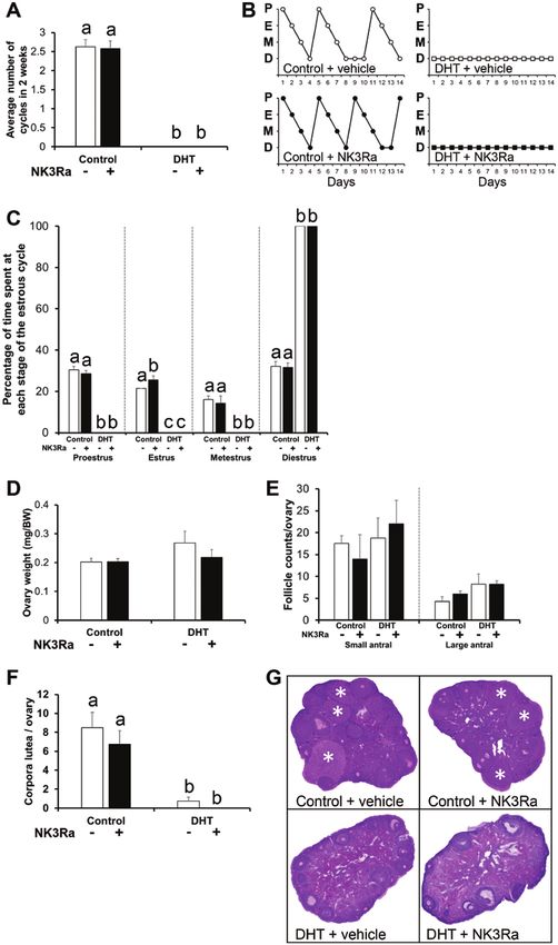
Figure 2. Estrous cycling and ovary weight and morphology. (A) Number of completed cycles in a 2-week period, showing dihydrotestosterone
(DHT)-induced acyclicity (P < .01) and NK3Ra had no influence on the number of completed cycles. n = 7-8 mice per experimental group. Data are
expressed as the mean ± standard error of the mean (SEM). (B) Estrous cycle pattern in representative females. P, proestrus; E, estrus; M, metestrus;
D, diestrus. (C) Percentage of time spent at each stage of the estrous cycle, showing significantly altered estrous cycles in DHT-exposed mice (P <
.01). n = 7-8 mice per experimental group. (D) Ovary weight, showing no significant differences between any experimental groups. n = 7-8 ovaries
per experimental group. Data expressed as the mean ± SEM. (E) Number of small antral and large antral follicles per ovary showing a main effect of
DHT with an accumulation of large antral follicles, but no significant effect of NK3Ra. n = 4 ovaries per experimental group. Data are expressed as
the mean ± SEM. (F) Number of corpora lutea per ovary showing DHT-induced anovulation (P < .01) but no significant effect of NK3Ra. n = 4 ovaries
per experimental group. Data are expressed as the mean ± SEM. (G) Histological sections of representative ovaries from control and DHT-induced
PCOS mice treated with and without the NK3R antagonist MLE4901. Star, corpora lutea. Different letters denote significant statistical differences.Endocrinology, 2021, Vol. 162, No. 5 7
observation of vaginal smears identified leukocytes as the pre- females displayed a 97% and 75% increase in adipocyte area
dominant cell type in PCOS-like mice, indicating that they were compared with their respective control females (Fig. 4A).
acyclic in pseudo-diestrus (P < .01) (Fig. 2C). NK3Ra treatment The degree of adipocyte hypertrophy observed in
for 4 weeks had no beneficial impact in overcoming cycle dis- parametrial fat pads of DHT + NK3Ra females was sig-
ruption, with all NK3Ra + DHT-exposed mice continuing to nificantly less than that exhibited in the DHT + vehicle
remain acyclic (Fig. 2A-2C). While ovary weight was not af- group (P < .01) (Fig. 4A and 4B). Additionally, circulating
fected by DHT exposure (Fig. 2D), DHT had a main effect on levels of the adipocyte-derived hormones adiponectin
the large antral follicle population (P < .05) with DHT-exposed and leptin, reported to be reduced (59) and elevated (60),
females displaying the PCOS-like ovarian characteristic of an respectively, in women with PCOS, were significantly
Downloaded from https://academic.oup.com/endo/article/162/5/bqab020/6125280 by Sydney College of Arts user on 09 April 2021
accumulation of antral follicles, evident by an increase in large reduced (P < .01) and significantly elevated (P < .01), re-
antral follicle numbers (Fig. 2E). NK3Ra treatment did not spectively, in DHT-exposed females (Fig. 4C and 4D).
overcome the effects of DHT treatment on increasing the antral Cotreatment of NK3Ra with DHT did not ameliorate
follicle population (Fig. 2E). the decrease in adiponectin levels in DHT-treated mice
In agreement with our previous studies (24, 53), DHT had a (Fig. 4C). In contrast, NK3Ra administration induced a
significant main effect (P < .01) with DHT-exposed ovaries ex- significant reduction in leptin levels in DHT-treated fe-
hibiting a drastic reduction in corpora lutea populations com- males (P < .01), albeit not to the levels in controls (Fig.
pared with control ovaries (Fig. 2F and 2G), consistent with 4D). Assessment of genes involved in metabolic pathways
ovulatory dysfunction, a key feature of PCOS. Treatment with in parametrial adipose tissue revealed a significant main
the NK3Ra did not significantly improve ovulatory dysfunc- effect of DHT, but not NK3Ra, with decreased mRNA
tion as there was no observed increase in corpora lutea popula- levels of Irs1 (P < .01) and AdipoR2 (P < .05) in DHT fe-
tions in DHT-exposed females treated with the NK3Ra (DHT males, relative to controls (Fig. 4E and 4F). Neither DHT
+ vehicle 0.8 ± 0.8; DHT + NK3Ra 0 ± 0, Fig. 2F). Hence, nor NK3Ra treatment significantly altered Pepck expres-
overall, NK3Ra treatment did not ameliorate reproductive sion (Fig. 4G).
PCOS-like traits in this mouse model of PCOS (Fig. 2A-2G).
NK3R Antagonism Did Not Alter Food Intake but
NK3R Antagonism Ameliorated Adiposity in a Increased the Respiratory Exchange Ratio in a
Peripubertal DHT-induced PCOS Mouse Model Peripubertal DHT-induced PCOS Mouse Model
DHT had a significant main effect on body weight, with DHT- Metabolic cage analysis of control and PCOS-like mice
exposed females exhibiting a significant increase in body treated with vehicle/NK3Ra, undertaken to identify
weight (P < .01) (Fig. 3A). NK3Ra treatment significantly underlying mechanisms for the observed amelioration in
reduced the magnitude of body weight increase in DHT + adiposity in PCOS-like mice treated with NK3Ra (Fig. 3),
NK3Ra mice (P < .05) (Fig. 3A). Similarly, DHT treatment had revealed no significant differences between any groups
a significant main effect on all fat pad weights, as it induced a for day and night food intake (Table 2). Similarly, no
significant increase in inguinal (P < .01), parametrial (P < .01), differences in day or night energy expenditure were ob-
retroperitoneal (P < .01), mesenteric (P < .01), and brown fat served between any group (Table 2). However, DHT had
pad weights (P < .01) (Fig. 3B-3F). NK3Ra administration a significant main effect (P < .05) on daytime locomotor
for 4 weeks completely overcame the impact of DHT treat- activity, with PCOS mice exhibiting lower day locomotor
ment on inguinal (P < .01) and mesenteric (P < .01) fat pad activity. This was also the case for night locomotor ac-
weights, and reduced the magnitude of increase in parametrial tivity with DHT mice exhibiting overall reduced activity
(P < .01) and retroperitoneal (P < .01) fat pads (Fig. 3B-3E). compared with control females (P < .05). Treatment with
Brown fat pad weight did not differ between DHT + vehicle NK3Ra did not significantly alter day or night locomotor
and DHT + NK3Ra groups (Fig. 3F). activity in DHT-exposed females (Table 2). However,
both DHT and NK3Ra significantly impacted the day
and night indirect calorimetry measurements of oxygen
NK3R Antagonism Reversed Adipocyte consumption, carbon dioxide production, and respira-
Hypertrophy in a Peripubertal DHT-induced PCOS tory exchange ratio (RER) (Table 2). Compared with
Mouse Model Control + vehicle mice, day (P < .01) and night (P < .01)
Histological analysis of parametrial fat pads revealed that RER was significantly reduced in DHT + vehicle mice
DHT exposure (P < .01) and NK3Ra (P < .01) induced (Table 2). In contrast, NK3Ra treatment significantly in-
significant main effects on parametrial fat depot adipocyte creased day (P < .01) and night (P < .01) RER in DHT +
area (Fig. 4A and 4B). DHT + vehicle and DHT + NK3Ra NK3Ra females compared with DHT + vehicle controls8 Endocrinology, 2021, Vol. 162, No. 5
Downloaded from https://academic.oup.com/endo/article/162/5/bqab020/6125280 by Sydney College of Arts user on 09 April 2021
Figure 3. Body weight and fat depot weights. (A) Body weight, showing dihydrotestosterone (DHT)-induced a significant increase in body weight (P
< .01) but NK3Ra caused a reduction in body weight in DHT + NK3Ra mice (P < .05). (B) Inguinal fat pad weight, showing DHT-induced an increase in
weight (P < .01) but this increase was completely ameliorated in DHT + NK3Ra mice (P < .01). (C) Parametrial fat pad weight, showing a DHT-induced
increase in weight (P < .01) but this increase was partially reversed in DHT + NK3Ra mice (P < .01). (D) Retroperitoneal fat pad weight, showing DHT-
induced a marked increase in weight (P < .01) but this increase was partially ameliorated in DHT + NK3Ra mice (P < .01). (E) Mesenteric fat pad weight,
showing a DHT-induced increase in weight (P < .01), which was completely reversed in DHT + NK3Ra mice (P < .01). (F) Brown fat depot weights,
showing a main effect of DHT (P < .01), but no beneficial impact of NK3Ra administration. For all graphs n = 7–8 per experimental group and data are
expressed as the mean ± standard error of the mean. Different letters denote significant statistical differences.
(Table 2), indicating that NK3Ra caused a shift to pref- an overall increase observed in DHT-exposed females com-
erential utilization of carbohydrates as the predominant pared with control females, but NK3Ra treatment had no
fuel source. effect regardless of DHT treatment (Fig. 5B). Peripubertal
DHT exposure significantly impacted fasting glucose levels
(P < .01), with both DHT + vehicle and DHT + NK3Ra fe-
Impact of NK3R Antagonism on Overall Glucose males exhibiting a significant increase in fasting glucose levels
Homeostasis in a Peripubertal DHT-induced PCOS compared with controls (Fig. 5C). However, NK3R antag-
Mouse Model onism had no effect on fasting glucose levels. A significant
There was no significant effect of DHT or NK3Ra treatment effect of DHT exposure was also apparent on overall glucose
on circulating cholesterol levels (Fig. 5A). In contrast, serum tolerance (P < .05). In addition, there was a trend (P = 0.09)
triglyceride levels differed by DHT treatment (P < .01), with to NK3Ra having a beneficial effect on glucose tolerance withEndocrinology, 2021, Vol. 162, No. 5 9
Downloaded from https://academic.oup.com/endo/article/162/5/bqab020/6125280 by Sydney College of Arts user on 09 April 2021
Figure 4. Fat depot histology, circulating adiponectin and leptin levels, and markers of adipocyte function. (A) Adipocyte size in parametrial fat
pads showing development of adipocyte hypertrophy in dihydrotestosterone (DHT)-exposed mice (P < .01), but a partial amelioration of adipocyte
hypertrophy in DHT + NK3Ra mice (P < .01). n = 5-6 per experimental group. Data are expressed as the mean ± standard error of the mean (SEM). (B)
Histological sections of representative parametrial fat pads from each treatment group showing the degree of adipocyte hypertrophy was reduced in
DHT + NK3Ra mice, compared to DHT + vehicle females. (C) Serum levels of adiponectin showing a DHT-induced decrease in levels (P < .01) and no
effect of NK3Ra treatment. n = 7-8 per experimental group. Data are expressed as the mean ± SEM. (D) Serum levels of leptin showing a DHT-induced
increase in levels (P < .01) and a partial restoration of levels in DHT + NK3Ra females (P < .01). n = 7-8 per experimental group. Data are expressed
as the mean ± SEM. Gene expression of Irs1 (E), AdipoR2 (F), and Pepck (F) in parametrial adipose tissue. n = 5-8 per experimental group. Data are
expressed as the mean ± SEM. Different letters denote significant statistical differences.10
Table 2. Metabolic state of control and PCOS mice with and without NK3Ra treatment
Day Night
Control + vehicle Control + NK3Ra DHT + vehicle DHT + NK3Ra Significance Control + vehicle Control + NK3Ra DHT + vehicle DHT + NK3Ra Significance
Food intake (g) 1.84 ± 0.33 1.79 ± 0.09 1.38 ± 0.14 1.80 ± 0.22 ns 2.08 ± 0.05 2.58 ± 0.26 2 ± 0.12 2.31 ± 0.24 ns
Locomotor activity 414.09 ± 37.37 424.96 ± 42.75 330.50 ± 26.85 340.71 ± 26.08 DHT main 860.76 ± 64.06a 983.20 ± 72.88a 833 ± 62.35a,b 636.94 ± 41.68b DHT × NK3Ra
(beam breaks) effect (P < .05) (P < .05)
Energy expenditure 12.97 ± 0.76 12.68 ± 0.47 13.78 ± 0.53 14.13 ± 0.29 ns 15.24 ± 1.09 15.16 ± 0.42 16.93 ± 0.64 16.46 ± 0.33 ns
(kcal/12 hours)
VO2 (mL/kg/hours) 4276.61 ± 88.01 4040.08 ± 66.05 4124.68 ± 73.69 3778.48 ± 51.44 DHT (P < .01) and 4743.04 ± 193.11a 4555.88 ± 80.69a 4801.66 ± 91.52a 4145.41 ± 57.95b DHT × NK3Ra
NK3Ra (P < .01) (P < .01)
main effects
VCO2 (mL/kg/hours) 3741.81 ± 79.06 3556 ± 64.28 3552.83 ± 64.40 3375.21 ± 48.84 DHT (P < .01) and 4158.27 ± 99.89 4038.95 ± 74.97 4092.43 ± 81.26 3742.83 ± 52.70 DHT (P < 0.05) and
NK3Ra (P < 0.01) NK3Ra (P < .01)
main effects main effects
RER (VCO2/VO2) 0.88 ± 0.0062a 0.88 ± 0.0049a 0.86 ± 0.0043bb 0.89 ± 0.0035c DHT × NK3Ra 0.87 ± 0.0045a 0.89 ± 0.0039b 0.85 ± 0.0037c 0.90 ± 0.0033d DHT × NK3Ra
(P < .01) (P < .01)
Measurements of indirect calorimetry by metabolic cages of control and PCOS mice with and without NK3Ra treatment on a standard chow diet, showing food intake (g) and energy expenditure (kcal/12 hours) do not differ
between groups. DHT had a main effect (P < .05) on locomotor activity (beam breaks), with DHT-induced PCOS mice displaying overall reduced day and night locomotor activity. Both DHT and NK3Ra treatments had sig-
nificant main effects on oxygen (O2) consumption (VO2), carbon dioxide (CO2) production (VCO2), and RER, with NK3Ra administration significantly reducing day (P < .05) and night (P < .01) RER in DHT-induced PCOS
mice. Data are the mean ± standard error of the mean; n = 4-5 mice. Different letters denote significant statistical differences.
Abbreviations: DHT, dihydrotestosterone; NK3R, neurokinin 3 receptor; PCOS, polycystic ovary syndrome; RER, respiratory exchange ratio.
Endocrinology, 2021, Vol. 162, No. 5
Downloaded from https://academic.oup.com/endo/article/162/5/bqab020/6125280 by Sydney College of Arts user on 09 April 2021Endocrinology, 2021, Vol. 162, No. 5 11
Downloaded from https://academic.oup.com/endo/article/162/5/bqab020/6125280 by Sydney College of Arts user on 09 April 2021
Figure 5. Serum cholesterol and triglyceride levels, fasting glucose levels, and glucose tolerance test. (A) Serum cholesterol levels showing no effect
of dihydrotestosterone (DHT0 exposure or NK3Ra treatment. (B) Serum triglyceride levels showing a main effect of DHT (P < .01), but no beneficial
impact of NK3Ra administration. (C) Fasting glucose levels showing a significant increase in glucose levels in DHT-exposed mice (P < .01), but no
significant effect of NK3Ra. (D) Incremental area under the curve analysis of glucose tolerance test showing DHT-induced an increase (P < .05) and
there was a trend (P = .09) for NK3Ra to have a beneficial effect on glucose tolerance in DHT + NK3Ra females. For all graphs n = 7-8 per experimental
group and data are expressed as the mean ± standard error of the mean. Different letters denote significant statistical difference.
glucose incremental area under the curve of DHT + NK3Ra for AR-mediated actions in establishing PCOS-associated
females being intermediate between control and DHT + ve- reproductive dysfunction. However, the downstream
hicle groups (Fig. 5D). AR-driven mechanisms involved remain to be established.
Previous findings from clinical investigations using NK3R
antagonists in non-PCOS (28-30, 64) and PCOS (35, 47)
Discussion populations revealed that NK3R antagonism decreases LH
The successful development of specific mechanism-based secretion, and LH hypersecretion and hyperandrogenism,
treatments for the management of PCOS would have a respectively, implicating involvement of the KNDy pathway.
significant impact on women’s reproductive health, as it In the present study, NK3R antagonism failed to overcome
would allow PCOS to be treated at its origin. In the cur- the reproductive deficits observed in the DHT-exposed fe-
rent study, we demonstrate that NK3R antagonism can males as they remained acyclic and anovulatory following
ameliorate a range of adverse metabolic PCOS traits in treatment with the NK3R antagonist MLE4901. Several
a peripubertal DHT-induced PCOS mouse model. These possibilities can be inferred from these findings. First, the
data provide evidence to support the therapeutic targeting data suggest that the development of AR-mediated repro-
of neuroendocrine actions in the development of novel ductive PCOS-like traits are not mediated via the NK3R
treatments to ameliorate the metabolic defects in PCOS. in this model, and that other mechanisms or pathways are
Induction of androgen excess replicates a wide range involved. Alternatively, NK3R may indeed be involved,
of endocrine, reproductive, and metabolic PCOS traits in but the antagonist was unable to overcome the chronically
a range of animal models (61, 62). Consistent with this, in elevated DHT of the model, or the antagonist dose was
the present study DHT-exposed females were completely not adequate. The latter is supported by the lack of im-
acyclic and displayed ovulatory dysfunction with a drastic pact on normal cycling in the Control + NK3Ra group.
reduction in corpora lutea numbers compared with con- Finally, these data also raise the possibility that the re-
trol mice. Both clinical (63) and experimental observations productive neuroendocrine axis is not hyperactive in the
(20, 22), where cycling regularity was restored by treat- chronic DHT model. Although evidence supports that the
ment with the AR antagonist flutamide, support a core role reproductive deficits of this model are mediated via excess12 Endocrinology, 2021, Vol. 162, No. 5
androgen signaling in the brain (24), it has yet to be de- potential role for SNS in PCOS requires further investigation.
termined whether LH pulse frequency is elevated in this A growing number of fat-borne factors, including adiponectin
model. A limitation of the present study is the lack of serial and leptin, have been shown to have an impact on metabolic as
blood sampling to measure pulsatile LH secretion, which well as reproductive function (75, 76). The reduction in serum
would indicate whether the model reflects a hyperactive adiponectin and AdipoR2 expression levels in adipose tissue
reproductive axis and whether the NKB antagonism was and increase in leptin levels in DHT-treated female mice mimic
able to impact LH pulse dynamics. In prenatally andro- the findings reported in PCOS women (59, 60). Moreover, the
genized mice that exhibit elevated LH pulsatility, blockade reduced leptin levels in PCOS-like females following NK3Ra
of inhibition of AR signaling reversed identified brain cir- treatment parallel the observed reduction in fat mass in DHT
Downloaded from https://academic.oup.com/endo/article/162/5/bqab020/6125280 by Sydney College of Arts user on 09 April 2021
cuit abnormalities, improved ovarian morphology and + NK3Ra mice.
restored reproductive cycling (20), supporting the theory The well-documented association of poor metabolic health
that blockade of androgen signaling, and its downstream and PCOS often presents in patients as dyslipidemia, poor
mechanisms, can reverse a PCOS phenotype. It will be im- glucose homeostasis, hyperinsulinemia, and insulin resistance
portant in future studies to assess the LH pulse dynamics (77, 78). Elevated fasting levels and incremental area under
in the chronic DHT model, and to determine whether NKB the curve of glucose were observed in DHT-exposed female
antagonism can ameliorate reproductive deficits in a model mice. While NK3R antagonism had no influence on fasting
in which androgen excess is endogenous versus exogenous. glucose levels, the NK3R antagonist, to a degree, reduced the
Compared with control females, vehicle-treated DHT- effect of DHT treatment, as blood glucose incremental area
exposed females exhibited a significant increase in body under the curve levels were similar to those observed in con-
weight and fat pad weights, which is in line with clinical trol females. Accumulating evidence suggests that the brain,
studies demonstrating that women with PCOS manifest particularly the hypothalamus, plays a key role in the homeo-
global adiposity (65). Strikingly, 4 weeks of NK3R antag- static regulation of energy and glucose metabolism and that
onist treatment completely overcame the impact of DHT defective crosstalk between the brain and peripheral meta-
exposure on inguinal and mesenteric fat pads and reduced bolic organs may contribute to the pathogenesis of type 2 dia-
the extent of increase in parametrial and retroperitoneal betes (79, 80). Our results are consistent with this theory and
fat pads. Our studies with metabolic cages revealed that imply neural mechanisms may contribute to the dysfunctional
NK3Ra treatment had no impact on day or night food glucose homeostasis in PCOS patients. Flutamide improves
intakes, energy expenditure, or locomotor activity. Thus, lipid profiles in women suffering from PCOS (81), supporting
ruling out a decrease in food intake or energy expenditure AR-driven pathways in the etiology of PCOS-associated
as a reason for the reduction in body and fat pad weights dyslipidemia. In the current study, circulating cholesterol and
in DHT + NK3Ra mice. However, NK3Ra treatment had triglyceride levels were not influenced by NK3R antagonism
an impact on RER, reflected as a significant shift in RER to suggesting that other AR mechanisms are involved in driving
higher values. This suggests a global change in metabolism dyslipidemia in this model of PCOS.
in the DHT + NK3Ra mice compared with DHT + vehicle The beneficial effect of NK3R antagonism on body weight,
females, and suggests that NK3Ra caused a change in fuel adiposity, and adipocyte hypertrophy in DHT-exposed mice
utilization, to one with carbohydrate as the predominant is in line with prior studies that highlight the importance of
fuel source (66). Taken together, these findings infer that, neuroendocrine actions in the development of PCOS fea-
rather than having an influence on food intake, NK3Ra tures (20, 24, 82). Neuron-specific AR deletion was found
had a beneficial influence on metabolic PCOS traits by al- to protect against DHT-induced PCOS-like characteristics,
tering the metabolic status of the PCOS-like females. including the elimination of increases in body weight, fat pad
It is well documented that impairments in adipose tissue weights, pronounced adipocyte hypertrophy, and fatty liver,
morphology and function are present in PCOS patients, with as well as hyperlipidemia (24). However, the AR-activated
evidence indicating that aberrations in adipose tissue play an downstream pathways inducing the development of these
important role in the metabolic dysfunction observed in PCOS traits remain to be elucidated. It has been speculated that
(67-72). In the present study, DHT-exposed females exhibited excessive AR signaling in hypothalamic neurons releasing
adipocyte hypertrophy, while treatment with an NK3R antag- neuronal peptides involved in controlling food intake, en-
onist partially ameliorated this trait, inferring that communi- ergy balance and metabolism, such as agouti-related peptide,
cation between the brain and adipose tissue is one contributor neuropeptide Y, or preproopiomelanocortin, may lead to the
in PCOS pathogenesis. How this beneficial effect is mediated AR-mediated establishment of PCOS-like metabolic dysfunc-
is unclear; however, there is evidence to support brain–adi- tion (83). This hypothesis is consistent with the observation
pose tissue crosstalk through sympathetic nervous system that in PCOS-like sheep, the AR antagonist flutamide pre-
(SNS) innervation of white adipose tissue (73). The SNS has vented prenatal DHT programming of agouti-related pep-
been associated with PCOS (74), but the relationship and a tide neuronal changes (83). The findings from this study alsoEndocrinology, 2021, Vol. 162, No. 5 13
support this hypothesis and suggest that the KNDy network 4. Dumesic DA, Oberfield SE, Stener-Victorin E, Marshall JC,
may be involved in mediating the AR-driven alterations in Laven JS, Legro RS. Scientific statement on the diagnostic cri-
metabolic function associated with PCOS. teria, epidemiology, pathophysiology, and molecular genetics of
polycystic ovary syndrome. Endocr Rev. 2015;36(5):487-525.
Substantial evidence supports a role for AR driven path-
5. Teede HJ, Misso ML, Costello MF, et al.; International PCOS
ways in the developmental origins of PCOS traits (12). In Network. Recommendations from the international evidence-
particular, neuroendocrine androgen actions have been based guideline for the assessment and management of poly-
highlighted as being important, with brain circuitry im- cystic ovary syndrome. Hum Reprod. 2018;33(9):1602-1618.
pairments identified across several species of PCOS animal 6. Moran LJ, Norman RJ, Teede HJ. Metabolic risk in PCOS:
models (82, 84). However, the precise neuroendocrine path- phenotype and adiposity impact. Trends Endocrinol Metab.
Downloaded from https://academic.oup.com/endo/article/162/5/bqab020/6125280 by Sydney College of Arts user on 09 April 2021
ways remain to be fully elucidated. The findings from the 2015;26(3):136-143.
7. Rubin KH, Glintborg D, Nybo M, Abrahamsen B, Andersen M.
current study have revealed that NK3R antagonism ameli-
Development and risk factors of type 2 diabetes in a nationwide
orates a range of adverse PCOS-associated metabolic, but
population of women with polycystic ovary syndrome. J Clin
not reproductive traits, in a PCOS mouse model, inferring Endocrinol Metab. 2017;102(10):3848-3857.
this pathway plays a role in mediating metabolic aspects of 8. Glintborg D, Rubin KH, Nybo M, Abrahamsen B, Andersen M.
PCOS pathogenesis. The observed beneficial effect of NK3R Cardiovascular disease in a nationwide population of Danish
antagonism on experimentally induced PCOS-like features women with polycystic ovary syndrome. Cardiovasc Diabetol.
provides strong evidence supporting the notion of manipu- 2018;17(1):37.
lation of neuroendocrine signaling as a viable new approach 9. Rocca ML, Venturella R, Mocciaro R, et al. Polycystic ovary syn-
drome: chemical pharmacotherapy. Expert Opin Pharmacother.
to treat PCOS and may hold great potential for the develop-
2015;16(9):1369-1393.
ment of novel therapeutic approaches to treat PCOS-related
10. Livadas S, Pappas C, Karachalios A, et al. Prevalence and im-
metabolic dysregulation. pact of hyperandrogenemia in 1218 women with polycystic
ovary syndrome. Endocrine. 2014;47(2):631-638.
11. Abbott DH, Dumesic DA, Levine JE. Hyperandrogenic ori-
Acknowledgments gins of polycystic ovary syndrome—implications for patho-
We thank Madeleine Cox for technical support with this project. physiology and therapy. Expert Rev Endocrinol Metab.
Financial Support: This work was supported by an Australian 2019;14(2):131-143.
National Health and Medical Research Council (NHMRC) Project 12. Walters KA, Rodriguez Paris V, Aflatounian A, Handelsman DJ.
Grant (APP1158540) and Fellowship (APP1117538), the Endocrine Androgens and ovarian function: translation from basic
Society of Australia, and by the School of Women’s and Children’s discovery research to clinical impact. J Endocrinol.
Health, University of New South Wales Sydney, including for a PhD 2019;242(2):R23-R50.
Scholarship awarded to Irene Sucquart. 13. Abbott DH, Rogers J, Dumesic DA, Levine JE. Naturally
occurring and experimentally induced rhesus macaque models
for polycystic ovary syndrome: translational gateways to clin-
Additional Information ical application. Med Sci (Basel). 2019;7(12):107.
Correspondence: Kirsty Walters, Fertility and Research Centre, 14. Cardoso RC, Padmanabhan V. Developmental programming
School of Women’s & Children’s Health, University of New South of PCOS traits: insights from the sheep. Med Sci (Basel).
Wales, Sydney, NSW 2052, Australia. Email: k.walters@unsw.edu.au. 2019;7(7):79.
Disclosures: I.E.S., R.N., M.C.E., V.R.P., A.A., M.J.B., R.E.C., R.B.G., 15. Hogg K, Wood C, McNeilly AS, Duncan WC. The in utero pro-
D.P.B., D.J.H., V.P., R.A.A., and K.A.W. have nothing to disclose. gramming effect of increased maternal androgens and a direct
Data Availability: Some or all datasets generated during and/or fetal intervention on liver and metabolic function in adult sheep.
analyzed during the current study are not publicly available but are PLoS One. 2011;6(9):e24877.
available from the corresponding author on reasonable request. 16. Siemienowicz KJ, Coukan F, Franks S, Rae MT, Duncan WC.
Aberrant subcutaneous adipogenesis precedes adult metabolic
dysfunction in an ovine model of polycystic ovary syndrome
References (PCOS). Mol Cell Endocrinol. 2021;519:111042.
1. March WA, Moore VM, Willson KJ, Phillips DI, Norman RJ, 17. Walters KA, Allan CM, Handelsman DJ. Rodent models
Davies MJ. The prevalence of polycystic ovary syndrome in a for human polycystic ovary syndrome. Biol Reprod.
community sample assessed under contrasting diagnostic cri- 2012;86(5):149, 1-,12.
teria. Hum Reprod. 2010;25(2):544-551. 18. Rittmaster RS. Antiandrogen treatment of polycystic
2. Bozdag G, Mumusoglu S, Zengin D, Karabulut E, Yildiz BO. ovary syndrome. Endocrinol Metab Clin North Am.
The prevalence and phenotypic features of polycystic ovary syn- 1999;28(2):409-421.
drome: a systematic review and meta-analysis. Hum Reprod. 19. Ryan GE, Malik S, Mellon PL. Antiandrogen treatment ameliorates
2016;31(12):2841-2855. reproductive and metabolic phenotypes in the letrozole-induced
3. Skiba MA, Islam RM, Bell RJ, Davis SR. Understanding vari- mouse model of PCOS. Endocrinology. 2018;159(4):1734-1747.
ation in prevalence estimates of polycystic ovary syndrome: 20. Silva MS, Prescott M, Campbell RE. Ontogeny and reversal of
a systematic review and meta-analysis. Hum Reprod Update. brain circuit abnormalities in a preclinical model of PCOS. JCI
2018;24(6):694-709. Insight. 2018;3(7):e99405.14 Endocrinology, 2021, Vol. 162, No. 5
21. Padmanabhan V, Veiga-Lopez A, Herkimer C, et al. 36. Smith JT. Sex steroid regulation of kisspeptin circuits. Adv Exp
Developmental programming: prenatal and postnatal an- Med Biol. 2013;784:275-295.
drogen antagonist and insulin sensitizer interventions 37. Quennell JH, Howell CS, Roa J, Augustine RA, Grattan DR,
prevent advancement of puberty and improve LH surge dy- Anderson GM. Leptin deficiency and diet-induced obesity reduce
namics in prenatal testosterone-treated sheep. Endocrinology. hypothalamic kisspeptin expression in mice. Endocrinology.
2015;156(7):2678-2692. 2011;152(4):1541-1550.
22. Sullivan SD, Moenter SM. Prenatal androgens alter GABAergic 38. Qiu J, Fang Y, Bosch MA, Rønnekleiv OK, Kelly MJ.
drive to gonadotropin-releasing hormone neurons: implica- Guinea pig kisspeptin neurons are depolarized by
tions for a common fertility disorder. Proc Natl Acad Sci U S A. leptin via activation of TRPC channels. Endocrinology.
2004;101(18):7129-7134. 2011;152(4):1503-1514.
Downloaded from https://academic.oup.com/endo/article/162/5/bqab020/6125280 by Sydney College of Arts user on 09 April 2021
23. Caldwell AS, Eid S, Kay CR, et al. Haplosufficient gen- 39. Smith JT, Acohido BV, Clifton DK, Steiner RA. KiSS-1 neur-
omic androgen receptor signaling is adequate to protect fe- ones are direct targets for leptin in the ob/ob mouse. J
male mice from induction of polycystic ovary syndrome Neuroendocrinol. 2006;18(4):298-303.
features by prenatal hyperandrogenization. Endocrinology. 40. Hiney JK, Srivastava VK, Pine MD, Les Dees W. Insulin-
2015;156(4):1441-1452. like growth factor-I activates KiSS-1 gene expression in
24. Caldwell ASL, Edwards MC, Desai R, et al. Neuroendocrine the brain of the prepubertal female rat. Endocrinology.
androgen action is a key extraovarian mediator in the develop- 2009;150(1):376-384.
ment of polycystic ovary syndrome. Proc Natl Acad Sci U S A. 41. Thorson JF, Prezotto LD, Adams H, et al. Energy balance
2017;114(16):E3334-E3343. affects pulsatile secretion of luteinizing hormone from
25. Abbott DH. Neuronal androgen receptor: Molecular gateway the adenohypophesis and expression of neurokinin B in
to polycystic ovary syndrome? Proc Natl Acad Sci U S A. the hypothalamus of ovariectomized gilts. Biol Reprod.
2017;114(16):4045-4047. 2018;99(2):433-445.
26. Walters KA, Edwards MC, Tesic D, et al. The role of 42. Merkley CM, Shuping SL, Nestor CC. Neuronal networks that
central androgen receptor actions in regulating the regulate gonadotropin-releasing hormone/luteinizing hormone
hypothalamic-pituitary-ovarian axis. Neuroendocrinology. secretion during undernutrition: evidence from sheep. Domest
2018;106(4):389-400. Anim Endocrinol. 2020;73:106469.
27. Navarro VM, Gottsch ML, Chavkin C, Okamura H, 43. Albalawi FS, Daghestani MH, Daghestani MH, Eldali A, Warsy AS.
Clifton DK, Steiner RA. Regulation of gonadotropin-releasing rs4889 polymorphism in KISS1 gene, its effect on polycystic ovary
hormone secretion by kisspeptin/dynorphin/neurokinin B syndrome development and anthropometric and hormonal param-
neurons in the arcuate nucleus of the mouse. J Neurosci. eters in Saudi women. J Biomed Sci. 2018;25(1):50.
2009;29(38):11859-11866. 44. Umayal B, Jayakody SN, Chandrasekharan NV,
28. Skorupskaite K, George JT, Veldhuis JD, Millar RP, Anderson RA. Wijesundera WS, Wijeyaratne CN. Polycystic ovary syndrome
Interactions between neurokinin B and kisspeptin in mediating (PCOS) and kisspeptin - A Sri Lankan study. J Postgrad Med.
estrogen feedback in healthy women. J Clin Endocrinol Metab. 2019;65(1):18-23.
2016;101(12):4628-4636. 45. Jeon YE, Lee KE, Jung JA, et al. Kisspeptin, leptin, and retinol-
29. Fraser GL, Ramael S, Hoveyda HR, Gheyle L, Combalbert J. binding protein 4 in women with polycystic ovary syndrome.
The NK3 receptor antagonist ESN364 suppresses sex hor- Gynecol Obstet Invest. 2013;75(4):268-274.
mones in men and women. J Clin Endocrinol Metab. 46. Daghestani MH. Evaluation of biochemical, endocrine, and
2016;101(2):417-426. metabolic biomarkers for the early diagnosis of polycystic ovary
30. Skorupskaite K, George JT, Veldhuis JD, Anderson RA. syndrome among non-obese Saudi women. Int J Gynaecol
Neurokinin B regulates gonadotropin secretion, ovarian follicle Obstet. 2018;142(2):162-169.
growth, and the timing of ovulation in healthy women. J Clin 47. Skorupskaite K, George JT, Veldhuis JD, Millar RP,
Endocrinol Metab. 2018;103(1):95-104. Anderson RA. Kisspeptin and neurokinin B interactions in
31. Clarkson J, Han SY, Piet R, et al. Definition of the hypothal- modulating gonadotropin secretion in women with polycystic
amic GnRH pulse generator in mice. Proc Natl Acad Sci U S A. ovary syndrome. Hum Reprod. 2020;35(6):1421-1431.
2017;114(47):E10216-E10223. 48. Osuka S, Iwase A, Nakahara T, et al. Kisspeptin in the hypo-
32. Moore AM, Coolen LM, Porter DT, Goodman RL, Lehman MN. thalamus of 2 rat models of polycystic ovary syndrome.
KNDy Cells Revisited. Endocrinology. 2018;159(9):3219-3234. Endocrinology. 2017;158(2):367-377.
33. Pennefather JN, Lecci A, Candenas ML, Patak E, Pinto FM, 49. Brown RE, Wilkinson DA, Imran SA, Caraty A, Wilkinson M.
Maggi CA. Tachykinins and tachykinin receptors: a growing Hypothalamic kiss1 mRNA and kisspeptin immunoreactivity
family. Life Sci. 2004;74(12):1445-1463. are reduced in a rat model of polycystic ovary syndrome
34. Topaloglu AK, Reimann F, Guclu M, et al. TAC3 and TACR3 (PCOS). Brain Res. 2012;1467:1-9.
mutations in familial hypogonadotropic hypogonadism reveal a 50. Cheng G, Coolen LM, Padmanabhan V, Goodman RL,
key role for Neurokinin B in the central control of reproduction. Lehman MN. The kisspeptin/neurokinin B/dynorphin (KNDy)
Nat Genet. 2009;41(3):354-358. cell population of the arcuate nucleus: sex differences and
35. George JT, Kakkar R, Marshall J, et al. Neurokinin B receptor effects of prenatal testosterone in sheep. Endocrinology.
antagonism in women with polycystic ovary syndrome: a ran- 2010;151(1):301-311.
domized, placebo-controlled trial. J Clin Endocrinol Metab. 51. Cernea M, Padmanabhan V, Goodman RL, Coolen LM,
2016;101(11):4313-4321. Lehman MN. Prenatal testosterone treatment leads to changesYou can also read