PROTEIN MISFOLDING AND AMYLOIDOSIS XIII - Miniworkshop and CIMN Meeting May 23-24, 2019 - Dipartimento di Fisica
←
→
Page content transcription
If your browser does not render page correctly, please read the page content below
Miniworkshop and CIMN Meeting
  PROTEIN MISFOLDING
  AND AMYLOIDOSIS XIII
             May 23-24, 2019
 Department of Physics, University of Genoa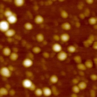
Miniworkshop and CIMN Meeting
PROTEIN MISFOLDING AND AMYLOIDOSIS XIII
Genoa, May 23-24, 2019
Department of Physics, Via Dodecaneso 33
Thursday, May 23
12:00-12:50 Welcome Buffet
13:00 – 13:10 Welcome from the Head of the Physics Department
Chair: Massimo Stefani
13:10 – 13:40 Marcus Fändrich (Institute of Protein Biochemistry, Ulm University) Cryo-EM
structures of fibrils from systemic amyloidosis.
13:40 – 14:00 Stefano Ricagno (Department of Biosciences, University of Milan) Cryo-EM structure
of cardiac amyloid fibrils from an immunoglobulin light chain AL amyloidosis patient.
14:00 – 14:20 Francesca Lavatelli (Fondazione IRCCS Policlinico San Matteo and Department of
Molecular Medicine, University of Pavia) Proteotoxicity of amyloidogenic cardiotropic light chains.
14:20 – 14:40 Giulia Fani (Department of Experimental and Clinical Biomedical Sciences, University
of Florence) Identification of membrane Ca2+ channels activated by protein misfolded oligomers.
14:40 – 15:00 Maria Elena Regonesi (Department of Biotechnology and Bioscience, University of
Milano-Bicocca) Characterization of CASA complex: role of HspB8 in protein quality control in
Spinocerebellar Ataxia type 3 model.
15:00-15:30 Coffee break
Chair: Patrizia Polverino de Laureto
15:30 – 15:50 Valeria Crippa (Department of Pharmacological and Biomolecular Sciences,
University of Milan) The clearance of aggregated TDP-43 responsible for ALS/FTD diseases
15:50 – 16:10 Margherita Romeo (Department of Molecular Biochemistry and Pharmacology,
Istituto di Ricerche Farmacologiche Mario Negri IRCCS, Milan) Investigating the spreading and
toxicity of tau using C. elegans.
16:10 – 16:30 Chiara Maria Giulia De Luca (Fondazione IRCCS Istituto Neurologico Carlo Besta,
Unit of Neurology 5 and Neuropathology, Milan) Efficient RT-QuIC seeding activity for α-synuclein in
olfactory mucosa samples of patients with Parkinson’s disease and Multiple System Atrophy.
Chair: Fabrizio Chiti
16:30 – 18:00 General discussion on key issues of amyloidogenesis
20:00 – Social dinner (I Tre Merli, Calata Cattaneo 17, Porto Antico)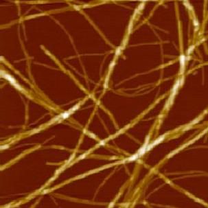
Friday, May 24
Chair: Luisa Diomede
9:00 – 9:20 Patrizia Polverino de Laureto (Department of Pharmaceutical and Pharmacological
Sciences, CRIBI Biotechnology Centre, University of Padova) Could polyphenol compounds be
promising drugs for Parkinson’s Disease?
9:20 – 9:40 Manuela Leri (Department of Experimental and Clinical Biomedical Sciences, University
of Florence) EVOO polyphenols can relieve autophagy dysregulation in Alzheimer's disease.
9:40 – 10:00 Giulia Faravelli (Department of Molecular Medicine, University of Pavia) C. elegans
expressing D76N beta-2microglobulin: a useful model for screening drug candidates targeting
amyloidosis.
10:00 – 10:20 Stefano Thellung (Section of Pharmacology, Department of Internal Medicine and
Centre of Excellence for Biomedical Research, University of Genoa) Prion protein fragment 90-231
impairs autophagic flux in neuronal cultures inducing cell death.
10:20 – 10:40 Andrea Danani (IDSIA- Dalle Molle Institute for Artificial Intelligence, Manno,
Switzerland) Structural polymorphism of Alzheimer’s beta amyloid fibrils.
10:40-11:00 Coffee break
Chair: Martino Bolognesi
11:00 – 11:20 Ciro Cecconi (Department of Physics, Informatics and Mathematics, University of
Modena and Reggio Emilia, and Institute of Nanoscience S3, CNR) Single-molecule studies of the
molecular mechanisms underlying the chaperone activity of HSPB8.
11:20 – 11:40 Claudio Canale (Department of Physics, University of Genoa) Unexpected asymmetry
in the aggregation of partially labeled peptides.
11:40 – 12:00 Alberto Diaspro (Department of Physics, University of Genoa and Nanoscopy, IIT)
Multi-Messenger Optical Microscopy to study biological macromolecules in cells.
12:00 Short talks:
12:00 – 12:10 Luca Oberti (Department of Biosciences, University of Milan) Rationally designed
mutant light chain highlights properties modulating the soluble toxicity
12:10 – 12:15 Davide Odino (Department of Physics, University of Genoa) Trodusquemine induces
changes in the properties of biomimetic membranes.
12:15 – 12:20 Mattia Siri (Department of Pharmaceutical Sciences, University of Genoa) Inhibition
of amyloid beta aggregation by multi-target compounds.
12:20 Final discussion
12:45 Buffet
Miniworkshop and CIMN Meeting
PROTEIN MISFOLDING AND AMYLOIDOSIS XIII
                Genoa, May 23-24, 2019
        Department of Physics, Via Dodecaneso 33

            ABSTRACTS
Miniworkshop and CIMN Meeting “Protein misfolding and amyloidosis XIII”
                             Genoa, May 23 – 24, 2019

                Cryo-EM structures of fibrils from systemic amyloidosis

                                         Fändrich, Marcus

                         Institute of Protein Biochemistry, Ulm University
        Helmholtzstrasse 8/1, 89081 Ulm, Germany, Email: marcus.faendrich@uni-ulm.de

Systemic amyloidosis is a group of protein misfolding disease in which the production site of the
fibril precursor protein does not necessarily correspond to the deposition site in the body. In
systemic AA amyloidosis, the fibril precursor serum amyloid A protein is produced mainly in liver
but deposits in spleen, kidney and other organs. In systemic AL amyloidosis, the fibril precursor is
an immunoglobulin light chain. Systemic AA amyloidosis, which affects humans and many animal
species, is one of the best cases of a prion-like disorder in mammals. We have used cryo electron
microscopy to determine the molecular structures of AL and AA amyloid fibrils purified from human
patient tissue as well as from AA amyloidotic mice. Our research provides insights into the
mechanisms of protein misfolding in vivo and species barrier during prion-like cross-seeding.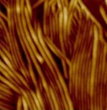
Miniworkshop and CIMN Meeting “Protein misfolding and amyloidosis XIII”
                             Genoa, May 23 – 24, 2019

Cryo-EM structure of cardiac amyloid fibrils from an immunoglobulin light chain AL
                               amyloidosis patient

Paolo Swuec 1,2, Francesca Lavatelli3, Masayoshi Tasaki3,4, Cristina Paissoni1, Paola Rognoni3,
   Martina Maritan1, Francesca Brambilla5, Paolo Milani3, Pierluigi Mauri 5, Carlo Camilloni 1,
       Giovanni Palladini3, Giampaolo Merlini3, Stefano Ricagno 1 & Martino Bolognesi 1,2

1 Dipartimento di Bioscienze, Università degli Studi di Milano, Via Celoria 26, 20133 Milano, Italy. 2
Centro di Ricerca Pediatrica Romeo ed Enrica Invernizzi, Università degli Studi di Milano, Via Celoria
26, 20133 Milano, Italy. 3 Amyloidosis Research and Treatment Center, Fondazione IRCCS Policlinico
San Matteo, and Department of Molecular Medicine, University of Pavia, P.le Golgi 19, 27100 Pavia,
Italy. 4 Department of Morphological and Physiological Sciences, Graduate School of Health Sciences,,
Kumamoto University, 4-24-1 Kuhonji, Kumamoto 862-0976, Japan. 5 Institute for Biomedical
Technologies-CNR, Via Fratelli Cervi 93, 20090 Segrate, Italy. These authors contributed equally:
Paolo Swuec, Francesca Lavatelli.

Systemic light chain amyloidosis (AL) is a life-threatening disease caused by aggregation and
deposition of monoclonal immunoglobulin light chains (LC) in target organs. Severity of heart
involvement is the most important factor determining prognosis. Here, we report the 4.0 Å
resolution cryo-electron microscopy map and molecular model of amyloid fibrils extracted from the
heart of an AL amyloidosis patient with severe amyloid cardiomyopathy. The helical fibrils are
composed of a single protofilament, showing typical 4.9 Å stacking and cross-β architecture. Two
distinct polypeptide stretches (total of 77 residues) from the LC variable domain (Vl) fit the fibril
density. Despite Vl high sequence variability, residues stabilizing the fibril core are conserved
through different cardiotoxic Vl, highlighting structural motifs that may be common to misfolding-
prone LCs. Our data shed light on the architecture of LC amyloids, correlate amino acid sequences
with fibril assembly, providing the grounds for development of innovative medicines.
Miniworkshop and CIMN Meeting “Protein misfolding and amyloidosis XIII”
Genoa, May 23 – 24, 2019
Proteotoxicity of amyloidogenic cardiotropic light chains
Giulia Mazzini1, Paola Rognoni1, Diletta Ami2, Esther Imperlini3, Paolo Mereghetti2, Antonino
Natalello2, Mario Nuvolone1, Masayoshi Tasaki4, Giovanni Palladini1, Giampaolo Merlini1,
Francesca Lavatelli1
1
Amyloidosis Research and Treatment Centre, Fondazione IRCCS Policlinico San Matteo and
Department of Molecular Medicine, University of Pavia, Pavia, Italy, 2Department of Biotechnology
and Biosciences, University of Milano-Bicocca, Milano, Italy, 3Ceinge Biotecnologie Avanzate,
Napoli, Italy, 4Department of Neurology, Graduate School of Medical Sciences, Kumamoto
University, Kumamoto, Japan
In vivo and in vitro evidence indicates that, in light chain (AL) amyloidosis, soluble amyloidogenic
light chains (LCs) concur with fibrils to generate organ damage, being proteotoxic to target cells.
Previous studies showed that cardiotropic LCs alter the viability of cultured cardiac cells and cause
proteome remodeling. Little is known, however, on the kinetics of damage and the sequence of
molecular events associated with toxicity.
Using patient-derived LCs with a characterized clinical phenotype (either isolated from patients’
urines or expressed as recombinant proteins), we are investigating the cell alterations and
amyloidogenic LCs trafficking in cultured human cardiac fibroblasts exposed to these proteins for
different lengths of time (30 min-6 days). A combination of cell biology, imaging and biophysics
approaches (in particular FTIR microspectroscopy supported by multivariate analysis) is being
used for this purpose. The biochemical features of natural fibrillar LCs are being studied by gel-
based and gel-free proteomics techniques, in order to detect features possibly involved in
pathogenicity, in particular post-translational modifications.
LCs are internalized at early timepoints; exposure to amyloidogenic cardiotropic LCs leads to time-
dependent decrease in MTT reduction, without significant cell death. Removal of cardiotropic LCs
did not translate into cell recovery in vitro. Preliminary FTIR results provided information on
changes in conformational properties of the cell proteome and on early modifications in the infrared
response of cell lipids, possibly modulating membrane fluidity. Deposited LCs, solubilised from ex-
vivo isolated fibrils, consist in a complex ensemble of fragments containing the variable LC region,
whose sites of C-terminal cleavage are being characterized.
Studying the effects of cardiotropic LCs on target cells for extended time will allow defining the time
course of damage, as well as the way LCs are trafficked and processed by cells. Characterization
of the LCs species in ex-vivo fibrils will be functional to a better understanding of the mechanisms
leading to aggregation and toxicity in vivo.Miniworkshop and CIMN Meeting “Protein misfolding and amyloidosis XIII”
Genoa, May 23 – 24, 2019
Identification of membrane Ca2+ channels activated by protein misfolded oligomers
Giulia Fani1,2, Benedetta Mannini2, Giulia Vecchi2, Roberta Cascella1, Cristina Cecchi1, Christopher
M. Dobson1, Michele Vendruscolo1 and Fabrizio Chiti1
1
Department of Experimental and Clinical Biomedical Sciences, University of Florence, Florence,
Italy
2
Centre for Misfolding Diseases, Department of Chemistry, University of Cambridge, Cambridge,
United Kingdom
An early biochemical modification following the interaction of protein oligomers with cells is the
disruption of intracellular calcium homeostasis. To this aim, we tried to identify specific Ca2+
channels involved in that abnormal influx of Ca2+ and whether it is channel-specific or non-specific.
The ability of the bacterial protein HypF-N to produce stable toxic oligomers structurally and
functionally similar to those associated with Alzheimer disease allows to benefit from large
amounts of these species, in addition to using toxic Aβ oligomers, to analyse the influx of Ca2+
caused by their interaction with the cell membrane. The potential role of five calcium channels,
previously identified as specifically interacting with toxic HypF-N oligomers, was investigated, using
specific inhibitors in N13 microglial and SH-SY5Y neuroblastoma cells. Moreover, other well-known
calcium receptors, such as AMPA and NMDA receptors, were examined. To obtain information on
the specific interaction between the channel and the oligomers, the FRET technique was used.
The data show that the Ca2+ channels interacting directly with the oligomers are not involved in the
influx of Ca2+. By contrast, AMPA and NMDA receptors appear to be involved in an immediate
influx, as a result of an oligomer-induced membrane destabilization that contributes to their
opening, in the absence of a direct interaction with the oligomers. Most of the late Ca2+ entrance is
nonspecific, with a direct passage of the Ca2+ ions through the destabilised membrane.Miniworkshop and CIMN Meeting “Protein misfolding and amyloidosis XIII”
Genoa, May 23 – 24, 2019
Characterization of the CASA complex: role of HspB8 in protein quality control in
Spinocerebellar Ataxia type 3 model.
Loredana Amigoni, Antonino Natalello, Diletta Ami, Serena Cecilia Pagliara, Marta Rivolta,
Annalisa D’Urzo, Marco Vanoni, Paolo Tortora, Maria Elena Regonesi
University of Milano-Bicocca, Department of Biotechnology and Bioscience, Milan.
It is known that several neurodegenerative diseases are triggered by the failure by protein quality
control mechanisms, which modulate protein turnover, folding and stability and prevent excessive
accumulation of protein aggregates. Neuronal damage is often due to low efficiency or impairment
of protein degradative mechanisms. Two pathways are involved in the clearance of misfolded
proteins and aggregates: the more selective ubiquitin proteasome system (UPS), and autophagy.
The “CASA” complex is a new chaperone assembly that stimulates degradation of protein
substrates by macroautophagy. The complex contains Bag3 (Bcl-2 associated athanogene protein
3), a member of the Bag family, a group of proteins having co-chaperone activity, characterized by
the presence of a Bag domain responsible for interactions with Hsp70. A further member is HspB8,
a sHsp, ATP-independent chaperone containing an α-crystallin domain, flanked by N-terminal and
C-terminal regions, involve in substrates recognition and interaction with other chaperons. This
complex seems to play a pivotal role in amyloid aggregates clearance in several
neurodegenerative disease models. Nevertheless, little is known about the mode of interaction
among the chaperones and their mechanism of action. We obtained HspB8 and Bag3 in purified
form and we studied their tendency to autoassemble and to form the complex. For this purpose, we
carried out size exclusion chromatography (SEC), circular dichroism (CD), Fourier Transform
Infrared Spectroscopy (FTIR), thioflavine T (ThT) analyses and solubility assays. CD and FTIR
analyses confirmed the IDP nature of both proteins. We demonstrated that HspB8 forms “reservoir”
in equilibrium with the dimeric form, which is the functional unit. In contrast, Bag3 did not show a
strong tendency to aggregate. Both proteins were unstable and subject to proteolysis when
incubated in isolation at 37°C. However, the complex was stable and FTIR spectra revealed that
HspB8 and Bag3 were associated in “native-like” conformation. The KD values obtained by surface
plasmon resonance displayed a much higher affinity of HspB8 to bind Bag3, rather than to form
homodimers, suggesting a major propensity of HspB8 to form the CASA complex.
Since it has been reported that HspB8 is able to interact with proteins involved in
neurodegenerative disease thus preventing their aggregation, we performed ThT analysis to
assess the chaperone-like activity of HspB8 on the Josephinic domain (JD), the region of the
ataxin-3 that triggers the aggregation. The ThT profiles showed that HspB8 is able to inhibit the JD
aggregation process, with a 35% of maximal reduction at a molar ratio of 1:0.25 (JD:HspB8). At
higher ratios, we observed a decrease in HspB8 inhibitory capability, likely due to its aggregation.
Further experimentation is intended to better clarify the rule(s) of HspB8 when included in the
complex.Miniworkshop and CIMN Meeting “Protein misfolding and amyloidosis XIII”
Genoa, May 23 – 24, 2019
The clearance of aggregated TDP-43 responsible for ALS/FTD diseases
Valeria Crippa1, Maria Elena Cicardi1,2, Elena Casarotto1, Barbara Tedesco1, Riccardo Cristofani1,
Veronica Ferrari1, Marta Chierichetti1, Paola Rusmini1, Mariarita Galbiati1, Margherita Piccolella1,
Elio Messi1, Serena Carra3, Angelo Poletti1
1
Dipartimento di Scienze Farmacologiche e Biomolecolari (DiSFeB), Department of excellence
2018-2022, Università degli Studi di Milano Via Balzaretti 9, 20133 Milano (Italy)
2
Jefferson Weinberg ALS Center, Vickie and Jack Farber Institute for Neuroscience, Department
of Neuroscience, Thomas Jefferson University Hospitals, 900 Walnut Street, Philadelphia, PA
19107, USA
3
Department of Biomedical, Metabolic and Neural Sciences, Department of excellence 2018-2022
University of Modena and Reggio Emilia, G. Campi 287, 41125, Modena (Italy)
Increasing biochemical and genetic evidence have suggested that Amyotrophic Lateral Sclerosis
(ALS) and Frontotemporal Dementia (FTD) are two related neurodegenerative diseases, with a
continuous clinical spectrum. Even if the neuronal population affected may differ between ALS and
FTD, a common feature is the mislocalization and aggregation of the TAR DNA binding protein 43
(TDP-43), in affected cells. TDP-43 aggregates contain C-terminal TDP-43 fragments of 35 kDa
(TDP-35) and 25 kDa (TDP-25), that are highly aggregation-prone, and are thought to be
neurotoxic. Therefore, TDP-35 or TDP-25 fragments must be efficiently cleared from cells to
prevent their aggregation and sequestration of other important neuronal components. The
clearance of aberrantly folded or misfolded proteins is mediated by the intracellular protein quality
control (PQC) system. The PQC system is composed of chaperone/co-chaperone proteins, which
recognize and bind to aberrant proteins for their refolding, but when this fails, they may target them
to the degradative systems, like the ubiquitin proteasome system and the autophagy. Also the
extracellular secretory pathway, mainly exosomes (EXOs) and microvesicles (MVs), might be
involved in misfolded proteins clearance from affected cells.
Using neuronal and muscle cell models and flies we have investigated the role of PQC and
extracellular vesicles in the clearance of TDP-43 and its C-terminal fragments. We have
demonstrated that all TDP-43 species are cleared by proteasome, but TDP-25 impairs autophagy.
We found that the routing of TDP fragments to proteasome, by overexpressing the co-chaperone
BAG1, or to autophagy, by overexpressing the neuroprotective small heat shock proteins HSPB8
or its co-chaperone BAG3 decreased the accumulation of both TDP43 fragments, possibly
reducing their toxicity. Finally, our preliminary results show that all TDP-43 species are secreted in
EVs, mainly in MVs. Very interestingly, we found that also HSPB8 is present in EVs, suggesting an
interplay between PQC and EVs.
GRANTS: Fondazione Cariplo, Italy (n. 2017_0747); Fondazione AriSLA, Italy (n. ALS_HSPB8;
ALS_Granulopathy; MLOpathy; Target-RAN), Università degli Studi di Milano e piano di sviluppo UNIMI
- linea B; Agenzia Italiana del Farmaco (AIFA) (Co_ALS); Italian Ministry of Health (n. GR-2011-
02347198); Fondazione Regionale per la Ricerca Biomedica (FRRB) (Regione Lombardia, TRANS_ALS,
project nr. 2015-0023).Miniworkshop and CIMN Meeting “Protein misfolding and amyloidosis XIII”
Genoa, May 23 – 24, 2019
Investigating the spreading and toxicity of tau using C. elegans
Margherita Romeo1, Maria Monica Barzago1, Gloria Vegliante2, Luca Colnaghi2, Carmina Natale1,
Luana Fioriti2, Arianna Cislaghi1, Ilaria Bertani2, Ada De Luigi1, Laura Colombo1, Roberto Chiesa2,
Elisa R. Zanier2, Luisa Diomede1
1
Department of Molecular Biochemistry and Pharmacology and 2Department of Neuroscience,
Istituto di Ricerche Farmacologiche Mario Negri IRCCS, Milan, Italy.
Abnormal tau phosphorylation and aggregation into bundles of filaments in the central nervous
system is a common feature of a heterogeneous group of pathologies called tauopathies. Similarly
to other misfolded and toxic proteins, hyperphosphorylated tau can spread in the brain exerting its
toxic function through a non-cell-autonomous mechanism.
This project aimed to develop a new method to investigate in vivo the spreading and toxicity of
hyperphosphorylated tau.
Cerebral homogenates from transgenic mice overexpressing human tau P301L was administered
to the nematode Caenorhabditis elegans. This worm has a short lifespan and many overlapping
proteins to humans. As such, it has the potential to serve as a research model from which large
volumes of data can be produced in a short space of time. Cerebral homogenates from traumatic
brain injured (TBI) mice, in which tau is hyperphosphorylated and propagates from the site of injury
to remote brain regions, were also employed.
We observed that homogenates from brains of both P301L and TBI-injured mice induced functional
deficits in C. elegans accompanied by specific neuromuscular synaptic damage. Similar results
were obtained after the administration to worms of recombinant human tau P301L in the
aggregated but not monomeric form.
These findings indicated that C. elegans represents a tractable model to establish in vivo a causal
relationship between misfolded/aggregated tau and neuronal dysfunction. This nematode may also
represent a novel platform to screen candidate agents able to modify the spreading properties of
hyperphosphorylated tau and impair its progression.Miniworkshop and CIMN Meeting “Protein misfolding and amyloidosis XIII”
Genoa, May 23 – 24, 2019
Efficient RT-QuIC seeding activity for α-synuclein in olfactory mucosa samples of
patients with Parkinson’s disease and Multiple System Atrophy
Chiara Maria Giulia De Luca1*, Antonio Emanuele Elia2*, Sara Maria Portaleone3, Federico Angelo
Cazzaniga1, Martina Rossi4, Edoardo Bistaffa1, Elena De Cecco4, Joanna Narkiewicz4, Giulia
Salzano4, Olga Carletta1, Luigi Romito2, Grazia Devigili2, Paola Soliveri2, Bernardino Ghetti5, Pietro
Tiraboschi1, Giuseppe Legname4, Fabrizio Tagliavini6, Roberto Eleopra2, Giorgio Giaccone1, Fabio
Moda1
1
Fondazione IRCCS Istituto Neurologico Carlo Besta, Unit of Neurology 5 and Neuropathology,
Milano, Italy. 2Fondazione IRCCS Istituto Neurologico Carlo Besta, Unit of Neurology I - Parkinson
and Movement Disorders Unit, Milano, Italy. 3Università degli Studi di Milano, Otolaryngology Unit,
San Paolo Hospital, Department of Health Sciences, Milano, Italy. 4Scuola Internazionale
Superiore di Studi Avanzati (SISSA), Laboratory of Prion Biology, Department of Neuroscience,
Trieste, Italy. 5Indiana University School of Medicine, Department of Pathology and Laboratory
Medicine, Indianapolis, USA. 6Fondazione IRCCS Istituto Neurologico Carlo Besta, Scientific
Directorate, Milano, Italy.
BACKGROUND
Parkinson’s disease (PD) is a neurodegenerative disorder whose diagnosis is often challenging
because symptoms may overlap with neurodegenerative parkinsonisms. PD is characterized by
intraneuronal accumulation of abnormal α-synuclein in brainstem while neurodegenerative
parkinsonisms might be associated with accumulation of either α-synuclein, as in the case of
Multiple System Atrophy (MSA) or tau, as in the case of Corticobasal Degeneration (CBD) and
Progressive Supranuclear Pasly (PSP), in other disease-specific brain regions. Definite diagnosis
of all these diseases can be formulated only neuropathologically by detection and localization of α-
synuclein or tau aggregates in the brain. Trace-amount of these proteins can appear in peripheral
tissues, including receptor neurons of the olfactory mucosa (OM), but their concentration is well
below the limits of detection of the conventional diagnostic techniques. We have optimized the
ultrasensitive Real Time Quaking Induced Conversion (RT-QuIC) assay for OM analysis.
MATERIALS AND METHODS
Samples of OM were collected from patients with a clinical diagnosis of PD, MSA, CBD and PSP
and analyzed by means of RT-QuIC. The experiments were performed at 42°C using a reaction
mix made of 140 μM human recombinant α-synuclein (αS), 40 mM PBS (pH 8.0), 170 mM NaCl
and 10 μM ThT. Final RT-QuIC products were biochemically (Western blot) and structurally
(Transmission Electron Microscopy) characterized.
RESULTS
OM collected from patients with synucleinopathies (PD and MSA) were able to induce αS
aggregation with higher efficiency than those collected from patients with tauopathies (CBD and
PSP). Moreover, the final RT-QuIC aggregates obtained from MSA and PD samples owned
peculiar biochemical and morphological features.
CONCLUSIONS
These results suggest that RT-QuIC can be efficiently seeded by OM samples. Therefore, their
analysis could be useful for improving the diagnostic workup for PD and neurodegenerative
parkinsonisms in the early stages of the disease before severe and irreversible brain damages
have occurred.Miniworkshop and CIMN Meeting “Protein misfolding and amyloidosis XIII”
Genoa, May 23 – 24, 2019
Could polyphenol compounds be promising drugs for Parkinson’s Disease?
Luana Palazzi1, Manuela Leri2, Elena Bruzzone2, Samuele Cesaro1, Massimo Stefani2,
Monica Bucciantini2, Patrizia Polverino de Laureto1
1
Dep. of Pharmaceutical and Pharmacological Sciences, CRIBI Biotechnology Centre, University
of Padova, 2 Dep. of Experimental and Clinical Biomedical Sciences "Mario Serio", university of
Firenze
Parkinson’s disease (PD) is the second most prevalent neurodegenerative disease. Until
now, drugs able to reverse the disease are not available. The gold standard is the levodopa, a
dopamine (DA) precursor that acts on symptoms, yet leading to severe side effects after prolonged
administration. Many efforts are underway to propose alternative targets that can be exploited for
the prevention or treatment of PD. One of the most promising target seems to be alpha-synuclein
(Syn). Some polyphenol compounds have been tested and have been shown to be able to
interfere with Syn aggregation in vivo and in vitro. This can be due to the antioxidant and
antinflammatory properties of polyphenols in neurodegeneration. Recently, we showed that,
Oleuropein aglycone (OleA), the main polyphenol in the olive oil, exhibits inhibitory activity towards
Syn aggregation. OleA is able to stabilize Syn monomer and to induce the formation of harmless,
off-pathway oligomers, by interacting and stabilizing the NAC and the C-terminal regions of Syn,
thus preventing its fibrillation.
In the present study, our research is focused on the interaction between Syn and
hydroxytyrosol (HT). HT is not only the main metabolite of OleA both in the olive oil and in the body
but is also one of the product, although in a minority part, of the metabolism of DA in vivo. We
investigated HT interferences on Syn aggregation by using biophysical and biological techniques.
Our results showed that HT is able to interfere with Syn aggregation process, in a dose dependent
manner, by interacting with Syn. The interaction is mediated by covalent and non-covalent binding
and the former is promoted by increasing HT concentration. Covalent interaction gives rise to the
formation of stable and not toxic oligomeric species and a highly stabilized dimer. The non-
covalent interaction stabilizes a less resistant and more prone to proteolysis oligomeric species.
HT does not change the natively unfolded structure of the protein but according to limited
proteolysis and H/D experiments, stabilizes specific regions of Syn leading to an inhibition of the
fibrillation process as seen for OleA. Cellular assays showed that HT reduces the toxicity and the
reactive oxygen species produced by Syn aggregates. Moreover, the aggregates grown in the
presence of HT show reduced interaction with membranes, an important and crucial factor for
prion-like properties of Syn on-pathway oligomers.
In conclusion, HT seems to be a good starting point for the design of a new drug able to
reverse PD. However, further studies are necessary to clarify the effect of HT in vivo.Miniworkshop and CIMN Meeting “Protein misfolding and amyloidosis XIII”
Genoa, May 23 – 24, 2019
EVOO polyphenols can relieve autophagy dysregulation in Alzheimer's disease
Manuela Leri1,2, Massimo Stefani1, Monica Bucciantini1
1
Dipartimento di Scienze Biomediche Sperimentali e Cliniche “Mario Serio”, Università degli Studi
di Firenze
2
NEUROFARBA Dipartimento di Neuroscienze, Psicologia, Area del Farmaco e Salute del
Bambino, Università degli Studi di Firenze
Autophagy is a key process involved in cell proteostasis, in regulating of lipid metabolism and
organelle turnover; it also contributes to clear materials of endogenous or exogenous origin (1).
Autophagy efficiency declines with age, with consequent accumulation of harmful protein
aggregates and damaged mitochondria, with increased ROS production. Accordingly,
abnormalities in the autophagic flux may contribute to many different pathophysiological conditions,
particularly those associated with ageing and neurodegeneration. In particular, any alteration of the
autophagy machinery is involved in the onset of Alzheimer’s disease (AD), the most common form
of dementia in the elderly increasingly occurring in developed countries (2). Indeed, a number of
studies have revealed that the maturation of autophagolysosomes and the inhibition of their
retrograde transport creates favourable conditions for accumulation of the A peptide whose
aggregation into extracellular plaques is considered the main responsible for neuronal damage in
AD (3). Recent data have shown that oleuropein aglycone (OleA), a key component of olive oil
(EVOO), stimulates cell defences against plaque-induced neurodegeneration and triggers
autophagy (4). After ingestion, OleA is metabolized to hydroxytyrosol (HT), the most powerful
antioxidant compound in the olive tree. Recent reports indicate that HT inhibits both enzymatic and
spontaneous oxidation of endogenous dopamine and mitigates its oxidation during monoamine
oxidase action (MAO) slowing the progression of Parkinson’s disease (5). Based on these
premises and considering that about 30% of OleA is converted to HT during digestion, we aimed to
investigate the molecular mechanism involved in autophagy activation by a mixture of OleA and
HT. Therefore, we performed a set of in vitro experiments in order to extend and to deepen the
knowledge on the molecular determinants of the beneficial properties of olive polyphenols. Our
results show that a mix of OleA/HT is able to activate the autophagy pathway more than the same
amounts (in molar terms) of OleA or HT. Moreover, a reduction of ROS production with a
significant recovery of cell viability was observed in cells exposed to toxic A1-42 oligomers. These
studies confirm and extend previous data and provide the rationale for consideration of these
molecules as promising candidates for prevention and long-term nutraceutical treatment of
neurodegeneration or as molecular scaffolds for further pharmacological development.
1. Yang Z and Klionsky DJ. Mammalian autophagy: core molecular machinery and signaling regulation. Curr Opin Cell
Biol 22: 124–131, 2010
2. Li L, Zhang X, Le W. Autophagy dysfunction in Alzheimer's disease. Neurodegener Dis. 2010;7(4):265-71.
3. Salminen A, Kaarniranta K, Kauppinen A, Ojala J, Haapasalo A, Soininen H, Hiltunen M. Impaired autophagy and APP
processing in Alzheimer's disease: The potential role of Beclin 1 interactome. Prog Neurobiol. 2013 106-107:33-54.
4. Grossi C, Rigacci S, Ambrosini S, Ed Dami T, Luccarini I, Traini C, Failli P, Berti A, Casamenti F, Stefani M. The
polyphenol oleuropein aglycone protects TgCRND8 mice against Aß plaque pathology,” PLoS One. 2013 Aug
8;8(8):e71702
5. Goldstein DS, Jinsmaa Y, Sullivan P, Holmes C, Kopin IJ, Sharabi Y. 3,4-Dihydroxyphenylethanol (Hydroxytyrosol)
Mitigates the Increase in Spontaneous Oxidation of Dopamine During Monoamine Oxidase Inhibition in PC12 Cells.
Neurochem Res. 2016 Sep;41(9):2173-8.Miniworkshop and CIMN Meeting “Protein misfolding and amyloidosis XIII”
Genoa, May 23 – 24, 2019
C. elegans expressing D76N beta-2microglobulin: a useful model for screening drug
candidates targeting amyloidosis.
Giulia Faravelli1, Sara Raimondi1, Loredana Marchese1, Valentina Mondani1, Frederick Partridge2,
Elia Di Schiavi3, Vittorio Bellotti1, David Sattelle2, Sofia Giorgetti1
1
Department of Molecular Medicine, Institute of Biochemistry, University of Pavia, 27100 Pavia,
Italy. 2Centre for Respiratory Biology, UCL Respiratory, Division of Medicine, University College
London, Gower Street, London, WC1E 6BT, United Kingdom. 3Institute of Bioscience and
Bioresources (IBBR), CNR, Via Pietro Castellino 111, Naples, Italy.
The availability of living organisms to study key molecular events underlying amyloidogenesis is
crucial for the exploration of new therapeutic avenues. The natural variant of β2-microglobulin
(D76N β2-m) is associated with a fatal familial form of amyloidosis. Recently C. elegans, a
nematode model well suited to the investigation of age-related diseases, was used in order to
establish three transgenic lines expressing the wild type and two highly amyloidogenic forms, but
the expression of the D76N variant was not possible with this system. Therefore, we engineered
the smg-1 temperature sensitive strain, in order to express the protein variant only at higher
temperatures. Using the INVertebrate Automated Phenotyping Platform (INVAPP) and an
algorithm, Paragon, we were able to rapidly detect growth and motility impairment in D76N β2-m
expressing worms that were incubated at 25°C. We also demonstrated that, by targeting the β2-m
gene using RNAi, we substantially rescued the defective phenotype. Moreover, the
INVAPP/Paragon system enabled demonstration of the efficacy of doxycycline, a drug able to
inhibit β2-m fibrillogenesis both in vitro and in vivo. Thus, a useful C. elegans model for D76N β2-m
related amyloidosis has been developed and the INVAPP/Paragon system provides a powerful tool
with which to undertake library-scale screening in the search for candidates able to combat
amyloid-induced toxicity.Miniworkshop and CIMN Meeting “Protein misfolding and amyloidosis XIII”
Genoa, May 23 – 24, 2019
Prion protein fragment 90-231 impairs autophagic flux in neuronal cultures inducing
cell death.
Stefano Thellung1, Alessandro Corsaro1, Katia Cortese2, and Tullio Florio1
1
Section of Pharmacology, Department of Internal Medicine (DiMI), and Centre of Excellence for
Biomedical Research (CEBR), University of Genova, Italy. 2Section of Human Anatomy,
Department of Experimental Medicine (DIMES), School of Medicine, University of Genova,
Genova, Italy
Intracellular processing trims pathogenic prion protein scrapie (PrPSc) at residues 88 to 91 and
generates C-terminal protease-resistant fragments whose aggregation produces neurotoxic
oligomers. A recombinant polypeptide that encompasses the amino acidic sequence 90 to 231 of
human prion protein (PrP90-231) is cytotoxic in vitro, accumulates as insoluble aggregates in
neuron cytoplasm and perturbs lysosomal and mitochondrial stability. We addressed the
hypothesis that PrP90-231 accumulation causes a derangement of proteostasis, mainly through
the alteration of autophagy competence. To this purpose we used mes-c-myc A1 (A1) cells that
derives from immortalization of mesencephalic neuronal progenitors; this cell line displays high
sensitivity toward PrP90-231 toxicity and remarkably efficient autophagy. Electron microscopy
analysis showed that A1 exposure to the peptide reduced autophagosomes/autolysosomes ratio,
leading to a significant increase of vesicles containing electron-dense material. In accordance,
PrP90-231 stimulated a time-dependent increase of p62 accumulation in the cytoplasm, indicating
that the digestion of the cargo material within autolysosomes is reduced by PrP90-231 load. The
level of autophagy flux, although elevated in A1 cells under basal conditions, can be furtherly
stimulated by deprivation of growth factors or pharmacological blockade of mTOR by rapamycin.
Both treatments protected cells against PrP90-231 toxicity; rapamycin also increased the number
of autophagosomes, counteracted p62 increase and reduced the amount of intracellular PrP90-231
aggregates. These data suggest that PrP90-231 internalization blocks the efficacy of autophagic
response in A1 neurons, leading to the accumulation and destabilization of autolysosomes.
Increasing autophagic flux by rapamycin restores the efficiency of the process and increases
neuronal resistance to PrP90-231 cytotoxicity.Miniworkshop and CIMN Meeting “Protein misfolding and amyloidosis XIII”
Genoa, May 23 – 24, 2019
Structural Polymorphism of Alzheimer’s Beta Amyloid Fibrils
Gianvito Grasso1, Marco Deriu2, Andrea Danani1
1
IDSIA- Istituto Dalle Molle di Studi sull’Intelligenza Artificiale, USI-Università della Svizzera Italiana
2
Department of Mechanical and Aerospace Engineering, Politecnico di Torino
Two principal variants of Aβ exist in human: Aβ1-40 and Aβ1-42. The former is the most abundant in
the plaques, while the latter is the most toxic species and forms fibrils more rapidly. Interestingly,
fibrils of Aβ1-40 peptides can only assume U-shaped conformations while Aβ1-42 can also arrange as
S-shaped three-stranded chains, as recently discovered. As alterations in protein conformational
arrangement correlate with cell toxicity and speed of disease progression, it is important to
characterize, at molecular level, the conformational dynamics of amyloid fibrils. Within this
framework, the higher toxicity of Aβ1–42 species compared to Aβ1–40 may be explained by their ability
to form a more stable S-shaped assembly.
The first part of this talk is focused on the conformational dynamics of U-shaped and S-shaped Aβ
small fibrils. The computational results provide evidences of the stability of the recently proposed
S-shaped model due to the to the maximized interactions involving the C-terminal residues,
suggesting that molecular architecture of the protein aggregates might play a pivotal role in
formation and conformational stability of the resulting fibrils.
In the second part we will show the mechanical performance of the U-shaped and S-shaped Aβ
fibrils. The computational study here presented highlights the superior mechanical behaviour of the
S-architecture, characterized by a Young’s modulus markedly higher than the U-shaped
architecture. The S-architecture showed a higher mechanical resistance to the enforced
deformation along the fibril axis, consequence of a better interchain hydrogen bonds’ distribution.
This study suggests the S-shaped Aβ1-42 species as a target of election in computational
screen/design/optimization of effective aggregation inhibitors.
- G. Grasso, M. Rebella, S. Muscat, U. Morbiducci, J. Tuszynski, A. Danani, M. Deriu, Conformational Dynamics and
Stability of U-Shaped and S-Shaped Amyloid β Assemblies, Int. J. Mol. Sci. 19 (2018) 571. doi:10.3390/ijms19020571.
- G. Grasso, M. Rebella, U. Morbiducci, J. A. Tuszynski, A. Danani, M.A Deriu, “The Role of Structural Polymorphism in
Driving the Mechanical Performance of the Alzheimer’s Beta Amyloid Fibrils”, submitted to Frontiers in Bioengineering
and Biotechnology,Miniworkshop and CIMN Meeting “Protein misfolding and amyloidosis XIII”
Genoa, May 23 – 24, 2019
Single-molecule studies of the molecular mechanisms underlying the chaperone
activity of HSPB8
Dhawal Choudhary1,2, Laura Mediani3, Mario J Avellaneda4, Simon Alberti5, Edgar Boczek5,
Sander J Tans4, Serena Carra3, Ciro Cecconi1,2
1
Department of Physics, Informatics and Mathematics, University of Modena and Reggio Emilia,
41125 Modena, Italy; 2Institute of Nanoscience S3, Consiglio Nazionale delle Ricerche, 41125
Modena, Italy; 3Department of Biomedical, Metabolic and Neural Sciences, and Centre for
Neuroscience and Neurotechnology, University of Modena and Reggio Emilia, via G. Campi 287,
41125 Modena, Italy, 4FOM institute AMOLF, Science Park 104, 1098 XG Amsterdam, The
Netherlands, 5Max Planck Institute of Molecular Cell Biology and Genetics, Pfotenhauerstr. 108, D-
01307 Dresden, Germany
We have investigated in vitro the effect of HSPB8 on the folding and aggregation processes of the
maltose binding protein (MBP) using optical tweezers. We have mechanically denatured
homotetramers of MBP and analyzed their folding and aggregation processes in the presence or
absence of HSPB8. Our results reveal a strong holdase activity of HSPB8, both at high (25 uM)
and low concentration (5 uM), which either prevents completely the aggregation of denatured MBP
molecules or allows the substrate to form only small and mechanically weak aggregates.
Moreover, and importantly, a careful analysis of the data also discloses an unexpected foldase
activity of HSPB8, which guides the folding process of denatured MBP domains into their native
states. Our findings highlight new mechanisms of interaction between HSPB8 and its substrates
and suggest a more complex physiological role for this chaperone than previously assumed.Miniworkshop and CIMN Meeting “Protein misfolding and amyloidosis XIII”
Genoa, May 23 – 24, 2019
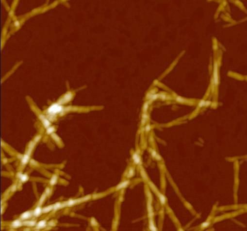
Unexpected asymmetry in the aggregation of partially labeled peptides.
Claudio Canale1, Michela Cosentino2, Paolo Bianchini2 and Alberto Diaspro1,2
1
Department of Physics, University of Genova, via Dodecaneso 33, 16146, Genova. Italy
2
Department of Nanophysics, Istituto Italiano di Tecnologia, via Morego 30, 16163, Genova, Italy
In the last decades, microscopy goes beyond the diffraction limit, thanks to innovative approaches
generally defined as super-resolution (SR) microscopy. SR microscopy provides new insights into
the biological processes at the, hitherto inaccessible, molecular scale.
Correlative nanoscopy is a new term indicating the integrated use of microscopy methods that
investigate the sample at the nanoscale. A super-resolution fluorescence microscope combined
with an atomic force microscope (AFM) is an example. The two techniques provide different data
sets from the sample, defining a new functionality of working and opening new scenarios in
biomolecular investigations.
We employed a stimulated emission depletion (STED) microscope coupled with an AFM to the
study of amyloid aggregates formation. We worked in vitro, inducing the aggregation of different
peptides, following standard methods, and labeling the samples at different dye to protein ratio,
and by using different conventional approaches.
The results obtained for insulin, Aβ1-42, and Aβ1-40 define the same scenario: only a fraction of
the fibrillar aggregates is visible in STED images, indicating that the labeled molecules were not
participating indistinctly to the aggregation process, and supporting the hypothesis that labeled
molecules are able to follow only selected pathways of aggregation. The results obtained by
correlative AFM-STED microscopy generate a warning: fluorescence techniques are not able to
characterise all the products derived from the aggregation of misfolded proteins. This statement
reveals the importance to correlate fluorescence data with other derived from label-free
techniques.Miniworkshop and CIMN Meeting “Protein misfolding and amyloidosis XIII”
Genoa, May 23 – 24, 2019
Multi-Messenger Optical Microscopy to study biological macromolecules in cells.
Alberto Diaspro1,2
1
Department of Physics, University of Genoa, 2Nanoscopy, Istituto Italiano di Tecnologia
The possibility of integrating different light-matter interactions to form images and to correlate
image data in optical microscopy is a key step for the design and implementation of a multi-
messenger optical microscope based on fluorescence and label-free contrast mechanisms within
the idea of “liquid tunable microscopy”[1]’. The multi-messenger microscope exploits the possibility
to “tune” the microscope across a large, almost unlimited, range of spatial and temporal resolution.
Such a multiscale and multimodal microscope is liquid because it aims to overlap in an efficient
and optimised way different mechanisms of contrast and it is tunable because it offers a real-time
tunability regarding spatial and temporal resolution. The multi messenger microscope operates
under image scannig conditions taking advantage of a new generation array of detectors [2]. It is
smart because it can adapt the current configuration to the scientific question and is open to
additional light-matter interaction modules. Interesting results can be obtained using STED-like
configurations [3] and label-free approaches, including Mueller matrix microscopy [4]. Expansion
[5] and light sheet microscopy [6] can be approached at the nanoscale. Moreover, for example, the
S(1,4)element of the Mueller matrix, named CIDS (Circular Intensity Differential Scattering), offers
label-free scouting of helical organisation motifs. As a perspective, the “liquid tunable microscopy”
concept suggests forming an image, L, made by a mix of C1...Cn images taken by different
mechanisms of contrast, reporting for each pixel a "decision" taken by an “enforced deep learning”
algorithm able to form, in real time, a “liquid” image, L=Cn+1.
[1] R. Won (2018) Nature Photonics, 12 (5): 259.
[2] M.Castello et al. (2019) Nature Methods, 16 (2): 175.
[3] G. Vicidomini et al. (2018) 15 (3): 173.
[4] A.Le Gratiet et al. (2018) OSA Continuum, 1 (3): 1068.
[5] L.Pesce et al. (2019) J.Biophotonics, in press.
[6] Cella Zanacchi et al. (2011) Nature Methods, 8 (12): 1047.Miniworkshop and CIMN Meeting “Protein misfolding and amyloidosis XIII”
Genoa, May 23 – 24, 2019
Rationally designed mutant light chain highlights properties modulating the soluble toxicity
Luca Oberti1, Martina Maritan1, Francesca Lavatelli2, Paola Rognoni2, Alberto Barbiroli3, Rosaria
Russo4, Pietro Sormanni5, Margherita Romeo6, Luisa Diomede6, Giovanni Palladini2, Giampaolo
Merlini2, Martino Bolognesi1,7, Stefano Ricagno1
1
Dipartimento di Bioscienze, Università degli Studi di Milano, Via Celoria 26 - 20133 Milano, Italy.
2
Amyloidosis Research and Treatment Center, Fondazione IRCCS Policlinico San Matteo, and
Department of Molecular Medicine, University of Pavia, P.le Golgi 19 - 27100, Pavia, Italy.
3
Dipartimento di Scienze per gli Alimenti, la Nutrizione e l'Ambiente, Università degli Studi di
Milano, 20133, Milano, Italy.
4
Dipartimento di Fisiopatologia Medico-Chirurgica e dei Trapianti, Università degli Studi di Milano,
Italy
5
Centre for Misfolding Diseases, Department of Chemistry, University of Cambridge, Cambridge
CB2 1EW, United Kingdom.
6
Department of Molecular Biochemistry and Pharmacology, Istituto di Ricerche Farmacologiche
Mario Negri IRCCS, Milan, 20156, Italy;
7
Centro di Ricerca Pediatrica Romeo ed Enrica Invernizzi, Università degli Studi di Milano, Via
Celoria 26 - 20133 Milano, Italy
Light chain amyloidosis (AL) is the most common form of systemic amyloidosis and it is
characterised by the deposition of immunoglobulin light chains (LCs) in target organs. Particularly,
heart is the most affected one with LC fibrillar deposits abundantly found in its tissues. Virtually,
because of genetic rearrangement and somatic hypermutation, each AL patient presents a
different amyloidogenic LC. Therefore, the understanding of the molecular mechanisms linked with
the LCs aggregation propensity requires investigating large sets of cases. Moreover, to date, it is
well known that soluble circulating amyloidogenic LCs exert proteo-toxicity. Indeed, lowering its
concentration in blood leads to an improvement of the heart physiology. However, the molecular
bases of the LC soluble toxicity have not been investigated in AL.
Herein, we present the comparative biophysical and structural characterisation of thirteen
sequence-diverse LCs. Eight of them were selected as responsible of cardiac AL in patients; while,
five of them were isolated from patients affected by multiple myeloma. Our results highlight that low
fold stability and high protein dynamics correlate with amyloidogenic LCs. On the contrary,
hydrophobicity, structural rearrangements and the LC dimeric association interface do not appear
to be linked with amyloid propensity.
Starting from these results, we aimed to rationally design a mutant (mH6) of a highly cardiotoxic LC
(H6), characterised by a higher stability. Structurally, H6 and mH6 match very closely; while,
opposite to H6, the biophysical profile of mH6 suggests that this protein displays a highly
cooperative fold, kinetic stability and lower molecular flexibility. Intriguingly, toxicity tests show that
mH6 proteotoxicity is lowered on human cardiac fibroblasts and C. elegans compared to the wild
type H6. These data, thus, strongly suggest a causative correlation of the above molecular
properties with the proteotoxicity observed in AL patients with cardiac involvement.Miniworkshop and CIMN Meeting “Protein misfolding and amyloidosis XIII”
Genoa, May 23 – 24, 2019
Trodusquemine induces changes in the properties of biomimetic membranes
Davide Odino1, Silvia Errico2, Claudio Canale1, Annalisa Relini3, Fabrizio Chiti2
1
Department of Physics, University of Genoa, Genoa, Italy; 2Department of Experimental and
Clinical Biomedical Sciences, University of Florence, Florence, Italy; 3Department of Chemistry and
Industrial Chemistry, University of Genoa, Genoa, Italy
Trodusquemine is an aminosterol which has been shown to decrease the toxicity of -synuclein
and A oligomers and has been proposed as a potential drug against Parkinson’s and Alzheimer’s
diseases [1,2]. There is evidence that trodusquemine inhibits oligomer binding to the cell
membrane [2]. To further elucidate this mechanism of action, we have studied the effects of
trodusquemine on biomimetic membranes. To this purpose, we have used atomic force
microscopy to investigate the morphological and mechanical properties of supported lipid bilayers
mimicking neuronal membranes, in the absence and in the presence of trodusquemine.
We found that at low concentrations of trodusquemine (5 µM) the bilayer still exhibits condensed
domains coexisting with the fluid phase. At higher concentrations (25 and 250 µM) the raft-like
domains disappear, strongly altering the distribution of negative charge which under normal
conditions is confined into the ordered domains. We also found that the contact angle of a drop of
liposomes deposited on mica shifts from hydrophilic to hydrophobic behavior with increasing
concentrations of trodusquemine, suggesting that this molecule has a strong influence on
interfacial tension.
Force spectroscopy experiments are in progress to characterize possible differences in the
mechanical properties of the membrane as a function of trodusquemine concentration.
[1] M. Perni et al., Multistep inhibition of alpha-synuclein aggregation and toxicity in vitro and in vivo
by trodusquemine. ACS Chem. Biol. 2018, 13, 2308−2319.
[2] R. Limbocker et al., Trodusquemine enhances A(42) aggregation but suppresses its toxicity by
displacing oligomers from cell membranes. Nat. Commun. 2019, 10, 225.Miniworkshop and CIMN Meeting “Protein misfolding and amyloidosis XIII”
Genoa, May 23 – 24, 2019
Inhibition of A aggregation by multi-target compounds
Mattia Siri1, Marta Campora1, Marco Catto2, Elena Gatta3, Claudio Canale3, Annalisa Relini4, Bruno
Tasso1, Michele Tonelli1
1
Department of Pharmacy, University of Genoa, Genoa, Italy, 2Department of Pharmacy-
Pharmaceutical Sciences, University of Bari, Bari, Italy, 3Department of Physics, University of
Genoa, Genoa, Italy; 4Department of Chemistry and Industrial Chemistry, University of Genoa,
Genoa, Italy
Alzheimer’s disease (AD) is a progressive neurodegenerative disorder, mainly characterized by the
accumulation of aggregates of -amyloid (A) and tau proteins, which lead to cell death with
particular loss of cholinergic function and consequent deficit of the neurotransmitter acetylcholine
(Ach). A beneficial strategy for the therapy of this multifactorial disease may be the design of multi-
target directed ligands, addressed with structural requirements for tackling at different levels the
intricate network of AD. In this context we investigated the structure-activity relationship of our first
series of quinone-based derivatives [1], developing new naphtho- and anthraquinone compounds
connected through a polymethylene chain to an aromatic or heteroaromatic ring. The novel
derivatives have been tested for the direct inhibition of Aβ aggregation and for the inhibition of both
cholinesterases (AChE and BChE). Most of the compounds have been found effective against Aβ
in the low micromolar range, and some of them also have proven to inhibit AChE and BChE,
confirming a multitarget mechanism of action. Atomic force microscopy was then employed to
assess the ability of four promising compounds to inhibit Aβ fibrillation and revealed clear
differences between Aβ1-42 aggregate morphologies obtained in the presence or absence of the
compounds. The same compounds have been assayed for their neuroprotective activity in primary
culture of cerebellar granule cells from postnatal rats (P7) and, in particular, one of them was more
effective in preventing Aβ1-42 toxicity. The results of these experiments will guide our further efforts
to design new multi-target agents for AD therapy.
[1] M. Tonelli, M. Catto, B. Tasso, et al. ChemMedChem 10 (2015) 1040-1053.You can also read