Carnosic Acid and Carnosol, Two Major Antioxidants of Rosemary, Act through Different Mechanisms1 OPEN
←
→
Page content transcription
If your browser does not render page correctly, please read the page content below
Carnosic Acid and Carnosol, Two Major Antioxidants of
Rosemary, Act through Different Mechanisms1[OPEN]
Margot Loussouarn,a,b Anja Krieger-Liszkay,c Ljubica Svilar,d Antoine Bily,b Simona Birtic ,b and
Michel Havaux a,2
a
  Commissariat à l’Energie Atomique et aux Energies Alternatives Cadarache, Centre National de la Recherche
Scientifique, Unité Mixte de Recherche 7265 Biologie Végétale et Microbiologie Environnementales, Aix
Marseille Université, Laboratoire d’Ecophysiologie Moléculaire des Plantes, F-13108 Saint-Paul-lez-Durance,
France
b
  Naturex, BP 81218, F-84911 Avignon cedex 9, France
c
  Institut de Biologie Intégrative de la Cellule, Centre National de la Recherche Scientifique, Commissariat à
l’Energie Atomique et aux Energies Alternatives Saclay, Institut de Biologie et de Technologie de Saclay,
Université Paris-Sud, 91191 Gif-sur-Yvette, France
d
  Criblage Biologique Marseille, Laboratoire Nutrition, Obésité et Risque Thrombotique, Aix-Marseille
Université, Institut National de la Recherche Agronomique, Institut National de la Santé et de la Recherche
Médicale, 13005 Marseille, France
ORCID IDs: 0000-0001-7141-4129 (A.K.-L.); 0000-0002-3219-0860 (L.S.); 0000-0003-3468-3818 (A.B.); 0000-0002-6434-393X (M.H.).

Carnosic acid, a phenolic diterpene specific to the Lamiaceae family, is highly abundant in rosemary (Rosmarinus officinalis). Despite
numerous industrial and medicinal/pharmaceutical applications of its antioxidative features, this compound in planta and its
antioxidant mechanism have received little attention, except a few studies of rosemary plants under natural conditions. In vitro
analyses, using high-performance liquid chromatography-ultraviolet and luminescence imaging, revealed that carnosic acid and
its major oxidized derivative, carnosol, protect lipids from oxidation. Both compounds preserved linolenic acid and
monogalactosyldiacylglycerol from singlet oxygen and from hydroxyl radical. When applied exogenously, they were both able to
protect thylakoid membranes prepared from Arabidopsis (Arabidopsis thaliana) leaves against lipid peroxidation. Different levels of
carnosic acid and carnosol in two contrasting rosemary varieties correlated with tolerance to lipid peroxidation. Upon reactive
oxygen species (ROS) oxidation of lipids, carnosic acid was consumed and oxidized into various derivatives, including into carnosol,
while carnosol resisted, suggesting that carnosic acid is a chemical quencher of ROS. The antioxidative function of carnosol relies on
another mechanism, occurring directly in the lipid oxidation process. Under oxidative conditions that did not involve ROS
generation, carnosol inhibited lipid peroxidation, contrary to carnosic acid. Using spin probes and electron paramagnetic
resonance detection, we confirmed that carnosic acid, rather than carnosol, is a ROS quencher. Various oxidized derivatives of
carnosic acid were detected in rosemary leaves in low light, indicating chronic oxidation of this compound, and accumulated in
plants exposed to stress conditions, in parallel with a loss of carnosic acid, confirming that chemical quenching of ROS by carnosic
acid takes place in planta.

  Carnosic acid is a labdane-type diterpene present in                        Hossain et al., 2010; Birtic et al., 2015). This lipid-soluble
plant species of the Lamiaceae family, such as rosemary                       compound is recognized for its high antioxidative capacities,
(Rosmarinus officinalis) and common salvia (Salvia officinalis;                 which have led to many industrial applications in the
                                                                              fields of foods and beverages, personal care, nutrition,
                                                                              and health (Birtic et al., 2015). The antioxidant proper-
    1
     M.L. was supported by a CIFRE studentship from the French                ties of carnosic acid, presumably due to the presence of
National Association for Research and Technology (ANRT).                      a catechol moiety (Supplemental Fig. S1), were evalu-
   2
     Address correspondence to michel.havaux@cea.fr.                          ated mainly in vitro in a large variety of artificial and/
   The author responsible for distribution of materials integral to the       or model systems. For instance, when tested in bulk and
findings presented in this article in accordance with the policy de-           emulsified lipid systems, carnosic acid was found to
scribed in the Instructions for Authors (www.plantphysiol.org) is:            protect fatty acids and triglycerides against oxidation
Michel Havaux (michel.havaux@cea.fr).
                                                                              (Hopia et al., 1996; Cuvelier et al., 1996). Carnosic acid
   M.L., S.B., and M.H. designed the experiments; M.L. performed
most experiments; A.K.-L. performed EPR analyses; L.S. performed
                                                                              also was observed to prevent low-density lipoprotein
LC-HRMS analyses; M.L., A.B., S.B., and M.H. interpreted the data;            oxidation in human aortic endothelial cells (Pearson
M.L. and M.H. wrote the article with some input from the other                et al., 1997) and lipid hydroperoxide-mediated oxi-
authors.                                                                      dative stress in Caco-2 cells (Wijeratne and Cuppett,
   [OPEN]
          Articles can be viewed without a subscription.                      2007). Inhibition of lipid peroxidation by carnosic acid
   www.plantphysiol.org/cgi/doi/10.1104/pp.17.01183                           was reported in rat liver microsomes and ox brain
Plant PhysiologyÒ, November 2017, Vol. 175, pp. 1381–1394, www.plantphysiol.org Ó 2017 American Society of Plant Biologists. All Rights Reserved. 1381

                                Downloaded on January 11, 2021. - Published by https://plantphysiol.org
                               Copyright (c) 2020 American Society of Plant Biologists. All rights reserved.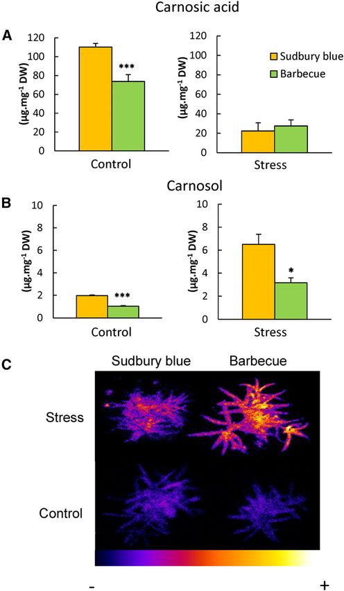
Loussouarn et al.

phospholipid liposomes (Aruoma et al., 1992; Romo                     tandem a peculiar and efficient antioxidant system in
Vaquero et al., 2013). Food material such as oil, raw and             planta. It is likely that this carnosic acid-based pro-
cooked meat, and cooked meat patties were protected                   tection mechanism is an important component in the
from oxidation by carnosic acid, in most cases with a                 ability of rosemary to withstand harsh climatic conditions
higher efficiency than synthetic antioxidants (Erkan                   that can prevail in its natural Mediterranean habitat.
et al., 2008; Zhang et al., 2010; Naveena et al., 2013;
Jordán et al., 2014). Carnosic acid also was described as
a scavenger of hydroxyl and 2,2-diphenyl-1-picrylhydrazyl             RESULTS
radicals (Aruoma et al., 1992; Luis and Johnson, 2005).               Lipid Protection by Carnosic Acid and Carnosol in Vitro
While the potential antioxidative activity of carnosic
acid is well documented, its exact mechanism of action                   The lipid monogalactosyldiacylglycerol (MGDG),
has not been studied extensively. In particular, little is            solubilized in methanol/chloroform, was oxidized
known of the interactions of carnosic acid with distinct              with singlet oxygen (1O2) generated by illuminating the
reactive oxygen species (ROS) or lipid radicals.                      photosensitizing agent Methylene Blue. As expected
Moreover, in most studies, in vitro oxidation was gen-                (Birtic et al., 2011), the MGDG solution became lumi-
erated by prolonged and artificial heating treatments, so              nescent after this oxidation treatment as imaged with a
that it is difficult to extrapolate the results to the in vivo         high-sensitivity cooled CCD camera (Fig. 1A). This
situation in plants. Surprisingly, the role of carnosic acid          photon emission originates from lipid peroxides whose
in plant leaves has received little attention, and the bio-           slow decomposition produces light-emitting species
logical role of this compound in plants is not firmly                  such as triplet carbonyls and singlet oxygen, with the
established.                                                          intensity of this signal being correlated with the extent
   Carnosic acid is present at very high concentrations,              of lipid peroxidation in the sample (Birtic et al., 2011;
up to several percent of dry weight, in leaves of the                 Cifra and Pospisil, 2014). When MGDG was supple-
Mediterranean half-shrub rosemary (Munné-Bosch and                    mented with carnosic acid during 1O2 oxidation, the
Alegre, 2001; del Baño et al., 2003; Luis and Johnson,                luminescence signal intensity was reduced noticeably
2005). Carnosic acid biosynthesis and accumulation take               (Fig. 1, A and B), indicating lower levels of MGDG
place exclusively in young rosemary leaves at the branch              oxidation and of lipid peroxides. Protection of MGDG
apices, with the diterpene molecule being partially con-              against oxidation also was observed with carnosol and
sumed during leaf development and aging (Hidalgo                      tocopherol, with the protective effect of the latter
et al., 1998; Brückner et al., 2014; Bozic et al., 2015). Be-       compound, however, appearing to be slightly lower
side carnosic acid, less abundant phenolic diterpenes can             than the protection provided by carnosol and carnosic
be measured in rosemary leaves, including carnosol                    acid. Lipid protection by carnosic acid and carnosol also
(Supplemental Fig. S1), the major oxidation product of                was obtained when the experiments were done with
carnosic acid. The antioxidative activity of the latter               linolenic acid (C18:3) instead of MGDG (Supplemental
compound, produced spontaneously from carnosic acid                   Fig. S2). Those observations show that, similar to to-
by nonenzymatic reaction, has been seldom investigated                copherols (Liebler et al., 1986), both carnosic acid and
(Aruoma et al., 1992; Zeng et al., 2001). Diterpene levels            carnosol are lipid protectors against attack by 1O2.
in field-grown rosemary plants displayed seasonal                      These effects were confirmed by analyzing hydroxy-
changes, with a tendency for carnosic acid losses in re-              octadecatrienoic acid (HOTE), an oxidation product
sponse to environmental stress conditions (Luis and                   of the main fatty acid in leaves, linolenic acid, and
Johnson, 2005). In particular, carnosic acid concentra-               hydroxyoctadecadienoic acid (HODE), the oxidation
tions in rosemary leaves under natural conditions were                product of linoleic acid, in the MGDG solution after 1O2
found to decrease at high temperatures and low pre-                   oxidation (Fig. 1C). Both HOTEs and HODEs were re-
cipitation rates in summer with concomitant increases in              duced substantially by carnosic acid and carnosol.
oxidized derivatives, suggesting that cellular oxidative                 MGDG also was oxidized by hydroxyl radicals pro-
stress is accompanied by the consumption of carnosic                  duced by hydrogen peroxide (H2O2) and iron (Fenton
acid (Munné-Bosch et al., 1999; Munné-Bosch and                       reaction), leading to luminescence emission (Fig. 2, A
Alegre, 2003). Both carnosic acid and carnosol accu-                  and B). The addition of carnosol to MGDG protects the
mulate in photosynthetic green tissues only (leaves,                  galactolipid solution against oxidation, as shown by the
sepals, and petals) and have be localized in the chlo-                marked decrease in luminescence (Fig. 2, B and D).
roplasts (Munné-Bosch and Alegre, 2001), although the                 Surprisingly, the addition of carnosic acid did not re-
synthesis of carnosic acid also has been reported in                  duce MGDG luminescence after oxidation by hydroxyl
glandular trichomes (Brückner et al., 2014).                          radicals (Fig. 2, A and C). On the contrary, carnosic acid
   Because the functions of the major rosemary diter-                 strongly increased luminescence, and this phenomenon
penes in plant leaves are poorly understood, we per-                  was still observed when carnosic acid concentrations
formed a comprehensive study of the antioxidant activity              were increased up to 600 mM. Actually, this lumines-
of carnosic acid and its oxidized derivative carnosol, both           cence enhancement was observed to be due to carnosic
in vitro and in rosemary plants. This study reveals dif-              acid itself, which became highly luminescent when in-
ferent modes of action for carnosic acid and carnosol                 cubated in the presence of free radicals (without lipid).
against ROS and lipid radicals, which make this diterpenoid           We checked that the mixture H2O2 + iron or a solution
1382                                                                                                          Plant Physiol. Vol. 175, 2017

                               Downloaded on January 11, 2021. - Published by https://plantphysiol.org
                              Copyright (c) 2020 American Society of Plant Biologists. All rights reserved.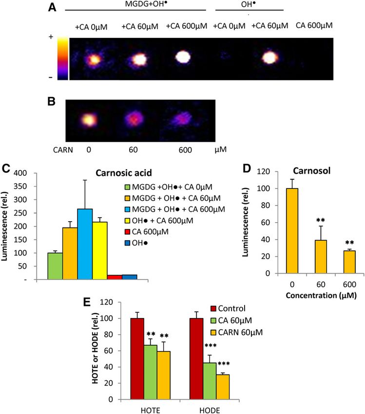
Antioxidant Functions of Carnosic Acid and Carnosol

                                                                                            Figure 1. Effects of carnosic acid, carnosol, and
                                                                                            a-tocopherol on in vitro oxidation of lipids by 1O2.
                                                                                            1
                                                                                             O2 was produced by a 30-min illumination of the
                                                                                            lipid solution (MGDG) in the presence of Methylene
                                                                                            Blue. 1O2 oxidation of MGDG was performed in the
                                                                                            presence of 60 or 120 mM carnosic acid, carnosol, or
                                                                                            a-tocopherol. A, Luminescence images of the oxi-
                                                                                            dized solutions. The color palette indicates signal
                                                                                            intensity from black (0) to white (highest values). B,
                                                                                            Quantification of the luminescence signals. Data are
                                                                                            normalized to the control signal values measured in
                                                                                            the absence of antioxidant. C, Hydroxy fatty acid
                                                                                            quantification (HOTE and HODE). Data are normal-
                                                                                            ized to the control HOTE values measured in the
                                                                                            absence of antioxidant. CA, Carnosic acid; CARN,
                                                                                            carnosol. Asterisks indicate significant differences
                                                                                            from control (0 mM) at P , 0.05 (*), P , 0.01 (**), and
                                                                                            P , 0.005 (***) by Student’s t test.

of carnosic acid in the absence of any ROS was not lu-                     spin probe (Hideg et al., 2011). The amplitude of the
minescent. The data of Figure 2A thus suggest that                         electron paramagnetic resonance (EPR) spectra of
carnosic acid reacts with free radicals, leading to its                    TEMPD was strongly reduced by carnosic acid, and this
oxidation and to the formation of light-emitting deriv-                    effect was visible even at the low concentration of 10 mM
atives. This is confirmed in Supplemental Figure S2: a                      (Fig. 4, A and B). As expected from the data of Figure 3,
drastic loss of carnosic acid occurred when lipids were                    EPR analyses showed that carnosol does not quench
oxidized by 1O2 or free radicals, whereas carnosol levels                  1
                                                                            O2: 60 mM carnosol had very little effect on the ampli-
were less affected. As a consequence, the lipid protec-                    tude of the 1O2 EPR spectrum (Fig. 4C). This result
tive action of carnosic acid cannot be assessed through                    confirms that carnosol is not able to eliminate 1O2 in the
autoluminescence measurements. HPLC analyses of                            micromolar concentration range, although it protects
HOTE and HODE levels can overcome this problem.                            lipids against oxidation in this concentration range. We
The data shown in Figure 2 revealed that carnosic acid,                    also examined the effects of a-tocopherol, a known
similar to carnosol, does protect MGDG from oxidation                      quencher of 1O2 (Foote et al., 1974; Di Mascio et al.,
by free radicals: the HOTE and HODE levels were re-                        1990). The quenching effect of tocopherol was visible at
duced noticeably in the presence of carnosic acid or                       concentrations in the millimolar range only (Fig. 4D),
carnosol.                                                                  indicating that tocopherol is a less efficient 1O2 quencher
                                                                           than carnosic acid.
                                                                              The spin probe 4-pyridyl-1-oxide-N-tert-butylnitrone
Interactions of Carnosol and Carnosic Acid with ROS                        (POBN) was used to measure the hydroxyl radical by
                                                                           EPR spectroscopy (Hideg et al., 2011). Carnosic acid
   The oxidative degradation of carnosic acid by the                       was able to quench this ROS (Fig. 5, A and B), while
hydroxyl radical is confirmed in Figure 3. When H2O2                        carnosol had virtually no effect on ROS concentration
and iron were added to a solution of carnosic acid, the                    (Fig. 5C). Taken together, the data of Figures 4 and 5
diterpene concentration fell rapidly, and an accumula-                     confirm that carnosic acid and carnosol differ in their
tion of carnosol was observed in parallel (Fig. 3A). The                   reaction with ROS, although both can protect lipids
same phenomena were found with 1O2, although the                           against ROS-induced lipid peroxidation (Fig. 1).
rates of carnosic acid disappearance and carnosol ac-                         Using liquid chromatography coupled with mass
cumulation were slower compared with the effect of                         spectrometry (LC-HRMS), we characterized oxidized
hydroxyl radicals. In striking contrast, carnosol was                      derivatives (besides carnosol) generated during the
resistant to this oxidation: the carnosol concentration                    in vitro oxidation of carnosic acid by 1O2 or hydroxyl
remained stable in the presence of hydroxyl radical or                     radical in solution (Supplemental Fig. S3). Authentic
1
 O2 (Fig. 3B). These findings indicate that carnosic acid                   standards were used to determine the retention times,
has a high reactivity toward ROS and is easily oxidiz-                     mass-to-charge ratios (m/z), full mass spectra, and tan-
able. Therefore, it is likely that the antioxidant activity                dem mass spectrometry (MS/MS) spectra of carnosic
of carnosic acid relies on chemical quenching of ROS.                      acid and carnosol (Supplemental Figs. S4 and S5),
   In Figure 4, 1O2 was produced from Rose Bengal in                       allowing unambiguous identification of those com-
the light and was quantified using 2,2,6,6-tetramethyl-                     pounds in oxidized solutions and in leaf extracts. Oxi-
4-piperidone hydrochloride (TEMPD), a 1O2-specific                          dation of carnosic acid was confirmed by a decrease in
Plant Physiol. Vol. 175, 2017                                                                                                                 1383

                                 Downloaded on January 11, 2021. - Published by https://plantphysiol.org
                                Copyright (c) 2020 American Society of Plant Biologists. All rights reserved.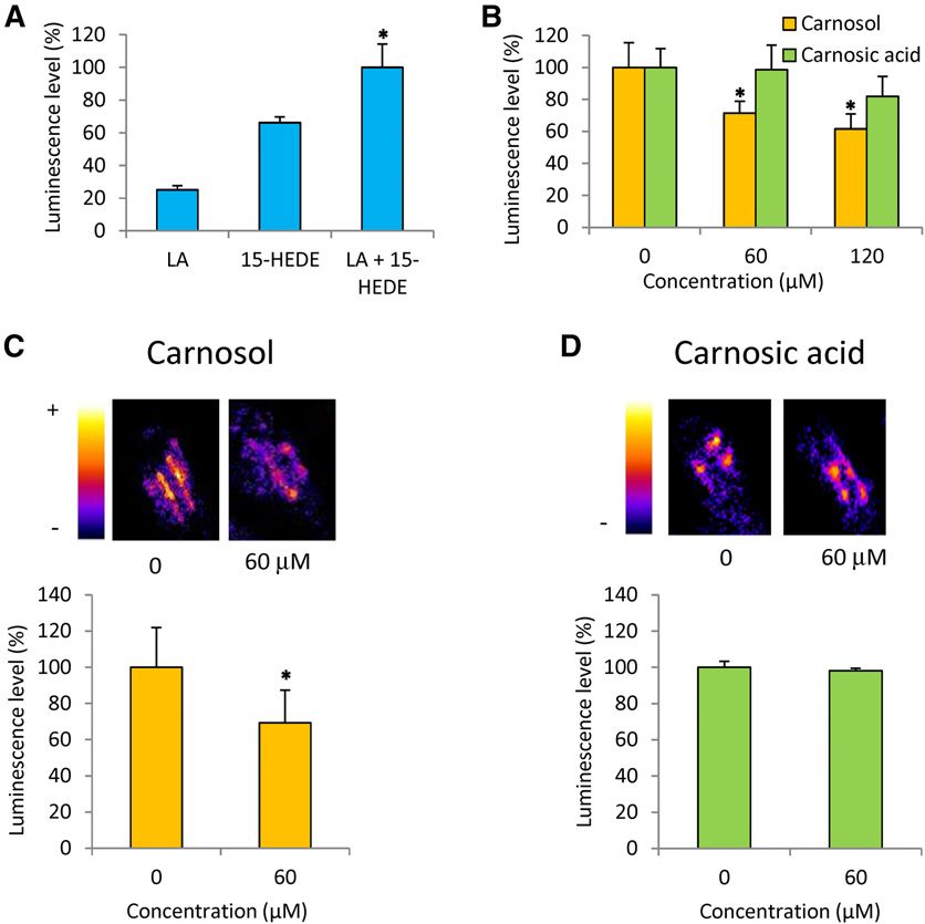
Loussouarn et al.

Figure 2. Effects of carnosic acid and carnosol on
in vitro oxidation of lipids by free radicals. Hydroxyl
radicals were produced by the Fenton reaction using
H2O2 + Fe2+ in the presence of 60 or 600 mM carnosic
acid (CA) or carnosol (CARN). A, Luminescence
imaging of MGDG oxidized by the hydroxyl radical
in the presence or absence of carnosic acid (60 and
600 mM). The luminescence signals of the mixture
H2O2 + iron (hydroxyl radicals) and of carnosol in the
presence or absence of hydroxyl radicals also were
measured as controls. B, Luminescence imaging of
MGDG oxidized by hydroxyl radical in the presence
of carnosol (60 and 600 mM). C, Quantification of the
luminescence signals shown in A. Data are normal-
ized to the signal values measured from oxidized
MGDG in the absence of antioxidant. D, Quantifi-
cation of the luminescence signals shown in B. Data
are normalized to the signal values measured from
oxidized MGDG in the absence of antioxidant. E,
Hydroxy fatty acid quantification (HOTE and HODE).
Data are normalized to the HOTE or HODE values
measured in the absence of antioxidant. Asterisks
indicate significant differences from control at P ,
0.01 (**) and P , 0.005 (***) by Student’s t test.

the carnosic acid peak and a concomitant production of                      and 12-o-methyl carnosic acid were confirmed by
carnosol. A variety of compounds obtained by the ox-                        matching their retention times and MS/MS spectra
idation of carnosic acid by 1O2 and by hydroxyl radical                     with those of the reference compounds (Supplemental
was detected. There was a strong overlap between the                        Figs. S6–S8), while rosmaridiphenol, 11912-o-methyl-
oxidation profiles of carnosic acid induced by 1O2 and by                    rosmanol, 7-methylisorosmanol, isorosmanol, rosmadial
the hydroxyl radical. Structures of rosmanol, isorosmanol,                  isomers, and 5,6,7,10-tetrahydroxyrosmariquinone were

Figure 3. Time course of the changes in car-
nosic acid and carnosol concentrations upon
exposure to 1O2 or hydroxyl radicals. 1O2 was
produced by illumination of Methylene Blue,
and hydroxyl radicals were produced by the
Fenton reaction using H2O2 + Fe2+. A and B,
Carnosic acid. C and D, carnosol.

1384                                                                                                                Plant Physiol. Vol. 175, 2017

                                     Downloaded on January 11, 2021. - Published by https://plantphysiol.org
                                    Copyright (c) 2020 American Society of Plant Biologists. All rights reserved.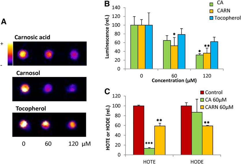
Antioxidant Functions of Carnosic Acid and Carnosol

                                                                                                        Figure 4. 1O2 quenching capacity of
                                                                                                        carnosic acid and carnosol. 1O2 was
                                                                                                        generated by a 5-min illumination
                                                                                                        of 100 mM Rose Bengal in the presence
                                                                                                        of the spin probe TEMPD. A, Effects of
                                                                                                        different concentrations of carnosic acid
                                                                                                        (CA) on the EPR spectra of TEMPD. B,
                                                                                                        Quantification of the decrease in the
                                                                                                        EPR signal amplitude induced by in-
                                                                                                        creasing concentrations of carnosic
                                                                                                        acid. C, Effects of carnosol (CARN) on
                                                                                                        the EPR spectra of TEMPD. D, Effects of
                                                                                                        a-tocopherol (TOC) on the EPR spectra
                                                                                                        of TEMPD. E, Quantification of the de-
                                                                                                        crease in the EPR spectra by increasing
                                                                                                        concentrations of a-tocopherol.

putatively identified by matching bibliography data                         Blue variety, in which carnosic acid represented up to
with the retention times and MS/MS spectra obtained                        10% of leaf dry weight under control growth conditions
experimentally with oxidized carnosic acid solutions                       (250 mmol photons m22 s21 and 25°C). The major oxi-
and with rosemary leaf extracts (Supplemental Figs.                        dized derivative of carnosic acid, carnosol, was less
S9–S13).                                                                   abundant (;2 mg mg21 dry leaf weight), neverthe-
                                                                           less representing about 0.2% of dry weight. We
                                                                           also analyzed the prenyl lipids, tocopherols and plas-
Carnosic Acid and Carnosol Levels in Rosemary Leaves                       tochromanol, which are both ubiquitous plastid anti-
                                                                           oxidants (Kruk et al., 2014, 2016). Both compounds
  Rosemary leaves are known to accumulate high                             were found in rosemary leaves at concentrations no-
amounts of carnosic acid (Birtic et al., 2015), as con-                   ticeably lower than carnosol: approximately 0.1 and
firmed in Figure 6A for young leaves of the Sudbury                         0.01 mg mg21, respectively.
Plant Physiol. Vol. 175, 2017                                                                                                               1385

                                 Downloaded on January 11, 2021. - Published by https://plantphysiol.org
                                Copyright (c) 2020 American Society of Plant Biologists. All rights reserved.
Loussouarn et al.
Figure 5. Quenching of hydroxyl radi-
cals by carnosic acid and carnosol.
Hydroxyl radicals were generated by the
Fenton reaction using H2O2 + Fe2+ in the
presence of the spin probe POBN. A,
Effects of different concentrations of
carnosic acid (CA) on the EPR spectra of
POBN. B, Quantification of the de-
crease in the EPR signal amplitude in-
duced by increasing concentrations of
carnosic acid. C, Effects of carnosol
(CARN) on the EPR spectra of POBN.
Growing rosemary plants for 4 weeks under harsh at the leaf surface and internal leaf tissues (Bozic et al.,
conditions of light and temperature (1,200 mmol pho- 2015). This partitioning was estimated by briefly
tons m22 s21 and 35°C/5°C [day/night]) led to a strong washing rosemary leaves (for 30 s) with dichloro-
decrease in carnosic acid compared with control con- methane in order to extract hydrophobic compounds
ditions (Fig. 6A). Concomitantly, the loss of carnosic from the trichomes. As shown in Figure 6C, this treat-
acid was associated with a marked increase (about 33) ment caused a complete emptying of the glandular tri-
in carnosol levels, suggesting consumption of the for- chomes while epidermal cells remained unaltered. The
mer compound during its antioxidant activity under solvent after leaf dipping was found to contain both car-
stress conditions with partial conversion to its oxidized nosic acid and carnosol (Fig. 6B), indicating storage of
metabolite carnosol. A strong accumulation of tocoph- those compounds in the trichomes. However, the amounts
erols and plastochromanol also was observed after ex- of diterpenes present in this fraction were relatively small,
posure of rosemary plants to high light and heat, thus representing less than 10% of total amounts. Thus, in the
exhibiting a behavior that contrasts with that of car- Sudbury Blue variety investigated here, carnosic acid and
nosic acid. This contrasting response was observed carnosol are stored mainly within the leaves. This parti-
previously for carnosic acid and a-tocopherol in sage tioning of carnosic acid and carnosol between trichomes
(Salvia officinalis) and rosemary exposed to natural and internal leaf tissues was not modified significantly by
drought conditions (Munné-Bosch and Alegre, 2003). growth in high light at high temperature (Fig. 6B).
Carnosic acid and carnosol are present in photo-
synthesizing green tissues only (Munné-Bosch and
Alegre, 2001; Luis and Johnson, 2005) and, in leaves, Oxidized Derivatives of Carnosic Acid in Planta
they have been found in the chloroplasts (Munné-Bosch
and Alegre, 2001). However, carnosic acid and carnosol Some of the compounds detected in vitro after ROS
also have been reported to partition between trichomes oxidation of carnosic acid (Supplemental Fig. S3) also
1386 Plant Physiol. Vol. 175, 2017
Downloaded on January 11, 2021. - Published by https://plantphysiol.org
Copyright (c) 2020 American Society of Plant Biologists. All rights reserved.Antioxidant Functions of Carnosic Acid and Carnosol
Figure 6. Carnosic acid and carnosol in leaves of
rosemary plants (Sudbury Blue variety) grown under
two different conditions of light and temperature
(250 mmol photons m22 s21 at 25°C/15°C day/night
[control] or 1,200 mmol photons m22 s21 at 35°C/5°C
[stress]). A, Carnosic acid, carnosol, a-tocopherol,
and plastochromanol-8 concentrations in leaves.
DW, Dry weight. Asterisks indicate significant differ-
ences from control at P , 0.05 (*) and P , 0.005 (***)
by Student’s t test. B, Carnosic acid and carnosol were
measured in leaves after organic solvent dipping and
in the organic solvent after leaf dipping (representing
the compounds stored in the trichomes). C, Glandular
trichomes before and after solvent dipping.
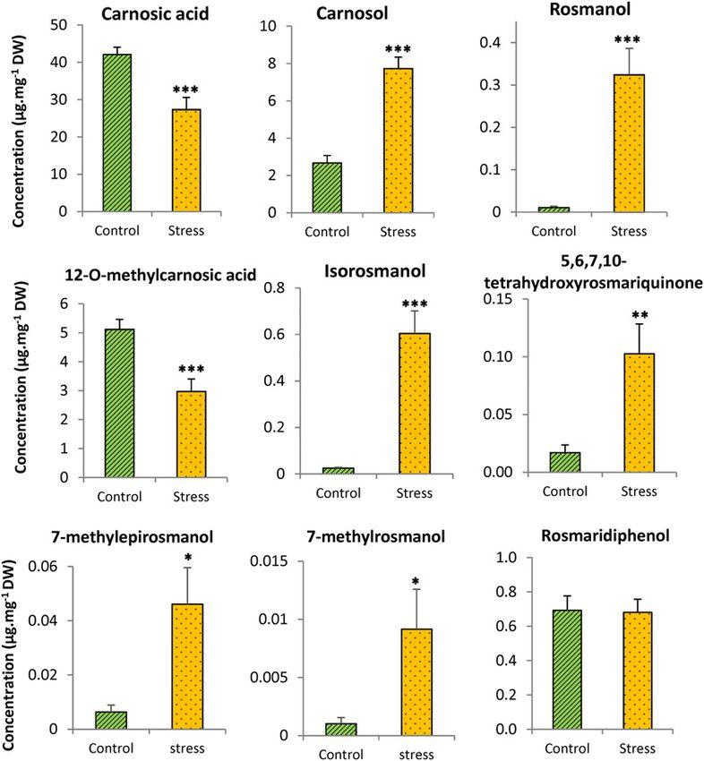
were found in rosemary leaves grown under control Exogenous Carnosic Acid and Carnosol Protect
conditions: rosmanol, isorosmanol, rosmaridiphenol, Thylakoid Membranes
7-methyl-epirosmanol, 7-methyl-rosmanol, 12-o-meth-
ylcarnosic acid, and 5,6,7,10-tetrahydroxyrosmar- Supplementing chloroplast membranes with carnosic
iquinone (Fig. 7; Supplemental Figs. S4–S12), indicating acid was shown previously to preserve a-tocopherol and
chronic oxidation of carnosic acid by ROS in leaves. In to reduce oxidative damage in high light (Munné-Bosch
line with this conclusion, a 30-h adaptation of rosemary and Alegre, 2003). Moreover, a marked consumption of
plants to darkness brought about a strong decrease in the exogenously applied carnosic acid was observed dur-
those compounds (Supplemental Fig. S14), confirming ing the high-light treatment. We performed a similar ex-
the link with light and the associated ROS production in periment with thylakoid membranes prepared from leaves
the chloroplasts. of Arabidopsis (Arabidopsis thaliana), a species that does not
Under stress conditions that caused a strong de- contain carnosic acid, supplemented with carnosol or
crease in carnosic acid and a concomitant accumula- carnosic acid. Thylakoid suspensions were exposed for 2 h
tion of carnosol (Fig. 6), the levels of several oxidation to high light (3,000 mmol photons m22 s21), causing pho-
products of carnosic acid, including rosmanol, iso- tooxidative damage, including the loss of chlorophyll and
rosmanol, 5,6,7,10-tetrahydroxyrosmariquinone, the accumulation of lipid peroxidation products (HOTE;
7-methyl-epirosmanol, and 7-methyl-rosmanol, in- Fig. 8). Adding carnosic acid or carnosol to the membrane
creased strongly in rosemary leaves (Fig. 7). The con- suspensions noticeably reduced photooxidative damage:
centration of other oxidized metabolites of carnosic loss of chlorophyll was reduced to 5% (versus 30% in
acid, such as rosmaridiphenol and 12-o-methylcarnosic control samples) by carnosic acid and carnosol, and lipid
acid, did not increase with the stress conditions. The peroxidation was very low. These results confirm that both
accumulation of rosmanol and isorosmanol, as well as carnosic acid and carnosol can protect biomembranes and
of methylated isorosmanol, was reported previously function as membrane lipid protectors in vivo.
in rosemary plants exposed to drought stress in the
field (Munné-Bosch et al., 1999). The accumulation of Direct Interaction of Carnosol with the Lipid
oxidized derivatives in rosemary leaves exposed to Peroxidation Process
high light and high temperature supports the idea that
the loss of carnosic acid observed under those condi- As shown above (Fig. 3), carnosol is resistant to direct
tions (Fig. 6) resulted from its oxidative degradation oxidation by ROS. However, when exposure to ROS took
by ROS. place in a lipid environment (linolenic acid; Supplemental
Plant Physiol. Vol. 175, 2017 1387
Downloaded on January 11, 2021. - Published by https://plantphysiol.org
Copyright (c) 2020 American Society of Plant Biologists. All rights reserved.Loussouarn et al.
Figure 7. Oxidation products of carnosic acid in
planta. Rosemary plants were grown under two dif-
ferent conditions (control and stress, described in the
legend of Fig. 6). The carnosic acid metabolites were
measured in rosemary leaves by ultra-performance
liquid chromatography-MS. Quantification of the
metabolites for which no standard is available was
done using the conversion factor of carnosic acid.
DW, Dry weight. Asterisks indicate significant differ-
ences at P , 0.05 (*), P , 0.01 (**), and P , 0.005
(***) by Student’s t test.
Fig. S2), some loss of carnosol was observed. This could The effect of carnosic acid in ROS-independent lipid
suggest that carnosol has the capacity to interact directly oxidation also was tested in vivo. Wounding is known to
with the lipid peroxidation mechanism itself and can be trigger lipoxygenase activity in leaves, causing enzymatic
degraded by reactions with some lipid oxidation-derived lipid peroxidation (Chauvin et al., 2013) and inducing the
products. It has been shown that lipid hydroperoxides are associated generation of photon emission (Birtic et al.,
capable of inducing membrane damage and lipid perox- 2011). In Figure 9C, leaves were injured with a scalpel in
idation in cell cultures (Wijeratne and Cuppett, 2006). darkness. As shown previously (Birtic et al., 2011), the
Based on this observation, in vitro oxidation of linolenic wounds can be visualized by the lipid oxidation-related
acid was triggered with a hydroxy fatty acid, 15- luminescence emission. The intensity of this luminescence
hydroxyeicosadienoic acid (15-HEDE), in darkness and signal was decreased significantly in leaves preinfiltrated
in the absence of ROS or ROS generator. Luminescence with carnosol compared with leaves preinfiltrated with a
from linolenic acid was noticeably increased by adding buffer that did not contain carnosol. Then, in line with the
15-HEDE (Fig. 9A), indicating oxidation of the fatty acid in vitro data shown in Figure 9B, carnosol can reduce lipid
molecule. The luminescence of HEDE was found to be peroxidation in planta through a mechanism different from
higher than that of linolenic acid (but lower than the ROS scavenging. Similar to what we observed with lino-
linolenic acid + 15-HEDE combination), probably due to lenic acid oxidized by 15-HEDE, carnosic acid was unable
the spontaneous decomposition of the hydroxy fatty acid to inhibit lipid peroxidation in wounded Arabidopsis
and the generation of light-emitting species. The addition leaves. Thus, taken together, our results show that the
of 60 or 120 mM carnosol to the mixture of linolenic acid + antioxidant activities of carnosic acid and carnosol rely on
15-HEDE decreased luminescence significantly (by 30% distinct mechanisms, involving direct interactions with
or 40%, respectively; Fig. 9B). This indicates that carnosol ROS or with the lipid oxidation process, respectively.
has a direct inhibitory effect on the lipid peroxidation
process. We checked that carnosol had no effect of the
15-HEDE intrinsic luminescence (data not shown), ex- Comparison of Two Rosemary Varieties Containing
cluding an action of the diterpene on 15-HEDE decom- Different Concentrations of Carnosic Acid and Carnosol
position products. In contrast with carnosol, carnosic acid
had no significant effect on the HEDE-induced oxidation As shown in Figure 10A, leaves of the Barbecue
of linolenic acid (Fig. 9B). variety contain substantially less carnosic acid and
1388 Plant Physiol. Vol. 175, 2017
Downloaded on January 11, 2021. - Published by https://plantphysiol.org
Copyright (c) 2020 American Society of Plant Biologists. All rights reserved.Antioxidant Functions of Carnosic Acid and Carnosol
Figure 8. Effects of carnosic acid or carnosol on Arabidopsis thylakoid membranes exposed to high light. Thylakoid suspensions
were exposed to white light of photon flux density (PFD) 1,500 mmol photons m22 s21 for 2 h. A, Decrease in chlorophyll content
after light treatment. B, HOTE with or without the addition of 50 mM carnosic acid (CA) or carnosol (CARN) to the membrane
suspensions. Asterisks indicate significant differences from control at P , 0.05 (*) and P , 0.005 (***) by Student’s t test.
carnosol than Sudbury Blue. As expected from the data and suggests that carnosic acid plays a protective role,
of Figure 6, exposure to high light at high-temperature not only under excess light energy, when ROS produc-
conditions caused a drastic loss of carnosic acid in both tion is expected to be elevated, but also in low light. This
varieties (Fig. 10A), which was accompanied by in- is in agreement with previous observations showing the
creased levels of carnosol (Fig. 10B). However, the latter presence of 1O2-specific degradation products of poly-
effect was less pronounced in the Barbecue variety unsaturated fatty acids in plant leaves in low light,
relative to Sudbury Blue. Photooxidative damage to reflecting continuous generation of 1O2 in illuminated
lipids in plants grown in high light or in control con- chloroplasts (Triantaphylidès et al., 2008). This phe-
ditions was visualized in both rosemary varieties by nomenon has led to the concept of lipid membranes
autoluminescence imaging (Fig. 10C). Interestingly, acting as supramolecular antioxidants that capture ROS
Barbecue plants exposed to stress conditions were no- (Schmid-Siegert et al., 2016). This concept could be ex-
ticeably more luminescent than Sudbury Blue plants, tended to the carnosic acid pool in rosemary leaves.
indicating more oxidative stress and lipid peroxidation Interestingly, carnosol, the major oxidized metabolite
in the former variety. The correlation found in the ex- of carnosic acid, was found to be an antioxidant and
periment of Figure 10 between the leaf content of car- lipid protector as efficient as carnosic acid. This result is
nosic acid and carnosol and the tolerance of rosemary to in line with early works that showed a protective effect
photooxidative stress is consistent with the lipid- of carnosol against lipid peroxidation in microsomal
protective functions of those diterpenes observed and liposomal systems (Aruoma et al., 1992). In previ-
in vitro (Figs. 1 and 8). However, the differential toler- ous studies, other carnosic acid-derived metabolites,
ance of Sudbury Blue and Barbecue to photooxidative such as rosmanol, epirosmanol, or rosmaridiphenol,
stress must be interpreted with caution, because the also were found to possess some antioxidative capac-
involvement of other factors in the responses of the two ities. For instance, carnosol, rosmanol, and epirosmanol
rosemary varieties cannot be excluded. were able to inhibit the oxidation of lipoproteins in vitro
(Zeng et al., 2001). Methyl carnosate was reported to be
even more active than carnosic acid in the protection of
DISCUSSION triglyceride emulsions at 60°C (Huang et al., 1996).
This study has confirmed that the phenolic diterpene Rosmanol and epirosmanol were reported to inhibit
carnosic acid is a potent antioxidant and has shown that mitochondrial and microsomal lipid peroxidation
this compound can efficiently protect lipids from oxi- (Haraguchi et al., 1995), and the antioxidative activity
dation, both in vitro (lipid solutions) and in vivo (bio- of rosmanol and 20-deoxocarnosol was observed using
membranes). This study also provides some insights into the 2,2-diphenyl-1-picrylhydrazyl antioxidant assay
the mechanism underlying the antioxidative activity of (Escuder et al., 2002). The in vitro antioxidant activity of
carnosic acid. This compound was found to have a very rosmanol, epirosmanol, and isorosmanol was found to
high reactivity toward ROS, being readily oxidized and be higher than that of a-tocopherol (Nakatani and
converted into a variety of metabolites in this process. Inatani, 1984). Thus, when scavenging ROS, carnosic
Thus, carnosic acid acts as a ROS scavenger that can acid can generate a variety of secondary antioxidants.
eliminate toxic ROS through its oxidation. Both singlet This cascade-type process is likely to amplify the anti-
oxygen, an excited form of oxygen, and free radicals can oxidative power of carnosic acid and to constitute an
be scavenged by carnosic acid, giving rise to overlapping effective defense mechanism. Moreover, ROS scav-
profiles of oxidized molecules. Oxidized derivatives of enging by carnosic acid can be fueled by the very large
carnosic acid were observed in rosemary leaves, both pools of this compound (representing several percent-
under control and stress conditions, and prolonged ad- ages of leaf dry weight) that rosemary plants are able to
aptation of rosemary plants to darkness brought about a accumulate in their leaves.
marked decrease in their concentrations. This indicates Carnosol was much more resistant to oxidation by
chronic oxidation of carnosic acid in plants in the light ROS than carnosic acid, although it protected lipids
Plant Physiol. Vol. 175, 2017 1389
Downloaded on January 11, 2021. - Published by https://plantphysiol.org
Copyright (c) 2020 American Society of Plant Biologists. All rights reserved.Loussouarn et al.
Figure 9. Inhibition of ROS-independent lipid per-
oxidation by carnosol. A, Induction of linolenic acid
(LA) oxidation by addition of the hydroxy fatty acid
15-HEDE in the dark, as measured by luminescence
emission. B, Effects of 60 and 120 mM carnosol or
carnosic acid on in vitro oxidation of linolenic acid
induced by the addition of 15-HEDE. C and D, Effects
of leaf infiltration with carnosol or carnosic acid on
wounding-induced lipid peroxidation in Arabidopsis
leaves in the dark, as measured by autoluminescence.
Top images show autoluminescence emission of
Arabidopsis leaves wounded with a scalpel. Leaves
were preinfiltrated with a buffer (10 mM MES, pH 5.6,
10 mM MgSO4, and 1% dimethyl sulfoxide) contain-
ing 0 or 60 mM carnosic acid or carnosol. Bottom
graphs show autoluminescence intensity of the
wounds in leaves infiltrated with 0 or 60 mM carnosic
acid or carnosol. Asterisks indicate significant differ-
ences from control (0 mM) at P , 0.05 by Student’s
t test.
from oxidation as efficiently as carnosic acid. Contrary recycling mechanism for carnosol that promotes the
to carnosic acid, carnosol could not lower the concen- recovery of its antioxidant activity under oxidative
tration of singlet oxygen or hydroxyl radical in solution. conditions. Also, it has been shown that the anti-
The chemical quenching capacities of carnosol thus oxidative efficiency of carnosol surpasses that of
appear to be weak compared with those of carnosic carnosic acid when assayed in model membranes
acid; therefore, the antioxidative activity of carnosol (Pérez-Fons et al., 2006, 2010). This effect was attributed
relies on a different mechanism that does not involve its to the enhanced lipid order by carnosol at the hydro-
direct oxidation by ROS. A possibility is that carnosol phobic core of the membrane, presumably contributing
reacts directly with lipid radicals and, hence, blocks the to membrane stabilization and the hindrance of radical
lipid peroxidation chain process. This idea was sup- propagation. Independently of the exact mechanism
ported by the inhibitory effect of carnosol on lipid underlying the antioxidative function of carnosol, the
peroxidation induced in vitro by a lipid hydroperoxide fact that the modes of action of carnosic acid and car-
or in vivo by lipoxygenase. Since this effect was ob- nosol differ widens the action spectrum of rosemary di-
served in darkness under conditions where ROS pro- terpenes in the defense of plants against oxidative stress.
duction was not induced, carnosol can act by Carnosic acid is present exclusively in some species
interfering with the lipid peroxidation process, playing of the Lamiaceae family, such as rosemary, sage, and
a lipid oxidation-blocking role like tocopherols oregano (Origanum vulgare; Hossain et al., 2010; Birtic
(Tavadyan et al., 2007). This phenomenon was not ob- et al., 2015). However, some Lamiaceae species, such as
served with carnosic acid. In the case of tocopherol, the basil (Ocimum basilicum) and thyme (Thymus vulgaris),
tocopheroxyl radical and tocopherol quinone formed in accumulate carnosol rather than carnosic acid. Most
this process are recycled either by reductants such as carnosic acid/carnosol-containing species are Medi-
ascorbate (Liebler et al., 1986; Szarka et al., 2012) or by terranean plants that can adapt to harsh climatic con-
enzymatically catalyzed reactions (Eugeni Piller et al., ditions and, therefore, need to protect themselves from
2014). A similar recycling mechanism could take place oxidative stress. Although the in vitro antioxidant
for carnosol. Interestingly, it has been shown that car- properties of carnosic acid have led to numerous
nosol quinone, an oxidized form of carnosol, is con- applications in food science and medicine (Birtic et al.,
verted into carnosol in water-containing solvent 2015), evidence for an antioxidative role in plants is
(Masuda et al., 2005). Similarly, thermal treatments of missing. The main source of information on this aspect
carnosol quinone in lipids can reform carnosol (Masuda is the pioneering work by Munné-Bosch and co-
et al., 2004). These results suggest the possibility of a workers, who showed the interdependence between
1390 Plant Physiol. Vol. 175, 2017
Downloaded on January 11, 2021. - Published by https://plantphysiol.org
Copyright (c) 2020 American Society of Plant Biologists. All rights reserved.Antioxidant Functions of Carnosic Acid and Carnosol
in Mediterranean climatic conditions. In rosemary, there
is a wide diversity of carnosic acid accumulation levels in
leaves (Wellwood and Cole, 2004). In a preliminary ex-
periment, we analyzed the carnosic acid concentrations in
leaves of a large range of rosemary varieties from various
geographic origins (data not shown). The Barbecue vari-
ety contained low levels of carnosic acid. When grown
under control conditions in a phytotron, the leaf concen-
tration in carnosic acid was lower by 40% in Barbecue
compared with the Sudbury Blue variety. Also, under
stress conditions, Barbecue was found to contain less
carnosol than Sudbury Blue. Interestingly, these lower
concentrations of carnosic acid and carnosol were corre-
lated with a lower resistance to photooxidative stress, in
line with a role for those diterpenes in the resistance of
rosemary plants to photooxidative stress. Moreover,
considering that carnosic acid functions as a chemical
quencher of ROS, the light-dependent presence of oxi-
dized carnosic acid derivatives in rosemary leaves and
their marked accumulation in plants exposed to stress
conditions indicate that the ROS-scavenging antioxidative
action of carnosic acid does operate in vivo.
The biosynthesis pathway of carnosic acid is currently
being elucidated. In particular, the enzymatic activities
responsible for the first three steps in the pathway have
been identified, and the synthesis of the carnosic acid
precursor ferruginol was achieved using yeast and Ni-
cotiana benthamiana expression systems (Bozic et al.,
2015). Subsequently, four P450 cytochromes have been
identified, the combined activities of which account for
all of the oxidation events leading to the biosynthesis of
carnosic acid when expressed in yeast (Ignea et al., 2016).
As a perspective, it could be envisaged from those results
to introduce the whole carnosic acid biosynthetic path-
way in model plants that are naturally deficient in car-
nosic acid, such as tobacco or Arabidopsis. It is clear that
a successful transformation of a vascular plant to express
the newly elucidated steps and, hence, to induce carnosic
acid accumulation would provide a useful tool to
Figure 10. Comparison of two rosemary varieties (Sudbury Blue and
confirm the antioxidative and lipid-protective activi-
Barbecue) containing different amounts of carnosic acid. Control,
Plants grown in low light; Stress, plants grown in high light at high
ties of carnosic acid and carnosol described here.
temperature (see legends of Fig. 6). A, Carnosic acid. B, Carnosol. C,
Autoluminescence imaging of lipid peroxidation in rosemary plants.
DW, Dry weight. Asterisks indicate significant differences at P , 0.05 (*) MATERIALS AND METHODS
and P , 0.005 (***) by Student’s t test.
Plant Material and Growth Conditions
Rosemary cuttings (Rosmarinus officinalis varieties Sudbury Blue and
the concentrations of carnosic acid and other low- Barbecue) were obtained from the plant nursery SARL du Tilleul at
molecular antioxidant molecules in rosemary leaves Chateaurenard, France. Plants were grown on a soil:sand mixture (70:30) in a
phytotron under a PFD of 250 mmol photons m22 s21, a photoperiod of 12 h,
(Munné-Bosch and Alegre, 2003) as well as the presence
and day/night temperatures of 25°C/19°C. Stress conditions were imposed
of oxidized abietane diterpenes in field-grown rose- by transferring plants to a high PFD of 1,200 mmol photons m22 s21 (photo-
mary plants in the summer (Munné-Bosch et al., 1999). period, 12 h) at a high day temperature of 35°C combined with a low night
Our work extends those previous studies and provides temperature of 5°C for 4 weeks. Young leaves at the top of plants aged
several arguments supporting that carnosic acid does 2 months were collected, weighed, frozen in liquid nitrogen, and stored at
280°C before analyses.
fulfill an antioxidant function in planta. First, both
carnosic acid and carnosol can protect chloroplast mem-
branes against high light-induced oxidation. Because bi- In Vitro Oxidation of Biological Molecules
omembranes are targets of high light, drought, and high Linolenic acid (1–3 mg mL21; obtained from Fluka), MGDG (1–2 mg mL21;
temperatures (Schwab and Heber, 1984; Conde et al., from Larodan), a-tocopherol (Naturex), carnosic acid (Extrasynthèse), and
2011), the accumulation of those antioxidants is beneficial carnosol (Sigma-Aldrich) were supplemented with Methylene Blue (final
Plant Physiol. Vol. 175, 2017 1391
Downloaded on January 11, 2021. - Published by https://plantphysiol.org
Copyright (c) 2020 American Society of Plant Biologists. All rights reserved.Loussouarn et al.
concentration, 0.1 mM). Oxidation of these molecules by 1O2 was induced by initial fluorescence level and Fm is the maximal fluorescence level and Fv is the
exposing the mixture to white light produced by HQI metal halide lamps difference between Fm and Fo. Fm was measured with an 800-ms pulse of intense
(Osram; PFD of 750 mmol photons m22 s21) at 7°C (except when specified white light, and Fo was measured with a 1-s pulse of far-red light.
otherwise). For the oxidation of linolenic acid (5–8 mg mL21 methanol), MGDG
(5 mg mL21 methanol/CHCl3), carnosic acid, and carnosol by hydroxyl radi-
cals, H2O2 and iron chloride (Fenton reaction) were added to the solutions and Lipid Peroxidation Imaging
left to react for 20 s. 15-HEDE also was used to oxidize linolenic acid in vitro:
Lipid peroxides were visualized by autoluminescence imaging (Havaux
15-HEDE in methanol was incubated at 60°C for 10 s and then mixed with
et al., 2006). Imaged autoluminescence signals are attributed to the spontaneous
linolenic acid (5 mg mL21 in methanol) at a final concentration of 10 mM.
decomposition of lipid peroxides (Birtic et al., 2011). Spontaneous photon
15-HEDE was prepared from eicosadienoic acid and soybean (Glycine max) li-
emission from whole rosemary plants was measured after 2.5 h of dark adap-
poxygenase according to the procedure described by Martini et al. (1994).
tation using a liquid N2-cooled CCD camera, as detailed previously (Birtic et al.,
2011). Acquisition time was 20 min, and pixel binning was 2 3 2. In vitro oxi-
Preparation of Thylakoid Membranes dation of lipid solutions (MGDG) also was measured by this method without
dark preadaptation and with a pixel binning of 5 3 5. The luminescence signals
Seven grams of leaves (fresh weight) was ground for 2 s in 50 mL of extraction were analyzed and quantified with ImageJ software.
buffer (330 mM sorbitol, 50 mM Tricine, 2 mM EDTA-Na2, 1 mM MgCl2, and 2 mM
ascorbate, pH 7.7) with 5 mM DTT in a Waring Blendor at low speed. The liquid
phase was removed and set aside, and 50 mL of extraction buffer was added for a
Biochemical Analysis of Lipid Peroxidation
second extraction. The extracts were filtered onto four Miracloth layers, and the In vitro oxidation solution with 30% (w/v) MGDG was ground with Ultraturax
filtrate was centrifuged for 4 min at 1,500g at 4°C. The pellet was washed twice T25 (IKA-Werk) in CHCl3:methanol (50:50, v/v) containing 5 mM triphenyl
with the extraction buffer and centrifuged for 4 min at 1,500g at 4°C. The washed phosphine, 1 mM butylated hydroxytoluene, and 1 M citric acid. 15-Hydroxy-
pellet was resuspended in 21 mL of lysis buffer, pH 7.8 (10 mM Tricine, 10 mM 11,13(Z,E)-eicosadienoic acid was added as an internal standard. After centrifu-
NaCl, and 10 mM MgCl2), with 1 mM phenylmethylsulfonyl fluoride with occa- gation at 700g for 5 min at 4°C, the organic phase (CHCl3) was evaporated under a
sional stirring for 15 min. The sample was centrifuged at 48,400g for 15 min. The stream of N2 at 40°C for 30 min. Then, the organic phase was resolubilized in
pellet was resuspended in 1.75 mL of storage buffer (100 mM Tricine, 10 mM NaCl, ethanol and NaOH (3.5 M). The sample was hydrolyzed at 80°C for 30 min. pH was
10 mM MgCl2, and 400 mM Suc, pH 7.8) and stored at 280°C before analyses. adjusted between 4 and 5 by the addition of citric acid (1 M), and hydroxy fatty
acids were then extracted with hexane:ether (50:50, v:v). HOTE isomers
(produced by the oxidation of linolenic acid) and HODE isomers (produced by
HPLC-UV Determination of Carnosic Acid and Carnosol the oxidation of linoleic acid) were separated and quantified by straight-
A total of 5 mL of methanol:H3PO4 (99.5:0.5, v/v) was added to 25 mg of phase HPLC-UV analysis, as described previously (Montillet et al., 2004).
leaves (fresh weight). The mix was ground for 1 min with an Ultra-Turrax T25
(IKA-Werke) at 24,000 rpm. After centrifugation at 4,500g for 10 min at 4°C, the 1
O2 and Hydroxyl Radical Detection by EPR Spin Trapping
pellet was resuspended in 2.5 mL of methanol:H3PO4 for a second extraction.
After filtration through a 0.45-mm polytetrafluoroethylene Costar filter, the Spin-trapping assays with POBN to detect the formation of hydroxyl radicals
extract was analyzed by HPLC-UV with a reverse-phase column (Waters were carried out using 50 mM H2O2 solution, 50 mM POBN, and 50 mM Fe-EDTA
NovaPak; 4 mm, 39 3 300 mm), isocratic elution with 65:34.8:0.2 (v/v/v) in the presence of carnosic acid, carnosol, or a-tocopherol. To detect singlet
acetonitrile:water:H3PO4 at a flow rate of 1 mL min21, and UV detection at oxygen, the spin probe TEMPD (100 mM) was illuminated for 2 min with red
230 nm. Quantification was done using authentic standards of carnosic acid and light (RG 630; 1,000 mmol photons m22 s 21) with Rose Bengal (100 mM) in the
carnosol. presence of carnosic acid, carnosol, or a-tocopherol. EPR spectra were recorded
at room temperature in a standard quartz flat cell using an ESP-300 X-band
spectrometer (Bruker). The following parameters were used: microwave fre-
Diterpene Extraction from Trichomes by Leaf Dipping quency, 9.73 GHz; modulation frequency, 100 kHz; modulation amplitude, 1 G;
in Solvent microwave power, 63 mW in TEMPD assays and 6.3 mW in POBN assays;
receiver gain, 2 3 104; time constant, 40.96 ms; number of scans, 16.
Carnosic acid and carnosol extraction from leaf trichomes was performed by
dipping detached rosemary leaves for 30 s in 1 mL of dichloromethane. The
solvent was then evaporated under nitrogen. A total of 250 mL of methanol with Mass Spectrometric Analysis of Metabolites
0.5% H3PO4 (v/v) was then added, and the solution was analyzed subsequently
by HPLC-UV, as described above. Diterpenes were extracted from the solvent- Mass spectrometry analyses were performed at the Criblage Biologique
dipped leaves as described above. Marseille (CRIBIOM) platform (Centre Hospitalier Universitaire Timone). The
LC-MS method was developed from Zhang et al. (2012) and Song et al. (2014).
Samples were diluted in acetonitrile:water (65:35 v:v) and then analyzed by
Prenyl Lipid Determinations ultra-performance liquid chromatography-HRMS and MS/MS.
The chromatographic separation was carried out on a Dionex Ultimate
A total of 60 mg of leaves was ground for 1 min in 2 mL of 100% ethyl acetate
3000 (Thermo Fisher Scientific) consisting of a rapid separation pump (LGP-3400
with an Ultra-Turrax at 24,000 rpm. After centrifugation for 3 min at 16,900g,
RS), an autosampler (WPS-3000 TRS), and a column compartment (TCC-3000
600 mL of extract was filtered with a 0.2-mm polytetrafluoroethylene filter. The
RS), all operated by Chromeleon 6.8 software. A Hypersil Gold reverse-phase
extract was evaporated under a stream of nitrogen, and 1 mL of methanol:hexane
column (100 nm 3 2.1 mm 3 1.9 mm; Thermo Scientific) was used for the
(17:1, v/v) was added to the tubes before analysis by HPLC-UV fluorescence. The
compound separation. Accurate mass measurements were performed on the
samples were submitted to reverse-phase HPLC using a Phenomenex Kinetex 2.6-
Q-Exactive Plus mass spectrometer (Thermo Fisher Scientific) with a heated
mm column (100 3 4.6 mm) operating in the isocratic mode with methanol:hexane
electrospray ionization probe. Thermo Xcalibur 3.0.63 software was used for the
(17:1, v/v) as a solvent system at a flow rate of 0.8 mL min21, as described pre-
instrument setup, control of the LC-MS system during acquisition, and data
viously (Ksas et al., 2015). Tocopherols and prenyl lipids, except oxidized
treatment. The Tune Q Exactive Plus 2.5 application was used for the direct
plastoquinone-9, were detected by their fluorescence at 330 nm with excitation at
control of the mass spectrometer. The column oven was maintained at 40°C,
290 nm. Plastoquinone-9 in the oxidized state was measured by its A255.
while the sample chamber temperature was set at 4°C. The mobile phase was
0.1% formic acid aqueous solution (v/v) (A) and acetonitrile containing 0.1%
Chlorophyll Fluorometry formic acid (B), eluting according to the following program: 0 to 10 min, 40% to
80% B, 10 to 12 min, 80% B, 12 to 12.1 min, 80% to 40% B, 12.1 to 18 min, 40% B.
Chlorophyll fluorescence emission from leaves attached to the plant was The flow rate was set at 0.4 mL min21, and the injection volume was 5 mL.
measured with a PAM-2000 modulated fluorometer (Walz), as described previ- LC-HRMS analyses were performed with external calibration in positive and
ously (Havaux et al., 2003). The maximal quantum yield of PSII photochemistry negative ionization modes, providing a mass precision lower than 3 ppm. The
was measured in dark-adapted samples by (Fm 2 Fo)/Fm = Fv/Fm, where Fo is the heated electrospray ionization probe and the transfer capillary temperatures
1392 Plant Physiol. Vol. 175, 2017
Downloaded on January 11, 2021. - Published by https://plantphysiol.org
Copyright (c) 2020 American Society of Plant Biologists. All rights reserved.Antioxidant Functions of Carnosic Acid and Carnosol
were kept at 310°C and 320°C, respectively. The spray voltage was set at 3,500 V, Supplemental Figure S11. LC-HRMS analysis of probable isomers of
and the S-lens RF (radio frequency) level was 55. Sheath and auxiliary gas were 5,6,7,10-tetrahydroxyrosmariquinone in a rosemary leaf extract and in
maintained at 30 and 8 arbitrary units. Mass resolving power was set to 70,000 a solution of oxidized carnosic acid.
full width at half maximum for m/z 200, the maximum injection time was set to
Supplemental Figure S12. LC-HRMS analysis of probable rosmaridiphe-
250 ms, and auto gain control was set to 10e6. LC-MS spectra were acquired in
nol in a rosemary leaf extract and in a solution of oxidized carnosic acid.
the mass range from m/z 80 to 700.
MS/MS analyses were performed on the Q-Exactive Plus mass spectrometer Supplemental Figure S13. LC-HRMS analysis of probable 11,12-o-dime-
(Thermo Fisher Scientific) using parallel reaction monitoring (HCD, higher energy thylrosmanol in a rosemary leaf extract and in a solution of oxidized
collision-induced dissociation) experiments. For this purpose, resolving power was carnosic acid.
set to 70,000 for m/z 200, auto gain control target was set to 1e6, and maximum
injection time was set to 250 ms. Precursor ions were isolated in the 2 m/z isolation Supplemental Figure S14. Effects of growth under dark adaptation (30 h)
window in the quadrupole and then fragmented in the higher collision energy on the levels of several oxidized metabolites of carnosic acid in rosemary
(HCD) cell under normalized collision energies determined previously. Thermo leaves.
Xcalibur 3.0.63 software was used for the instrument setup and control of the
LC-MS system during acquisition as well as for data treatment. Carnosic acid and
isorosmanol were obtained from Sigma-Aldrich, carnosol was purchased from
ACKNOWLEDGMENTS
Extrasynthèse, and rosmanol and 12-o-methyl carnosic acid were obtained from
Phytolab. Carnosic acid-derived metabolites for which standards are not available We are grateful to the members of the Phytotec platform (Commissariat à
were quantified using the conversion factor of carnosic acid. l’Energie Atomique et aux Energies Alternatives Cadarache) for help in grow-
ing plants under control and stress conditions, to Bernard Genty (Commissariat
à l’Energie Atomique et aux Energies Alternatives Cadarache) for help with the
Environmental Scanning Electron Microscopy autoluminescence imaging technique, to Brigitte Ksas (Commissariat à
l’Energie Atomique et aux Energies Alternatives Cadarache) for advices in
Rosemary leaves were dipped in dichloromethane for 30 s, and the leaf HPLC analyses, to Jerzy Kruk (Jagiellonian University, Krakow) for the gift
surface was examined with an FEI QUANTA 200 FEG environmental scanning of plastochromanol standard, and to Renaud Podor (Commissariat à l’Energie
electron microscope operating at 30 kV. The sample was placed in a 5-mm- Atomique et aux Energies Alternatives Marcoule) for help with environmental
diameter platinum crucible inside the microscope analysis chamber at a water scanning electron microscopy analyses. We also thank Alain Tissier (Leibniz
vapor pressure of 600 Pa at 2°C. Institute of Plant Biochemistry, Halle) and Jean-Luc Montillet (Commissariat à
l’Energie Atomique et aux Energies Alternatives Cadarache) for useful
discussions.
Statistical Analyses Received August 31, 2017; accepted September 13, 2017; published September
15, 2017.
All experiments were done at least in triplicate. Statistical differences between
measurements on different treatments were analyzed following Student’s t test.
Differences were considered significant at P , 0.05. One, two, or three asterisks
was assigned to 0.01 , P , 0.05, 0.005 , P , 0.01, and P , 0.005, respectively.
LITERATURE CITED
Aruoma OI, Halliwell B, Aeschbach R, Löligers J (1992) Antioxidant and
pro-oxidant properties of active rosemary constituents: carnosol and
Supplemental Data carnosic acid. Xenobiotica 22: 257–268
Birtic S, Dussort P, Pierre FX, Bily AC, Roller M (2015) Carnosic acid.
The following supplemental materials are available.
Phytochemistry 115: 9–19
Supplemental Figure S1. Chemical structures of carnosic acid, carnosol, Birtic S, Ksas B, Genty B, Mueller MJ, Triantaphylidès C, Havaux M
rosmanol, isorosmanol, 7-methyl-epirosmanol, 7-methyl-rosmanol, 12-o- (2011) Using spontaneous photon emission to image lipid oxidation
methylcarnosic acid, 5,6,7,10-tetrahydroxyrosmariquinone, 11-12-di-o- patterns in plant tissues. Plant J 67: 1103–1115
methylisorosmanol, rosmadial, and rosmaridiphenol. Bozic D, Papaefthimiou D, Brückner K, de Vos RC, Tsoleridis CA, Katsarou D,
Supplemental Figure S2. In vitro oxidation of linolenic acid with 1O2 or Papanikolaou A, Pateraki I, Chatzopoulou FM, Dimitriadou E, et al (2015)
with hydroxyl radicals in the presence of carnosic acid or carnosol. Towards elucidating carnosic acid biosynthesis in Lamiaceae: functional
characterization of the three first steps of the pathway in Salvia fructicosa
Supplemental Figure S3. List of carnosic acid derivatives measured after and Rosmarinus officinalis. PLoS ONE 10: e0124106
in vitro oxidation of carnosic acid by singlet oxygen or hydroxyl radical. Brückner K, Bozic D, Manzano D, Papaefthimiou D, Pateraki I, Scheler U,
Supplemental Figure S4. LC-HRMS analysis of carnosic acid in a standard Ferrer A, de Vos RCH, Kanellis AK, Tissier A (2014) Characterization of
product solution, a solution of oxidized carnosic acid, and a rosemary two genes for the biosynthesis of abietane-type diterpenes in rosemary
leaf extract. (Rosmarinus officinalis) glandular trichomes. Phytochemistry 101: 52–64
Chauvin A, Caldelari D, Wolfender JL, Farmer EE (2013) Four 13-lipoxygenases
Supplemental Figure S5. LC-HRMS analysis of carnosol in a standard contribute to rapid jasmonate synthesis in wounded Arabidopsis thaliana
product solution, a solution of oxidized carnosic acid, and a rosemary leaves: a role for lipoxygenase 6 in responses to long-distance wound
leaf extract.
signals. New Phytol 197: 566–575
Supplemental Figure S6. LC-HRMS analysis of 12-o-methylcarnosic acid Cifra M, Pospisil P (2014) Ultra-weak photon emission from biological
in a standard product solution, a solution of oxidized carnosic acid, and samples: definitions, mechanisms, properties, detection and applica-
a rosemary leaf extract. tions. J Photochem Photobiol B Biol 139: 2–10
Conde A, Chaves MM, Gerós H (2011) Membrane transport, sensing and signaling
Supplemental Figure S7. LC-HRMS analysis of isorosmanol in a standard
in plant adaptation to environmental stress. Plant Cell Physiol 52: 1583–1602
product solution and a solution of oxidized carnosic acid.
Cuvelier ME, Richard H, Berset C (1996) Antioxidative activity and phe-
Supplemental Figure S8. LC-HRMS analysis of rosmanol in a standard nolic composition of pilot-plant and commercial extracts of sage and
product solution and a solution of oxidized carnosic acid. rosemary. J Am Oil Chem Soc 73: 645–652
del Baño MJ, Lorente J, Castillo J, Benavente-García O, del Río JA, Ortuño A,
Supplemental Figure S9. LC-HRMS analysis of probable 7-methyl-
Quirin KW, Gerard D (2003) Phenolic diterpenes, flavones, and rosmarinic
rosmanol and 7-methyl-epirosmanol in a rosemary leaf extract and in
acid distribution during the development of leaves, flowers, stems, and roots of
a solution of oxidized carnosic acid.
Rosmarinus officinalis: antioxidant activity. J Agric Food Chem 51: 4247–4253
Supplemental Figure S10. LC-HRMS analysis of probable rosmadial iso- Di Mascio P, Devasagayam TP, Kaiser S, Sies H (1990) Carotenoids, to-
mers in a rosemary leaf extract and in a solution of oxidized carnosic copherols and thiols as biological singlet molecular oxygen quenchers.
acid. Biochem Soc Trans 18: 1054–1056
Plant Physiol. Vol. 175, 2017 1393
Downloaded on January 11, 2021. - Published by https://plantphysiol.org
Copyright (c) 2020 American Society of Plant Biologists. All rights reserved.You can also read