Hepatic resistance to cold ferroptosis in a mammalian hibernator Syrian hamster depends on effective storage of diet-derived α-tocopherol - Nature
←
→
Page content transcription
If your browser does not render page correctly, please read the page content below
ARTICLE
                  https://doi.org/10.1038/s42003-021-02297-6                OPEN

                  Hepatic resistance to cold ferroptosis in a
                  mammalian hibernator Syrian hamster depends on
                  effective storage of diet-derived α-tocopherol
                  Daisuke Anegawa1,2, Yuki Sugiura3, Yuta Matsuoka4, Masamitsu Sone1, Mototada Shichiri5, Reo Otsuka1,
                  Noriko Ishida5, Ken-ichi Yamada 4, Makoto Suematsu3, Masayuki Miura 2 & Yoshifumi Yamaguchi 1,6,7 ✉

                  Mammalian hibernators endure severe and prolonged hypothermia that is lethal to non-
                  hibernators, including humans and mice. The mechanisms responsible for the cold resistance
1234567890():,;

                  remain poorly understood. Here, we found that hepatocytes from a mammalian hibernator,
                  the Syrian hamster, exhibited remarkable resistance to prolonged cold culture, whereas
                  murine hepatocytes underwent cold-induced cell death that fulfills the hallmarks of ferrop-
                  tosis such as necrotic morphology, lipid peroxidation and prevention by an iron chelator.
                  Unexpectedly, hepatocytes from Syrian hamsters exerted resistance to cold- and drug-
                  induced ferroptosis in a diet-dependent manner, with the aid of their superior ability to retain
                  dietary α-tocopherol (αT), a vitamin E analog, in the liver and blood compared with those of
                  mice. The liver phospholipid composition is less susceptible to peroxidation in Syrian ham-
                  sters than in mice. Altogether, the cold resistance of the hibernator’s liver is established by
                  the ability to utilize αT effectively to prevent lipid peroxidation and ferroptosis.

                  1 Hibernation Metabolism, Physiology and Development Group, Institute of Low Temperature Science, Hokkaido University, Sapporo, Hokkaido, Japan.
                  2 Department   of Genetics, Graduate School of Pharmaceutical Sciences, The University of Tokyo, Bunkyo-ku, Tokyo, Japan. 3 Department of Biochemistry,
                  Keio University School of Medicine, Shinjuku-ku, Tokyo, Japan. 4 Physical Chemistry for Life Science Laboratory, Faculty of Pharmaceutical Sciences, Kyushu
                  University, Higashi-ku, Fukuoka, Japan. 5 Biomedical Research Institute, National Institute of Advanced Industrial Science and Technology (AIST), Ikeda,
                  Osaka, Japan. 6 Global Station for Biosurfaces and Drug Discovery, Global Institution for Collaborative Research and Education (GI-CoRE), Hokkaido
                  University, Sapporo, Japan. 7 Inamori Research Institute for Science Fellowship (InaRIS), Kyoto, Japan. ✉email: bunbun@lowtem.hokudai.ac.jp

                  COMMUNICATIONS BIOLOGY | (2021)4:796 | https://doi.org/10.1038/s42003-021-02297-6 | www.nature.com/commsbio                                              1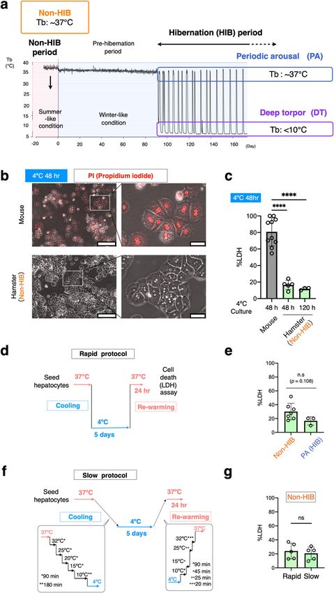
ARTICLE                                                               COMMUNICATIONS BIOLOGY | https://doi.org/10.1038/s42003-021-02297-6

M
           ammalian hibernators survive harsh seasons by invok-              of cancer medicine29. Ferroptosis sensitivity is also affected by
           ing a depressed metabolism and a prolonged low body               cellular lipid profiles, at least in several human cancer cell lines.
           temperature (Tb)1–3. Small mammalian hibernators,                 Manipulation of some metabolic enzymes of glycerophospholipids
including ground squirrels, chipmunks, marmots, bats, and                    (hereafter, simply phospholipids (PLs)) or treatment with certain
hamsters, reduce their core Tb to below 10 °C during hibernation             fatty acids changes cellular lipid composition and affects the sus-
(HIB) (Fig. 1a). This severe hypothermic state is called deep                ceptibility to ferroptosis in human cancer cell lines30–33. Lipid
torpor (DT) and continues for several days, sometimes over                   composition also affects membrane fluidity and function at dif-
1 week. DT is interrupted by periodic arousal (PA), in which                 ferent temperatures. Hence, it is of interest to compare the com-
animals leave the hypothermic state and become normothermic                  position of lipids, mainly of PLs constituting cellular membranes,
by rewarming through both non-shivering and shivering ther-                  between non-hibernators and hibernators.
mogenesis. Animals remain in the normothermic state for a                       Syrian hamsters are ideal model animals for studying the
period of time (typically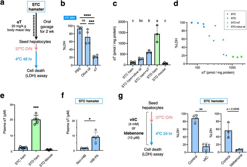
COMMUNICATIONS BIOLOGY | https://doi.org/10.1038/s42003-021-02297-6                                                             ARTICLE

cells derived from summer-like hamsters and those from winter-                 of evidence indicate that Syrian hamster hepatocytes possess
like euthermic hamsters in the PA phase during the HIB period.                 intrinsic cell-autonomous resistance to cold culture, regardless
Hepatocytes exhibited resistance to cold-rewarming stresses                    of HIB.
irrespective of HIB, although the resistance tended to be
enhanced during the HIB period (Fig. 1e). The cold resistance
of the hamsters was not affected by body mass of the animals                   Diet- and species-dependent cold resistance. Over the course of
used for hepatocytes isolation (Supplementary Fig. 1). These lines             exploring the mechanisms of cold resistance, we unexpectedly

COMMUNICATIONS BIOLOGY | (2021)4:796 | https://doi.org/10.1038/s42003-021-02297-6 | www.nature.com/commsbio                                  3
ARTICLE COMMUNICATIONS BIOLOGY | https://doi.org/10.1038/s42003-021-02297-6
Fig. 1 Intrinsic cold resistance in primary cultured hepatocytes of Syrian hamsters. a Schematic representation of changes in the core body temperature
during hibernation in Syrian hamsters. Animals raised in summer-like (warm and long photoperiod) conditions are regarded as animals in non-HIB period.
Several months after exposure to winter-like (cold and short photoperiodic) conditions, the animals begin to hibernate; the animals entered into deep
torpor (DT) that lasted about 4–5 days. DT was spontaneously interrupted by periodic arousal (PA), which lasted about 12–24 h. Cycles of DT and PA were
repeated continuously during HIB period. b Phase-contrast images of cultured hepatocytes from mouse and Syrian hamsters. Dead cells were stained with
propidium iodide (PI). Scale bars; left, 250 μm. Right, 50 μm. c The amount of cell death determined by LDH release assay after 48 or 120 h of cold culture.
****p < 0.0001 (Two-tailed Welch’s t-test). d–g An experimental procedure to recapitulate the DT-PA (cold-rewarming) process in cultured cells (d, f). No
significant difference (ns) in the amount of cell death was found after both rapid and slow cold-rewarming stress, as determined by LDH assay, between
summer-like euthermic animals and winter-like periodically aroused hamsters (e, g). Two-tailed unpaired t-test. Data are represented as the mean ±
standard deviation (SD) and each data point in (c) and (d) represents an independent sample replicate.
found that the cold resistance of hamster hepatocytes diminished cold culture after the pre-culture significantly increased TBARS in
when certain changes were made to the hamsters’ diets. Hepa- STC hamsters, but not in STD hamsters, within 8 h (Fig. 2e),
tocytes from hamsters raised on a standard diet (STD hamsters suggesting that cold-induced extensive lipid peroxidation only
hereafter) exhibited remarkable cold resistance (as shown in occurred in the hepatocytes of STC hamsters. Recent studies have
Figs. 1 and 2a) when cultured at 4 °C, whereas those from animals proposed that specific oxidized lipids, including oxidized
raised on a stock diet (STC hamsters) did not (Fig. 2a and Sup- phosphatidylethanolamine (PE) (18:0_20:4), act as ferroptotic
plementary Data 2). The STD and STC diets are usually used to death sensitizers and serve as ferroptosis signatures30,31. Hence,
maintain Syrian hamsters and mice in our laboratory, respec- we examined whether oxidized PE (18:0_20:4) was also produced
tively, and differ in their ingredients (see “Methods”). It should be during CICD. For this purpose, the 18O2 labeling method was
noted, however, that the both mice and STD hamsters used in this utilized again, using LC–MS to selectively detect the lipid
study were fed an STD diet. Nevertheless mouse hepatocytes did peroxidation that occurred during the cold culture (Fig. 2b).
not exhibit cold resistance (Figs. 1b, c and 2d). Thus, cold This analysis demonstrated that oxidized PE (38:4) species, those
resistance in hepatocytes is exerted in an STD diet-dependent corresponds with oxidized PE (18:0_20:4), were substantially
manner in hamsters, but not in mice. produced during the cold culture by both mouse and STC
hamster hepatocytes, whereas few signals indicating oxidized PE
(38:4) were detected in the cold-subjected hepatocytes from STD
Hamster’s resistance to cold-induced lipid peroxidation and hamsters (Fig. 2f, g). Thus, cold culture induces lipid peroxidation
ferroptosis. To gain insight into the mechanisms of diet- with a ferroptosis signature in hepatocytes from mice and STC
dependent cold resistance in hamster hepatocytes, we tried to hamsters, but hepatocytes from STD hamsters resist such
characterize the hallmarks of CICD in hepatocytes. MitoB, a mass ferroptosis-like lipid peroxidation.
spectrometry probe for oxidative stress in mitochondria39,40, was These observations prompted an investigation into whether
utilized to examine whether mitochondrial ROS production hamster hepatocytes were also resistant to ferroptosis-inducing
occurs under cold stress in hepatocytes from STC hamsters and agents under euthermic 37 °C conditions. In certain types of cancer
STD hamsters. MitoB accumulates in the mitochondrial matrix, cells, ferroptosis is triggered by the inhibition of glutathione
due to its cationic and lipophilic moiety and reacts with H2O2 to peroxidase 4 (Gpx4), which reduces lipid hydroperoxide at the
form a stable product, mitoP. To discriminate the cold-induced expense of glutathione42. When the ferroptosis inducers RSL3 and
oxidation of mitoB from autoxidation during sample preparation buthionine sulfoximine (BSO), which inactivate Gpx4 and deplete
and storage, the cold culture was conducted in the presence of glutathione, respectively, were applied to hepatocytes, both chemicals
18O gas in order to specifically label the mitoP generated during induced a large amount of cell death in hepatocytes from STC
2
the cold culture (Fig. 2b). The mitoP (18O)/mitoB ratio increased hamsters and mice. However, hepatocytes from STD hamsters were
markedly under cold stress in STC hamster hepatocytes, whereas not affected in this way (Fig. 2h and Supplementary Data 2). These
this increase was much less substantial in STD hamster hepato- results indicate that STD hamster hepatocytes resist ferroptosis as
cytes (Fig. 2c and Supplementary Data 2). This result suggests well as cold-induced ferroptosis in a diet-dependent manner.
that in STD hamsters, cold-induced mitochondrial ROS pro-
duction is suppressed, and/or the produced ROS are rapidly
eliminated through antioxidant mechanisms. Hamster hepatocytes contain less highly unsaturated fatty acid
In non-hibernator mammals, prolonged cold stress triggers (HUFA) in phosphatidylcholine (PC) and PE. To explore the
CICD, which accompanies lipid peroxidation mediated by ROS. mechanisms of resistance to cold-induced ferroptosis and lipid
CICD is inhibited by lipophilic radical scavengers like α- peroxidation in hamsters, we examined the difference in lipid
tocopherol (αT) or the iron-chelator deferoxamine in rat composition among mouse, STC hamster, and STD hamster cells,
hepatocytes and human cancer cell lines26,41. These chemicals as changes in lipid profiles can affect ferroptosis sensitivity30–32.
have been recently recognized as inhibitors of ferroptosis, a form In fact, the amount of PE (18:0_20:4), from which ferroptosis-
of regulated cell death associated with lipid peroxidation29. We signature-oxidized PE (18:0_20:4) species are generated during
then tested whether CICD in hepatocytes could be prevented by ferroptosis, was greater in freshly isolated hepatocytes of mice
ferroptosis inhibitors, including deferoxamine (an iron-chelator than in those of hamsters (Fig. 2i and Supplementary Data 2).
and antioxidant) and ferrostatin-1 and Trolox (free-radical This raises the possibility that a higher intrinsic amount of PE-
scavengers). As predicted, these compounds effectively inhibited 38:4 (18:0_20:4) in mouse hepatocytes might have contributed to
CICD in hepatocytes from STD mice and STC hamsters (Fig. 2d). the increase in oxidized PE (18:0_20:4) in mouse hepatocytes
Next, lipid peroxidation, a hallmark of ferroptosis, was examined under cold culture (Fig. 2f, g). On the other hand, the amount of
by measuring the amount of TBARS (2-thiobarbituric acid PE-38:4 (18:0_20:4) was not significantly different between
reactive substance) in cold-cultured hepatocytes (Fig. 2e). Imme- hepatocytes of STC hamsters and those of STD hamsters (Fig. 2i),
diately after isolation, this measurement was not significantly which did not explain the different levels of vulnerability to cold
different between hepatocytes from STC and STD hamsters. The stress between the two groups. A comprehensive analysis of total
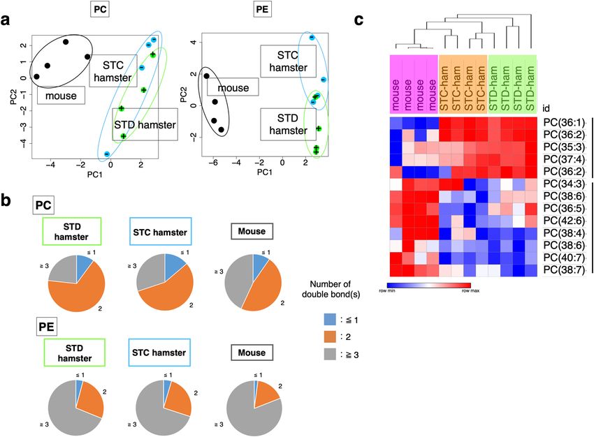
4 COMMUNICATIONS BIOLOGY | (2021)4:796 | https://doi.org/10.1038/s42003-021-02297-6 | www.nature.com/commsbioCOMMUNICATIONS BIOLOGY | https://doi.org/10.1038/s42003-021-02297-6 ARTICLE lipids extracted from freshly isolated hepatocytes was also con- axis were highly unsaturated fatty acids (HUFAs) containing ducted to compare two major PL classes, PL-PC, and PL-PE. more than two carbon–carbon double bonds (Supplementary Principle component analysis revealed a clear difference in the PC Table. 1). Indeed, the ratio of HUFAs to the analyzed total lipid and PE lipid compositions between the mice and the two groups species in the hepatocytes was greater in mice than in hamsters of hamsters along the PC1 axis (Fig. 3a and Supplementary for both PC (Fig. 3b, c) and PE (Fig. 3b and Supplementary Data 3), even when the mice and STD hamsters were given the Data 3), indicating that hamster hepatocytes contained fewer same STD diet. Most lipids that largely contributed to the PC1 HUFAs in their PC and PE than do mouse hepatocytes. HUFAs COMMUNICATIONS BIOLOGY | (2021)4:796 | https://doi.org/10.1038/s42003-021-02297-6 | www.nature.com/commsbio 5
ARTICLE COMMUNICATIONS BIOLOGY | https://doi.org/10.1038/s42003-021-02297-6
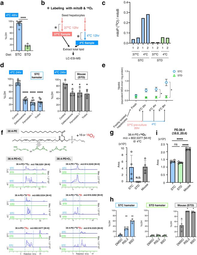
Fig. 2 Diet-dependent resistance to cold-induced ferroptosis-like cell death in Syrian hamster hepatocytes. a Cold resistance was observed only in
hepatocytes isolated from Syrian hamsters fed with the STD diet (STD), but not those fed with the STC diet (STC). ****p < 0.0001 (Two-tailed Welch’s
t-test). b An experimental procedure for mitoB and 18O2 labeling to detect mitochondrial H2O2 production and lipid peroxidation under cold-culture
conditions. c Increase in the ratio of 18O-containing mitoP to mitoB under cold culture. Results from two independent samples (1 and 2) have been shown.
d Inhibition of cold-induced cell death by ferroptosis inhibitors (100 μM of deferoxamine, 1 μM of ferrostatin-1, or 100 μM of Trolox) in hepatocytes from
STC hamsters and mice. ****p < 0.0001, *p < 0.05 versus Control (One-way ANOVA with the Tukey’s multiple comparison test). e Lipid peroxidation
(TBARS assay) occurred only in cold-cultured hepatocytes from STC hamsters. TBARS levels were measured immediately after hepatocyte isolation, after
pre-culture at 37 °C for 20 h, and after cold culture for 4 or 8 h. n = 3 (STC) or n = 4 (STD) independent sample replicates. *p < 0.05 (One-way ANOVA
with the Tukey’s multiple comparison test). f LC–MS identification of oxidized 38:4-PE in cold-cultured hepatocytes. Upper, structure of 38:4-PE and its
possible oxidized forms. Lower, comparison of extracted ion chromatogram of oxidized lipid species between STC and STD at different temperature (37 or
4 °C). Multiple peaks for oxidized 38:4-PE species (inside dashed lines) dominantly appeared in STC hepatocytes and those labeled with 18O were detected
only in STC hepatocytes at 4 °C. g Relative quantification of PE (38:4) + 18O2, an oxidized PE (38:4) with two 18O atoms. It was detected by LC–MS
analysis in hepatocytes from STC hamsters or mice, but not from STD hamsters, after 12-h cold culture. N.D. (not detected). h Ferroptosis was induced by
2-μM RSL3 or 1-mM BSO in hepatocytes from STC hamsters and mice, but not from STD hamsters, under 37 °C culture conditions. **p < 0.01, *p < 0.05
versus Control (One-way ANOVA with the Tukey’s multiple comparison test). i The amount of PE (38:4) in freshly isolated hepatocytes measured by
LC–MS analysis. ****p < 0.0001, ns p > 0.05 (One-way ANOVA with the Tukey’s multiple comparison test). Data are represented as the mean ± SD and
each data point in (a), (b), (g), (h) and (i) represents an independent sample replicate.
Fig. 3 Lipidome analysis revealed different phospholipid compositions of hepatocytes between mouse and hamster. a Principle component analysis
(PCA) of PL-PC and PL-PE species in freshly isolated hepatocytes from mouse, STC hamster, and STD hamster. b Pie charts showing the average
percentage of lipid species classified by the number of carbon–carbon double bonds. c Hierarchical clustering of the relative amount of PC species among
distinct groups of animals. Relative abundance of each lipid species among samples is shown by blue red gradient.
are targets of oxidation and become the origin of lipid perox- STD and STC diets, the difference in the amount of the dietary
idation. Taken together, differences in the amounts of HUFAs, antioxidant vitamin E is notable. According to the manufacturer’s
including PE (38:4), between hamsters and mice may contribute information, the STD diet contains five times more vitamin E
to the difference in cold-induced oxidative stress sensitivity than that found in the STC diet (STD: 208.3 mg/kg, STC:
between hepatocytes of these two species. 45.2 mg/kg). Vitamin E is a lipophilic radical scavenger, acting as
a chain-breaking antioxidant to prevent lipid peroxidation and
ferroptosis. Therefore, a rescue experiment was conducted to test
Diet-derived αT confers cold resistance on hamster hepato- whether a high intake of dietary vitamin E was sufficient for
cytes. Finally, we determined which nutrient in the STD diet was granting cold resistance to cold-vulnerable hepatocytes from STC
responsible for granting cold resistance to hamster hepatocytes. hamsters. An appropriate amount of αT, a major form of the
Although the amounts of many ingredients differ between the vitamin E analogs, was estimated as 20-μg/g body mass per a day
6 COMMUNICATIONS BIOLOGY | (2021)4:796 | https://doi.org/10.1038/s42003-021-02297-6 | www.nature.com/commsbioCOMMUNICATIONS BIOLOGY | https://doi.org/10.1038/s42003-021-02297-6 ARTICLE Fig. 4 Intake of sufficient dietary αT is required for the cold resistance of hamster hepatocytes. a Schemes for oral administration of αT to hamsters. 20-μg αT/g body mass dissolved in olive oil was administered to STC hamsters once a day for 2 weeks. b The amount of cell death after 48-h cold culture among hamsters administered with PBS, olive oil, or αT. ****p < 0.0001, ***p < 0.001, ns p > 0.05 (One-way ANOVA with Tukey’s multiple comparison test). c αT content in freshly isolated hepatocytes. αT level was normalized to the total protein amount in the hepatocyte lysates. Letters a, b, c refer to the significant differences of αT level from each other—if two columns do not share a letter, they are significantly different (p < 0.05) (One-way ANOVA with the Tukey’s multiple comparison test). d Relationship between the amount of cell death and αT content in (c). e, f αT concentration in plasma of STC hamsters, STD hamsters, and STD mice in summer-like (non-HIB) condition, and of STC hamsters in summer-like, non-HIB, or at periodic arousal during HIB (HIB-PA). ***p < 0.001 (One-way ANOVA with the Tukey’s multiple comparison test), *p < 0.05 (Welch’s t-test). g Inhibition of cold-induced cell death by 4-mM vitamin C or 10-μM idebenone in STC hamster hepatocytes. **p < 0.01 (Two-tailed paired t-test). A scheme for treating hepatocytes with vitamin C or idebenone is shown. Data are represented as the mean ± SD and each data point in (b–g) represents an independent sample. and orally administered to STC hamsters for 2 weeks, and their αT absorbed from diets is redistributed via liver into whole hepatocytes were subsequently subjected to cold culture. As a body44. Thus, hamsters, but not mice, have the capacity to retain result, CICD was significantly suppressed in hepatocytes from a high amount of diet-derived αT in their hepatocytes as well as in αT-treated hamsters compared with those from the vehicle con- the blood circulation, which contributes to the cold resistance of trol, olive-oil-treated hamsters (Fig. 4a, b and Supplementary hamster hepatocytes by preventing lipid peroxidation. Interest- Data 4). Thus, the cold resistance of hamster hepatocytes is ingly, the plasma αT concentration of euthermic STC hamsters reliant on a sufficient amount of dietary αT. To examine how at PA during the HIB period was about fivefold higher (8.85 ± dietary αT contributed to cold resistance in hamster hepatocytes, 3.23 μM) than those in the non-HIB period (1.57 ± 0.24 μM) αT content was measured in freshly isolated hepatocytes43. This (Fig. 4f), implying the existence of systems to compensate for the analysis revealed that hepatocytes from αT-treated hamsters low amount of αT taken from the diet during the HIB period in contained a higher amount of αT compared with those from Syrian hamsters. olive-oil-treated hamsters or non-treated STC hamsters (Fig. 4c). αT is regenerated from its oxidized form, αT radical, by vitamin C STD hamsters exhibited a much higher αT content and lower or ubiquinol (Coenzyme QH2) in a distinct chemical reaction45–47. frequency of cell death than that exhibited by αT-treated STC Given that αT is consumed to prevent lipid peroxidation during cold hamsters, and the preventive effect on cell death correlated with culture, it is expected that cold-vulnerable STC hepatocytes contain- the αT content of hepatocytes (Fig. 4c, d and Supplementary ing small amounts of αT may exhibit improved cold resistance when Data 3). Interestingly, the amount of αT in mouse hepatocytes αT regeneration is enhanced. Consistent with this idea, the addition was about 1/10th of that in hamster hepatocytes, despite the fact of vitamin C or idebenone, a Coenzyme QH2 analog, effectively that the mice were fed with the same STD diet as the STD inhibited CICD in hepatocytes from STC hamsters (Fig. 4g). Taken hamsters (Fig. 4c). Plasma αT concentration was also higher in together, these data suggest that the cold resistance of hamster the STD hamsters than in the mice fed with STD diet or the STC hepatocytes is established via retention of a large amount of αT, hamsters (Fig. 4e), which is consistent with the fact that most of which is utilized to prevent lipid peroxidation. COMMUNICATIONS BIOLOGY | (2021)4:796 | https://doi.org/10.1038/s42003-021-02297-6 | www.nature.com/commsbio 7
ARTICLE                                                             COMMUNICATIONS BIOLOGY | https://doi.org/10.1038/s42003-021-02297-6

Discussion                                                                 [PE-38:4(18:0_20:4)] (a known ferroptosis signature), and inhi-
One of the greatest and longest-lasting mysteries of HIB is                bition of cell death by deferoxamine (an iron chelator) and αT (a
how mammalian deep hibernators endure severe hypothermia                   lipophilic radical scavenger). Pathophysiological conditions such
(
COMMUNICATIONS BIOLOGY | https://doi.org/10.1038/s42003-021-02297-6                                                                                       ARTICLE

remained in place until the animals experienced a PA36. Under this condition, most       Cell death assay. Hepatocytes were incubated with 1-μg/mL PI (Sigma P4170) for
animals start HIB 2–4 months after cold exposure34. For sampling animals in the          about 30 min and observed under a DMi8 microscope (Leica Microsystems). The
PA phase during the HIB period, we observed animal status every morning by               amount of LDH in the supernatant was measured using an LDH cytotoxicity
visual inspection with the sawdust method mentioned above to check whether the           detection kit (Takara MK401) following the manufacturer’s instructions. The
hibernating animals were in the PA or DT phase. Euthermic animals in sponta-             amount of LDH in each sample was normalized against that of hepatocytes fully
neous PA phases, judged by locomotion in cages and the reaction to handling, were        lysed with Triton X-100 (final 1% in PBS). The samples were individually mixed
anesthetized and used for hepatocyte culture around 12:00–16:00 (ZT2-6) (see             with reagents on microplates, and the absorbance was measured at 490 and 630 nm
isolation of hepatocytes for details). In this study, Tb and duration during PA were     (as a control) using 2030 ARVOTM X (Perkin Elmer) or Multiskan GO (Thermo
not determined, as a Tb logger (iButton) was not implanted into the animals used         Fisher Scientific) after a 30-min incubation at room temperature.
for hepatocyte culture because of the concern that surgical operation of Tb loggers
into the body cavity might affect liver physiology and hepatocyte culture. Animals       18O
                                                                                             2 labeling. Three hours after seeding the hepatocytes, the culture medium was
were sacrificed under anesthesia with 4.5% isoflurane for hepatocytes isolation by
                                                                                         changed to the new basal medium containing 5-μM mitoB (Cayman Chemical,
reperfusion or for blood collection by decapitation at 13–18 weeks of age, except for
                                                                                         17116). To replace oxygen with 18O2 as much as possible, a bubbling treatment was
Figs. 1d, e and 4f in which the animals were at 31–36 weeks of age.
                                                                                         carried out with 18O2-containing gas in the new basal medium [Gas A (N2: 75%,
    Male C57BL/6 mice were purchased from SLC, Inc, Japan. Animals were reared           18O : 20%, CO : 5%) for 37 °C culture and Gas B (N : 80%, 18O : 20%) for 4 °C
under summer-like conditions (light condition = 14L:10D cycle, lights on                     2           2                                      2          2

06.00–20.00, ambient temperature = 22–25 °C) and had ad libitum access to STD            culture]. Gas mixing was completed by a custom-made gas mixer. After the medium
diet (MR standard diet, Nihon Nosan, Japan) and water in this experiment.                change, the cell culture plates were put into a chamber that had been incubated at
Because we did not have information on the diet the mice had been fed in the             37 °C in advance. Then, the chamber was filled with 18O2-containing gas for 30 min
breeding company, we purchased the mice at 8 weeks of age and fed the STD diet           at a flow rate of 20 cc/min, which was presumed to replace the gas in the chamber
for over 2 months and used for hepatocyte culture experiments at 16–21 weeks             completely. After that, each chamber was placed in a 37 °C incubator or a 4 °C
of age.                                                                                  refrigerator and cultured for 12 h. Then, the cells were washed with PBS once and
    All animal care and experimental procedures were approved by the Ethics              homogenized in 1-mL MeOH (5 × 10⁵ cells/mL). These homogenates were stored at
Committees of the University of Tokyo (Ethical Approval no. P28-11) and                  −80 °C and used for measurements of mitoP/B39,40 and lipids within 2 weeks.
Hokkaido University (Ethical Approval no. 18-0140), and conducted according to
the ethics guidelines of the University of Tokyo and Hokkaido University.                Measurement of mitochondrial hydrogen peroxide in vitro. We prepared MitoB
                                                                                         (C25H23BBrO2P, molecular weight 477.14, Sigma) stock solution in sterile saline.
                                                                                         Diluted MitoB solution (0.5 mg/mL in a 1:1 solution of DMSO:PBS) was added to
Chemicals. Reagents used in this study are as follows; Deferoxamine (Sigma               the hepatocyte culture. The production of mitochondrial hydrogen peroxide during
Aldrich, D9533), Ferrostatin-1 (Sigma Aldrich, SML0583), Trolox (Cayman Che-             incubation was assessed by determining the MitoP/MitoB ratio using LC–MS/MS
mical, 10011659), vitamin C (Ascorbic acid) (Wako, 012-04802), RSL3 (Sigma               (LC-MS8040, Shimadzu Corporation), by monitoring positive ion transitions of m/z
Aldrich, SML2234), BSO (Cayman Chemical, 14484).                                         397.1 > 183.0 and m/z 369.1 > 183.0 for MitoB and MitoP, respectively40. The
                                                                                         samples were resolved on the LUNA Phenyl-Hexyl column (100 × 2.0 mm I.D ×
                                                                                         100 mmL, 3-μm particle, Shimadzu GLC), using a step gradient with mobile phase
Isolation of hepatocytes. Hepatocytes from the livers of hamsters and mice were          A (0.1% formate) and mobile phase B (0.1% acetonitrile) at ratios of 90:10 (0–2
prepared by the two-step collagenase perfusion technique, according to previous          min), 65:35 (2–5 min), 50:50 (5–10 min), 0:100 (10–14 min), and 90:10 (14–20 min),
studies with minor modifications60. Briefly, after anesthesia with 4.5% isoflurane,         at a flow rate of 0.2 mL/min and a column temperature of 40 °C.
the livers were exposed and perfused with a solution containing 1-mM EGTA
(ethylene glycol tetraacetic acid) in Ca2+/Mg2+-free Hank’s balanced salt solution
                                                                                         TBARS assay. Lipid peroxidation levels in the hepatocytes were measured by a
for about 5 min, followed by a solution containing 1-mg/mL collagenase with
                                                                                         TBARS (TAC method) assay kit (Cayman Chemical). For the fresh samples, 1 ×
Ca2+/Mg2+ in Hank’s balanced salt solution for 2–3 min at a flow rate of 13 mL/
                                                                                         106 hepatocytes were washed immediately after isolation with PBS (40 × g or 100 ×
min for hamsters and 6 mL/min for mice. The livers were gently removed and put
                                                                                         g, 2 min) and homogenized with 200 μL of homogenizing buffer (50-mM phos-
into EMEM (Sigma Aldrich, M4655) containing 10% FBS. Hepatocytes were
                                                                                         phate buffer, 1-mM EDTA, 1% Triton X-100). For the 4 °C samples at 0, 4, and 8 h,
obtained after mechanical dissociation by shaking the liver tissue gently in the
                                                                                         1 × 10⁶ hepatocytes cultured on 60-mm dishes were washed with PBS after 37 °C
EMEM, grabbing with tweezers with a circular tip, and subsequently filtering with a
                                                                                         pre-culture or cold culture and homogenized with 200 μL of homogenizing buffer.
100-μm mesh cell strainer (TOKYO SCREEN CO., LTD). The cells were collected
                                                                                         These homogenates were stored at −80 °C until use. A TBARS assay was con-
via centrifugation at 40 × g for 1 min. Then, the cells were resuspended in 24.5 mL of
                                                                                         ducted using 100 μL of these homogenates following the manufacturer’s instruc-
complete Percoll medium (composed of 12.5 mL of L-15 [Gibco 11415-064] med-
                                                                                         tions. Briefly, the homogenates were mixed with reagents and incubated at 98 °C
ium supplemented with 0.429-g/L HEPES, 2-g/L BSA [Sigma A1470], 1 × 10⁻⁷-M
                                                                                         for 1 h. After being incubated for 10 min on ice, the samples were centrifuged at
insulin [Wako 093-06471], 1.2 mL of 10× HBSS(-) and 10.8 mL of Percoll [GE
                                                                                         1600 × g for 10 min at 4 °C. Then, absorbance was measured at 540 nm using
Healthcare 17-5445-02]), and live parenchymal cells were purified via centrifugation
                                                                                         Multiskan GO (Thermo Fisher Scientific). The same homogenates were used to
at 60 × g for 10 min. Then, the cells were washed twice in EMEM containing 10%
                                                                                         quantify the total protein amount by the BCA method, and the TBARS level was
FBS via centrifugation at 40 × g for 2 min, and finally filtered with a 40-μm cell
                                                                                         normalized to the protein amount.
strainer (BD Falcon). Cell viability (>80%) was assessed by the trypan blue
exclusion test.
                                                                                         Lipidome analysis and identification of lipid peroxidation with LC–MS. Freshly
                                                                                         isolated hepatocytes were centrifuged and the pellets were stored in 1.5-mL
Preparation of collagen-coated plate. Atelocollagen acidic solution (KOKEN,              Eppendorf tube at −80 °C until use. For total lipid extraction, 100 µL of 1-butanol/
IPC-30) was diluted at 0.5% in PBS. The diluted collagen solution was added to           methanol (1:1, v/v) with 5-mM ammonium formate was added into the pellets, and
tissue culture-treated plates (Corning) and incubated 5 min at room temperature.         the mixture was vortexed for 10 s, sonicated for 15 min in a sonic water bath, and
The plates were washed with PBS once and placed at 37 °C until they were used.           then centrifuged (16,000 × g, 10 min, 20 °C). The supernatant was transferred into a
                                                                                         0.2-mL glass insert with Teflon insert cap for analysis by LC ESI-MS61.
                                                                                             For lipidomic analysis, an orbitrap type MS (Q-Exactive focus, Thermo Fisher
Primary culture of hepatocytes. The basal medium for culturing hepatocytes was           Scientific, San Jose, CA), that enables us to perform highly selective and sensitive
DMEM/F12 (Gibco 21041-025) supplemented with 5-mM HEPES (Gibco 15630-                    metabolite quantification owing to the Fourier Transfer MS principle, was
080), 30-mg/L L-proline (Wako 161-04602), 0.5% BSA (Sigma A1470), 10-ng/mL               connected to a HPLC (Ultimate3000 system, Thermo Fisher Scientific). LC and MS
epidermal growth factor (Sigma E4127), 1% Insulin, Transferrin, Selenium, Etha-          conditions were based on Růžička et al.62. Briefly, the samples were resolved on the
nolamine Solution (ITS-X) (Gibco 51500056), 1 × 10⁻⁷-M dexamethasone (Wako               Thermo Scientific Accucore C18 column (2.1 × 150 mm, 2.6 μm) with mobile phase
047-18863), 10-mM nicotinamide (Wako 141-01202), 1-mM L-ascorbic acid 2-                 A (10-mM ammonium formate in 50% acetonitrile (v) and 0.1% formic acid (v))
phosphate (Wako 323-44822), and 1% Penicillin-Streptomycin (Wako 168-23191).             and mobile phase B (2-mM ammonium formate in acetonitrile/isopropyl alcohol/
Hepatocytes were seeded on collagen-coated plates and cultured at 37 °C in a 5%          water, ratios of 10:88:2 (v/v/v) with 0.02% formic acid (v)), using step gradient at
CO2 incubator. A 10% FBS-supplemented basal medium was used as the seeding               ratios of 65:35 (0 min), 40:60 (0–4 min), 15:85 (4–12 min), 0:100 (12–21 min),
medium. Three to four hours after seeding, the medium was replaced with serum-           0:100 (21–24 min), 65:35 (24–24.1 min) and 100:0 (24.1–28 min), at a flow rate of
free basal medium, and the cells were cultured for about 16 h for stabilization.         0.4 mL/min and a column temperature of 35 °C.
Then, cold culture was conducted in a 4 °C refrigerator using the basal medium               The Q-Exactive focus mass spectrometer was operated under an ESI positive
containing 100-mM HEPES (pH 7.4). This formulation is sufficient for keeping a            and negative mode. Full mass scan (m/z 250–1100) followed by three rapid data-
pH of 7.5 at 4 °C, excluding the possibility that pH dysregulation affects cell via-     dependent MS/MS, was operated at resolutions of 70,000 and 17,500, respectively.
bility. For the slow protocol, additional refrigerators and another cell culture         The automatic gain control target was set at 1 × 10⁶ ions, and maximum ion
incubator were set to 10, 15, 20, 25, and 32 °C, respectively, and cell culture dishes   injection time was 100 ms. Source ionization parameters were as follows; spray
were transferred successively from the incubators to refrigerators.                      voltage at 3 kV, transfer tube temperature at 285 °C, S-Lens level at 45, heater

COMMUNICATIONS BIOLOGY | (2021)4:796 | https://doi.org/10.1038/s42003-021-02297-6 | www.nature.com/commsbio                                                                9
ARTICLE COMMUNICATIONS BIOLOGY | https://doi.org/10.1038/s42003-021-02297-6
temperature at 370 °C, Sheath gas at 60, and auxilliary gas at 20. Acquired data 7. Giroud, S. et al. The torpid state: recent advances in metabolic adaptations and
were analyzed by Qual browser for oxidized lipid analysis, and by LipidSearch protective mechanisms(dagger). Front. Physiol. 11, 623665 (2020).
software (Mitsui Knowledge Industry, Tokyo, Japan) for major PLs with following 8. Arnold, W., Ruf, T., Frey-Roos, F. & Bruns, U. Diet-independent remodeling
parameters; search parameters: precursor mass tolerance = 3 ppm, product mass of cellular membranes precedes seasonally changing body temperature in a
tolerance = 7 ppm, m-score threshold = 3. hibernator. PLoS ONE 6, e18641 (2011).
9. Ruf, T. & Arnold, W. Effects of polyunsaturated fatty acids on hibernation and
Oral administration of αT. The Syrian hamsters ate about 385-g/kg body mass of torpor: a review and hypothesis. Am. J. Physiol. Regul. Integr. Comp. Physiol.
diet per week in our laboratory. According to the manufacturer’s information, the 294, R1044–R1052 (2008).
difference in daily intake of vitamin E between the STD diet and the STC diet was 10. Giroud, S. et al. Membrane phospholipid fatty acid composition regulates
estimated to be about 9.0-μg/g body mass. To compensate for this difference and cardiac SERCA activity in a hibernator, the Syrian hamster (Mesocricetus
avoid overdose, αT (Tokyo Chemical Industry, T2309) dissolved in olive oil (Wako, auratus). PLoS ONE 8, e63111 (2013).
150-00276) at a concentration of 20 μg/μL was administered at 1-μL/g body mass 11. Lindell, S. L. et al. Natural resistance to liver cold ischemia-reperfusion injury
to hamsters fed with the STC diet for more than a month. As controls, olive oil or associated with the hibernation phenotype. Am. J. Physiol. Gastrointest. Liver
PBS were given to other groups of animals. Administration of αT or the controls Physiol. 288, G473–G480 (2005).
was conducted by oral gavage once daily between 11:00 and 13:00 for 14 con- 12. Otis, J. P., Pike, A. C., Torrealba, J. R. & Carey, H. V. Hibernation reduces
secutive days before hepatocytes were cultured from the animals. cellular damage caused by warm hepatic ischemia-reperfusion in ground
squirrels. J. Comp. Physiol. B 187, 639–648 (2017).
Quantification of αT with high-performance liquid chromatography (HPLC). 13. Kurtz, C. C., Lindell, S. L., Mangino, M. J. & Carey, H. V. Hibernation confers
Freshly isolated hepatocytes were washed with PBS and stored at −80 °C until αT resistance to intestinal ischemia-reperfusion injury. Am. J. Physiol.
extraction. Plasma was obtained by centrifuging blood immediately after the col- Gastrointest. Liver Physiol. 291, G895–G901 (2006).
lection from a finger of hamster or a tail of mouse using heparin-coated capillary 14. Yan, J., Barnes, B. M., Kohl, F. & Marr, T. G. Modulation of gene expression in
tubes. 1/100 volume of 13% EDTA-2K was added to the collected blood imme- hibernating arctic ground squirrels. Physiol. Genom. 32, 170–181 (2008).
diately and mixed, and then the blood was centrifuged at 1200 × g for 10 min at 15. Buck, C. L. & Barnes, B. M. Annual cycle of body composition and
room temperature. The supernatant was collected as plasma, immediately frozen in hibernation in free-living Arctic ground squirrels. J. Mammal. 80, 430–442
liquid N2, and stored at −80 °C until use. The collected blood was extraction and (1999).
measurement of αT was conducted as per a previously described method43. Briefly, 16. Bogren, L. K., Olson, J. M., Carpluk, J., Moore, J. M. & Drew, K. L. Resistance
cell pellets or plasma were homogenized using a bead mixer with PBS, and lysates to systemic inflammation and multi organ damage after global ischemia/
were used for the protein quantification with a BCA assay and for vitamin E reperfusion in the arctic ground squirrel. PLoS ONE 9, e94225 (2014).
measurement. Extraction of total lipids from the lysates or plasma was done by 17. Bhowmick, S., Moore, J. T., Kirschner, D. L. & Drew, K. L. Arctic ground
vigorously mixing them with chloroform/methanol (2:1 by volume) containing squirrel hippocampus tolerates oxygen glucose deprivation independent of
100 μM of butylated hydroxytoluene to avoid the in vitro oxidation. Hepatic αT hibernation season even when not hibernating and after ATP depletion,
contents were measured using HPLC with an amperometric electrochemical acidosis, and glutamate efflux. J. Neurochem. 142, 160–170 (2017).
detector (ECD) (Nanospace SI-1; Osaka Soda, Japan) set at 700 mV, with an ODS 18. Lyman, C. P., Willis, J. S., Malan, A., Wang, L. C. H. Hibernation and Torpor
column (Wakosil-II 5C18 RS, 5 μm, 250 × 4.6 mm; Fuji film Wako, Osaka, Japan, in Mammals and Birds. (Academic Press, 1982).
or) followed by a reducing column (RC-10, 15 × 4 mm; Shiseido) with methanol 19. Talaei, F. et al. Serotonin and dopamine protect from hypothermia/rewarming
containing 50-mM sodium perchlorate as eluent at 0.7 ml/min. αT in plasma was damage through the CBS/H2S pathway. PLoS ONE 6, e22568 (2011).
measured using HPCD-ECD equipped with a reducing column (ECD-700, Eicom, 20. Cooper, S. T. et al. The hibernating 13-lined ground squirrel as a model
Japan). Methanol containing 50-mM sodium perchlorate, 2% of water, and 0.1% organism for potential cold storage of platelets. Am. J. Physiol. Regul. Integr.
sodium acetate was used as eluent. Comp. Physiol. 302, R1202–R1208 (2012).
21. Jain, S., Keys, D., Martin, S., Edelstein, C. L. & Jani, A. Protection from
Statistics and reproducibility. Statistical analyses were conducted using Graph- apoptotic cell death during cold storage followed by rewarming in 13-lined
Pad Prism unless otherwise mentioned. We repeated same experiments at least two ground squirrel tubular cells: the role of prosurvival factors X-linked inhibitor
and typically over three times, except data on PL metabolomics (Fig. 3) and mitoP/ of apoptosis and PhosphoAkt. Transplantation 100, 538–545 (2016).
B labeling (Fig. 2c) due to cost limitation and restriction. Each sample size is 22. Hendriks, K. D. W. et al. Differences in mitochondrial function and
indicated in the figure or legend. morphology during cooling and rewarming between hibernator and non-
hibernator derived kidney epithelial cells. Sci. Rep. 7, 15482 (2017).
Reporting summary. Further information on research design is available in the Nature 23. Ou, J. et al. iPSCs from a hibernator provide a platform for studying cold
Research Reporting Summary linked to this article.
adaptation and its potential medical applications. Cell 173, 851–863.e16
(2018).
24. Hendriks, K. D. W. et al. Hibernator-derived cells show superior protection
Data availability and survival in hypothermia compared to non-hibernator cells. Int. J. Mol. Sci.
Source data underlying figures are presented in Supplementary Data 1–4. All other data 21, https://doi.org/10.3390/ijms21051864 (2020).
are available upon reasonable request to the corresponding author Y.Y. 25. Singhal, N. S. et al. Cytoprotection by a naturally occurring variant of ATP5G1
in Arctic ground squirrel neural progenitor cells. Elife 9, https://doi.org/
10.7554/eLife.55578 (2020).
Received: 28 November 2020; Accepted: 3 June 2021; 26. Hattori, K. et al. Cold stress-induced ferroptosis involves the ASK1-p38
pathway. EMBO Rep. 18, 2067–2078 (2017).
27. Nakamura, T. et al. The mitochondrial Ca(2+) uptake regulator, MICU1, is
involved in cold stress-induced ferroptosis. EMBO Rep. e51532, https://doi.
org/10.15252/embr.202051532 (2021).
References 28. Dixon, S. J. et al. Ferroptosis: an iron-dependent form of nonapoptotic cell
1. Andrews, M. T. Molecular interactions underpinning the phenotype of death. Cell 149, 1060–1072 (2012).
hibernation in mammals. J. Exp. Biol. 222, https://doi.org/10.1242/jeb.160606 29. Dixon, S. J. & Stockwell, B. R. The hallmarks of ferroptosis. Annu. Rev. Canc
(2019). Biol. 3, 35–54 (2019).
2. Mohr, S. M., Bagriantsev, S. N. & Gracheva, E. O. Cellular, molecular, and 30. Doll, S. et al. ACSL4 dictates ferroptosis sensitivity by shaping cellular lipid
physiological adaptations of hibernation: the solution to environmental composition. Nat. Chem. Biol. 13, 91–98 (2017).
challenges. Annu. Rev. Cell Dev. Biol. 36, 315–338 (2020). 31. Kagan, V. E. et al. Oxidized arachidonic and adrenic PEs navigate cells to
3. Carey, H. V., Andrews, M. T. & Martin, S. L. Mammalian hibernation: cellular ferroptosis. Nat. Chem. Biol. 13, 81–90 (2017).
and molecular responses to depressed metabolism and low temperature. 32. Magtanong, L. et al. Exogenous monounsaturated fatty acids promote a
Physiol. Rev. 83, 1153–1181 (2003). ferroptosis-resistant cell state. Cell Chem. Biol. 26, 420–432.e9 (2019).
4. Aloia, R. C. & Raison, J. K. Membrane function in mammalian hibernation. 33. Beatty, A. et al. Ferroptotic cell death triggered by conjugated linolenic acids is
Biochim. Biophys. Acta 988, 123–146 (1989). mediated by ACSL1. Nat. Commun. 12, 2244 (2021).
5. Orr, A. L., Lohse, L. A., Drew, K. L. & Hermes-Lima, M. Physiological 34. Chayama, Y., Ando, L., Tamura, Y., Miura, M. & Yamaguchi, Y. Decreases in
oxidative stress after arousal from hibernation in Arctic ground squirrel. body temperature and body mass constitute pre-hibernation remodelling in
Comp. Biochem. Physiol. A Mol. Integr. Physiol. 153, 213–221 (2009). the Syrian golden hamster, a facultative mammalian hibernator. R. Soc. Open
6. Ma, Y. L. et al. Absence of cellular stress in brain after hypoxia induced by Sci. 3, 160002 (2016).
arousal from hibernation in Arctic ground squirrels. Am. J. Physiol. Regul. 35. Trefna, M. et al. The influence of sex and diet on the characteristics of
Integr. Comp. Physiol. 289, R1297–R1306 (2005). hibernation in Syrian hamsters. J. Comp. Physiol. B 187, 725–734 (2017).
10 COMMUNICATIONS BIOLOGY | (2021)4:796 | https://doi.org/10.1038/s42003-021-02297-6 | www.nature.com/commsbioCOMMUNICATIONS BIOLOGY | https://doi.org/10.1038/s42003-021-02297-6 ARTICLE
36. Janský, L., Haddad, G., Kahlerova, Z. & Nedoma, J. Effect of external factors 62. Ruzicka, J., Mchale, K. & Peake, D. A. Data acquisition parameters
on hibernation of golden-hamsters. J. Comp. Physiol. 154, 427–433 (1984). optimization of quadrupole orbitrap for global lipidomics on LC-MS/MS time
37. Chayama, Y. et al. Molecular basis of white adipose tissue remodeling that frame. (2014).
precedes and coincides with hibernation in the syrian hamster, a food-storing
hibernator. Front Physiol. 9, 1973 (2018).
38. Russell, T. L. et al. Medullary respiratory circuit is reorganized by a seasonally- Acknowledgements
induced program in preparation for hibernation. Front. Neurosci. 13, 376 We are grateful to T. Miyake, H. Kusuhara, and Y. Ito for instructing the primary
(2019). hepatocyte cultures, and N. Kono, S. Shuji, and all members of Miura’s and Yamaguchi’s
39. Cocheme, H. M. et al. Measurement of H2O2 within living Drosophila during laboratories for helpful discussion. We also thank Y. Chayama, L. Ando, Y. Sato,
aging using a ratiometric mass spectrometry probe targeted to the T. Fujimoto, H. Taii, H. Makino, K. Sone, and S. Enju for experimental assistance. This
mitochondrial matrix. Cell Metab. 13, 340–350 (2011). work was supported by grants from the Japan Science and Technology Agency
40. Cocheme, H. M. et al. Using the mitochondria-targeted ratiometric mass (JPMJPR12M9 to Y.Y.) and from the Japanese Society for the Promotion of Science, and
spectrometry probe MitoB to measure H2O2 in living Drosophila. Nat. Protoc. the Ministry of Education, Culture, Sports, Science, and Technology in Japan
7, 946–958 (2012). (JP20H05766, JP19H04046, and JP18K19321 to Y.Y., JP18K14884 to Y.M., JP17H03977
41. Rauen, U., Polzar, B., Stephan, H., Mannherz, H. G. & de Groot, H. Cold- and 18K19405 to K.Y., and JP16H06385 to M.M.). This work was also supported by
induced apoptosis in cultured hepatocytes and liver endothelial cells: grants from the Japan Agency for Medical Research and Development (AMED) to Y.Y.
mediation by reactive oxygen species. FASEB J. 13, 155–168 (1999). (JP20gm6310019), M.M. (JP17gm061004 and JP19gm5010001), and K.Y.
42. Yang, W. S. et al. Regulation of ferroptotic cancer cell death by GPX4. Cell (JP19gm0910013), by a grant from AMED Moonshot R&D Program to Y.S.
156, 317–331 (2014). (21zf0127003h001), and by grants to Y.Y. from the Cell Science Foundation, Sekisui
43. Shichiri, M. et al. alpha-Tocopherol suppresses lipid peroxidation and Chemical Innovations Inspired by Nature Research Support Program, the Sumitomo
behavioral and cognitive impairments in the Ts65Dn mouse model of Down Foundation for Basic Research Projects, the Takeda Science Foundation, Toray Science
syndrome. Free Radic. Biol. Med. 50, 1801–1811 (2011). foundation, the Naito Foundation, the Uehara Memorial Foundation, the Mochida
44. Kono, N. et al. Impaired alpha-TTP-PIPs interaction underlies familial Memorial Foundation for Medical and Pharmaceutical Research, Terumo Life Science
vitamin E deficiency. Science 340, 1106–1110 (2013). Foundation, Inamori Foundation, and NIBB Collaborative Research Program (16-408
45. Doll, S. et al. FSP1 is a glutathione-independent ferroptosis suppressor. Nature and 20-101). D.A. was a research fellow of the Japan Society for the Promotion of
575, 693–698 (2019). Science.
46. Halpner, A. D., Handelman, G. J., Harris, J. M., Belmont, C. A. & Blumberg, J.
B. Protection by vitamin C of loss of vitamin E in cultured rat hepatocytes. Author contributions
Arch. Biochem. Biophys. 359, 305–309 (1998). D.A., Y.S. and Y.Y. conceived the experiments. D.A., Y.S., Y.M., M. So., N.I., M. Sh., R.O.
47. Bersuker, K. et al. The CoQ oxidoreductase FSP1 acts parallel to GPX4 to and Y.Y. performed the experiments. D.A., Y.S., Y.M., M. So., R.O. and Y.Y. analyzed
inhibit ferroptosis. Nature 575, 688–692 (2019). data and wrote the manuscript. M.M., K.Y., M. Su. and Y.Y. supervised the study.
48. Hazel, J. R. Thermal adaptation in biological membranes: is homeoviscous
adaptation the explanation? Annu. Rev. Physiol. 57, 19–42 (1995).
49. Gaschler, M. M. & Stockwell, B. R. Lipid peroxidation in cell death. Biochem. Competing interests
Biophys. Res. Commun. 482, 419–425 (2017). The authors declare no competing interests.
50. Frank, C. L., Dierenfeld, E. S. & Storey, K. B. The relationship between lipid
peroxidation, hibernation, and food selection in mammals. Am. Zool. 38,
341–379 (1998).
Additional information
Supplementary information The online version contains supplementary material
51. Giroud, S. et al. Dietary lipids affect the onset of hibernation in the garden
available at https://doi.org/10.1038/s42003-021-02297-6.
dormouse (Eliomys quercinus): implications for cardiac function. Front.
Physiol. 9, 1235 (2018).
Correspondence and requests for materials should be addressed to Y.Y.
52. Burla, B. et al. MS-based lipidomics of human blood plasma: a community-
initiated position paper to develop accepted guidelines. J. Lipid Res. 59, Peer review information Communications Biology thanks the anonymous reviewers for
2001–2017 (2018). their contribution to the peer review of this work. Primary handling editor: George Inglis.
53. Bowden, J. A. et al. Harmonizing lipidomics: NIST interlaboratory Peer reviewer reports are available.
comparison exercise for lipidomics using SRM 1950-metabolites in frozen
human plasma. J. Lipid Res. 58, 2275–2288 (2017). Reprints and permission information is available at http://www.nature.com/reprints
54. Li, J. et al. Ferroptosis: past, present and future. Cell Death Dis. 11, 88 (2020).
55. Tsurusaki, S. et al. Hepatic ferroptosis plays an important role as the trigger
Publisher’s note Springer Nature remains neutral with regard to jurisdictional claims in
for initiating inflammation in nonalcoholic steatohepatitis. Cell Death Dis. 10,
published maps and institutional affiliations.
449 (2019).
56. Tøien, O., Drew, K. L., Chao, M. L. & Rice, M. E. Ascorbate dynamics and
oxygen consumption during arousal from hibernation in Arctic ground
squirrels. Am. J. Physiol. Regul. Integr. Comp. Physiol. 281, R572–R583 (2001). Open Access This article is licensed under a Creative Commons
57. Ma, Y. L. et al. Ascorbate distribution during hibernation is independent of Attribution 4.0 International License, which permits use, sharing,
ascorbate redox state. Free Radic. Biol. Med. 37, 511–520 (2004). adaptation, distribution and reproduction in any medium or format, as long as you give
58. Okamoto, I. et al. Up-regulation of an extracellular superoxide dismutase-like appropriate credit to the original author(s) and the source, provide a link to the Creative
activity in hibernating hamsters subjected to oxidative stress in mid- to late Commons license, and indicate if changes were made. The images or other third party
arousal from torpor. Comp. Biochem Physiol. C. Toxicol. Pharm. 144, 47–56 material in this article are included in the article’s Creative Commons license, unless
(2006). indicated otherwise in a credit line to the material. If material is not included in the
59. Frank, C. L., Gibbs, A. G., Dierenfeld, E. S., Kramer, J. V. Life in the Cold. (eds article’s Creative Commons license and your intended use is not permitted by statutory
Klingenspor, M. & Heldmaier, G.) 207–213 (Springer, 2000). regulation or exceeds the permitted use, you will need to obtain permission directly from
60. Katsuda, T. et al. Conversion of terminally committed hepatocytes to the copyright holder. To view a copy of this license, visit http://creativecommons.org/
culturable bipotent progenitor cells with regenerative capacity. Cell Stem Cell licenses/by/4.0/.
20, 41–55 (2017).
61. Alshehry, Z. H. et al. An efficient single phase method for the extraction of
plasma lipids. Metabolites 5, 389–403 (2015). © The Author(s) 2021
COMMUNICATIONS BIOLOGY | (2021)4:796 | https://doi.org/10.1038/s42003-021-02297-6 | www.nature.com/commsbio 11You can also read