CPA-seq reveals small ncRNAs with methylated nucleosides and diverse termini
←
→
Page content transcription
If your browser does not render page correctly, please read the page content below
Wang et al. Cell Discovery (2021)7:25
                                    https://doi.org/10.1038/s41421-021-00265-2
                                                                                                                                                                                                        Cell Discovery
                                                                                                                                                                                                     www.nature.com/celldisc

                                     ARTICLE                                                                                                                                                    Open Access

                                    CPA-seq reveals small ncRNAs with methylated
                                    nucleosides and diverse termini
                                    Heming Wang1,2,3, Rong Huang1,2,3, Ling Li1, Junjin Zhu1,3, Zhihong Li4, Chao Peng                                                             4
                                                                                                                                                                                     , Xuran Zhuang1, Haifan Lin5,
                                    Shuo Shi6 and Pengyu Huang 1,3,7

                                      Abstract
                                      High-throughput sequencing reveals the complex landscape of small noncoding RNAs (sRNAs). However, it is limited
                                      by requiring 5′-monophosphate and 3′-hydroxyl in RNAs for adapter ligation and hindered by methylated nucleosides
                                      that interfere with reverse transcription. Here we develop Cap-Clip acid pyrophosphatase (Cap-Clip), T4 polynucleotide
                                      kinase (PNK) and AlkB/AlkB(D135S)-facilitated small ncRNA sequencing (CPA-seq) to detect and quantify sRNAs with
                                      terminus multiplicities and nucleoside methylations. CPA-seq identified a large number of previously undetected
                                      sRNAs. Comparison of sRNAs with or without AlkB/AlkB(D135S) treatment reveals nucleoside methylations on sRNAs.
                                      Using CPA-seq, we profiled the sRNA transcriptomes (sRNomes) of nine mouse tissues and reported the extensive
                                      tissue-specific differences of sRNAs. We also observed the transition of sRNomes during hepatic reprogramming.
                                      Knockdown of mesenchymal stem cell-enriched U1-5′ snsRNA promoted hepatic reprogramming. CPA-seq is a
                                      powerful tool with high sensitivity and specificity for profiling sRNAs with methylated nucleosides and diverse termini.
                  1234567890():,;
1234567890():,;
                  1234567890():,;
1234567890():,;

                                    Introduction                                                                                        other sRNAs. However, methylated nucleosides, which
                                      Small noncoding RNAs (sRNAs) of 15–40 nucleotides                                                 are abundant in tsRNA, often result in pauses, stops, or
                                    in length comprise a large family of microRNA (miRNA),                                              misincorporations during reverse transcription6–8.
                                    small interfering RNA (siRNA), PIWI-interacting RNA                                                 Moreover, the commonly used sRNA-seq methods
                                    (piRNA), as well as tsRNA, rsRNA, snsRNA, snosRNA,                                                  usually require 5′-monophosphate (5′-P) and 3′-hydroxyl
                                    and lncsRNA that are processed from tRNA, ribosomal                                                 (3′-OH) in the RNA molecule for adapter ligation9.
                                    RNA (rRNA), small nuclear RNA (snRNA), small                                                        Recently, there have been increasing reports showing the
                                    nucleolar RNA (snoRNA) and long noncoding RNA                                                       existence of 5′-hydroxyl (5′-OH), 5′-cap, 5′-triphosphate
                                    (lncRNA), respectively1–3. sRNA profiling by high-                                                   (5′-ppp), 3′-phosphate (3′-P), 2′,3′-cyclic phosphate (3′-
                                    throughput sRNA sequencing (sRNA-seq) provides                                                      cP), and 3′-aminoacyl (3′-aa) in eukaryotic sRNAs, which
                                    insights into the intricate landscape of sRNAs4. To gen-                                            hampered adapter ligation10–12. Thus, significant sub-
                                    erate sRNA libraries for high-throughput sequencing,                                                populations of sRNAs are not detected by commonly used
                                    sRNA molecules are usually ligated to 3′ and 5′ adapters                                            sRNA-seq methods; these sRNAs may form a hidden layer
                                    followed by reverse transcription and PCR amplification5.                                            of the transcriptome.
                                    This prevailing sRNA-seq method has been widely used                                                  Several strategies have been developed to overcome the
                                    for quantitative studies of miRNAs and piRNAs, as well as                                           obstacles of sequencing RNAs with terminus multi-
                                                                                                                                        plicities and nucleoside methylations. Alkali treatment
                                                                                                                                        was used to remove aminoacyl residues from charged
                                    Correspondence: Shuo Shi (shishuo@shanghaitech.edu.cn) or
                                    Pengyu Huang (huangpengyu@yeah.net)                                                                 tRNAs13. Multiple decapping enzymes were used to
                                    1
                                     School of Life Science and Technology, ShanghaiTech University, Shanghai                           hydrolyze the phosphoric acid anhydride bonds in the
                                    201210, China
                                    2                                                                                                   triphosphate bridge of the cap structure to generate 5′-P
                                     University of Chinese Academy of Sciences, Beijing 100049, China
                                    Full list of author information is available at the end of the article                              termini for 5′ adapter ligation, including for example
                                    These authors contributed equally: Heming Wang, Rong Huang, Ling Li

                                    © The Author(s) 2021
                                                        Open Access This article is licensed under a Creative Commons Attribution 4.0 International License, which permits use, sharing, adaptation, distribution and reproduction
                                                        in any medium or format, as long as you give appropriate credit to the original author(s) and the source, provide a link to the Creative Commons license, and indicate if
                                    changes were made. The images or other third party material in this article are included in the article’s Creative Commons license, unless indicated otherwise in a credit line to the material. If
                                    material is not included in the article’s Creative Commons license and your intended use is not permitted by statutory regulation or exceeds the permitted use, you will need to obtain
                                    permission directly from the copyright holder. To view a copy of this license, visit http://creativecommons.org/licenses/by/4.0/.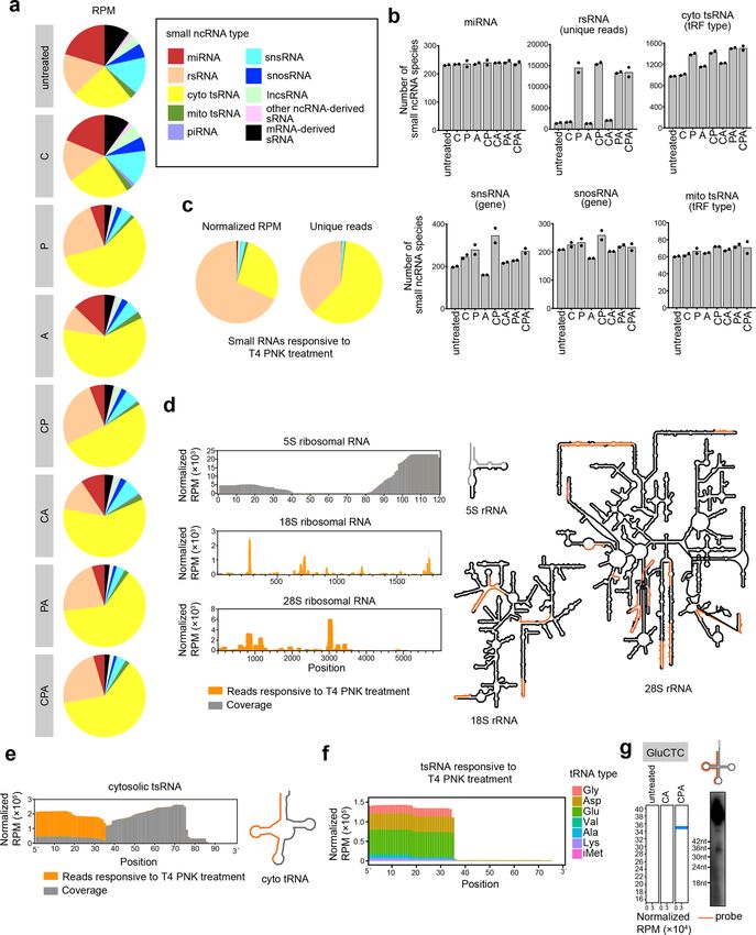
Wang et al. Cell Discovery (2021)7:25                                                                          Page 2 of 16

tobacco acid pyrophosphatase (TAP), RNA 5′ pyropho-           Results
sphatase (RppH), and Cap-Clip acid pyrophosphatase            Overview of CPA-seq
(Cap-Clip), all of which are capable of decapping both 7-       Here, we developed CPA-seq to overcome common
methylguanosine (m7G) and 2,2,7-trimethylguanosine            obstacles described above that impede preparation of
(m3G) caps14–18. Moreover, pyrophosphatases are also          sRNA libraries (Fig. 1a). First, we incubated sRNAs in
capable of generating 5′-P termini from 5′-tripho-            deacylation buffer (pH = 9.0) to remove aminoacyl resi-
sphorylated RNA19.                                            dues in aminoacyl-tRNA-derived 3′-tsRNAs (Fig. 1b and
  Another enzyme used to reduce terminus multiplicity is      Supplementary Fig. S1a)13. Second, we used Cap-Clip to
T4 polynucleotide kinase (PNK), which catalyzes the           remove the 5′-cap and 5′-ppp from RNAs to generate 5′-P
phosphorylation of 5′-OH termini to generate 5′-P and         termini. We compared two commercially available dec-
removal of phosphoryl groups from 3′-P and 2′,3′-cyclic       apping enzymes, RppH and Cap-Clip. In our hands, Cap-
phosphate (3′-cP) termini to generate 3′-OH20,21. Dicer       Clip was superior to RppH for preserving RNA integrity
and Drosha each generate 5′-P and 3′-OH termini in            (Supplementary Fig. S1b). Cap-Clip efficiently removed
miRNAs; several other ribonucleases, such as Angiogenin,      the 5′-m7G cap from a synthetic 5′-m7G-capped short
produce 5′-OH, 3′-P, or 3′-cP termini in other sRNA           RNA to enable 5′-adapter liagation, showing high decap-
types10,22,23. Thus, T4 PNK has been employed for             ping efficiency15,35 (Fig. 1c and Supplementary Fig. S1c).
sequencing of tsRNA24 and cyclic phosphate-containing-          Third, we used T4 PNK to reduce terminus multiplicities
RNAs25, as well as circulating lncsRNA and mRNA-              (Fig. 1d, e and Supplementary Fig. S1d, e). T4 PNK efficiently
derived sRNAs26.                                              phosphorylated 5′-OH and removed phosphoryl groups from
  To overcome the obstacles in reading through methy-         3′-termini of synthetic RNA oligos bearing 5′-OH and 3′-P,
lation sites during reverse transcription, two strategies     thereby enabling efficient adapter ligation (Fig. 1d, e and
have been developed. One strategy is to pre-treat RNA         Supplementary Fig. S1d). Fourth, a mixture of AlkB and AlkB
with demethylase. In previous studies, AlkB has been          (D135S) (AlkB mix) was used to remove methylations in
reported to efficiently remove methylations in N1-             m1A, m3C, and m1G. We optimized the AlkB mix reaction
methyladenosine (m1A), N3-methylcytosine (m3C), and           for demethylation efficiency while retaining RNA integrity
the AlkB(D135S) variant can efficiently demethylate N1-        (Fig. 1f and Supplementary Fig. S1f–k). After sequential
methylguanosine (m1G); both of these have been used to        deacylation and CPA treatments, sRNAs were ligated to 5′
facilitate sequencing of tRNAs and their derivants27,28.      and 3′ degenerate adapters containing unique molecular
Another strategy is to use reverse transcriptases with high   identifiers (UMIs)36, reverse transcribed by TGIRT-III, and
processivity in reverse transcription of highly structured    followed by PCR amplification (Fig. 1a).
or heavily modified RNAs, such as thermostable group II          We tested the sensitivity of CPA-seq. CPA-seq of
intron reverse transcriptase (TGIRT) and an evolved form      25–100 ng of small RNA extracted from HEK293T cells
of the HIV-1 reverse transcriptase, both of which can         revealed comparable species numbers of different sRNA
introduce misincorporation at methylation sites29–31.         types (Supplementary Fig. S1l–n).
  Using Cap-Clip, T4 PNK, and AlkB/AlkB(D135S)-
facilitated small ncRNA sequencing (CPA-seq), we pro-         Performance comparisons among CPA-seq and
filed the sRNome of human embryonic kidney cells               commercial sRNA-seq methods
(HEK293T), and revealed sRNAs with terminus multi-              To evaluate the performance of CPA-seq, we sequenced
plicities and nucleoside methylations. Comparing sRNA         sRNAs from HEK293T cells using CPA-seq and using
with or without treatment of AlkB mix, we estimated the       three commercially available library preparation methods
methylation status of tsRNAs. We also profiled the sRNA        for sRNA-seq (NEBNext, QIAseq, and TruSeq, collective
transcriptomes (sRNomes) of nine mouse tissues. CPA-          shortened to “NQT-seq”; Supplementary Fig. S2a). Note
seq revealed similar tissue-specific expression patterns of    that among the NQT-seq methods, QIAseq uses UMI-
miRNAs as in previous reports3,32–34. However, compared       containing RT primer to reduce bias.
to previously reported sRNA atlases across different            Sequencing reads corresponding to sRNAs of 16–40
mouse tissues generated by the conventional sRNA-seq          nucleotides (nt) in length were used for subsequent ana-
methods, we observed more complex sRNA profiles                lyses. The sequencing reads were aligned sequentially to
across mouse tissues32–34. We found that a large number       known miRNA, rRNA, cytosolic tRNA, piRNA, mito-
of tsRNAs, snsRNAs, snosRNAs, and lncsRNAs also               chondrial tRNA, lncRNA, snRNA, snoRNA, and other
showed tissue-specific expression patterns. The expres-        ncRNA types (“Materials and methods” and Supplemen-
sion patterns of sRNAs in specific cell types could be         tary Fig. S3).
remodeled upon cell fate conversion. Thus, the sRNomes          The distributions of sRNA types varied using different
generated using CPA-seq in this study could facilitate        sRNA library preparation methods (Supplementary Fig.
studies of sRNAs in mammalian tissues.                        S2b, Table S1). MiRNAs are known to contain extensive
Wang et al. Cell Discovery (2021)7:25                                                                                                   Page 3 of 16

 Fig. 1 CPA-seq. a The workflow of sRNA library preparation for CPA-seq. Purified small RNAs are incubated in deacylation buffer to remove 3′-
 aminoacyl (3′-aa), treated with Cap-Clip to remove 5′ m7G and m3G caps, then treated with T4 PNK to convert 5′-OH to 5′-P, and to convert 3′-P and
 3′-cP to 3′-OH, followed by treatment with a mix of AlkB and AlkB(D135S) to remove methylations in m1G, m3C, and m1A. The pretreated small RNAs
 were ligated with 3′ and 5′ adapters, reverse transcribed by TGIRT-III, and then PCR amplified for sequencing. b Northern blotting of RNA samples
 from HEK293T with/without treatment of deacylation buffer. c Cap-Clip treated synthetic 5′-m7G-RNA (31 nt) was ligated with a 5′-adapter (26 nt).
 d T4 PNK-treated synthetic 5′-OH RNA (27 nt) was ligated with a 5′-adapter (26 nt). e T4 PNK-treated synthetic 3′-P RNA (27 nt) was ligated with 3′-
 adapter (29 nt). f LC-MS/MS analysis showed that sequential treatments with deacylation buffer, Cap-Clip, T4 PNK, and AlkB mix (CPA) efficiently
 removed methylations in m1G, m3C, and m1A of small RNAs extracted from HEK293T cells (n = 3).

5′-P and 3′-OH termini, and can be captured by all of the                    detection of non-miRNA sRNAs. The sRNA profile
tested sRNA-seq methods. We observed that the miRNA                          detected by CPA-seq matched the northern blotting
species detected with all of the tested methods shared                       banding pattern more closely than the profiles generated
large overlap (Supplementary Fig. S2c). However, CPA-                        using the NQT-seq methods (Supplementary Fig. S4).
seq revealed much more species of tsRNA, lncsRNA,
snsRNA, snosRNA, rsRNA, mRNA-derived, and other                              CPA-seq reveals sRNAs with diverse termini
ncRNA-derived sRNAs, suggesting the terminus multi-                            Next, we performed sRNA sequencing of small RNA
plicities and nucleoside methylation in non-miRNA                            extracted from HEK293T cells that we process with the
sRNAs (Supplementary Fig. S2c).                                              full CPA-seq process or with various combinations of the
  We performed Northern blotting to verify the perfor-                       Cap-Clip, T4 PNK, and AlkB mix enzymes. Unsurpris-
mance of different library preparation methods for the                       ingly, distinct distributions of the various sRNA types
Wang et al. Cell Discovery (2021)7:25 Page 4 of 16
were detected upon these different treatments (Fig. 2a, b (Fig. 3b). Cap-clip responsive snsRNAs were mainly
and Supplementary Table S1). The miRNAs revealed by derived from Sm-class snRNAs, including RNU1 and
different treatments showed a high correlation (Supple- RNVU1, which are well-characterized to be 5′-capped
mentary Fig. S5a), suggesting the low terminus multi- with m3G38 (Fig. 3c). Using a probe recognizing the 5′
plicity of the miRNAs. Thus, we normalized the RPM parts of U1 snRNA, we found that the Cap-Clip-
value to total miRNA RPM in the following analyses to decapped U1 snRNAs and their 5′ snsRNAs (U1-5′
estimate the amount of different sRNA species revealed snsRNAs) ran slightly faster in electrophoresis and were
by sRNA-seq with different treatments. readily digested by XRN-1, a 5′→3′ exoribonuclease
We first compared the sRNAs detected in CA and CPA requiring 5′-P (Fig. 3d). This result validated the exis-
groups to analyze the sRNAs responsive to T4 PNK tence of 5′-capped U1-5′ snsRNAs in HEK293T cells.
treatment, which putatively contain 5′-OH, 3′-P, or 3′-cP. We surprisingly observed a few Cap-Clip responsive
A majority of the sRNAs responsive to T4 PNK treatment tsRNAs with 5′ cleavage sites within the 5′-leader sequence
(unique reads that were highly detected in CPA group, but of tRNA precursor or 5′ parts of mature tRNA (Fig. 3e). As
lowly detected in CA group with the fold change >30) these Cap-Clip-responsive tsRNAs do not contain the 5′-
were found to be rsRNAs and tsRNAs (Fig. 2b, c). termini of their corresponding tRNA precursors, they are
Surprisingly, we found that the majority of the rsRNAs possibly de novo capped or triphosphorylated.
that could be captured without T4 PNK treatment were
derived from 5S ribosomal RNA. On the contrary, the CPA-seq reveals methylated sRNAs
majority of the 18S and 28S ribosomal RNA-derived The commonly used reverse transcriptases tend to stop
rsRNAs require T4 PNK treatment to be captured by at m1A, m3C, and m1G sites. Thus, it is difficult to
sRNA-seq, and putatively contain 5′-OH, 3′-P, or 3′-cP sequence sRNAs derived from tRNAs and rRNAs con-
termini (Fig. 2d). The 18S and 28S ribosomal RNA- taining m1A, m3C, and m1G sites28 (Fig. 4a). Our CPA-seq
derived rsRNAs are preferentially generated by the clea- method uses TGIRT-III, a highly processive reverse
vages at bubble region of the RNA (Fig. 2d). The distinct transcriptase that has been shown to significantly increase
5′ or 3′ termini of 5S and 18S/28S rRNA-derived sRNAs the detection of sRNAs derived from tRNAs containing
suggested different mechanisms for generation of these m1A, m3C, and m1G sites8,29,39 (Figs. 2a, 4b). During
rsRNAs. reverse transcription, TGIRT-III tends to introduce mis-
tsRNAs that are responsive to T4 PNK treatment are incorporations and stops at m1A, m3C, and m1G sites,
mapped mainly to 5′ parts of tRNAs (Fig. 2e–g). The T4 providing us an opportunity to estimate m1A, m3C, and
PNK-responsive tsRNAs are mostly generated by the m1G stoichiometries at individual sites across the
cleavage at the anticodon loop of the tRNA (Fig. 2e). sRNome.
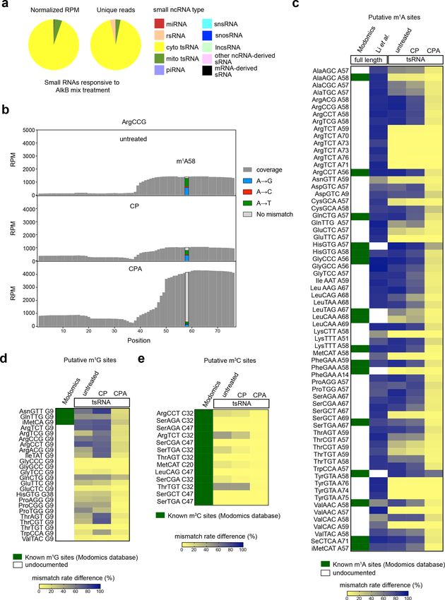
Previous studies have reported Angiogenin, a stress- We compared the sRNAs detected in CP and CPA
activated endonuclease, cleaves tRNAs within the antic- groups to analyze the sRNAs responsive to AlkB mix
odon loop to generate 5′ tsRNA under stress10,23. The treatment, which we expected to include RNAs contain-
Angiogenin is also known to generate 5′-P and 3′-cP, ing m1A, m3C, or m1G sites. The majority of the sRNAs
which hamper the ligation of adapters to tsRNAs gener- responsive to AlkB mix treatment are derived from tRNAs
ated by Angionenin37. However, the large amount of T4 (Fig. 4a). We also compared the extent of misincorpora-
PNK-responsive 5′ tsRNA from HEK293T cells without tion events in the “untreated”, CP, and CPA groups. The
stress cannot be simply explained by Angiogenin-mediated significantly reduced number of misincorporation events
biogenesis of tsRNA. Whether other mechanisms are at putative m1A, m3C, and m1G sites in the CPA group
involved in the biogenesis of tsRNAs with 5′-OH, 3′-P, and confirmed the high demethylation efficiency of AlkB mix
3′-cP termini should be investigated in the future. treatment (Fig. 4b–e). We compared the putative m1A
sites in tsRNAs with known m1A sites (Modomics data-
CPA-seq reveals 5′-capped sRNAs base) and putative m1A sites of their corresponding full-
Next, we compared the sRNAs detected in the PA and length tRNAs revealed by a previous study using the same
CPA group to analyze the sRNAs responsive to Cap-Clip TGIRT reverse transcriptase as we used in CPA-seq40,41.
treatments; these were expected to include RNAs contain All of the putative m1A, m1G, and m3C sites on tsRNAs
a 5′-cap or 5′-ppp. In consistent with a previous study revealed in this study were either known m1A sites or
using TAP to sequence 5′-capped sRNAs16, we also putative m1A sites of their corresponding full-length
observed many snsRNAs, lncsRNAs, and snosRNAs that tRNAs revealed by Li et al.40 (Fig. 4c–e and Supplemen-
were responsive to Cap-Clip treatment (Fig. 3a). We tary Fig. S5b–f). However, we also observed a few putative
found that the lncsRNA, snosRNA, and snsRNA reads m1A sites revealed by Li et al.40 were not methylated at
mapped mainly to 5′ parts of their corresponding full- the corresponding sites of tsRNAs, suggesting lower m1A
length RNAs, which usually contain 5′-caps or 5′-ppp methylation frequency of tsRNAs (Fig. 4c).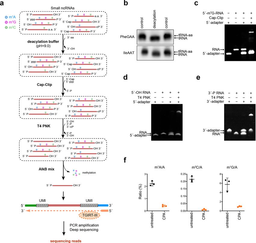
Wang et al. Cell Discovery (2021)7:25                                                                                                      Page 5 of 16

 Fig. 2 CPA-seq reveals sRNAs with diverse termini. a Distribution of different types of sRNAs extracted from HEK293T cells that we process with
 the full CPA-seq process or with various combinations of the Cap-Clip, T4 PNK, and AlkB mix enzymes (n = 2). b The number of sRNA species revealed
 with different treatments (n = 2). c Distribution of different types of sRNAs responsive to T4 PNK treatment (unique reads that were highly detected in
 CPA group, but lowly detected in CA group with the fold change > 30, n = 2). d Reads of sRNA responsive to T4 PNK treatment mapping to 5S, 18S,
 and 28S ribosomal RNAs have been combined to show detection of rsRNAs containing diverse termini (rsRNAs with RPM > 300 are shown in the
 structural map). e Reads of sRNA responsive to T4 PNK treatment mapping to cytosolic tsRNAs have been combined to show detection of tsRNAs
 containing diverse termini. f Reads of tsRNAs responsive to T4 PNK treatment. g Northern blotting of GluCTC 5′tsRNAs that are responsive to T4 PNK
 treatment.
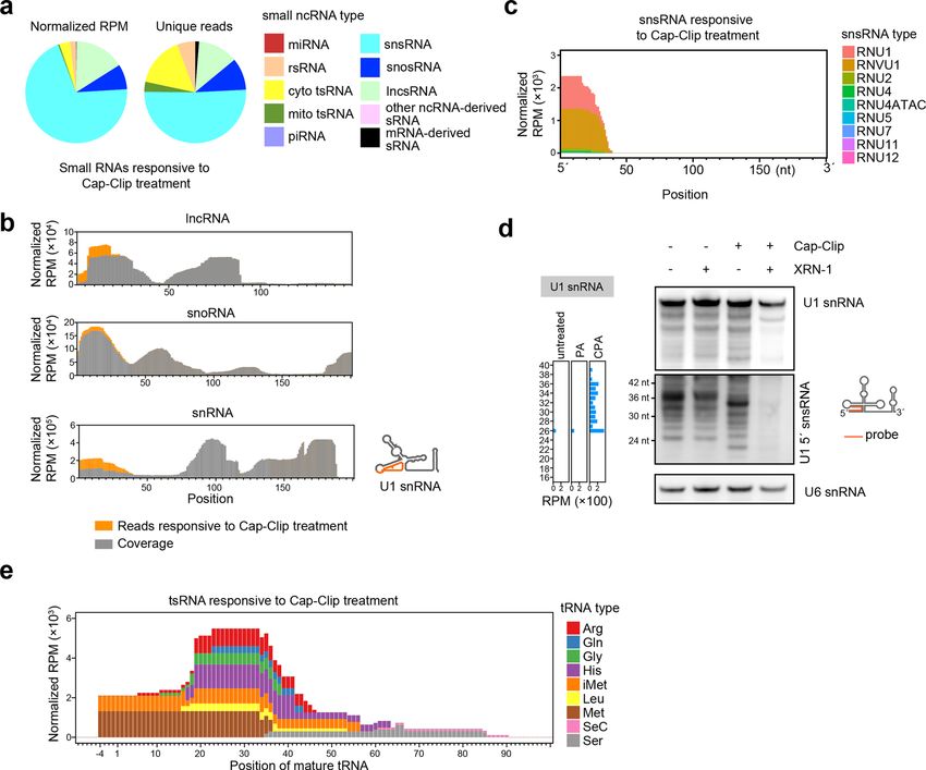
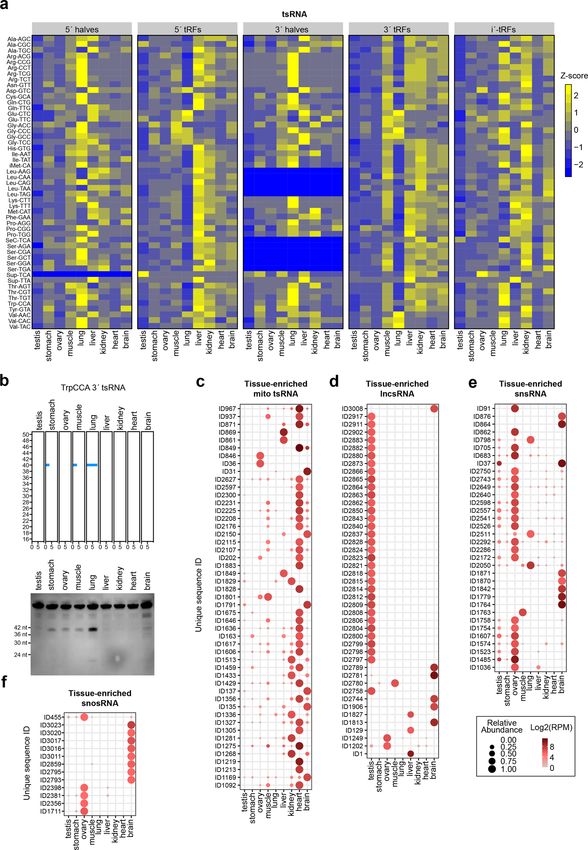
Wang et al. Cell Discovery (2021)7:25                                                                                                       Page 6 of 16

 Fig. 3 CPA-seq reveals sRNAs with 5′-cap and 5′-ppp. a Distribution of different types of sRNAs responsive to Cap-Clip treatment (unique reads
 that were highly detected in CPA group, but lowly detected in PA group with the fold change >30, n = 2). b Reads of sRNA responsive to Cap-Clip
 treatment mapping to lncRNAs, snoRNAs, and snRNAs have been combined to show the detection of lncsRNAs, snosRNAs, and snsRNAs putatively
 containing 5′-caps or 5′-ppp. c Reads of snsRNAs responsive to Cap-Clip treatments. d Northern blotting of sRNAs treated with the indicated
 enzymes, using a probe complementary to U1-5′snsRNA. Cap-Clip removed the 5′-cap from U1-5′snsRNAs and enabled U1-5′snsRNAs to be digested
 by XRN-1 (lane 4), a 5′→3′ exoribonuclease that requires 5′-P. Removal of the 5′-cap by Cap-Clip leads to a slight shift of the bands representing U1-5′
 snsRNAs (lane 3). U6 snRNA is used as a reference. e Reads of tsRNAs responsive to Cap-Clip treatments.

Profiling sRNomes of mouse tissues by CPA-seq                                   in mitochondria (Fig. 5a). snsRNA and mRNA-derived
   We used CPA-seq to profile the sRNomes of nine                               sRNA were more abundant in the brain (Fig. 5a).
mouse tissues including testis, stomach, ovary, muscle,                          Next, we determined the overall sRNA expression pat-
lung, liver, kidney, heart, and brain (from 9- to 10-week-                     terns across different tissues. Hierarchical cluster analysis
old C57BL/6J mice). The sRNA library for each tissue                           of unique reads of different sRNA types showed that a
contained between 4.42 and 13.07 million unique reads                          large number of sRNAs were differentially expressed in
(Supplementary Table S2, Fig. S6a).                                            different mouse tissues (Fig. 5b). In the t-distributed
   We then mapped the sequencing reads to different sRNA                       stochastic neighbor embedding (t-SNE) projection plot,
types. In total, miRNA contributed to 1.83%–12.32% of all                      sRNAs were separated according to their tissue type,
the detected CPA-seq RNA reads in mouse tissues, a small                       representing distinct sRNA expression patterns across
proportion mirroring our findings from the HEK293T cells.                       various tissues (Supplementary Fig. S6b).
rsRNA and tsRNA were the most prominent sRNA types
among all mouse tissues profiled with CPA-seq. The rela-                        Tissue-enriched sRNAs
tive abundance of other sRNA types varied in different                            To identify tissue-enriched sRNAs, we calculated the
tissues. For example, mitochondrial tsRNAs were abundant                       tissue specificity index (TSI) for each detected sRNA
in the heart, and energy-demanding organ known to be rich                      using a previously described method42. miRNAs are the
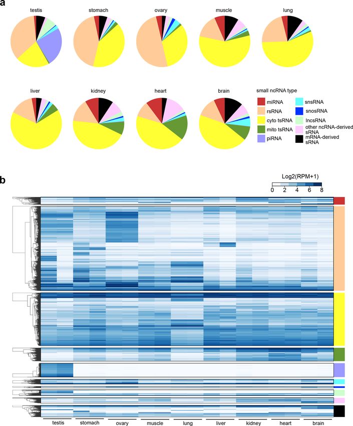
Wang et al. Cell Discovery (2021)7:25                                                                                                   Page 7 of 16

 Fig. 4 Methylome of sRNAs. a Pie diagrams showing the distribution of different types of sRNAs responsive to AlkB mix treatment (unique reads
 that were highly detected in CPA group, but lowly detected in CP group with the fold change >30, n = 2). b Misincorporation and coverage plot for
 putative m1A sites identified in this study. c Estimation of m1A methylome by the mismatch frequency at A sites of tRNAs. Treatment of AlkB mix
 demethylates m1A and reduces TGIRT-induced misincorporation at m1A sites. The known m1A sites (Modomics database) and putative m1A sites of
 full-length tRNAs revealed by Li et al.40 are shown accompanied by the m1A site of tsRNAs revealed by this study. d The m1G sites of tsRNAs revealed
 by CP-seq and CPA-seq. e The m3C sites of tsRNAs revealed by CP-seq and CPA-seq.
Wang et al. Cell Discovery (2021)7:25                                                                                                     Page 8 of 16

 Fig. 5 sRNomes across different mouse tissues. a Coverage of sRNA types across different mouse tissues as profiled using CPA-seq. b Hierarchical
 clustering of unique reads (RPM > 20 in at least one tissue) from the indicated mouse tissues. For hierarchically-clustered analysis, the unique reads
 were first classified according to the sRNA type. Then the average Euclidean distance for log2 transformed unique reads of each type of sRNA was
 computed.

most extensively studied sRNA type, and our CPA-seq                           miR-34c), and several brain-enriched miRNAs (miR-9,
method successfully identified many well-described tis-                        miR-124, and miR-128) (Supplementary Fig. S6c)32–34.
sue-enriched miRNAs, including for example the liver-                         The well-matched tissue-specific miRNA expression pat-
enriched miRNA (miR-122), the heart-enriched miRNA                            terns between the sequencing results of CPA-seq and
(miR-208a), the testis-enriched miRNA (miR-34b and                            previous reports using conventional sRNA-seq methods
Wang et al. Cell Discovery (2021)7:25 Page 9 of 16
suggested relatively low multiplicity of 5′/3′-termini and significantly promoted the induction of hepatic marker
nucleoside methylations in miRNAs. genes, suggesting a potentially important role of U1-5′
Recently reports have emphasized the presence and snsRNA in hepatic reprogramming (Fig. 7f and Supple-
functional impacts of tsRNAs in diverse cell types43,44. mentary Fig. S7e).
However, as highlighted by our profiling results above, it is
clear that previous surveys conducted with conventional Discussion
sRNA-seq methods have almost certainly missed very large In this study, we developed CPA-seq for profiling
numbers of tsRNAs, and especially those tsRNAs contain- sRNAs with terminus multiplicities and nucleoside
ing 5′-OH, 3′-P, or methylated nucleosides. We used CPA- methylations. This technique enables sensitive identifica-
seq to systematically analyze the distributions of tsRNAs in tion of a significant fraction of sRNAs that are missed by
mouse tissues. We noted that the lung showed a distinct conventional sRNA-seq methods. These newly detected
pattern of tsRNAs (Figs. 5b and 6a, b), which were mainly sRNAs represent a hidden layer of the sRNome.
mapped to 5′-tRNA halves and 3′-tRNA halves (Fig. 6a). Recently, many efforts have been made to improve the
Another trend was that the tsRNAs derived from different sRNA sequencing protocols to retrieve previously unde-
cytosolic tRNA isodecoders showed distinct tissue-specific tectable sRNA sequences. TGIRT-seq showed higher
expression patterns (Fig. 6a). processivity in RNA-seq but has not been optimized for
Overall, the heart contained the highest numbers of detection of sRNAs with multiple modifications29. Using
tissue-enriched mitochondrial tsRNAs (Fig. 6c); the testis AlkB, ARM-seq enables sequencing of 3′ tsRNAs con-
is enriched for tissue-enriched lncsRNAs (Fig. 6d). taining m1A28. However, owing to the multiplicity of 5′/3′
Moreover, we observed that the ovary and brain con- termini, ARM-seq is insensitive for sequencing 5′ tsRNAs.
tained the highest numbers of tissue-enriched snsRNAs T4 PNK has been used to facilitate adapter ligation in
and snosRNAs among the tested tissues (Fig. 6e, f and preparation of sRNA libraries47. However, T4 PNK is not
Supplementary Table S3). capable of removing the 5′-cap structure, which is known
to be abundant in 5′ snsRNAs. Thus, compared to cur-
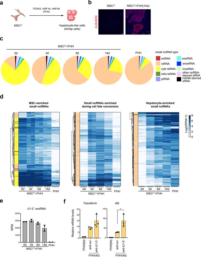
Reprogramming of sRNomes during hepatic rently available sRNA-seq methods, CPA-seq reveals a
transdifferentiation more complete view of diverse sRNA species in mam-
As many sRNAs showed tissue-specific expression pat- malian cells including but not limited to tsRNAs and
terns, we questioned whether the sRNome may get repro- snsRNAs. CPA-seq is a powerful tool with high sensitivity
grammed as cell fate changes occur. We used a previously for profiling sRNAs that can reveal a more complete
described strategy to convert human mesenchymal stem cells picture of the sRNome.
expressing SV40 large T antigen (MSCLT) into hepatocytes We used CPA-seq to profile the sRNomes of nine
(hiHep cells) by enforced expression of FOXA3, HNF1A, and mouse tissues and revealed the abundant presence of
HNF4A (FHH) (Fig. 7a)45. Confirming the success of the tsRNA, snsRNA, snosRNA, and lncsRNA, as well as
conversion, liver cell marker genes were gradually induced in miRNA. These sRNAs showed tissue-specific expression
the MSCLT upon FHH expression (Fig. 7b and Supplemen- patterns. Accumulating reports are emphasizing the
tary Fig. S7a). We performed CPA-seq on MSCLT under- essential biological functions of non-miRNA sRNAs2. The
going hepatic cell fate conversion and found that the sRNomes of different tissues characterized in the present
composition of sRNAs transit toward that of primary human study will likely deepen our understanding of the func-
hepatocyte (PHH) (Fig. 7c and Supplementary Fig. S7b, c). tional roles of sRNAs in diverse tissues.
Supporting the anticipated sRNome reprogramming, we Moreover, we gained a deeper insight into the repro-
found that FHH expression gradually decreased the abun- gramming of sRNA expression profiles during cell fate
dance of tsRNAs and increased the abundance of rsRNAs conversion by using CPA-seq. The expression of FHH in
(Fig. 7d). Interestingly, the number of rsRNAs increased, MSCLT converted MSCLT-like sRNome to PHH-like
while the expression of LeuCAG 3′tsRNA, a ribosomal bio- sRNomes. Importantly, we observed a 5′-capped sRNA,
genesis regulator, dramatically decreased upon hepatic U1-5′ snsRNA, functioned as an inhibitor for hepatic
reprogramming43,46 (Supplementary Fig. S7d). reprogramming. Knockdown of U1-5′ snsRNA by U1-5′
It was also interesting to observe a significant low- ASO can promote hepatic reprogramming.
expression level of U1-5′ snsRNA, a 5′-capped sRNA, in Although we optimized the sRNA library preparation of
primary human hepatocytes (PHH) as compared to that of CPA-seq, degradation of full-length ncRNA may still
MSCLT (Fig. 7e). The expression levels of U1-5′ snsRNA contribute to the CPA-seq reads. For example, nucleoside
in MSCLT gradually decreased after transfection of FHH demethylation could lead to the fragility of full-length
(Fig. 7e). To investigate the potential role of U1-5′ tRNAs48. Using pre-size-selected small RNA for CPA-seq
snsRNA in hepatic reprogramming, we treated MSCLT could further eliminate the contamination of full-length
with an U1-5′ antisense oligonucleotides (ASO), which RNA degradations.Wang et al. Cell Discovery (2021)7:25 Page 10 of 16 Fig. 6 Tissue-enriched sRNAs. a Heatmap of the relative abundance of tsRNA\s of different types in the indicated mouse tissues. b Detection of lung-enriched TrpCCA 3′tsRNAs by Northern blotting. c–f Dot plot showing expression patterns of tissue-enriched mitochondrial tsRNAs (TSI > 0.95, RPM > 70 in at least one tissue) (c), lncsRNAs (TSI > 0.95, RPM > 50 in at least one tissue) (d), snsRNAs (TSI > 0.95, RPM > 50 in at least one tissue) (e), and snosRNAs (TSI > 0.95, RPM > 30 in at least one tissue) (f).
Wang et al. Cell Discovery (2021)7:25                                                                                                    Page 11 of 16

 Fig. 7 Reprogramming of sRNomes during hepatic reprogramming. a Schematic illustration of the strategy used for direct conversion of human
 MSCLT to hepatocytes. b Fluorescent immunostaining of MSCLT and MSCLT infected with FOXA3, HNF1A, and HNF4A (FHH) for 14 days. Scale bar:
 100 μm. c Distribution of different types of sRNAs in PHH and MSCLT infected with FHH. d Hierarchical clustering of unique reads (RPM > 20 in at least
 one sample) from sequenced samples showing the decreased expressions of MSCLT-enriched sRNAs and increased expression of PHH-enriched
 sRNAs during hepatic reprogramming. e Differential expression levels of U1-5′snsRNA in MSCLT and PHH. The expression levels of U1-5′snsRNA
 gradually decreased in MSCLT cells infected with FHH. f Treatment of U1-5′ ASO promoted the induction of hepatic marker genes in MSCLT cells
 overexpressing FHH.
Wang et al. Cell Discovery (2021)7:25                                                                            Page 12 of 16

  There are still several types of sRNAs, such as NAD-          injection of 50 mg/kg pentobarbital sodium and then
capped RNA, that cannot be captured by CAP-seq.                 sacrificed by cervical dislocation. Mice were pinned down
Improvement of CPA-seq in the future could provide new          onto dissecting tray and the ventral surfaces were sprayed
insight into the compositions of sRNAs.                         with 70% ethanol. We then opened the chest and abdom-
                                                                inal cavity of male mice, and used precooled PBS to wash
Materials and methods                                           the residual blood from the heart into the body circulation.
Molecular cloning and lentivirus production                     The testes, stomachs, muscles, lungs, livers, kidneys, hearts,
   Plasmids used for expression of SV40 large T, FOXA3,         the whole brains of male mice, and the ovaries of female
HNF1A, and HNF4A were described in previous pub-                mice were obtained for RNA extraction. The use and care
lication45. Constructed plasmids were introduced into           of animals complied with the guideline of the Biomedical
HEK293FT cells together with packaging plasmid psPAX2           Research Ethics Committee of ShanghaiTech University.
(Addgene) and envelop plasmid pMD2.G (Addgene).
After 48 h incubation, the medium containing lentiviruses       Tissue handling and RNA extraction
was collected and passed through 0.45-μm filter.                   Upon collection, tissue samples were sectioned into
                                                                smaller pieces and submerged in RNAlater® Solution
Cell culture and RNA preparation                                (ThermoFisher) for 1 h. Then the tissues were removed
  Human embryonic kidney HEK293T cells and human                from solution and preserved in Lysis/Binding buffer (Life
mesenchymal stem cells were obtained from the Amer-             Technologies) until further processing. Small RNA was
ican Type Culture Collection (ATCC). HEK293T cells              isolated from 100 mg tissues using mirVana miRNA Iso-
were maintained in DMEM (Thermo) medium supple-                 lation Kit according to the manufacturer’s instructions.
mented with 10% FBS and 1% 100× penicillin-                     For primary human hepatocytes (PHH), RNA was
streptomycin (Gibco) with 5% CO2 at 37 °C and human             extracted from PHH cultured for 48 h in HMM medium.
bone marrow-derived mesenchymal stem cells were                 RNA integrity number (RIN) values were used to measure
maintained according to the manufacturer’s instructions.        RNA integrity. RIN values of mouse tissue samples were
Cryopreserved human hepatocytes from three individuals          assessed by an Agilent 2100 Bioanalyzer (Agilent Bio-
were provided by Research Institute for Liver Diseases          technologies Ltd., USA, Supplementary Table S5).
(Shanghai) Co. Ltd and Lonza Walkersville Inc. One
donor is a 25-year-old Caucasian male, with no history of       Conversion of MSCLT to hepatocytes
smoking and drinking alcohol. The second donor is a 51-           To induced hepatic cell fate conversion, 2 × 10⁵ human
year-old Hispanic male with a history of drinking alcohol       MSCLT were mixed with lentivirus expressing FOXA3,
and no history of smoking. The third donor is a 2-month-        HNF1A, and HNF4A (MOI = 2 for each virus) and seeded
old Caucasian boy. Mycoplasma contamination tests were          on a collagen I-coated 6-cm dish. Two days later, the
performed routinely. Total or Small RNA was prepared            medium was changed with HMM medium. The HMM
from cells using mirVana miRNA isolation kit (Invitro-          medium was replaced every 2 days. RNA integrity number
gen) according to the manufacturer’s instructions. Small        (RIN) values of samples were assessed by an Agilent 2100
RNA used for Northern blotting was purified by RNAiso            Bioanalyzer (Supplementary Table S5).
for (Takara) according to the manufacturer’s instructions.
                                                                Quantification of ribonucleosides by LC-MS/MS
Medium                                                            Hundred and fifty nanograms of RNA was first digested
   Hepatocyte maintenance medium (HMM) is DMEM/F12              by nuclease P1 (NEB, 1U) in 17 μL 1× P1 digestion buffer
(Gibco) supplemented with 0.544 mg/L ZnCl2 (Sinopharm),         containing 25 mM NaCl, 2.5 mM ZnCl2 at 42 °C for 2 h.
0.75 mg/L ZnSO4·7H2O (Sinopharm), 0.2 mg/L CuSO4·5H2O           Next, 1 μl FastAP Thermosensitive Alkaline Phosphatase
(Sinopharm), 0.025 mg/L MnSO4 (Sinopharm), 2 g/L Bovine         (ThermoFisher) and 2 μl 10× FastAP buffer (Thermo-
serum albumin (Sigma-Aldrich), 2 g/L Galactose (Sigma-          Fisher) were added to the reaction and incubated at 37 °C
Aldrich), 0.1 g/L Ornithine, 0.03 g/L Proline, 0.61 g/L Nico-   for 2 h. Reactions were added 20 μl acetonitrile for futher
tinamide, 1× Insulin-transferrin-sodium selenite media sup-     detection. After centrifuged at 14,000 rpm for 15 min, the
plement (Sigma-Aldrich), 40 ng/mL TGFα (Peprotech),             supernatant was aspirated for LC-MS/MS analysis. The
40 ng/mL EGF (Peprotech), 10 µM dexamethasone, 10 µM            LC-MS/MS analysis was performed on Agilent 1290
Y-27632 (MCE), 0.5 µM A-83-01 (Tocris), 3 µM CHIR99021          UPLC (Agilent, USA) coupled to AB Sciex 6500 triple
(Sigma-Aldrich).                                                quadrupole mass spectrometer (AB Sciex, USA) with the
                                                                electrospray ionization (ESI) source. A Thermoscientific
Animals                                                         Hypersil GOLD aQ column (3 µm, 2.1 × 150 mm) was
  Wild-type 9–10-week-old C57BL/6J mice (Charles River          used for ribonucleosides separation with a flow rate at
Laboratories, China) were anesthetized by intraperitoneal       0.4 ml/min and column temperature of 35 °C. The mobile
Wang et al. Cell Discovery (2021)7:25 Page 13 of 16
phases were comprised of (A) 0.1% formic acid in 100% Preparation of probes
water and (B) 0.1% formic acid in 100% acetonitrile. The The DNA probes were labeled by digoxigenin (DIG)
gradient elution was carried out as follows: 0–6 min at 0% using DIG Oligonucleotide Tailing Kit (2nd Generation,
B; 6–8 min at 0%–1% B; 8–10 min at 1%–6% B; 10–11 min Roche) according to the manufacturer’s instructions.
at 6% B; 11–13 min at 6%–50% B; 13–15 min at 50%–70% Short tail-labeled probes were generated with 2–3
B; 15–18 min at 75% B; 18–19 min at 75%–0% B; and nucleotides consisting of DIG-dUTP. A mixture of 2 μl of
19–24 min at 0% B. The injection volume was set to 2 μL. reaction buffer, 2 μl of CoCl2-solution, 0.5 μl of DIG-
The mass parameters were as follows: ion spray voltage dUTP solution, 0.5 μl of 400 U Terminal transferase, and
was 5500 V, ion source temperature was 500 °C, collision 100 pmol of oligonucleotides was prepared and briefly
gas was set to Medium, ion source gas 1 was 50 psi, ion centrifuged, followed by incubation at 37 °C for 15 min
source gas 2 was 60 psi, curtain gas was 35 psi. Multiple and cool down on ice. The probes were stored at −20 °C.
reaction monitoring (MRM) was used to monitor target
ribonucleosides in the positive ion mode. The detailed Northern blotting analysis
MRM transitions were as follow: A, m/z 268 → 136; m1A, For Northern blotting, small RNA sample was mixed with
m/z 282 → 150; G, m/z 284 → 152; m1G, m/z 298 → 166; Gel loading buffer II (Invitrogen) and incubated at 90 °C for
C, m/z 244 → 112;m3C, m/z 258 → 126. The dwell time for 5 min. Then the samples were incubated on ice for 3 min
each ribonucleoside was 100 ms. The declustering poten- and loaded into denaturing 15% polyacrylamide gel con-
tial and collision energy were 20 and 15 V, respectively. taining 8 M Urea. The RNAs were transferred onto a posi-
Data acquisition and processing were performed using tive charged nylon membrane, and UV cross-linked at
Analyst (version 1.6, SCIEX). 150 mJ/cm2. Then the membrane was pre-hybridization for
1 h and blotted with DIG-labeled DNA probes against target
qRT-PCR RNA subsequently, and incubated overnight at 35 °C. The
RNA was reverse transcribed into cDNA with HiScript membranes were washed three times with low stringent
II 1st Strand cDNA Synthesis Kit (Vazyme) according to buffer (2× SSC buffer with 0.1%wt/vol SDS) at room tem-
manufacturer’s instructions. Quantitative real-time PCR perature for 10 min each, then rinsed three times with high
was performed with ChamQ Universal SYBR qPCR stringent buffer (0.1× SSC buffer with 0.1%wt/vol SDS) for
Master Mix (Vazyme) on ABI QuantStudio 7 real-time 10 min each, finally rinsed in 1× DIG washing buffer (Roche)
PCR system (Applied Biosystems). Primer sequences are for 10 min. Following the washes, the membranes were
provided in Supplementary Table S4. incubated with 1× blocking buffer (dilute the 10× blocking
solution with 1× Maleic acid buffer, Roche) at room tem-
ASO administration into cells perature for 1–2 h, after which the DIG antibody (Anti-
The antisense oligonucleotides (ASOs), targeting U1-5′ Digoxigenin-AP Fab fragments, Roche) was added into the
snsRNA (5′-GCAGGGGAGATACCATGATCAC-3′), or blocking buffer at a ratio of 1:10,000 and incubated for
negative control were synthesized from RiboBio additional 2 h at room temperature. The membranes were
(Guangzhou, China). 10 nM final concentration ASO then washed three times in DIG washing buffer for 15 min
were transfected using Lipofectamine 2000 (Life Tech- each and rinsed in 1× DIG detection buffer (Roche) for
nologies) according to the manufacturer’s instructions. 5 min, and then soaked with CSPD ready-to-use reagent
After 72 h, the transfected cells were harvested for total (Roche) before imaging using a GE AI680 imaging system.
RNA preparation using mirVana miRNA Isolation Kit. The probe sequences were listed in Supplementary Table S4.
Immunofluorescence staining tRNA aminoacylation analysis
For immunofluorescent staining, the cells were fixed with tRNA aminoacylation was determined by acid urea
4% paraformaldehyde for 15 min at room temperature, and polyacrylamide gel electrophoresis (acid urea PAGE) fol-
then incubated with 3% BSA-PBS containing 0.25% Triton lowed by northern blotting method49. Briefly, RNA sam-
X-100 (Sigma) for 15 min. Cells were then washed three ples were isolated with Trizol and dissolved in 10 mM
times with PBS. After being blocked by 3% BSA in PBS for sodium acetate solution (pH = 5.2). Then samples were
60 min at room temperature, cells were incubated with treated with 0.1 M Tris (pH = 9.0) at 37 °C for 45 min,
Goat anti-Human Albumin Antibody (Bethyl Laboratories, then the treated and control RNA samples were pre-
Inc.) for 2 h at room temperature, washed three times with cipitated with 2.5 volume of ethanol and 1/10 volume of
TBST, and then incubated with Cy3-conjugated AffiniPure 3 M sodium acetate solution (pH 5.2), and resuspended in
Donkey Anti-Goat secondary antibody (Jackson) for 10 mM sodium acetate (pH = 5.2). Two micrograms of
60 min at room temperature in dark. Nuclei were stained RNA samples were loaded into 6% acid (pH = 5.2) urea
with DAPI (Sigma). Primary and secondary antibodies were polyacrylamide gel, then the RNAs were separated by
diluted in PBS containing 3% BSA. electrophoresis using 0.1 M sodium acetate (pH = 5.2) asWang et al. Cell Discovery (2021)7:25 Page 14 of 16
electrophoresis buffer. After electrophoresis, the RNAs in sample of HEK293T cells and mouse tissues were mixed
the gel were transferred onto a positive charged nylon with 10 pmol of adenylated 3′ DNA adapter and nuclease-
membrane for further northern blotting analysis. free water to a volume of 4 µL, and preincubated at 70 °C
for 2 min. The reaction was transferred to ice and incu-
Pretreatment of small RNA for CPA-seq bated for 5 min. The reaction was initiated by adding
Two micrograms of small RNA from each sample was 200 U T4 RNA ligase 2 truncated KQ (NEB), 40 U murine
incubated with deacylation buffer (pH = 9.0) at 37 °C for RNase inhibitor (NEB), 3 µL 50% PEG 8000 (NEB), and
45 min and followed by ethanol precipitation. The small 1 µL of 10× T4 RNA ligase reaction buffer (NEB) to a final
RNA was recovered in 20 µL nuclease-free water volume of 10 µL. The reaction was incubated at 25 °C for
(Ambion). Then the recovered small RNA was treated 2 h. To remove 3′ adapters, 50 U of 5′ Deadenylase (NEB)
with TURBO DNA-free kit (Ambion) for DNA con- was added to the reaction, and the reaction was incubated
tamination removing. The small RNA was purified from at 30 °C for 1 h, followed by adding 1 µL of RecJf (NEB)
the reaction by ethanol precipitation. Then the recovered and incubating at 37 °C for 1 h. Then the reaction was
small RNA was incubated with 1 U Cap-Clip Acid Pyr- incubated at 70 °C for 20 min to inactivate enzymes used
ophosphatase (Cellscript) in 1× Cap-Clip Acid Pyropho- in 3′ adapter ligation. Before 5′ adapter ligation, the 5′
sphatase reaction buffer (Cellscript) at 37 °C for 30 min. RNA adapters were denatured at 70 °C for 2 min and
Then, the reaction was added with 20 U T4 PNK (NEB) in immediately transferred to ice. To initiate 5′ adapter
1× T4 PNK reaction buffer (NEB) and 1 mM ATP (NEB) ligation, the reaction resulting from 3′ adapter ligation
and incubated at 37 °C for 30 min. The small RNA was was mixed with 1 µL of 25 μM denatured 5′ RNA adapter,
purified from the reaction by phenol-chloroform extrac- 1 µL of 10 mM ATP (NEB), 1 µL of T4 RNA Ligase 1
tion and ethanol precipitation. Then the purified small (ssRNA Ligase, 30 units/µL, NEB) to a volume of 15 µL,
RNA was treated with 2× molar ratio of AlkB and 4× and incubated at 37 °C for 2 h. Next, the reaction was
molar ratio of AlkB (D135S) at 25 °C for 1 h with 300 mM mixed with 1 µL of 10 µM reverse transcription (RT)
KCl, 2 mM MgCl2, 10 µM of (NH4)2Fe(SO4)2·6H2O, primers, heated at 75 °C for 5 min, and then incubated at
300 µM 2-ketoglutarate (2-KG), 2 mM L-ascorbic acid, 37 °C for 15 min, followed by incubation at 25 °C for
50 µg/ml BSA, 50 mM MES buffer (pH 5.0). The reaction 15 min to hybridize the RT primers. To perform reverse
was quenched by addition of 5 mM EDTA. After phenol- transcription, the reaction resulting from hybridization of
chloroform extraction and ethanol precipitation, the small RT primers was mixed with 1.7 µL of 5 M NaCl, 1 µL of
RNA was recovered in 3 µL nuclease-free water (Ambion). 25 mM dNTPs (an equimolar mix of 25 mM dATP,
dCTP, dGTP, and dTTP), 1 µL of 100 mM Dithiothreitol,
Library preparation for commercially available sRNA 0.7 µL of murine RNase inhibitor, and 1 µL of 200 units/
library preparation kits µL TGIRT-III enzyme (Index) to a final volume of
For sequencing of equimolar synthetic small RNAs 21.4 µL, and incubated at 57 °C for 2 h. The cDNAs
(Universal Reference, MACS), 5 fmol of equimolar syn- resulting from reverse transcription were mixed with 2×
thetic small RNAs were used for library preparation. For TBE-Urea loading buffer and incubated at 90 °C for 5 min.
sequencing of small RNAs from HEK293T cells, 50 ng of Then the samples were incubated on ice for 3 min, and
small RNA were used for library preparation of each loaded into denaturing 15% polyacrylamide gel containing
sample. NEBNext Small RNA kit (NEB), TruSeq Small 8 M Urea for electrophoresis. The bands corresponding to
RNA Library Prep Kit and QIAseq miRNA Library Kit libraries of RNA between 15 and 50 nt were sized selected,
were used for preparation small RNA libraries according and purified by ethanol precipitation. The purified cDNAs
to manufacturer’s recommendations. were recovered in 23 µL nuclease-free water.
To perform PCR amplification, 23 µL of purified cDNAs
Preparation of adenylated 3′ DNA adapter were mixed with 25 µL of NEBNext Ultra II Q5 Master
The 3′ DNA adapter was adenylated according to the Mix, 1 µL of 10 mM SR Primer for Illumina, 1 µL of
introduction of the 5′ DNA Adenylation Kit (NEB). 10 mM Index Primer to a final volume of 50 µL. The PCR
10 pmol synthetic 5′-phosphate 3′ DNA adapter with 2 µL reaction was performed for 15 cycles of 98 °C for 10 s,
10× 5′ DNA adenylation reaction Buffer, 2 µL 1 mM ATP, 61 °C for 30 s, and 72 °C for 15 s. The PCR products were
2 µL Mth RNA Ligase in 20 µL reaction solution were electrophoresed in a 6% polyacrylamide gel. The band
incubated at 65 °C for 3 h. The adenylated 3′ DNA adapter corresponding to PCR products between 140 and 200 bp
was stored at −80 °C until use. was size selected and purified by ethanol precipitation.
Then the PCR products were sequenced using Illumina
Library preparation for CPA-seq HiSeq X10 paired-end 2 × 150 bp sequencing. The
For 3′ adapter ligation, 5 fmol of equimolar synthetic sequences of adapters and primers were listed in Sup-
small RNAs or 50 ng of pretreated small RNA from each plementary Table S4.Wang et al. Cell Discovery (2021)7:25 Page 15 of 16
Preprocessing and read counting of sRNA-seq data compute the Euclidean Distance for log2 transformed
Trim_galore (0.6.4) was used to remove the adapter unique reads to get the hierarchically-clustered heatmap.
sequences and sequencing reads with QC < 30. The We plot the t-distributed stochastic neighbor embedding
sequences corresponding to small RNAs between 15 and (t-SNE) of different tissues by an R package (Rtsne) with
40 nt were used for subsequent analyses. For sequencing initial_dims = 100 and maxiter = 1000. Only the unique
reads from CPA-seq, umitools50 was used to remove PCR reads with RPM > 20 in at least one tissue are used for
duplicates and generate unique UMI reads. To accelerate clustering, t-SNE plot, and correlation analysis. R (3.6.2)
the sequence alignment, identical sequences were collapsed and Python (3.6.7) were used for statistical analysis.
together for read counting and subsequent mapping.
Tissue specificity index
sRNA annotation Tissue specificity index (TSI) was used to evaluate the
Bowtie (version 1.0.0, --norc -k 1) was used for the reads expression variability of each sRNA across different
mapping. We allowed zero mismatch for miRNA map- mouse tissues as previously described42. The formula used
ping to avoid the misannotation of some tsRNAs that for computing the TSI is:
share similar sequences with miRNAs. For other reads
mapping, one mismatch was allowed. The sequencing PN
reads were mapped to the miRNA, rRNA, cytosolic tRNA, x
i¼1 j;i
N max xj;i
tRNA precursor, piRNA cluster, ncRNA, and genome in TSIj ¼ i
order. For miRNA mapping, reads between 16 and 28 nt N 1
were selected and mapped to the miRBase. To generate
reference sequences for mature cytosolic tRNAs, we Where N is the total number of tissues measured and xj,i
added a CCA sequence to the 3′-ends of all tRNA refer- is the expression intensity of sRNA j in tissue i.
ence sequences and a G to the 5′-ends of histidine tRNAs. Acknowledgements
To generate reference sequences for tRNA precursors, we We thank all staff of the high-performance computing (HPC) platform of
extracted sequences from 100 bp upstream to 100 bp ShanghaiTech University and the molecular and cell biology core facility, SLST,
ShanghaiTech University. P.H. is funded by the Ministry of Science and
downstream of the tRNAs in genome. To annotate the Technology of China (MoST; 2019YFA0801501, 2016YFA0100500), and National
types of tsRNAs, we used MINTmap51. For piRNA Natural Science Foundation of China grants (31970687, 31571509, 31522038).
mapping, reads between 24 and 32 nt were selected and X.Z. is funded by Shanghai Sailing Plan for the Young Scientific Talents
(19YF1434000).
mapped to the piRNA clusters. The reference genome
sequences (human: hg38, mouse: mm10) were down- Author details
loaded from the UCSC (https://genome.ucsc.edu/). The 1
School of Life Science and Technology, ShanghaiTech University, Shanghai
miRNA reference sequences were from miRbase v22.0 201210, China. 2University of Chinese Academy of Sciences, Beijing 100049,
China. 3CAS Center for Excellence in Molecular Cell Science, Chinese Academy
(http://www.mirbase.org/). The tRNA reference sequen- of Sciences, Shanghai 200031, China. 4National Facility for Protein Science in
ces were from GtRNAdb (http://gtrnadb.ucsc.edu/). The Shanghai, Zhangjiang Lab, Shanghai Advanced Research Institute, Chinese
5S rRNA reference sequences were from 5S rRNA data- Academy of Science, Shanghai 201210, China. 5Yale Stem Cell Center and
Department of Cell Biology, Yale University School of Medicine, New Haven, CT
base (http://combio.pl/rrna/). The 5.8S, 18S, 28S rRNAs, 06520, USA. 6Shanghai Institute for Advanced Immunochemical Studies (SIAIS),
and 45S rRNA reference sequences were from SILVA ShanghaiTech University, Shanghai 201210, China. 7Institute of Biomedical
(https://www.arb-silva.de/) and NCBI (https://www.ncbi. Engineering, Chinese Academy of Medical Sciences and Peking Union Medical
College, Tianjin 300192, China
nlm.nih.gov/). The snRNA, snoRNA, lncRNA, and other
ncRNA reference sequences were from Ensembl Author contributions
(https://asia.ensembl.org/index.html). The piRNA cluster P.H. and S.S. designed the project. P.H., H.L., and S.S. supervised the project. H.
reference sequences were from piRNA Cluster DataBase W. developed the CPA-seq method and performed bioinformatics analysis. H.
W. and L.L. prepared the small RNA libraries. R.H., L.L., P.H., and J.Z. performed
(https://www.smallrnagroup.uni-mainz.de/piCdb/). northern blotting experiments. Z.L. and C.P. performed quantification of
ribonucleosides based on LC-MS analysis. P.H. and R.H. wrote the manuscript.
Analysis of sRNA expression All authors revised and approved the manuscript.
To compare the expressions of sRNAs, we used the RPM
Data availability
(reads per million mapped reads). We then find the Raw sequencing data were stored in Sequence Read Archive (SRA), under
highest expression sequences in residual sequences as the accession number PRJNA633608.
next parental sequence until we find out all sequences.
Conflict of interest
Considering the efficiency during the human MSCLT to The authors declare no competing interests.
hepatocyte-like cells, we select small RNAs which are
highly detected in primary human hepatocyte compared to
Publisher’s note
human MSCLT (t-test, P value < 0.05 and fold change > 2) Springer Nature remains neutral with regard to jurisdictional claims in
to describe the sRNomes in hepatic reprogramming. We published maps and institutional affiliations.Wang et al. Cell Discovery (2021)7:25 Page 16 of 16
Supplementary information The online version contains supplementary 24. Honda, S. et al. Sex hormone-dependent tRNA halves enhance cell pro-
material available at https://doi.org/10.1038/s41421-021-00265-2. liferation in breast and prostate cancers. P Natl Acad. Sci. USA 112,
E3816–E3825 (2015).
Received: 11 March 2021 Accepted: 25 March 2021 25. Honda, S., Morichika, K. & Kirino, Y. Selective amplification and sequencing of
cyclic phosphate-containing RNAs by the cP-RNA-seq method. Nat. Protoc. 11,
476–489 (2016).
26. Giraldez, M. D. et al. Phospho-RNA-seq: a modified small RNA-seq method
that reveals circulating mRNA and lncRNA fragments as potential biomarkers
References in human plasma. EMBO J. 38, https://doi.org/10.15252/embj.2019101695
1. Cech, T. R. & Steitz, J. A. The noncoding RNA revolution-trashing old rules to (2019).
forge new ones. Cell 157, 77–94 (2014). 27. Zheng, G. et al. Efficient and quantitative high-throughput tRNA sequencing.
2. Kumar, P., Kuscu, C. & Dutta, A. Biogenesis and function of transfer RNA-related Nat. Methods 12, 835–837 (2015).
fragments (tRFs). Trends Biochem. Sci. 41, 679–689 (2016). 28. Cozen, A. E. et al. ARM-seq: AlkB-facilitated RNA methylation sequencing
3. Grivna, S. T., Beyret, E., Wang, Z. & Lin, H. A novel class of small RNAs in mouse reveals a complex landscape of modified tRNA fragments. Nat. Methods 12,
spermatogenic cells. Genes Dev. 20, 1709–1714 (2006). 879–884 (2015).
4. Giraldez, M. D. et al. Comprehensive multi-center assessment of small RNA- 29. Mohr, S. et al. Thermostable group II intron reverse transcriptase fusion pro-
seq methods for quantitative miRNA profiling. Nat. Biotechnol. 36, 746–757 teins and their use in cDNA synthesis and next-generation RNA sequencing.
(2018). RNA 19, 958–970 (2013).
5. Wang, Z., Gerstein, M. & Snyder, M. RNA-Seq: a revolutionary tool for tran- 30. Safra, M. et al. The m1A landscape on cytosolic and mitochondrial mRNA at
scriptomics. Nat. Rev. Genet. 10, 57–63 (2009). single-base resolution. Nature 551, 251–255 (2017).
6. Zhang, X., Cozen, A. E., Liu, Y., Chen, Q. & Lowe, T. M. Small RNA modifications: 31. Zhou, H. et al. Evolution of a reverse transcriptase to map N(1)-methylade-
integral to function and disease. Trends Mol. Med. 22, 1025–1034 (2016). nosine in human messenger RNA. Nat. Methods 16, 1281–1288 (2019).
7. McCloskey, J. A. & Rozenski, J. The small subunit rRNA modification database. 32. Guo, Z. et al. Genome-wide survey of tissue-specific microRNA and tran-
Nucleic Acids Res. 33, D135–D138 (2005). scription factor regulatory networks in 12 tissues. Sci. Rep. 4, 5150 (2014).
8. Clark, W. C., Evans, M. E., Dominissini, D., Zheng, G. & Pan, T. tRNA base 33. Lagos-Quintana, M. et al. Identification of tissue-specific microRNAs from
methylation identification and quantification via high-throughput sequen- mouse. Curr. Biol. 12, 735–739 (2002).
cing. RNA 22, 1771–1784 (2016). 34. Isakova, A., Fehlmann, T., Keller, A. & Quake, S. R. A mouse tissue atlas of small
9. Silber, R., Malathi, V. G. & Hurwitz, J. Purification and properties of bac- noncoding RNA. Proc. Natl Acad. Sci. USA 117, 25634–25645 (2020).
teriophage T4-induced RNA ligase. Proc. Natl Acad. Sci. USA 69, 3009–3013 35. Neri, F. et al. Intragenic DNA methylation prevents spurious transcription
(1972). initiation. Nature 543, 72–77 (2017).
10. Emara, M. M. et al. Angiogenin-induced tRNA-derived stress-induced RNAs 36. Jayaprakash, A. D., Jabado, O., Brown, B. D. & Sachidanandam, R. Identification
promote stress-induced stress granule assembly. J. Biol. Chem. 285, and remediation of biases in the activity of RNA ligases in small-RNA deep
10959–10968 (2010). sequencing. Nucleic Acids Res. 39, e141 (2011).
11. Terns, M. P. & Dahlberg, J. E. Retention and 5’ cap trimethylation of U3 snRNA 37. Shapiro, R., Riordan, J. F. & Vallee, B. L. Characteristic ribonucleolytic activity of
in the nucleus. Science 264, 959–961 (1994). human angiogenin. Biochemistry 25, 3527–3532 (1986).
12. Abdelhamid, R. F. et al. Multiplicity of 5’ cap structures present on short RNAs. 38. Will, C. L. & Luhrmann, R. Protein functions in pre-mRNA splicing. Curr. Opin.
PLoS ONE 9, e102895 (2014). Cell Biol. 9, 320–328 (1997).
13. Evans, M. E., Clark, W. C., Zheng, G. & Pan, T. Determination of tRNA ami- 39. Zubradt, M. et al. DMS-MaPseq for genome-wide or targeted RNA structure
noacylation levels by high-throughput sequencing. Nucleic Acids Res 45, e133 probing in vivo. Nat. Methods 14, 75–82 (2017).
(2017). 40. Li, X. et al. Base-resolution mapping reveals distinct m(1)A methylome in
14. Fromont-Racine, M., Bertrand, E., Pictet, R. & Grange, T. A highly sensitive nuclear- and mitochondrial-encoded transcripts. Mol. Cell 68, 993–1005
method for mapping the 5’ termini of mRNAs. Nucleic Acids Res 21, 1683–1684 (2017).
(1993). 41. Boccaletto, P. et al. MODOMICS: a database of RNA modification pathways.
15. Song, M. G., Bail, S. & Kiledjian, M. Multiple Nudix family proteins possess mRNA 2017 update. Nucleic Acids Res. 46, D303–D307 (2018).
decapping activity. RNA 19, 390–399 (2013). 42. Ludwig, N. et al. Distribution of miRNA expression across human tissues.
16. Affymetrix, E. T. P. & Cold Spring Harbor Laboratory, E. T. P. Post-transcriptional Nucleic Acids Res. 44, 3865–3877 (2016).
processing generates a diversity of 5’-modified long and short RNAs. Nature 43. Kim, H. K. et al. A transfer-RNA-derived small RNA regulates ribosome bio-
457, 1028–1032 (2009). genesis. Nature 552, 57–62 (2017).
17. Djebali, S. et al. Landscape of transcription in human cells. Nature 489, 44. Schorn, A. J., Gutbrod, M. J., LeBlanc, C. & Martienssen, R. LTR-retrotransposon
101–108 (2012). control by tRNA-derived small RNAs. Cell 170, 61–71 (2017).
18. Nguyen, Q. et al. Target-enrichment sequencing for detailed characterization 45. Huang, P. et al. Direct reprogramming of human fibroblasts to functional and
of small RNAs. Nat. Protoc. 13, 768–786 (2018). expandable hepatocytes. Cell Stem Cell 14, 370–384 (2014).
19. Almeida, M. V., de Jesus Domingues, A. M., Lukas, H., Mendez-Lago, M. & 46. Kim, H. K. et al. A tRNA-derived small RNA regulates ribosomal protein S28
Ketting, R. F. RppH can faithfully replace TAP to allow cloning of 5’-tripho- protein levels after translation initiation in humans and mice. Cell Rep. 29,
sphate carrying small RNAs. MethodsX 6, 265–272 (2019). 3816–3824 (2019).
20. Richardson, C. C. Phosphorylation of nucleic acid by an enzyme from T4 47. Krishna, S. et al. Dynamic expression of tRNA-derived small RNAs define cel-
bacteriophage-infected Escherichia coli. Proc. Natl Acad. Sci. USA 54, 158–165 lular states. EMBO Rep. 20, e47789 (2019).
(1965). 48. Motorin, Y. & Helm, M. tRNA stabilization by modified nucleotides. Biochemistry
21. Cameron, V. & Uhlenbeck, O. C. 3’-Phosphatase activity in T4 polynucleotide 49, 4934–4944 (2010).
kinase. Biochemistry 16, 5120–5126 (1977). 49. Janssen, B. D., Diner, E. J. & Hayes, C. S. Analysis of aminoacyl- and peptidyl-
22. St Clair, D. K., Rybak, S. M., Riordan, J. F. & Vallee, B. L. Angiogenin abolishes cell- tRNAs by gel electrophoresis. Methods Mol. Biol. 905, 291–309 (2012).
free protein synthesis by specific ribonucleolytic inactivation of 40S ribosomes. 50. Fu, Y., Wu, P. H., Beane, T., Zamore, P. D. & Weng, Z. Elimination of PCR
Biochemistry 27, 7263–7268 (1988). duplicates in RNA-seq and small RNA-seq using unique molecular identifiers.
23. Yamasaki, S., Ivanov, P., Hu, G. F. & Anderson, P. Angiogenin cleaves tRNA BMC Genomics 19, 531 (2018).
and promotes stress-induced translational repression. J. Cell Biol. 185, 35–42 51. Loher, P., Telonis, A. G. & Rigoutsos, I. MINTmap: fast and exhaustive profiling of
(2009). nuclear and mitochondrial tRNA fragments from short RNA-seq data. Sci. Rep.
7, 41184 (2017).You can also read


