The cooperative function of arginine residues in the Prototype Foamy Virus Gag C-terminus mediates viral and cellular RNA encapsidation - Hamann ...
←
→
Page content transcription
If your browser does not render page correctly, please read the page content below
The cooperative function of arginine residues in
the Prototype Foamy Virus Gag C-terminus
mediates viral and cellular RNA encapsidation
Hamann et al.
                                         Hamann et al. Retrovirology 2014, 11:87
                                  http://www.retrovirology.com/content/11/1/87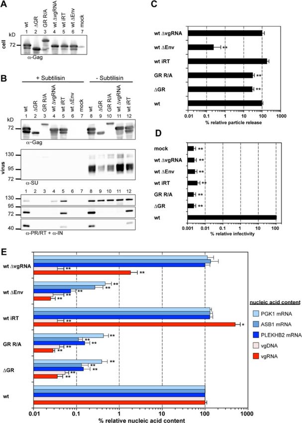
Hamann et al. Retrovirology 2014, 11:87
http://www.retrovirology.com/content/11/1/87

 RESEARCH                                                                                                                                        Open Access

The cooperative function of arginine residues in
the Prototype Foamy Virus Gag C-terminus
mediates viral and cellular RNA encapsidation
Martin V Hamann1,2†, Erik Müllers1,2,5†, Juliane Reh1,2, Nicole Stanke1,2, Gregory Effantin3,4, Winfried Weissenhorn3,4
and Dirk Lindemann1,2*

  Abstract
  Background: One unique feature of the foamy virus (FV) capsid protein Gag is the absence of Cys-His motifs,
  which in orthoretroviruses are irreplaceable for multitude functions including viral RNA genome recognition and
  packaging. Instead, FV Gag contains glycine-arginine-rich (GR) sequences at its C-terminus. In case of prototype
  FV (PFV) these are historically grouped in three boxes, which have been shown to play essential functions in genome
  reverse transcription, virion infectivity and particle morphogenesis. Additional functions for RNA packaging and Pol
  encapsidation were suggested, but have not been conclusively addressed.
  Results: Here we show that released wild type PFV particles, like orthoretroviruses, contain various cellular RNAs in
  addition to viral genome. Unlike orthoretroviruses, the content of selected cellular RNAs in capsids of PFV vector
  particles was not altered by viral genome encapsidation. Deletion of individual GR boxes had only minor negative
  effects (2 to 4-fold) on viral and cellular RNA encapsidation over a wide range of cellular Gag to viral genome ratios
  examined. Only the concurrent deletion of all three PFV Gag GR boxes, or the substitution of multiple arginine residues
  residing in the C-terminal GR box region by alanine, abolished both viral and cellular RNA encapsidation
  (>50 to >3,000-fold reduced), independent of the viral production system used. Consequently, those mutants also
  lacked detectable amounts of encapsidated Pol and were non-infectious. In contrast, particle release was reduced to a
  much lower extent (3 to 20-fold).
  Conclusions: Taken together, our data provides the first identification of a full-length PFV Gag mutant devoid in
  genome packaging and the first report of cellular RNA encapsidation into PFV particles. Our results suggest that the
  cooperative action of C-terminal clustered positively charged residues, present in all FV Gag proteins, is the main viral
  protein determinant for viral and cellular RNA encapsidation. The viral genome independent efficiency of cellular RNA
  encapsidation suggests differential packaging mechanisms for both types of RNAs. Finally, this study indicates that
  analogous to orthoretroviruses, Gag – nucleic acid interactions are required for FV capsid assembly and efficient particle
  release.
  Keywords: Foamy virus, RNA packaging, Gag, Assembly

* Correspondence: dirk.lindemann@tu-dresden.de
†
 Equal contributors
1
 Institute of Virology, Medical Faculty “Carl Gustav Carus”, Technische
Universität Dresden, Fetscherstr. 74, 01307 Dresden, Germany
2
 CRTD/DFG-Center for Regenerative Therapies Dresden - Cluster of Excellence,
Technische Universität Dresden, Fetscherstr. 105, 01307 Dresden, Germany
Full list of author information is available at the end of the article

                                         © 2014 Hamann et al.; licensee BioMed Central Ltd. This is an Open Access article distributed under the terms of the Creative
                                         Commons Attribution License (http://creativecommons.org/licenses/by/4.0), which permits unrestricted use, distribution, and
                                         reproduction in any medium, provided the original work is properly credited. The Creative Commons Public Domain
                                         Dedication waiver (http://creativecommons.org/publicdomain/zero/1.0/) applies to the data made available in this article,
                                         unless otherwise stated.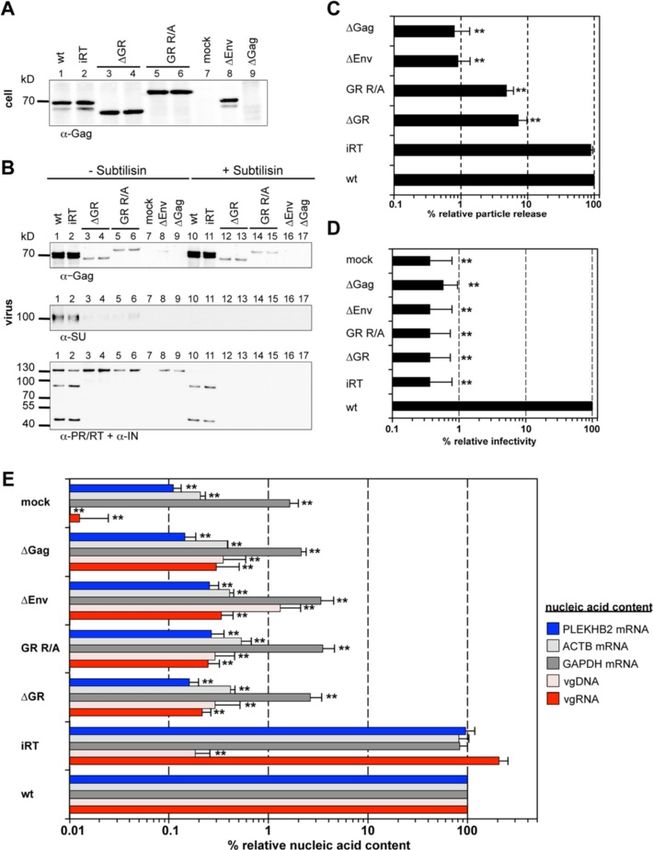
Hamann et al. Retrovirology 2014, 11:87                                                                          Page 2 of 16
http://www.retrovirology.com/content/11/1/87

Background                                                      replication strategy, some of which resemble features of
Gag is the main structural protein of retroviruses that         HBV (reviewed in [15]). For example, unlike orthoretro-
orchestrates the highly regulated process of particle as-       viruses the FV Gag proteins are not processed into the
sembly (reviewed in [1]). In addition to its function in        canonical matrix (MA), capsid (CA) and NC subunits
assembly, Gag also secures the selective packaging of di-       during particle morphogenesis. FV particles, which are
meric viral genomic RNAs (vgRNAs) from a cytoplasmic            released in an FV Env-dependent manner [16], contain a
pool that consists of a substantial excess of non-viral         capsid of immature morphology consisting of Gag pre-
and spliced viral RNAs (reviewed in [2-5]).                     cursor (p71Gag for PFV) and a large processing product
   Orthoretroviral Gag proteins mediate the interaction         (p68Gag for PFV) that is derived from the precursor by
with nucleic acids largely but not entirely by the C-           proteolytic processing through the viral protease at a
terminal nucleocapsid (NC) domain (reviewed in [5]).            single cleavage site. Furthermore, FV Gag proteins lack
They contain conserved motifs of regularly spaced cyst-         the characteristic orthoretroviral Cys-His motifs [17]. It
eine and histidine residues (Cis-His), which are import-        is believed that glycine-arginine-rich (GR) motifs in the
ant for recognition of the complex structured packaging         FV Gag C-terminus represent a functional equivalent of
signal (called Psi) in the vgRNA [6]. This specific mech-       the orthoretroviral Cys-His motifs (reviewed in [18]). In-
anism allows selective vgRNA encapsidation in orthoretro-       deed, others and we have previously shown that these
viral particles (reviewed in [2,7]). However, orthoretroviral   FV Gag motifs, which are grouped in three GR boxes
Gag proteins also possess additional non-specific RNA           (GRI-III) in PFV Gag, have essential functions in gen-
binding features and several types of retroviruses were         ome RTr, virion infectivity as well as particle morpho-
found to encapsidate cellular RNAs constituting up to           genesis [19,20]. However, up to date it remains unclear if
50% of the particle-associated nucleic acid content [8].        individual FV GR motifs also mediate RNA packaging.
Most non-viral RNAs are packaged in a non-selective             Initial studies proposed for the PFV Gag GR box I (GRI)
fashion, although in some cases a selective encapsidation       similar functions in the selective encapsidation of
is observed leading to their enrichment similar to vgRNA.       vgRNAs as assigned to retroviral NC domains and their
The potential biological role of encapsidated host-cell         Cys-His motifs [19,21]. Other studies failed to confirm a
RNAs for viral replication is largely unknown. Besides its      role of individual PFV Gag GR boxes for RNA packaging
participation in recognition and encapsidation of vgRNA         and only PFV Gag proteins with large C-terminal trun-
the orthoretroviral NC domain has nucleic acid chaperone        cations lacking all GR boxes were reported to have lost
activities as well as additional functions in particle assem-   vgRNA packaging capacity [20,22,23]. Further contro-
bly and release, and timing of reverse transcription (RTr)      versy arises from the fact that only the different simian
(reviewed in [2,9]).                                            FV (SFV) Gag proteins and PFV Gag contain clustered
   Hepatitis B virus (HBV), another virus encapsidating a       GR boxes, while no GR boxes were assigned for the
RNA genome, which undergoes reverse transcription               non-primate FV Gag proteins [24,25]. However in gen-
and shares several features of its replication cycle with       eral, all FV Gag proteins contain a high proportion of
FVs, seems to have another mechanism of RNA pack-               glycine and arginine residues in their C-termini. Unfor-
aging. It encodes a core protein with N-terminal assem-         tunately, only for PFV their function in viral replication
bly and C-terminal nucleic acid binding domain, the             was experimentally examined to date.
latter being strongly enriched in basic amino acids               Similarly debated is the question of how FVs encapsi-
(reviewed in [10]). In contrast to orthoretroviruses, en-       date their Pol protein, which is translated as a separate
capsidation of HBV pregenomic RNA (pgRNA) requires              protein and not as an orthoretroviral-like Gag-Pol fusion
HBV polymerase (P-protein) coexpression, preferentially         protein. It is hypothesized that Gag as well as the Pol pre-
in cis, and P-protein binding to a 5′ RNA stem loop             cursor both bind to vgRNA, where the RNA serves as a
structure within the pgRNA molecule [11,12]. However,           bridging molecule for Pol encapsidation [19,20,22,26-28].
in vivo the specificity of pgRNA packaging is further in-       However, another study challenged this view and instead
fluenced by the phosphorylation status of the core pro-         proposed Pol encapsidation to require an accessory, direct
tein, although it was demonstrated that it possesses            Gag-Pol protein interaction involving positively charged
phosphorylation-independent unspecific RNA binding              residues of GRI [23].
activities in vitro [13,14]. Whether released HBV parti-          Thus, despite being addressed in several studies, the
cles also contain encapsidated cellular RNAs has not            determinants for FV RNA packaging and Pol encapsida-
been investigated to date.                                      tion still remain unclear. As previous studies indicated
   FVs or spumaviruses constitute the only genus in a           that not an individual GR box motif mediates FV RNA
separate subfamily of retroviruses, the spumaretroviri-         packaging [20,22,23], we hypothesized that RNA pack-
nae. This is because FVs, although closely related to           aging might be a cooperative function of the whole FV
orthoretroviruses show several unique features in their         Gag C-terminus and in particular the arginine residues
Hamann et al. Retrovirology 2014, 11:87                                                                          Page 3 of 16
http://www.retrovirology.com/content/11/1/87

therein. We therefore, adopted a more “global” GR-box            The differences in relative vgRNA content (normalized
view and analyzed the combined role of positively charged      for Gag content) of the various mutant particles in com-
arginine residues within the GRI-III boxes of PFV Gag in       parison to wild type became more apparent when each
viral replication. We find that the arginine residues in the   sample set (varying in their amount of transfer vector)
PFV Gag C-terminus cooperatively mediate viral and cel-        was examined separately (Figure 2B). Independently of
lular RNA encapsidation and that Gag – nucleic acid in-        the amount of transfer vector, all individual GR box de-
teractions are required for capsid assembly, efficient         letions showed only a very moderate (2 to 4-fold) reduction
particle release as well as Pol precursor packaging.           in vgRNA encapsidation (Figure 2B, red bars). The differ-
                                                               ences to wild type were statistical significant (p < 0.05) for
                                                               ΔGRI and ΔGRIII samples in the 0.5 μg and 0.05 μg trans-
Results                                                        fer vector group.
Minor contribution of individual GR boxes for vgRNA              In summary, these results demonstrate that individual
encapsidation                                                  GR box sequences contribute only moderately to viral
An initial study suggested GRI as the main determinant         genome encapsidation, independently of the ratio of cap-
for PFV vgRNA packaging [19]. Using an extensive, struc-       sid to vgRNA present in the cell during particle assem-
tured analysis comprising all three individual PFV Gag GR      bly. This is in line with our previous findings at even
boxes mutated separately we recently highlighted novel         higher cellular concentrations of packageable vgRNA
GR box functions in RTr and particle morphogenesis [20].       [20]. Thus, we are unable to reproduce the reported
In this study, which employed an expression-optimized 4-       major role of GRI for PFV vgRNA packaging [19].
component PFV vector system, we found only minor con-
tributions of individual GR boxes for functions in vgRNA       Global mutants in the PFV Gag GR-rich C-terminal domain
packaging. To fully exclude that effects of individual GR      display reduced particle release capacity and are
boxes on vgRNA packaging were missed due to the rather         non-infectious
high concentration of the provided transfer vector, we re-     As individual GR box motifs contribute only very little
evaluated the potential contribution of individual GR          to viral genome encapsidation, even under conditions of
boxes for packaging of vgRNA using an optimized qPCR           limited availability of packageable vgRNA, we hypothe-
setup and additionally extended the analysis to also in-       sized that instead the whole Gag C-terminus, and the
clude conditions of limited availability of packageable        positively charged residues in particular, might mediate
virus genome. To avoid any influence of the differential       RNA encapsidation. In line with this hypothesis, non-
intra-particle RTr capacities of the individual GR box Gag     simian FVs do contain a high number of arginine resi-
mutants on the quantitative analysis [20] we used an RT-       dues in their C-terminus, but lack the typical primate
deficient PFV Pol packaging construct for the production       FV clustering into GR boxes [24,25]. Furthermore, Lee
of all particle samples.                                       et al. reported positively charged residues as the main
   We characterized the nucleic acid composition of            determinants of GRI box function in Pol encapsidation
three sets of wild type (wt) and mutant PFV particles          [23]. To examine the functional role of GR-rich se-
harboring individual GR box deletions (ΔGRI, ΔGRII,            quences in the PFV Gag C-terminus for vgRNA encapsi-
ΔGRIII) (Figure 1A), generated by transfecting cells with      dation we generated packaging constructs for two
different amounts of transfer vector, ranging over three       “global” PFV Gag GR-box mutants (Figure 1A). One
orders of magnitude (Figure 2). The amount of Gag, Pol         mutant (ΔGR) has all three PFV Gag GR boxes deleted
and Env packaging constructs was kept constant in all          whereas in a second (GR R/A) 23 arginine residues in
samples, which resulted in a similar virus production          the C-terminal part of Gag containing GRI to GRIII
and particle release in all samples ([20], data not shown).    were substituted by alanine.
   For particles containing wild type Gag a good correl-         In order to avoid potential secondary effects of the intro-
ation between the amount of transfer vector, the cell-         duced mutations on vgRNA structure and the expression
associated (normalized for total RNA), and the particle-       pattern of the viral proteins we used a 4-component PFV
associated vgRNA copies (normalized for Gag content)           vector system to characterize the mutant Gag phenotype
was observed (Figure 2A, wt). In contrast to wild type,        in the first place. When 293T cells were co-transfected with
particles derived from Gag proteins with individual GR         transfer vector as well as packaging constructs for Env,
box deletions showed a slightly reduced (~2-fold) vgRNA        Pol and the respective Gag proteins, both Gag mutants
encapsidation capacity relative to the corresponding cellu-    were expressed at wild type levels and released as viral
lar levels (Figure 2A, ΔGRI, ΔGRII, ΔGRIII). Only ΔGRIII       particles, albeit in significantly reduced amounts com-
mutant particles showed an up to 8-fold reduced relative       pared to wt (3 to 4-fold) (Figure 3A-C). We did not ob-
encapsidation capacity at the lowest amount of transfer        serve mutant Gag processing neither in the cell lysates
vector (Figure 2A, ΔGRIII 0.05).                               (Figure 3A, lane 2, 3) nor in the viral supernatants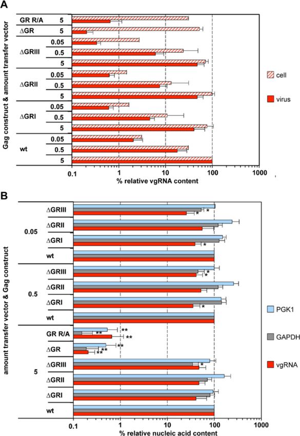
Hamann et al. Retrovirology 2014, 11:87                                                                                               Page 4 of 16
http://www.retrovirology.com/content/11/1/87

 Figure 1 PFV Gag mutants used in the study. (A) Schematic outline of the PFV Gag precursor protein. A solid arrow marks the primary
 processing site in p71Gag. The enlargement shows the protein sequences of the C-termini of wild type PFV Gag (wt) p68Gag subunit and the
 established GR box deletion mutant (ΔGR) as well as the mutant having 23 C-terminal arginine residues substituted by alanine (GR R/A). Amino
 acid residues introduced to or altered in comparison to the wild type sequence are indicated in red bold typeset. Differentially shaded boxes
 highlight C-terminal structural and functional domains. A, assembly domain; GRI-III, glycine-arginine-rich boxes I to III. The described minimal
 chromatin-binding sequence is underlined [32,33]. The numbers indicate the amino acid position in the wt Gag protein. (B) Schematic outline of
 the PFV transfer vector puc2MD9 used. CAS: cis acting sequences I to III required for productive transduction. CMV: cytomegalovirus immediate
 early promoter, R: long terminal repeat (LTR) repeat region; U5: LTR unique 5′ region; ΔU3: enhancer – promoter deleted LTR unique 3’ region;
 partial coding sequences of PFV Gag, Pol and Env overlapping the CAS sequences are indicated by dashed boxes and marked with Δ. SFFV U3:
 spleen focus forming virus U3 enhancer promoter; EGFP: enhanced green fluorescent protein ORF; numbers indicate nucleotide positions in the
 HSRV2 RNA genome. Below the schematic outline the amplicons generated by the qPCR primer – probes sets (Table 1) specific for PFV pol or
 egfp ORFs are shown. Numbers indicate nucleotide position in the HSRV2 RNA genome or the egfp ORF.

(Figure 3B, lane 2, 3, 9, 10). Interestingly, both mutant                  particle-associated (Figure 3B, lane 2, 3, 9, 10) as demon-
Gag proteins (ΔGR and GR R/A) migrated higher than                         strated previously [30].
their expected molecular weight, a phenomenon we                              In line with the Pol encapsidation and Gag processing
previously observed already for Gag ΔGRII mutants                          defect of the PFV Gag ΔGR and GR R/A particles, we
(Figure 3A + B, lane 2, 3, 9, 10) [20]. The released Gag                   could not detect any infectivity of the respective viral su-
protein was associated with Env and the relative                           pernatants (Figure 3D). Thus, the PFV Gag ΔGR and
amounts of both proteins correlated well (Figure 3B,                       GR R/A mutant show at least a 10,000-fold decrease in
lane 9, 10). The subtilisin resistance of Gag but not Env                  virion infectivity, although their particle release was di-
SU in the particle samples indicated that mutant Gag is                    minished only 3 to 4-fold (Figure 3C + D).
released as membrane enveloped particulate material                           Both Gag mutants were also examined in context of
and not as free protein (Figure 3B) [29].                                  proviral expression constructs with sequences of authen-
  In contrast to Gag and Env, no mature Pol subunits                       tic codon-usage to ensure that the observed phenotypes
(>50-fold reduced) were detectable in particle prepara-                    are not due to potentially altered uncoupled viral expres-
tions of the Gag ΔGR and GR R/A mutants. Only re-                          sion generated by the 4-component PFV vector system
leased Pol precursor was detected in the samples, which                    (Figure 4). In general the proviral expression system dis-
was readily digested by subtilisin and was therefore not                   plays a roughly 10-fold lower wild type particle release
Hamann et al. Retrovirology 2014, 11:87                                                                                                       Page 5 of 16
http://www.retrovirology.com/content/11/1/87

 Figure 2 Viral and non-viral RNA encapsidation by Gag mutants with individual GR box deletions. 293T cells were co-transfected with
 puc2MD9, pcoPP, pcoPE and either pcoPG4 (wt), pcoPG ΔGRI (ΔGRI), pcoPG ΔGRII (ΔGRII), pcoPG ΔGRIII (ΔGRIII), pcoPG4 ΔGR (ΔGR), or pcoPG4 GR
 R/A (GR R/A). The amount of packaging constructs was kept constant whereas different amounts of transfer vector puc2MD9 (5, 0.5, 0.05 μg) were
 used as indicated. The total amount of DNA used for transfection was kept constant by filling up with pUC19. Subsequently extracted particle-associated
 nucleic acids and total cellular RNA samples were subjected to qPCR analysis using different primer-probe sets as summarized in Table 1. (A) Cellular and
 viral particle-associated levels of vgRNA. Viral particle and cellular nucleic acid content was determined by qPCR using specific primer – probes
 sets for PFV Pol. Mean values and standard deviation (n = 3-6) are shown as relative values compared to the wild type control. Viral particle
 values were normalized for Gag content, cellular values were normalized per ng of total RNA. (B) Viral particle-associated levels of vgRNA and
 selected cellular mRNAs. Viral nucleic acid content was determined by qPCR using specific primer – probes sets for PFV Pol, as well as human
 PGK1 and GAPDH mRNAs. Mean values and standard deviation (n = 3-6) are shown as relative values compared to the wild type control in
 each set of transfections varying in their amount of transfer vector as indicated. Viral particle values were normalized for Gag content. Differences
 between means of the wild type and the individual mutants were analyzed by Welch’s t test (*, p < 0.05; **, p < 0.01).

than achieved using the 4-component vector system                              comparison to wild type (Figure 4B + C). Of note Western
[31]. Overall the analysis revealed a similar phenotype in                     blot analysis of particle-associated mature Pol proteins in
respect to Gag processing, Pol incorporation and viral                         samples generated with the proviral expression constructs
infectivity for both Gag mutations (Figure 4A-D). Only                         shown in Figure 4B was close to the detection limit due to
their particle release deficiency was more pronounced in                       the reduced particle release of this system compared to
comparison to the 4-component vector system as particle-                       the 4-component vector system [31]. Nevertheless, using
associated Gag levels were reduced up to 20-fold in                            larger amounts proviral construct derived, pelleted viral
Hamann et al. Retrovirology 2014, 11:87                                                                                                       Page 6 of 16
http://www.retrovirology.com/content/11/1/87

 Figure 3 Analysis of the GR-rich PFV Gag C-terminus in context of a replication-deficient vector system. 293T cells were co-transfected
 with pMD9, pcziPol, pcoPE and either pcoPG4 (wt), pcoPG4 ΔGR (ΔGR), or pcoPG4 GR R/A (GR R/A). As controls, cells were transfected with
 pcoPE, pcoPG4 and pcziPol (wt ΔvgRNA), with pMD9, pcoPE, pcoPG4 and pcziPol iRT (wt iRT), with pMD9, pcoPG4 and pcziPol (wt ΔEnv) or
 only with pcDNA3.1 zeo + (mock). (A, B) Representative Western blot analysis of viral particles (virus) purified from 293T cell culture supernatant by
 ultracentrifugation through 20% sucrose and 293T cell lysates (cell). PFV proteins were detected using antibodies specific for PFV Gag (α-Gag), for PFV
 Pol PR/RT and IN (α-PR/RT + α-IN), or for PFV Env SU (α-SU). (C) Viral particle release was determined by quantitative Western blot analysis of viral
 particles. Mean values and standard deviations (n = 3-6) are shown as relative values compared to the wild type control and normalized for cellular
 expression levels. (D) Infectivity analysis of PFV particle containing cell culture supernatants. The values obtained using wild type PFV Gag expression
 plasmids were arbitrarily set to 100%. Relative means and standard deviations from six independent experiments are shown. Absolute titers of wt
 supernatants ranged between 2 × 10⁶ and 1.3 × 10⁷ eGFP ffu/ml. (E) Viral particle nucleic acid content was determined by qPCR using specific primer
 – probes sets for PFV Pol, as well as human PGK1, ASB1 and PLEKHB2 mRNAs as summarized in Table 1. Mean values and standard deviation (n = 3-6)
 are shown as relative values compared to the wild type control. Values were not normalized for Gag content. Differences between means of the wild
 type and the individual mutants in (C)-(E) were analyzed by Welch’s t test (*, p < 0.05; **, p < 0.01).
Hamann et al. Retrovirology 2014, 11:87                                                                                                         Page 7 of 16
http://www.retrovirology.com/content/11/1/87

 Figure 4 Analysis of the GR-rich PFV Gag C-terminus in context of replication-competent proviral expression constructs. 293T cells were
 transfected with pczHSRV2 (wt), pczHSRV2 ΔGR (ΔGR), or pczHSRV2 GR R/A (GR R/A). As controls, cells were transfected with variants pczHSRV2
 iRT (iRT), expressing a Pol protein with enzymatically inactive RT domain; pczHSRV2 iEnv (ΔEnv), with inactivated Env translation start; pczHSRV2
 M78 (ΔGag), with inactivated Gag translation start; or only with pUC19 (mock). (A, B) Representative Western blot analysis of viral particles (virus)
 and 293T cell lysates (cell). PFV proteins were detected using antibodies specific for PFV Gag (α-Gag), for PFV Pol PR/RT and IN (α-PR/RT + α-IN),
 or for PFV Env SU (α-SU). (C) Viral particle release was determined by quantitative Western blot analysis of viral particles. Mean values and standard
 deviations (n = 3-6) are shown as relative values compared to the wild type control and normalized for cellular expression levels. (D) Infectivity analysis
 of PFV particle-containing cell culture supernatants. The values obtained using wild type PFV proviral expression plasmids were arbitrarily set to 100%.
 Relative means and standard deviations from three independent experiments are shown. Absolute titers of wt supernatants ranged between 6 × 10³
 and 7 × 10⁴ eGFP ffu/ml. (E) Viral particle nucleic acid content was determined by qPCR using specific primer – probes sets for PFV Pol, as well as
 human ACTB, GAPDH and PLEKHB2 mRNAs as summarized in Table 1. Mean values and standard deviation (n = 4-6) are shown as relative values
 compared to the wild type control. Values were not normalized for Gag content. Differences between means of the wild type and the individual
 mutants in (C)-(E) were analyzed by Welch’s t test (**, p < 0.01).

particles mature IN subunits were detectable in 20-fold di-                        In summary, the removal of all PFV Gag GR boxes or
luted wt samples but were absent in virion samples of                            replacement of their residing arginine residues results
both mutants ΔGR and GR R/A (see Additional file 1).                             in a reduced release of particles that are non-infectious
Hamann et al. Retrovirology 2014, 11:87 Page 8 of 16
http://www.retrovirology.com/content/11/1/87
and lack any detectable amounts of encapsidated Pol and probes specific to PFV pol (Figure 3E, compare
proteins. ‘ΔGR’ and ‘GR R/A’ to ‘wt ΔEnv’ and ‘wt’) nor when
using primers specific to the egfp ORF (data not shown).
Positively charged amino acids of the PFV Gag GR-rich This indicated a functional defect at the level of vgRNA
motifs are essential for efficient viral genome encapsidation for both mutants. Again a similar analysis
encapsidation of both Gag mutants in context of proviral expression
To characterize the viral genome encapsidation features constructs revealed an identical phenotype in respect to
of the two global Gag GR box mutants, the nucleic acid viral genome encapsidation (Figure 4E).
composition of wild type and mutant particles as well as These results strongly suggest that the cooperative ac-
several controls was determined by qPCR using different tion of clustered positively charged amino acids in the
primer – probe sets specific for viral vector genome Gag C-terminus rather than individual GR-box motifs is
(Figure 1B, Table 1). essential for viral genome encapsidation.
Consistent with the infectivity defect of the PFV Gag
ΔGR or GR R/A mutants, we were unable to detect any PFV particles encapsidate cellular RNAs in a Gag GR-rich
particle-associated viral genomic nucleic acids (vgRNA, motif-dependent manner
vgDNA) above background levels (>3,000-fold reduc- Interestingly, our analysis of particle-associated nucleic
tion) in respective particle preparations generated by the acids indicated that wild type FV particles can encapsi-
4-component vector system, neither when using primers date low levels (2% of wt) of subgenomic viral RNAs
Table 1 qPCR Primer/probe sets
Target Primer/Probe 5′-3′ sequencea Cycle conditions
PFV genome (Integrase 1) fwd CTTCAACCTTTGCTGAATG 95°C, 8 min, 1×
rev TAATACAGGGCTATAGGTGT 95°C, 30 s, 40×
probe FAM-TTGGAATTCAGTACTCCTTATCACCC-BHQ1 58°C, 30 s, 40×
PFV genome (Integrase 2) fwd TGCAATTCCAAAGGTGATTC 95°C, 8 min, 1×
rev TACCTCTTTCCTTTGCCCAT 95°C, 30 s, 40×
probe FAM-TCAAGGTGCAGCATTCACTTCTTCAA-BHQ1 59°C, 30 s, 40×
EGFP fwd GCAGTGCTTCAGCCGCTAC 95°C, 8 min, 1×
rev AAGAAGATGGTGCGCTCCTG 95°C, 30 s, 40×
probe HEX-CCGACCACATGAAGCAGCACGACTT-BHQ2 59°C, 30 s, 40×
72°C, 45 s, 40×
GAPDH fwd CATCAATGGAAATCCCATCA 95°C, 45 s, 40×
rev GACTCCACGACGTACTCAGC 95°C, 30 s, 40×
probe FAM-TCCAGGAGCGAGATCCCTCCA-BHQ1 59°C, 30 s, 40×
72°C, 30 s, 40×
ACTB fwd TGGACTTCGAGCAAGAGATG 95°C, 8 min, 1×
rev GAAGGAAGGCTGGAAGAGTG 95°C, 30 s, 40×
probe FAM-CGGCTGCTTCCAGCTCCTCC-BHQ1 59°C, 30 s, 40×
72°C, 30 s, 40×
ASB1 primer/probe mix HS00211548_m1 Gene Expression Kit Applied Biosystems 95°C, 10 min, 1×
95°C, 15 s, 40×
60°C, 1 min, 40×
PGK1 primer/probe mix HS99999906_m1 Gene Expression Kit Applied Biosystems 95°C, 1 min, 1×
95°C, 15 s, 40×
60°C, 1 min, 40×
PLEKHB2 primer/probe mix HS00215820_m1 Gene Expression Kit Applied Biosystems 95°C, 10 min, 40×
95°C, 15 s, 40×
60°C, 1 min, 40×
a
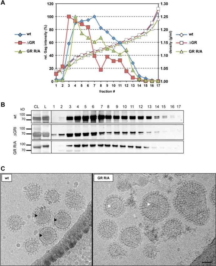
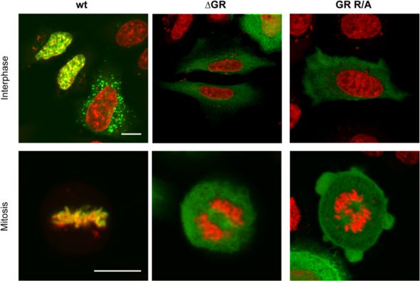
Fam: 6-carboxyfluorescein; HEX: hexachloro-fluorescein; BHQ1: Black Hole Quencher 1; BHQ2: Black Hole Quencher 2.Hamann et al. Retrovirology 2014, 11:87 Page 9 of 16 http://www.retrovirology.com/content/11/1/87 containing the authentic pol ORF (detected by the pol- amounts of transfer vector used (p < 0.05) (Figure 2B, specific qPCR, Figure 1B) in the absence of vgRNA grey bars). (Figure 3E, wt ΔvgRNA). However, this Pol packaging In summary, our analyses demonstrate that PFV wt construct-derived RNA was not reverse transcribed since Gag particles also contain cellular mRNAs. Furthermore, at the same time no vDNA was detectable (Figure 3E, wt they show that the GR-rich sequences of PFV Gag but ΔvgRNA). As other retroviruses are known to encapsidate not individual GR-box motifs are essential for both viral subgenomic vRNAs and cellular RNAs by different mech- and non-viral RNA encapsidation. anisms in addition to the viral genome [8], it seemed likely, although not previously examined, that FVs might PFV Gag GR-rich motif mutants lack chromatin tethering also encapsidate cellular RNAs. Furthermore, since our functions Gag ΔGR and GR R/A mutants showed no detectable Sequences of GRII were shown to be involved in tether- vgRNA packaging we were intrigued by the question if ing PFV Gag to the host cell chromatin during mitosis they might package cellular RNAs or if the mutations [32,33]. We therefore examined the intracellular distri- abolished RNA encapsidation in general. bution of the two “global” PFV Gag GR-box mutants by We therefore examined the encapsidation of cellular confocal fluorescence microscopy of cells transfected RNAs into released wild type and mutant FV particles. with C-terminally EYFP tagged Gag variants. In contrast For this purpose we determined by qPCR the presence to wt Gag, PFV Gag ΔGR protein did not colocalize with of selected RNA species, some of which were reported host cell chromatin during mitosis (Figure 5, compare previously to be packaged into murine leukemia virus left and middle panels), showing the same phenotype as (MLV) or human immunodeficiency virus (HIV) parti- reported previously for a PFV Gag ΔGRII mutant pro- cles [8], in nucleic acid preparations of different PFV tein [20,32,33]. Interestingly, the PFV Gag GR R/A mutant particle samples generated using the 4-component vector displayed an identical phenotype as ΔGR (Figure 5, right system or proviral expression constructs (Figure 3E + panel) and ΔGRII [20], indicating that the C-terminal ar- Figure 4E). Relative copy numbers of cellular PGK1, ginine residues contribute to chromatin binding, although ASB1, PLEKHB2, ACTB and GAPDH mRNAs of wild only two of them (R540 and R542) are located in the min- type PFV particle preparations were at least 50 to 1,000- imal chromatin binding site (CBS) as characterized by fold higher than those of mock particle preparations de- Tobaly-Tapiero and colleagues [32]. The identical pheno- rived from 293T cells transfected with pUC19 (mock) or type of these Gag mutants suggests that changing as little particle preparations of samples not expressing PFV Env as two arginine residues in the PFV Gag CBS are sufficient (ΔEnv) (Figure 3E + Figure 4E). Our results show for the to inactivate its chromatin tethering function. In line with first time that FVs can also package cellular RNAs. Inter- this hypothesis, Schneider et al. [34] recently demon- estingly, the level of the cellular mRNAs in PFV particles strated in context of chimeric MLV Gag proteins an was not significantly influenced by the presence or ab- inactivation of the chromatin tethering function of an sence of vgRNA (Figure 3E, compare ‘wt’ and ‘wt iRT’ to inserted minimal PFV Gag CBS by mutating its two argin- ‘wt ΔvgRNA’). Most importantly however, we were unable ine residues. to detect those cellular RNAs in Gag ΔGR and GR R/A derived particle preparations (Figure 3E + Figure 4E, com- PFV GR box mutant particles display capsid assembly pare ‘ΔGR’ and ‘GR R/A’ to ‘wt’) indicating that these mu- defects tants lost RNA packaging capacity in general. In vivo particle assembly of some orthoretroviruses (e.g. This observation led us to also analyze the cellular MLV), but not others (HIV), is strongly dependent on RNA content of mutant particles with individual GR Gag – nucleic acid interactions (reviewed in [5]). To in- box deletions, since specific GR boxes may convey cel- vestigate whether FV capsid assembly might be influ- lular RNA encapsidation. In contrast to the “global” enced by Gag – nucleic acid interactions we performed PFV Gag GR-box mutants ΔGR and GR R/A, particle- isopycnic ultracentrifugation in order to compare the associated cellular mRNA contents (PGK1, GAPDH) of buoyant density of wild type and mutant viral particles particles with individual GR box deletions was within a (Figure 6A + B). The analysis revealed a different density 2 to 3-fold range of wild type and not influenced by the profile of mutant viral particles in comparison to wild amount encapsidated vgRNA (Figure 2B). Although type. Fractions with the highest Gag signal ranged from below statistical significance, GRII deleted particles 3 to 5 in mutant samples, whereas the Gag peak in the consistently showed a slightly increased PGK1 mRNA wt preparation was located between fraction 6 and 7. content (Figure 2B, light blue bars). Furthermore, Hence, both mutants seem to have slightly lighter par- ΔGRIII box deleted particles consistently showed the ticle densities, indicating aberrant particle morphology. lowest content in GAPDH mRNA of all mutants that This was further supported by cryo electron microscopy was statistically different to wild type at all three analysis. Virus particles were produced by transiently
Hamann et al. Retrovirology 2014, 11:87                                                                                           Page 10 of 16
http://www.retrovirology.com/content/11/1/87

 Figure 5 Altered intracellular distribution of mutant Gag proteins. Intracellular distribution of C-terminal eYFP-tagged Gag wt (wt), Gag ΔGR
 (ΔGR) or Gag GR R/A (GR R/A) proteins in HeLa cells 48 h post transfection. Samples were fixed and stained with DAPI. The images show a
 merged image of the eYFP (green) and the DAPI (red) channel. Scale bar: 10 μm.

transfected 293T cells expressing the PFV Gag wild type                   PFV Gag mutant GR R/A, having 23 arginine residues
or GR R/A mutant proteins in the context of the 4-                        (out of 65 total arginine or lysine residues present in
component vector system and concentrated by ultracen-                     p71Gag) in the C-terminus of Gag replaced by alanine, is
trifugation followed by size exclusion filtration prior to                the first full-length PFV Gag mutant reported to have no
electron microscopy analysis. Gag expression levels of wild               vgRNA packaging capacity. Harboring “only” 23 amino
type and Gag ΔGR or GR R/A expressing cells were                          acid changes, this mutant indicates that the clustering of
comparable (Figure 3A) and particle release was readily                   positively charged residues is the main Gag determinant
detectable for both mutants, though at a reduced level                    required for vgRNA encapsidation, independent of the
(Figure 3B + C). Pronounced morphological differences                     viral production system (replication-deficient vectors vs.
of GR R/A mutant particles in comparison to wild type                     replication-competent proviruses) used for analysis. A
particles were detectable in cryo electron micrographs                    general requirement of positively charged residue en-
(Figure 6C). Unlike wild type PFV particles, which con-                   richment instead of their clustering into GR boxes as
tained significant numbers of virions with a clearly vis-                 RNA-binding motifs can elegantly explain how non-
ible regular shaped capsid structure (Figure 6C left,                     primate FVs package vgRNA while lacking GR boxes. In
black arrowheads), no such structures were detectable                     line with this hypothesis the Gag C-termini of feline FV,
in GR R/A particle preparations (Figure 6C right). In                     equine FV, and bovine FV contain 20, 23, and 22 argin-
addition, GR R/A particles appeared to be more heteroge-                  ine residues respectively (reviewed in [18]).
neous in size and some contained irregularly shaped elec-                    We also report here for the first time the encapsidation
tron dense material (Figure 6C right, white arrowheads).                  of various cellular RNAs into PFV particles. Notably, un-
   Thus, PFV Gag – nucleic acid interactions seem to be                   like reported for MLV and HIV-1 we did not observe any
important for correct capsid assembly that might be re-                   significant difference in their copy numbers whether wild
quired for efficient particle release in vivo.                            type PFV vector particles contained or lacked vgRNA [8].
                                                                          This suggests that vgRNA and non-viral RNAs do not
Discussion                                                                compete with each other, which might indicate different
In this study we characterized the PFV Gag determi-                       mechanisms of encapsidation of both types of RNA into
nants essential for vgRNA encapsidation. We demon-                        PFV particles. Since the positively charged residues in the
strate that independent of the intracellular ratio of Gag                 Gag C-terminus also appear to control packaging of all
to vgRNA the whole GR-rich Gag C-terminus rather                          non-viral RNAs examined, the basis of selective encapsida-
than individual GR box motifs mediates vgRNA encapsi-                     tion of vgRNA remains to be defined. In general, however,
dation. In line with our results Stenbak and colleagues                   our results indicate that by not relying on specific Gag
reported that truncation of the whole Gag C-terminus                      motifs FVs seem to have a fundamentally different RNA
comprising the GR-rich sequences and the p3 domain                        packaging strategy than other retroviruses. The analogy to
can abolish vgRNA packaging [22]. The “global” GR-box                     Hepadnaviruses, which share some features in their
Hamann et al. Retrovirology 2014, 11:87 Page 11 of 16 http://www.retrovirology.com/content/11/1/87 Figure 6 Buoyant density and electron microscopy analysis of the mutant PFV particles. 293T cells were co-transfected with either pcoPG4 (wt), pcoPG4 ΔGR (ΔGR), or pcoPG4 GR R/A (GR R/A) in combination with pcoPP, pcoPE and puc2MD9 to yield the respective wt and mutant particles. (A) Concentrated virus supernatant was loaded onto an iodixanol step gradient ranging from 15 to 40%. After centrifugation at 197,000 g for 3 h the gradient was split into 17 fractions and their density determined by refractometry measurements (right ordinate) and their Gag content examined by Western blot analysis (left ordinate). The highest Gag signal intensity in each gradient was set to 100%. (B) Western blot analysis of the Gag content in individual gradient fractions (1-17), pelleted virus particles (L) and cell lysates of transfected 293T cells (CL) using polyclonal antibodies specific for PFV Gag (α-Gag) (see above). (C) Cryo-electron microscopy analysis of wild type PFV (left) and of GR R/A mutant (right) particles. The virus particles were concentrated via a 20% sucrose gradient centrifugation step followed by size exclusion column filtration. Black arrowheads indicate regular Gag assemblies in the wild type virus and white arrows mark putatively aberrant Gag assemblies in the GR R/A mutant. Scale bar: 60 nm. replication strategy with FVs, may indicate that the specifi- Although we have not formally shown that both global city of vgRNA encapsidation is influenced by the FV Gag Gag GR box mutants are completely devoid of nucleic phosphorylation status [13,14]. However, except for cellular acids the absence of all cellular mRNAs examined (five in PFV Gag being phosphorylated predominantly at serine total) suggests that this is indeed the case. Therefore an- residues reported by Enssle and colleagues [35] neither other interesting task to be examined in the future is the specific phosphorylation sites of Gag have been identified determination of the repertoire and abundance of coding nor the influence of the Gag phosphorylation status on or non-coding, non-viral RNAs co-packaged into wild type viral replication studied. FV virions on a global scale and its comparison to other
Hamann et al. Retrovirology 2014, 11:87                                                                         Page 12 of 16
http://www.retrovirology.com/content/11/1/87

retroviruses. The PFV Gag GR box mutants described here          export. Differential levels of Gag and Env expression by
will be an important tool/control for this kind of analysis.     FV vector and proviral expression systems used in our
Furthermore, elucidation of the potential roles of various       study may modulate the interaction capacity of the mutant
particle-associated non-viral RNAs in the retroviral replica-    capsids with the glycoprotein and explain the differences
tion cycle is a field that has not been widely studied so far.   in the particle release deficiency observed for both virus
   Together with a lack of viral genome packaging we did         production systems.
not detect particle-associated mature PFV Pol subunits
in virus samples of PFV Gag ΔGR and GR R/A mutants               Conclusions
generated by different expression systems. In line with          In summary, our data sheds light on the close connection
the absence of particle-associated Pol no processing of          of RNA packaging, Pol encapsidation, capsid assembly,
particle-associated mutant Gag protein was observed.             and as previously shown RTr in FV replication. We char-
Thus, we find that Pol is not encapsidated if no RNA is          acterized here the first full-length PFV Gag protein mu-
packaged. While, Lee and colleagues previously sug-              tant deficient in vgRNA encapsidation independent of the
gested GRI as a direct Gag-Pol interaction motif [23], we        virus production system used. Furthermore, we show for
showed that deletion or substitution of GRI alone does           the first time that like their orthoretroviral cousins FVs
neither abolish Pol nor RNA packaging [20]. Although             also encapsidate various non-viral RNAs. More import-
we cannot fully exclude accessory contributions of Gag-          antly, we demonstrate that the positively charged residues
Pol protein interactions our data strongly implies that Pol      in the whole PFV Gag C-terminus act cooperatively to
packaging does not require direct Gag-Pol interaction but        package vgRNA but also all cellular mRNAs examined, in-
rather occurs through an essential RNA bridge [27].              dicating an RNA packaging mechanism different to other
   In case of MLV and Rous sarcoma virus (RSV) the Gag           retroviruses. The nucleic acid binding abilities of Gag in-
NC domain is strictly required for particle production           fluence PFV particle assembly and egress. While cellular
[36,37], while several studies showed that the arginine-         RNAs seem to be able to complement functions of vgRNA
rich C-terminus of PFV Gag is not absolutely required to         for particle assembly and egress they cannot complement
assemble and release viral particles [22,38,39]. We ob-          its function for Pol packaging. These findings on PFV
served release of the “global” GR-box Gag mutants ΔGR            Gag nucleic acid interactions and encapsidation also
and GR R/A, however, at a lower level than wild type, with       provide additional lines of evidence strongly supporting
the extent of the reduction being influenced by the ex-          the current model of PFV Pol packaging by a mechanism
pression system used. Similarly, in HIV-1 certain muta-          involving simultaneous binding of Gag and Pol to vgRNA.
tions in the Cys-His motifs significantly reduce particle
production due to impaired Gag multimerization [40,41].          Methods
Albeit retroviral Gag multimerization does not require           Cells and culture conditions
binding of nucleic acids it is greatly facilitated by their      The human kidney cell line 293T [48], the human epi-
presence [42-44]. The absence of regularly shaped capsid         thelium HeLa cell line [49] and the human fibrosarcoma
structures in electron micrographs of released GR R/A            cell line HT1080 [50] as well as the clonal variant
mutant particles, the larger heterogeneity in size and the       HT1080 PLNE thereof [31] were cultivated in Dulbecco’s
altered density profiles of mutant particles are in line with    modified Eagle’s medium (DMEM) supplemented with
assembly and/or oligomerization defects of PFV Gag pro-          10% heat-inactivated fetal calf serum and antibiotics.
teins lacking nucleic acid binding capacity. Therefore, we       HeLa cells used for confocal laser scanning microscopy
like to suggest that also wild type PFV Gag multimeriza-         were cultivated in phenol red free media.
tion is facilitated by interactions of Gag with nucleic acids
thereby enhancing capsid assembly and particle release.          Recombinant plasmid DNAs
However, in the absence of vgRNA its structural function         A 4-component PFV vector system, consisting of the
in PFV particle assembly and egress seems to be comple-          expression-optimized packaging constructs pcoPG4 (PFV
mented by non-viral RNAs, as wild type particles lacking         Gag), pcoPE (PFV Env), pcoPP (Pol) or its authentic ORF
vgRNA (wt ΔvgRNA) still contain all non-viral RNAs ex-           containing variant pcziPol (PFV Pol), and the enhanced
amined and show wild type-like capsid assembly and par-          green fluorescent protein (eGFP)-expressing PFV transfer
ticle release. In contrast, efficient Pol packaging seems to     vectors pMD9 or puc2MD9 (Figure 1B), has been de-
be absolutely dependent on the presence of vgRNA.                scribed previously [20,26,29]. In some experiments pre-
   As FV Gag proteins lack a membrane targeting signal           viously described variants of the PFV Pol packaging
virus egress is dependent on capsid interactions with the        construct, containing expression-optimized (pcoPP2) or
glycoprotein leader peptide [16,45-47]. These interactions       authentic (pcziPol iRT) ORFs with catalytically inactive
might be strongly influenced by Gag oligomerization and          reverse transcriptase (Pol iRT, YVDD312–315GAAA mu-
capsid assembly and thereby alter the efficiency of particle     tation), were used [20,51].
Hamann et al. Retrovirology 2014, 11:87                                                                     Page 13 of 16
http://www.retrovirology.com/content/11/1/87

  All PFV Gag packaging constructs used in this study         incubated in a digestion mix containing final concentra-
are based on the parental pcoPG4 vector or its C-             tions of 1 mM CaCl2, 50 mM Tris-HCl pH 8.0 and
terminal EYFP tagged variant pcoPG4 CeYFP [29]. The           25 μg/ml subtilisin. The mock treated other half was in-
PFV Gag packaging constructs used in this study are           cubated with the digestion mix including PBS instead of
depicted in Figure 1A. The packaging constructs encod-        subtilisin. The digestion was stopped after 2 h at 37°C by
ing mutant Gag protein with deletion in individual GR         adding phenylmethylsulfonylfluoride (PMSF) at a final
boxes (pcoPG4 ΔGRI, pcoPG4 ΔGRII, pcoPG4 ΔGRIII)              concentration of 100 μg/ml to each reaction prior to
have been described previously [20]. Two additional           addition of 2× sodium dodecyl sulfate (SDS) protein
PFV Gag packaging constructs were generated for this          sample buffer (PPPC; 100 mM Tris-HCl [pH 6.8], 24%
study. The ΔGR mutant (pcoPG4 ΔGR, pcoPG4 CeYFP               glycerol, 8% SDS, 2% dithiothreitol, 0.02% Coomassie
ΔGR) has all three GR boxes simultaneously deleted and        blue G-250).
each replaced by a TGAS peptide sequences as found in
the original individual GR box deletion mutants [20]. In      Infectivity analysis
the GR R/A mutant (pcoPG4 GR R/A, pcoPG4 CeYFP                Transduction efficiency of recombinant, eGFP-expressing
GR R/A) 23 arginine residues between aa 485 and 614 of        PFV vector particles by fluorescence reporter-gene trans-
PFV Gag were replaced by alanine.                             fer assay was analyzed as described previously [55]. Virus
  The CMV-driven proviral expression vector pczHSRV2          particles generated by use of proviral expression plasmids
(wt) and its variants pczHSRV2 M69 (iRT), expressing a Pol    were titrated on HT1080 PLNE cells harboring a Tas-
protein with enzymatically inactive RT domain (YVDD312-       inducible nuclear egfp ORF in their genome as described
315GAAA mutation), and pczHSRV2 M78 (ΔGag), having            previously [31]. All transduction experiments were per-
the Gag translation start inactivated (M1L, ATG to TTG;       formed at least three times. In each independent experi-
S3Stop, TCA to TAA mutation) were described previously        ment the values obtained with the wild type construct
[51,52]. For this study the variants pczHSRV2 ΔGR (ΔGR),      pcoPG4 and pczHSRV2, respectively, were arbitrarily set
having all three GR boxes deleted and each replaced by        to 100% and values obtained with other constructs were
TGAS peptide sequences, pczHSRV2 GR R/A (GR R/A),             normalized as a percentage of the wild type values.
having 23 arginine residues between aa 485 and 614 of PFV
Gag replaced by alanine, and pczHSRV2 iEnv (ΔEnv) having      Western blot analysis
the Env translation start inactivated (M1T, ATG to ACG,       Cells from a single transfected 100-mm cell culture dish
M5T, ATG to ACG; M16T, ATG to ACG mutation) were              were lysed in detergent-containing buffer and the lysates
generated. All constructs were verified by sequencing         were subsequently centrifuged through a QIAshredder
analysis. Primer sequences and additional details are         column (QIAGEN). Protein samples from cellular lysates
available upon request.                                       or purified particulate material were separated by SDS-
                                                              PAGE on a 10% polyacrylamide gel and analyzed by im-
Transfection and virus production                             munoblotting as described previously [16]. Polyclonal
Cell culture supernatants containing recombinant viral        rabbit antisera specific for PFV Gag [56] or the amino
particles were generated by transfection of 293T cells with   acids (aa) 1 to 86 of the PFV Env leader peptide (LP),
the corresponding plasmids using polyethyleneimine (PEI)      [16] as well as hybridoma supernatants specific for PFV
as described previously [20,31]. For subsequent Western       RT (clone 15E10) or PFV integrase (IN) (clone 3E11)
blot analysis the supernatant generated by transient trans-   [57] were employed. After incubation with a horseradish
fection was harvested, passed through a 0.45-μm filter and    peroxidase (HRP)-conjugated secondary antibody, the
centrifuged at 4°C and 25,000 rpm for 3 h in a SW40 or        blots were developed with Immobilon Western HRP
SW28 rotor (Beckman) through a 20% sucrose cushion.           substrate. The chemiluminescence signal was digitally
The particulate material was resuspended in phosphate-        recorded using a LAS-3000 (Fujifilm) imager and quan-
buffered saline (PBS). For cryo electron microscopy ana-      tified using ImageGauge (Fujifilm).
lysis viral particles were produced in serum-free medium
and a further concentration step using Amicon Ultra           Confocal microscopy
0.5 ml 100 K Concentrators was included following the         The analysis of the intracellular distribution of C-
first concentration by ultracentrifugation through 20% su-    terminal eYFP-tagged Gag constructs using confocal mi-
crose similar as described recently [53].                     croscopy was done as described previously [33]. Briefly,
                                                              HeLa cells were plated at a concentration of 6 × 10⁴ cells
Subtilisin digest                                             per well on cover slips in 12-well plates one day before
Subtilisin treatment of concentrated particles was per-       transfecting them with 0.1 μg Gag C-terminal eYFP fu-
formed as previously described [30,54]. Briefly, half of      sion expression plasmid (pcoPG4 CeYFP or mutants
each purified particle pellet resuspended in PBS was          thereof as indicated) using FuGENE HD transfection
Hamann et al. Retrovirology 2014, 11:87 Page 14 of 16
http://www.retrovirology.com/content/11/1/87
reagent (Roche). At 48 h post transfection the cells Absolute copy numbers corresponding to 0.5 ml plain
were washed with cold PBS, fixed with 3% paraformal- supernatant of wild type samples ranged from 3.4 × 10⁵
dehyde, and the cell nuclei were stained with DAPI for to 6.2 × 10⁷ with a detection limit of 1.2 × 10³ for viral
5 min. Finally the cells were covered with Mowiol. Con- genomic RNA, 2.3 × 10⁶ to 2.8 × 10⁷ with a detection
focal laser scanning images were obtained on a Zeiss limit of 1.2 × 10³ for viral genomic DNA, 7.0 × 10⁵ to
LSM 510 using a Zeiss Apochromat 63×, NA 1.4 oil 6.9 × 10⁶ with a detection limit of 1.8 × 10³ for GAPDH
immersion objective. Fluorescence images were evalu- mRNA, 2.4 × 10⁴ to 1.3 × 10⁵ with a detection limit of
ated using ImageJ software. 1.8 × 10² for ASB1 mRNA, 2.3 × 10⁵ to 1.3 × 10⁶ with a
detection limit of 1.8 × 10³ for PGK1 mRNA, 8.2 × 10⁴
Quantitative PCR analysis to 4.6 × 10⁵ with a detection limit of 1.8 × 10² for
Preparation of particle samples for qPCR analysis was PLEKHB2 mRNA, and 8.0 × 10⁶ to 9.3 × 10⁶ with a de-
performed as previously described [20,54]. Furthermore, tection limit of 1.1 × 10² for ACTB. Whether the nucleic
cellular nucleic acids were extracted by trypsinizing tran- acid copy number values of the different particle sam-
siently transfected 293T cells of one 100-mm cell culture ples were normalized for Gag content by quantitative
dish. After terminating the trypsinisation reaction by Western blot analysis is mentioned in the legend of the
addition of 4 ml complete medium the homogenous individual figures. Determined RNA values of cellular
cells suspension was pelleted at 300 g for 5 min. Subse- samples were calculated as copies/ng total RNA and
quently, the supernatant was aspirated and cellular total expressed as percentage of the wild type.
RNA was extracted from the cell pellet with the RNeasy
Mini kit (QIAGEN) according to the manufacturers Cryo-electron microscopy
protocol. This included the recommended addition of Wild type PFV and the GR R/A mutant samples were both
beta-mercaptoethanol to buffer RLT, the use of QIAsh- observed by cryo electron microscopy (cryo-EM) following
redder columns for cell homogenization and omission of the same procedure except that wild type PFV particles
the optional DNaseI digestion on the spin column. Re- were first inactivated for at least 1 h in 4% paraformalde-
verse transcriptase reaction including DNaseI digest was hyde before being processed. In summary, 4 μl of sample
essentially done as for the particle samples, using 250 ng containing 10 nm gold beads was applied to Quantifoil
of total RNA and 10 pmol oligo (dT)30 or random hex- holey carbon grid and the grid was plunge frozen in liquid
amer primers in a total volume of 40 μl. Finally, for ethane with a Vitrobot (FEI, the Netherlands). The frozen
qPCR analysis of RNA content 4.5 μl of each reverse grid was transferred to a FEI F20 FEG cryo electron micro-
transcriptase reaction was analyzed in duplicates in a scope. Images were recorded at a nominal magnification of
total reaction volume of 25 μl using Maxima Probe 29,000 on a 4 k by 4 k Eagle CCD camera. The gold beads
qPCR Master Mix including ROX dye (Thermo Scien- were computationally removed from the field of view
tific) and a StepOnePlus (Applied Biosystems) quantita- shown in Figure 6 for clarity.
tive PCR machine. Primers, Taqman probes and cycling
conditions for specific quantification of PFV genome, Density centrifugation
EGFP, or human GAPDH and ACTB are summarized in Density centrifugation was essentially performed as de-
Table 1. Cellular and particle-associated copy numbers scribed before [30]. Briefly, particle preparations were
of ASB1, PGK1 and PLEKHB2 mRNAs were determined concentrated from 60 ml culture supernatant of 293T
using the Taqman Gene Expression Assay Kits cells transiently co-transfected using the 4-component
(HS00211548_m1, HS99999906_m1, HS00215820_m1 re- vector system and resuspended in 175 μl PBS. This virus
spectively, from Applied Biosystems) according to the concentrate was then overlaid onto a 1.8 ml iodixanol-
manufacturers manual. All values obtained were referred PBS (OptiPrep®) gradient consisting of nine 220 μl layers
to a standard curve consisting of 10-fold serial dilutions of ranging from 15% to 40%. Following ultracentrifugation
respective reference plasmids containing the target se- at 197,000 g and 4°C for 3 h (TLS55 rotor; Beckman)
quences (puc2MD9qP for viral genomic sequences and 17× 127 μl fractions were collected from top to bottom.
EGFP, pCR2.1-TOPO-GAPDH, pCR.2.1-TOPO-ACTB, Subsequently, the density of the fractions was deter-
pCR2.1-TOPO-ASB1, pCR2.1-TOPO-PGK1 and pCR2.1- mined by refractometry, as well as the Gag protein con-
TOPO-PLEKHB2 respectively). All sample values included tent examined by Western blot analysis.
were in the linear range of the standard curves with a span
from 10 to 109 copies. The values for the DNA or RNA Additional file
content of viral particle samples obtained by the qPCR
analysis are expressed as percentage of the wild type (gen- Additional file 1: Analysis of Pol particle encapsidation in context
erated by transfection of cells with pcoPG4, pcoPP/pcziPol, of proviral expression constructs using highly concentrated viral
particle samples. Particle preparations derived from supernatants of
pcoPE and puc2MD9 or alternatively pczHSRV2).You can also read