Epidemiology and pathology of avian malaria in penguins undergoing rehabilitation in Brazil
←
→
Page content transcription
If your browser does not render page correctly, please read the page content below
Vanstreels et al. Veterinary Research (2015) 46:30
DOI 10.1186/s13567-015-0160-9
                                                                                                                         VETERINARY RESEARCH

 RESEARCH ARTICLE                                                                                                                                Open Access

Epidemiology and pathology of avian malaria in
penguins undergoing rehabilitation in Brazil
Ralph Eric Thijl Vanstreels1*, Rodolfo Pinho da Silva-Filho2, Cristiane Kiyomi Miyaji Kolesnikovas3,
Renata Cristina Campos Bhering4, Valeria Ruoppolo1,5, Sabrina Epiphanio6, Marcos Amaku7,
Francisco Carlos Ferreira Junior8, Érika Martins Braga8 and José Luiz Catão-Dias1

  Abstract
  Seabird rehabilitation is a valuable strategy to mitigate the impacts of oil pollution and other anthropogenic factors,
  and can significantly contribute to the conservation of penguins. However, infectious diseases such as avian malaria
  (Plasmodium spp.) can hamper the success of rehabilitation efforts. We combined morphological and molecular
  diagnostic methods to investigate the epidemiology and pathology of Plasmodium in Magellanic penguins (Spheniscus
  magellanicus) at rehabilitation centers along 2500 km of the coastline of Brazil. True prevalence of malarial parasites
  was estimated between 6.6% and 13.5%. We identified five species, three of which had not been described
  infecting penguins (P. cathemerium, P. nucleophilum, P. unalis); an additional five distinct Plasmodium lineages were
  also distinguished, and albeit unidentified these clearly correspond to species that also have not yet been reported in
  penguins. Our results indicate that the diversity of plasmodia that may infect these birds is greater than previously
  recognised. Considering the well-defined seasonality observed in this study, it is clear that rehabilitation centers could
  benefit by narrowing their preventative efforts on penguins maintained or admitted during the Austral spring-summer,
  particularly by preventing mosquitoes from coming into contact with penguins.

Introduction                                                                          Falkland Islands [9], and are susceptible to the disease.
Avian malaria is a disease caused by mosquito-transmitted                             There are reports of Plasmodium sp infections in
protozoans of the genus Plasmodium, and more than 60                                  Magellanic penguins (MPs) at zoos in the United States
species are known to infect birds [1]. Plasmodium infec-                              [10], South Korea [11] and Brazil [12], and at rehabili-
tions tend to be asymptomatic or pose only minor impact                               tation centers in Chile [13] and Brazil [8,14]. In each of
on fitness and survival of most species of birds, however a                           these reports, avian malaria has led to rapid outbreaks
few avian groups are considered highly susceptible and                                with high morbidity and mortality of MPs. In contrast,
may develop severe disease when exposed to these para-                                none of the studies examining blood or tissue samples
sites [2]. Penguins (Sphenisciformes) are one such group,                             of wild MPs have found evidence of Plasmodium sp or
and avian malaria is an important infectious diseases for                             other blood parasites, nor have studies on sympatric
these birds, especially in a captive environment [3,4].                               populations of other penguin species [5,6].
Nearly all cases of avian malaria in penguins have been                                 The fact that there are reports of Plasmodium sp in
attributed to P. relictum and/or P. elongatum [3,5,6],                                MPs in captivity and undergoing rehabilitation but not
with only isolated reports of infection by P. juxtanu-                                in the wild suggests the MPs are Plasmodium-free when
cleare [7] and P. tejerai [8].                                                        admitted to rehabilitation centers, and acquire the infec-
   Magellanic penguins (Spheniscus magellanicus) breed                                tion during their time in these facilities. We examine
along the coast of Argentina, Chile and the Malvinas-                                 this hypothesis by conducting a broad investigation for
                                                                                      Plasmodium spp. in MPs at rehabilitation centers along
* Correspondence: ralph_vanstreels@yahoo.com.br                                       the coast of Brazil, determining spatial and temporal dis-
1
 Departamento de Patologia, Faculdade de Medicina Veterinária e Zootecnia,            tribution of malarial infections, mortality and lineages.
Laboratório de Patologia Comparada de Animais Selvagens, Universidade
de São Paulo, Avenida Orlando Marques de Paiva 87, São Paulo, SP, 05088–000,
Brazil
Full list of author information is available at the end of the article

                                        © 2015 Vanstreels et al.; licensee BioMed Central. This is an Open Access article distributed under the terms of the Creative
                                        Commons Attribution License (http://creativecommons.org/licenses/by/4.0), which permits unrestricted use, distribution, and
                                        reproduction in any medium, provided the original work is properly credited. The Creative Commons Public Domain
                                        Dedication waiver (http://creativecommons.org/publicdomain/zero/1.0/) applies to the data made available in this article,
                                        unless otherwise stated.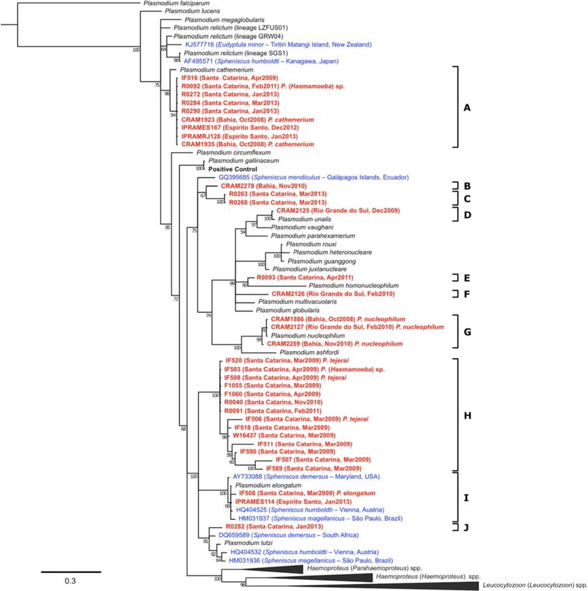
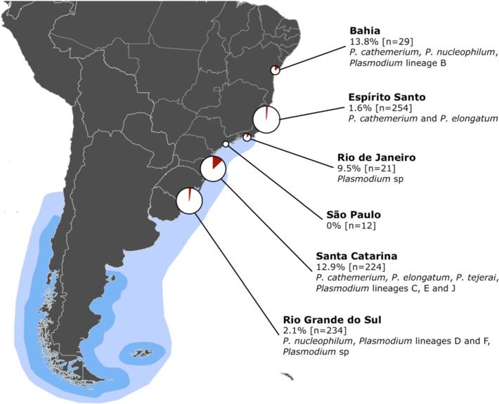
Vanstreels et al. Veterinary Research (2015) 46:30                                                                                         Page 2 of 12

Materials and methods                                                         individuals and over an extended time period (“system-
Study locations and data collection                                           atic sampling”). Other samples were collected from
We studied MPs received for rehabilitation at five orga-                      other facilities over a short period and/or from penguins
nizations along the coast of Brazil (Figure 1): CRAM-                         showing clinical or necropy findings suggestive of infec-
FURG (Rio Grande, Rio Grande do Sul - 32°01’34”S 52°                          tious disease (“opportunistic sampling”).
06’21”W), CETAS Florianópolis (Florianópolis, Santa                             Samples and biological information collected within 0–3
Catarina - 27°35’51”S 48°26’20”W), FUNDAMAR (São                              days from intake to the facility were considered “intake”,
Sebastião, São Paulo - 23°49’21”S 45°24’53”W), CETAS                          whereas those collected 0–7 days before death or release
Unimonte (São Vicente, São Paulo - 23°56’50”S 46°                             were considered “exit”. Each individual was categorised in
23’39”W), and IPRAM (Cariacica, Espírito Santo - 20°                          relation to their “age group” on intake (juvenile, adult),
19’54”S 40°21’38”W). Each organization receives pen-                          “oiling” on intake (oiled, not oiled), “survival” during re-
guins rescued along the coastline of their state and                          habilitation (survivor, deceased) and “diagnosis” (positive,
neighboring states; additionally, CRAM-FURG also                              negative). Rehabilitation records were used to determine
receives then releases penguins that were rehabilitated                       “warm season period”, i.e. the number of days spent in the
at other states (Bahia, Espírito Santo, Rio de Janeiro).                      facility (from intake to death or release) that occurred
Considering this dynamic, each penguin was assigned a                         within the period 01 October to 31 March.
“location” (Bahia, Espírito Santo, Rio de Janeiro, São
Paulo, Santa Catarina, Rio Grande do Sul) based on the                        Study design
facility in which they were subjected to rehabilitation                       Screening for Plasmodium combined one or more of the
(not rescue nor release location).                                            following diagnostic methods: thin blood smears (TBS),
  Samples were collected in different time periods at                         nested polymerase chain reaction (PCR) and histopathology
each location (Table 1). Samples were collected system-                       (HP).
atically from January 2009 to December 2012 at Rio                              Plasmodium screening was divided in two stages. The
Grande do Sul and from March 2009 to February 2013                            following criteria were adopted in the first stage: (a) all TBS
at Santa Catarina; in these periods, MPs were evenly                          collected at all study sites were examined; (b) for “survivor”
sampled without bias towards clinically ill or healthy                        penguins, the “exit” blood sample was tested with PCR; (c)

 Figure 1 Geographic distribution of the sampling effort, detection and lineages of Plasmodium spp. Pie charts represent sampling effort
 (size) and percentage of positive results (red fraction). Blue areas represent the wintering (light blue) and breeding (darker blue) distribution of
 Magellanic penguins [9].
Vanstreels et al. Veterinary Research (2015) 46:30                                                                                               Page 3 of 12

Table 1 Sample sizes examined using different diagnostic tests to screen for Plasmodium sp infections
Latitude       Study site (time period)                       Blood smears + PCR           PCR           Blood smears          Histopathology         Total
13°00’S        Bahia (1999–2008)‡                             7 (1)                        -             7 (1)                 -                      8 (2)
13°00’S        Bahia (Jun2009-Dec2012)‡                       21 (2)                       -             -                     -                      21 (2)
20°20’S        Espírito Santo (1999–2008)‡                    2                            -             17 (1)                -                      19 (1)
20°20’S        Espírito Santo (Sep2012)                       86                           -             111                   -                      197
20°20’S        Espírito Santo (Sep2012-Feb2013)               18 (2)                       20 (1)        -                     -                      38 (3)
22°50’S        Rio de Janeiro (1999–2008)‡                    6                            1 (1)         5                     1 (1)                  13 (2)
22°50’S        Rio de Janeiro (Jan2009-Dec2012)‡              2                            -             6                     -                      8
23°58’S        São Paulo (Aug2010-Sep2010)                    1                            11            -                     -                      12
27°36’S        Santa Catarina (Mar2009-Feb2013)†              106 (19)                     81 (8)        37 (2)                -                      224 (29)
32°02’S        Rio Grande do Sul (1999–2008)                  -                            -             11                    2 (2)                  13 (2)
32°02’S        Rio Grande do Sul (Jan2009-Dec2012)†           192 (3)                      8             21                    -                      221 (3)
Total          Systematic sample collection                   298 (22)                     89 (8)        58 (2)                -                      445 (32)
               Opportunistic sample collection                143 (5)                      32 (2)        151 (2)               3 (3)                  329 (12)
               Grand total                                    441 (27)                     121 (10)      209 (4)               3 (3)                  774 (44)
Values within parenthesis indicate the number of positive samples. “†” indicates that sample collection was systematic, i.e. was not conducted in a manner that
could favor sick or healthy individuals. “‡” indicates samples collected upon admission to CRAM-FURG from penguins that had been rehabilitated at
other facilities.

for “deceased” penguins from which frozen tissue samples                          internal enclosures in which rehabilitation is conducted
had been collected at necropsy, these tissues were tested                         were not entirely protected against mosquitoes and were
with PCR; (d) for “deceased” penguins from which frozen                           within 500 metres from bodies of freshwater and/or frag-
tissue sampled were not collected at necropsy, the last                           ments of Atlantic forest. Penguins were physically re-
blood sample collected before death was tested with PCR;                          strained and blood samples were collected from the jugular
(e) for “deceased” penguins from which neither blood nor                          or metatarsal veins. Body mass was determined with a scale
frozen tissue samples were available, tissue samples in                           with ± 5 g precision; when these data were collected upon
formalin were examined by HP. At this stage, blood smear                          intake, it was referred to as “intake mass”. Thin blood
examination was blind to PCR results and vice-versa.                              smears and heparin capillaries were prepared immediately
   The second stage of Plasmodium screening used the                              after blood collection; the remaining blood was stored in
criteria: (a) if a penguin obtained a positive or inconclusive                    tubes with heparin or without anticoagulants, then frozen. In
result for one or more samples in the first step, all samples                     some cases, hematocrit was determined through centrifuga-
available from that individual were tested with PCR; (b) if                       tion in heparin capillaries at 16 000 g for 5 min; total plasma
a penguin obtained a positive result, all samples from                            protein was determined with a clinical refractometer.
other individuals that had been at the same facility at the                          Blood smears were dried at room temperature, fixed
same date or three weeks before or following to the date                          with absolute methanol, stained with Giemsa or Wright-
of collection of the positive sample were tested with PCR.                        Rosenfeld stain, and examined under 1000× magnification
Because distinct sets of samples were available for each                          (field of view area = 0.126 mm2). A minimum 150 fields
individual, different combinations of diagnostic tests were                       (~30 000 erythrocytes) were examined during the
used to detect Plasmodium among study sites (Table 1).                            first stage of screening and an additional 250 fields
   Finally, individuals with positive results in the previous                     (~50 000 erythrocytes) were examined during the
steps were further tested: (a) positive samples were sub-                         second stage. Blood parasites were morphologically
jected to sequencing of the cyt-b gene; (b) histopathology of                     characterized [16] and quantified with the assistance
all available tissue samples were evaluated to determine                          of digital image analysis to count 10 000 erythrocytes
microscopic lesions and determine the occurrence of                               [17]; parasite forms were differentiated into four cat-
exoerythrocytic meronts; (c) all available blood smears were                      egories (trophozoite, meront, microgametocyte and
used to characterize parasite morphology.                                         macrogametocyte).
                                                                                     Whenever possible, penguins that died during rehabili-
Sample collection, hematology and pathology                                       tation were refrigerated and examined within 12 to 24 h
MPs at the study facilities are subjected to standardized re-                     after death; when this was not possible, carcasses were
habilitation protocols under the supervision of veterinarians                     frozen for later examination. Gross lesions were photo-
[15]. At all facilities involved in the study, external and                       graphed and noted, and samples of organs and tissues
Vanstreels et al. Veterinary Research (2015) 46:30 Page 4 of 12
were fixed in 10% buffered formalin. Formalin-fixed total number of individuals infected by a given Plasmo-
tissues were embedded in paraffin and sections of 3 or dium lineage.
5 μm were obtained, stained with hematoxylin-eosin and Chi-Square test was used to compare diagnosis (dependent
examined under light microscopy. variable – DV) among laboratory methods (independent
variable – IV) (TBS + PCR, PCR, TBS; histopathology was
Molecular biology and phylogenetic analysis not included due to small sample size).
Frozen samples of blood and tissues (lung, spleen or All subsequent analyses in this subsection were re-
liver) were used for molecular analyses. DNA extraction stricted to data obtained from systematically sampled
was conducted using the DNEasy Blood and Tissue Kit and PCR-tested individuals. True prevalence was esti-
(#69506, Qiagen) and was verified and quantified through mated from apparent prevalence using Blaker’s 95%
UV spectrophotometry (Nanodrop 2000, Thermo Fisher confidence interval [24] assuming a 80% sensitivity
Scientific). We used a nested polymerase chain reaction [25,26] and 100% specificity.
targeting the mitochondrial cytochrome b (cyt-b) gene of Mann–Whitney tests were used to determine if warm
Haemoproteus and Plasmodium [18] with 3 ng/μL of season period or intake masses (DV) were different be-
sample DNA, 0.6 μM of each primer, and GoTaq Green tween categories of location and oiling (IV). Linear re-
Master Mix 2x (M7122, Promega). Blood samples from gression was used to determine if there was correlation
chicken experimentally infected with Plasmodium between warm season period and intake masses. Fisher’s
gallinaceum and samples from chickens raised in exact test was used to compare survival (DV) between
arthropod-free environments were used as positive and categories of diagnosis (IV), either overall or within data
negative controls, respectively. Gel electrophoresis was subsets. All tests were two-tailed and used a significance
conducted to visualize amplification products, using level of 0.05.
2% agarose gel, SYBR Safe (S33102, Invitrogen), and a Binary logistic regression was employed to determine
high-resolution imaging system (Gel Doc EZ System which independent variables (IV) such as location, warm
170–8270, Bio-Rad). PCR amplification products of posi- season period, oiling and intake mass had a significant
tive samples were purified with Polyethylene Glycol 8000. effect in determining diagnosis (DV). The order of inclu-
Bi-directional sequencing with dye-terminator fluorescent sion of variables in the logistic model and best model
labeling was performed through automated sequencing selection was based on the P-value of the Slope-equal-
(ABI Prism 3100, Applied Biosystems); forward and to-zero test (only variables with P > 0.1 were included)
reversed chromatograms were edited and consensus and Pearson’s Goodness-of-fit test.
sequences were deposited in GenBank (Additional file 1). All procedures in this study were approved by the Ethics
Phylogenetic relationships among Plasmodium lineages Committee on Animal Use of the School of Veterinary
identified in this study and related hemosporidian parasites Medicine and Animal Science of the University of São
were inferred by using sequences from reference lineages Paulo (CEUA 601415) and were authorized by Brazilian
from the MalAvi database [19], for which species was authorities (SISBIO 20825-6).
identified based on studies using morphological evidence,
as well as penguin-infecting Plasmodium lineages from Results
published studies (Additional file 1). Sequences were Epidemiology of Plasmodium in penguins at rehabilitation
aligned using ClustalW [20] as implemented in MEGA centers
5.2.2 [21]. A Bayesian phylogenetic tree for the parasite Forty-four of the 774 MPs (5.68%) undergoing rehabili-
sequences was produced using MrBayes 3.2.2 [22] with the tation at facilities in six states along the coast of Brazil
GTR + I + G model of nucleotide evolution, as recom- were identified as positive to Plasmodium (Figure 1 and
mended by ModelTest [23]. We ran two Markov chains Table 1). Positive individuals were identified at all states
simultaneously for 5 million generations that were sampled except São Paulo, and no individuals were positive upon
every 1000 generations. The first 1250 trees (25%) were intake. In all positive cases, clinical history and diagnos-
discarded as a burn-in step and the remaining trees were tic results were consistent with the hypothesis that infec-
used to calculate the posterior probabilities. tion occurred during the stay at rehabilitation facilities.
Details on individual rehabilitation history and clinical
Statistical analysis parameters of Plasmodium-positive individuals are pro-
“Apparent prevalence” was defined as the number of vided in Additional file 2. A substantial proportion of
positive individuals divided by the number of individuals positive cases (39%: 17/44) were concentrated in a single
tested. “Survival ratio” was defined as the number of outbreak that occurred at Santa Catarina in March-April
“survivor” individuals divided by the total number of in- 2009. All avian malaria cases were first identified as
dividuals in a given data subset. “Lethality” was defined positive between the months October and April, inclu-
as the number of “deceased” individuals divided by the sive (Figure 2).Vanstreels et al. Veterinary Research (2015) 46:30 Page 5 of 12 Figure 2 Monthly distribution of Plasmodium infections in Magellanic penguins at rehabilitation facilities in Brazil. Line represents the incidence of Plasmodium infections (number of cases first recorded at each month; right vertical axis). Bars represent the susceptible population (number of penguins that spent one or more days at the rehabilitation facilities in a given month; left vertical axis). Data is combined for all facilities and years. Individuals tested with PCR or with a combination of Plasmodium-negative individuals, with 66.6% of the PCR and TBS were more frequently determined to be Plasmodium-positive MPs dying during rehabilitation positive (7.50% and 6.32%, respectively) than those whereas just 46.21% of the Plasmodium-negative died tested with TBS alone (1.79%) (P = 0.022). All samples (P = 0.037). identified as positive by TBS were positive in the corre- sponding PCR test, whereas only 45.5% (10/22) PCR- Plasmodium lineages infecting penguins positive samples were identified as positive in the Morphological characterization of parasites revealed the corresponding TBS. occurrence of at least four different morphospecies in 16 When only individuals sampled systematically and blood smear-positive individuals: P. (Novyella) nucleo- tested with PCR (whether in combination with TBS or philum, P. (Haemamoeba) cathemerium, P. (Haemamoeba) not) are considered, apparent prevalence was 7.75% tejerai and P. (Huffia) elongatum (photomicrographs pro- (30/387) (Table 2). Based on this result, true preva- vided in Additional file 3 and in references [8] and [14]). In lence was estimated between 6.6% and 13.5%. Logistic the blood smears of P. cathemerium, it should be noted that regression of this data subset (Log-likelihood = −43.029, in addition to the well-defined elongated rod-shaped P < 0.001; Pearson Goodness-of-fit test: χ2 = 145.68, pigment granules with pointed ends that are unique to df = 321, P > 0.99) revealed the following variables were this species, unusual morphological characteristics were significant determinants of positivity to Plasmodium: also noted: meronts were relatively small with scanty location (Santa Catarina in relation to Rio Grande do Sul: cytoplasm and young macrogametocytes frequently pre- Z = 1.75, P = 0.080, b = 1.437, bCI95% = −0.172 – 3.048, sented relatively large vacuoles surrounded by small OR = 4.21, ORCI95% = 0.84 – 21.06), warm season period round pigment granules. (Z = 4.97, P < 0.001, b = 0.030, bCI95% = = 0.018 – 0.042, Cyt-b sequences were obtained from 34 of the 36 PCR- OR = 1.03, ORCI95% = 1.02 – 1.04) and intake mass (Z = 2.47, positive individuals (see Additional file 1), and phylogen- P = 0.013, b = 0.0014, bCI95% = = 0.0003 – 0.0025, OR = 1.01, etic analysis revealed that these lineages can be classified ORCI95% = 1.01 – 1.01). Oiling did not have a significant in 10 distinct clusters (Figure 3). Four clusters were con- effect (not oiled in relation to oiled; Z = 0.59, P = 0.554, firmed as morphospecies based on parasite morphology in b = 0.47, bCI95% = = −1.10 – 2.05, OR = 1.61, ORCI95% = blood smears: P. cathemerium (cluster A), P. nucleophilum 0.33 – 7.75). (cluster G), P. tejerai (cluster H), and P. elongatum (cluster Strong association/correlation was present amongst I). Additionally, even though the morphology of Plasmo- variables: between location and oiling (P < 0.001), warm dium lineage D (obtained from penguin CRAM2125) season period (P < 0.001), and intake mass (P = 0.029); could not be observed in blood smears, the sequences between oiling and warm season period (P < 0.001) and from this lineage neatly clustered with a reference P. intake mass (P = 0.005); and between warm season period (Novyella) unalis lineage, with high probability (100) and and intake mass (P = 0.001, R2 = 0.039, b > 0). Survival was sequence identity (444/445 nucleotides = 99.76%). Five significantly different between Plasmodium-positive and phylogenetic lineages (B, C, E, F and J) did not cluster with
Vanstreels et al. Veterinary Research (2015) 46:30 Page 6 of 12
Table 2 Details of the diagnostic results in relation to sample collection and testing strategy, age group, oiling and
survival
Sampling and screening Age Oiling Died Survived Total
group
Positive Negative Positive Negative
Systematically sampled Juvenile Oiled 3 92 0 81 176
and PCR-tested individuals Not oiled 13 62 9 67 151
Adult Oiled 0 5 0 37 42
Not oiled 4 6 1 7 18
Opportunistically sampled Juvenile Oiled 1 2 0 9 12
and/or non-PCR-tested individuals Not oiled 7 129 5 213 354
Adult Oiled 1 5 0 11 17
Not oiled 0 0 0 4 4
Total 29 301 15 429 774
any reference lineages nor with lineages previously towards the cause of death could not be determined.
obtained from penguins. Diffuse interstitial pneumonia occurred in all exam-
ined cases (n = 22) and was most frequently granulo-
Pathology of avian malaria in penguins cytic (82%) (Figure 4C). Multifocal hepatitis occurred
Twenty-two Plasmodium-positive cases were examined in all examined cases (n = 20) as was most frequently
by histopathology, and exoerythrocytic meronts were mononuclear (70%) (Figure 4D); hepatic necrosis and
observed in 19 cases (86.4%). Meronts were present in ductal hyperplasia were present in 20% and 25% of
macrophages and endothelial cells (Figure 4A), and cases, respectively. Splenitis occurred in 75% of cases
occurred in a broad variety of tissues, especially in the (n = 20) and was most often granulocytic (67%); necrotiz-
heart, liver, lungs, spleen and kidneys. ing splenitis and/or lymphocytolysis was observed in 65%
P. tejerai was lethal to 75% of penguins infected (12/16), of cases (Figure 4E), and massive splenic hemorrhages
and tissue meronts were observed in all six P. tejerai cases were observed in two cases (10%). Hematopoiesis (60%)
examined by histopathology. P. cathemerium was identi- and hemosiderosis (90%) were frequently observed in the
fied in 9 MPs, three of which died (33% lethality); another spleen and/or liver (n = 20). Myocarditis was observed in
two were euthanized for other reasons. Only one of three 33.3% of cases (n = 21) and was most frequently granulo-
P. nucleophilum-infected MPs died, and necropsy revealed cytic (57%).
there were no tissue meronts but high numbers of
intraerythrocytic parasites were present within blood Discussion
vessels (Figure 4B); the concurrence of other significant Avian malaria has been widely recognised as one of the
pathological processes (severe splenic amyloidosis, hel- most significant infectious diseases for wild and cap-
minthes within lungs and liver parenchyma and intes- tive penguins [3,4,27]. Our findings demonstrate that
tinal blood vessels) did not allow for a conclusion as Plasmodium spp. infect Magellanic penguins at several
to whether or not avian malaria was the cause of death. rehabilitation centers along the Brazilian coast, leading
P. elongatum was identified in one dead MP, which did to substantial levels of mortality, and limiting the
present tissue meronts. P. unalis was identified in one success of rehabilitation efforts for this species at these
dead penguin, but no tissue meronts were observed and centers.
severe respiratory lesions indicated that aspergillosis
was the primary cause of death. Plasmodium sp lineages Epidemiology of avian malaria in penguins at
E and J were each identified in one individual, and tissue rehabilitation centers
meronts were present in each case. Plasmodium lineage While the overall apparent prevalence ranged from 2.1%
C was identified in two euthanized individuals; tissue to 13.8% among rehabilitation centers, when only sam-
meronts were present in both cases but concurred with ples collected and tested in a systematic manner with
other significant lesions (severe aspergillosis accom- highly sensitive diagnostic methods were considered, the
panied by necrotizing pancarditis; amyloidosis and hel- apparent prevalence was 7.8%. With 95% confidence,
minthes within air sacs). true prevalence was estimated between 6.6% and 13.5%.
Only one case of mixed infection resulted in death The only comparable data in the literature are provided by
(P. elongatum + P. tejerai), however histopathology was the Southern African Foundation for the Conservation of
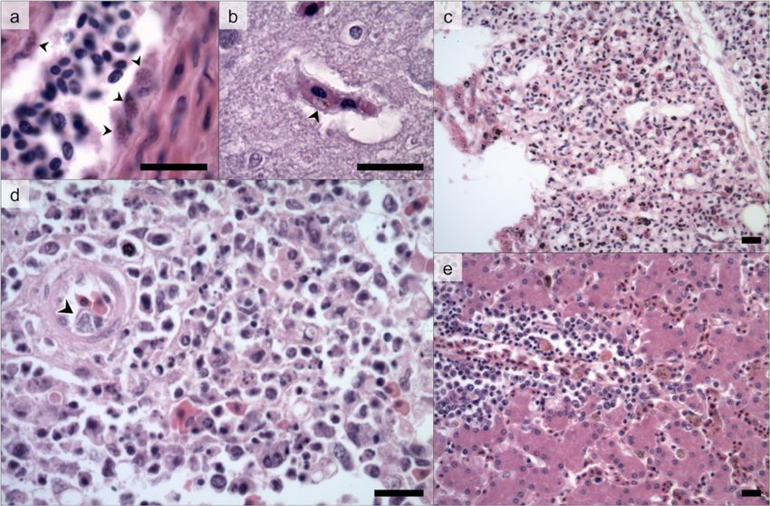
not conducted and therefore the role of avian malaria Coastal Birds (SANCCOB), an organization that rehabilitatesVanstreels et al. Veterinary Research (2015) 46:30 Page 7 of 12 Figure 3 Phylogenetic tree of the Plasmodium spp. lineages identified in penguins. (red) Magellanic penguins undergoing rehabilitation along the coast of Brazil (this study), (blue) published penguin-infecting lineages, (black) reference lineages. Branch lengths are drawn proportionally to the extent of changes (scale bar is shown). African penguins (Spheniscus demersus) in South Africa; 17- proportion of animals released of Plasmodium-positive 34% of the African penguins admitted by SANCCOB in individuals is not significantly different from that of the 2001–2002 were blood smear-positive to Plasmodium at overall rehabilitation population [27]. some point during their rehabilitation [27]. There is, however, a critical difference to be considered We found that 5% of MPs that survived through rehabili- when comparing our findings with those of SANCCOB: tation were Plasmodium-positive at some point, compared there are no records of Plasmodium infections in wild to 10.8% of the deceased MPs, with a 44% higher mortality MPs, whereas African penguins are infected in the wild in Plasmodium-positive penguins compared to Plasmo- [5]. Therefore, whilst we found no evidence to indicate dium-negative (66.6% vs. 46.2%). These results contrast with that the MPs we studied carried the infection from the the observed epidemiology at SANCCOB, where the wild, at SANCCOB 30-35% of the Plasmodium-positive
Vanstreels et al. Veterinary Research (2015) 46:30                                                                                                Page 8 of 12

 Figure 4 Histological findings associated with avian malaria in Magellanic penguins. (a) exoerythrocytic meronts in endothelial cells (arrowheads)
 within a liver arteriole (R0040, P. tejerai); (b) parasitized erythrocyte (arrowhead) within a cerebral blood vessel (CRAM2127, P. nucleophilum); (c) diffuse
 granulocytic interstitial pneumonia, congestion and edema (IF584, P. tejerai); (d) diffuse necrotizing splenitis with an exoerythrocytic meront
 within an endothelial cell of a central arteriole (arrowhead) (R0290, P. cathemerium); (e) multifocal perivascular mononuclear hepatitis, congestion and
 hemosiderosis (R0093, Plasmodium sp lineage E). Hematoxilin-Eosin. Scale bars = 15 μm.

African penguins are already positive upon intake [27].                           for release in the subsequent winter; a lower number of
In this context, the epidemiology of avian malaria in                             individuals are also admitted for rehabilitation during
Magellanic penguins in rehabilitation centers may re-                             summer.
semble more that of captive penguins in the Northern                                Surprisingly, body mass upon intake was found to be a
Hemisphere than that of their African counterparts.                               positively-correlated predictor of Plasmodium infection,
  In the Northern Hemisphere, it is well established that                         indicating that MPs admitted with higher body mass
the occurrence of avian malaria in penguins is strongly                           have a higher probability of becoming infected with
modulated by seasonality, with cases concentrated in the                          Plasmodium. A possible explanation is that MPs admit-
Boreal spring-summer due to climate-mediated fluctua-                             ted during summer months are often admitted due to
tions in mosquito abundance [10,28,29]. We observed                               moult problems and as a result may have relatively
a consistent concentration of all Plasmodium-positive                             higher body mass (RETV, personal observations).
cases in the Austral spring-summer (October to April),                              The rehabilitation facility studied in Santa Catarina
with the probability of infection being positively corre-                         had a substantially higher Plasmodium incidence than the
lated with the total number of days spent in rehabilita-                          other facilities involved in this study. The facility is located
tion during that time of the year. In contrast, most MPs                          within a State Park (Parque Estadual do Rio Vermelho)
are received for rehabilitation along the Brazilian coast                         with penguin enclosures directly under tree cover of
during winter months (June to September) (Figure 2),                              Atlantic forest and less than 10 metres away from a
which relates to the species’ wintering ecology [9]. Current                      large freshwater lake (Lagoa da Conceição). This pro-
rehabilitation protocols require the release of MPs is to be                      vides an ideal environment for mosquito proliferation
avoided during the summer months in recognition of the                            and close proximity to an abundant and diverse avi-
species’ migration [15]. As a result, individuals that were                       fauna. Additionally, this facility rehabilitates not only
unable to meet the release criteria until spring are retained                     marine animals but also terrestrial wildlife, including
Vanstreels et al. Veterinary Research (2015) 46:30                                                                   Page 9 of 12

birds apprehended from illegal trade. The higher ap-            consistently high sequence identity and phylogenetic prox-
parent prevalence of MPs at this facility may reflect           imity with a reference lineage of P. cathemerium, along
not a regional difference in susceptibility or lineage          with a failure to retrieve sequences of another Plasmodium
virulence, but is more likely to be the consequence of          lineage in the same samples, we believe there is sufficient
an increased frequency of inoculation due to close              evidence to identify these lineages as P. cathemerium and
contact with mosquitoes and other birds acting as               that hypothesis (a) or (b) are most likely.
proximal reservoirs of infection.
                                                                Pathogenicity of Plasmodium to penguins
Plasmodium lineages infecting penguins and their                Even though penguins seem to be susceptible to infection
pathology                                                       by a variety of Plasmodium lineages occurring regionally,
It has been generally accepted that P. relictum and P.          we cannot say that all lineages have similar epidemiology or
elongatum are the two most significant species of Plas-         pathogenicity. In this study we found that P. tejerai and
modium responsible for avian malaria in penguins                P. cathemerium were lethal (75% and 33% of MPs in which
[3,5,6], in addition to an isolated record of P. juxtanu-       they were detected, respectively) whilst other lineages had
cleare [7]. In recent studies [8,14] we have documented         only a few cases recorded and/or could not be demon-
the role of P. tejerai in causing an avian malaria out-         strated as the leading factor causing death. These findings,
break in MPs at Santa Catarina.                                 combined with previous reports that albeit less frequent
   Our results demonstrate that a broad variety of Plasmo-      P. relictum tends to produce more severe disease than
dium species can be found in penguins undergoing                P. elongatum in penguins [29,31], raises the question on
rehabilitation in South America, including three species        whether the Plasmodium subgenus Haemamoeba is more
that had not yet been demonstrated to infect penguins           pathogenic to these birds than other subgenera of Plasmo-
(P. cathemerium, P. nucleophilum and P. unalis) and 5           dium. Comparative pathology through experimental inocu-
lineages that albeit unidentified clearly correspond to         lation in domestic birds may assist in clarifying whether
Plasmodium species that also have not yet been reported         pathogenicity is intrinsically higher for these lineages or if it
in penguins (lineages B, C, E, F and J). Such diversity of      reflects a susceptibility bias present in penguins.
plasmodia corroborates the interpretation that the infection      Overall, however, the histopathological lesions observed
of captive penguins results from local mosquitoes inocula-      in this study were generally consistent among lineages.
ting penguins with Plasmodium spp. acquired from other          The most prominent pathological processes were granulo-
birds surrounding of the penguin exhibits [12,29-31].           cytic pneumonia and splenitis and mononuclear hepatitis;
   It is worth noting that P. cathemerium, P. elongatum,        these were probably the effect of vasculitis associated with
P. nucleophilum and P. relictum are renowned as gener-          the proliferation of Plasmodium within endothelial cells of
alist parasites with low host-specificity, infecting a broad    these tissues. In most cases, death likely culminated as a
range of avian species in several taxonomic orders [32].        result of respiratory insufficiency from the marked pneu-
In this sense, our findings suggest that the predomin-          monia, congestion and edema. These lesions are not
ance of P. relictum in Europe and Asia and P. relictum          unlike those reported in P. relictum and P. elongatum
and P. elongatum in North America might not necessar-           infections in penguins in zoos and aquaria in the Northern
ily indicate a particular susceptibility of penguins to         Hemisphere [10,11,33].
those parasites, but perhaps reflects their natural abun-         In contrast, no exoerythrocytic meronts were observed
dance in those regions.                                         in the tissues of the only MP that died with a P. nucleo-
   Although we observed mature P. cathemerium microga-          philum infection, whereas a high number of intraery-
metocytes with well-defined elongated rod-shaped pigment        throcytic parasites was observed within blood vessels.
granules with pointed ends, which are the defining features     Similarly, the only individual infected with P. unalis did
of P. (Haemamoeba) cathemerium [16], in all cases we also       not present detectable exoerythrocytic meronts. This
observed late trophozoites and young macrogametocytes           distinct pattern may indicate a different stage of infec-
with relatively large vacuoles surrounded by small round        tion or a distinct pathogenesis. Future studies will be wel-
pigment granules and relatively small meronts with scanty       come to clarify whether this is a consistent pattern in
cytoplasm (Additional file 3), which are uncharacteristic to    infections by these lineages in penguins and which patho-
P. cathemerium. These findings may be interpreted as: (a) a     physiological mechanisms are involved.
host-specific morphological variation of P. cathemerium,
(b) a variant or subspecies of P. cathemerium, (c) co-          Concurrent diseases
infection with a secondary unidentified lineage, or (d) a       Two of the Plasmodium-positive MPs identified in this
novel and yet undescribed Plasmodium species whose mor-         study had also been identified as positive to Avipoxvirus in
phological characteristics overlap with those of P. cathemer-   a previous study [34]. This is probably not uncommon as
ium. Considering the molecular evidence indicating a            both pathogens are mosquito-borne, however it may
Vanstreels et al. Veterinary Research (2015) 46:30 Page 10 of 12
confuse interpretation of pathological findings. In particu- These individuals were clinically healthy and passed the
lar, the only two individuals studied by Niemeyer et al. standard release criteria [15] and were blood smear-
[34] that presented necrotizing splenitis were found to be negative or, in a few cases, blood samples were collected
Plasmodium-positive in this study. This might indicate but not examined in time before release. In the cases
that such severe lesions were related to avian malaria and where blood smears were negative, it must be considered
not poxvirosis, and that Avipoxvirus might not have been that even non-parasitemic penguins can relapse if
as pathogenic as originally thought. treated with corticosteroids – and presumably the same
Other concurrent diseases included aspergillosis, gastro- would occur if they became stressed – due to the per-
intestinal helminthiasis, spleen amyloidosis, cholestasis, sistence of exoerythrocytic meronts [30]. In the cases
unidentified myocardium cysts, and helminthes in the where samples could not be tested before release, this
lungs, liver, air sacs and skin (Additional file 3). Some of exposes a potential dillema that is common in oil spill
these findings, such as aspergillosis and gastrointestinal responses, where it is not always feasible to test the large
helminthiasis have been previously reported in penguins numbers of individuals in a brief period [36,37].
with avian malaria [10,35]. However, there have been no Brossy et al. [38] expressed concern on the potential of
reports of helminthes in the respiratory system, skin or air rehabilitation centers releasing African penguins with
sacs of MPs [3]. Furthermore, the myocardium cysts blood parasites, and perhaps this concern should be even
herein observed clearly were not Plasmodium and could greater for MPs considering that Plasmodium has yet to
correspond to either protozoan or metazoan parasites. be recorded in this species in the wild. In the case of MPs,
Additional studies will be conducted to clarify the identity however, because climate and environmental conditions
and significance of these parasites. are generally adverse and mosquitoes occur very scarcely
in the southeastern coast of Argentina [39,40] and are
Implications for rehabilitation and conservation absent at the Malvinas-Falkland Islands [41], the probabi-
The prevention of avian malaria in penguins in Northern lity of Plasmodium transmission from a rehabilitated
Hemisphere zoos has largely relied on the oral adminis- penguin to a wild penguin in these regions is very low.
tration of primaquine during summer [15]. In Brazil, Even so, it is important to emphasize that pathogen spill-
primaquine commerce is restricted by the government over to wild populations should remain a prime and
due to concerns of potential resistance in human mal- critical concern for rehabilitation centers, and that even a
aria, and therefore this drug cannot be acquired or used low non-zero probability is nonetheless a significant risk
by rehabilitation centers. As a result, the centers are to be considered and addressed.
forced to employ other prevention strategies, namely the Rehabilitation facilities on the Pacific coast of South
physical isolation of penguins from mosquitoes, which is America may be in a different situation. There are reports
often challenging and costly. of avian malaria in MPs undergoing rehabilitation in Chile
Our study sheds light on a positive aspect of the epidemi- [13], and even though no studies have detected blood para-
ology of this disease at rehabilitation centers, namely that sites in penguins in Chile [5,6], both ecological models [40]
the periods in which Plasmodium infections occur (sum- and blood parasite studies in other avian species [42] con-
mer) is directly opposite to the period in which there are sistently indicate that mosquitoes are abundant on the
the most penguins in rehabilitation (winter). Consequently, Southwestern coast of South America. Studies examining
one of the key strategies for the prevention of avian malaria the occurrence of blood parasites in the Chilean popula-
in these facilities might be to develop rehabilitation proto- tions of MPs are therefore urgently required, and rehabilita-
cols that allow the shortening of the time needed by the tion facilities in the region should remain cautious of
penguins to achieve the fit-to-release criteria, so that they potentially releasing Plasmodium-positive individuals back
can be released before the summer. As a result, these into the wild.
facilities would benefit from narrowing their malaria-
prevention efforts to a relatively lower number of indivi-
Additional files
duals (those received and/or maintained between October
and April), becoming more effective in the prevention and Additional file 1: Public database ascension numbers. Genbank and
early diagnosis. This is a relevant implication not only for MalAvi ascension numbers for the sequences obtained or included in the
permanent rehabilitation efforts, but also for oil spill analyses. Taxonomic names within brackets indicate the taxon to which
the species is presumed to correspond on the basis of phylogenetic
responses involving penguins, when the physical and hu- analyses of the cytochrome b mitochondrial gene.
man resources required for malaria prevention, diagnosis Additional file 2: Individual details of Plasmodium-positive Magellanic
and treatment may be substantial [35] and potentially penguins. Spreadsheet with rehabilitation history and clinical parameters of
beyond the capacity of those involved. Plasmodium-positive Magellanic penguins identified in this study.
A number of MPs herein examined were released des- Additional file 3: Plasmodium spp. in Giemsa-stained blood smears
of Magellanic penguins. Photomicrographs: P. nucleophilum (CRAM2127):
pite having been Plasmodium-positive at some point.Vanstreels et al. Veterinary Research (2015) 46:30 Page 11 of 12
(a,b) trophozoites, (c,d) meronts, (e) coinfection by erythrocytic meront Received: 7 October 2014 Accepted: 4 February 2015
and microgametocyte, (f) macrogametocyte, (g) microgametocyte, (h)
co-infection by macro and microgametocyte; P. cathemerium (CRAM1923): (i)
trophozoite, (j,k) meronts, (l-n) macrogametocytes, (o,p) microgametocytes.
Scale bar = 5 μm. References
1. Martinsen ES, Perkins SL (2013) The diversity of Plasmodium and other
Haemosporidians: The interesection of taxonomy, phylogenetics and
Abbreviations genomics. In: Carlton JM, Perkins SL, Deitsch KW (ed) Malaria parasites:
DV: Dependent variable; HP: Histopathology; IV: Independent variable; comparative genomics, evolution and molecular biology. Caister Academic
MPs: Magellanic penguins; PCR: nested polymerase chain reaction; Press, Norfolk, pp 1–15
SANCCOB: Southern African Foundation for the Conservation of Coastal 2. Levin II, Parker PG (2011) Hemosporidian parasites: impacts on avian hosts.
Birds; TBS: Thin blood smears. In: Miller E, Fowler M (ed) Fowler’s zoo and wild animals medicine. Elsevier
Saunders, Missouri, pp 356–363
3. Clarke JR, Kerry KR (1993) Diseases and parasites of penguins. Kor J Polar Res
Competing interests
4:79–96
The authors declare that they have no competing interests.
4. Jones HI, Shellam GR (1999) Blood parasites in penguins, and their potential
impact on conservation. Mar Ornithol 27:181–184
Authors’ contributions 5. Jones HI, Shellam GR (1999) The occurrence of blood-inhabiting protozoa in
RETV collected and prepared samples, performed hematological exams, captive and free-living penguins. Polar Biol 21:5–10
necropsies, molecular testing, histopathological analyses, and drafted the 6. Quillfeldt P, Martínez J, Hennicke J, Ludynia K, Gladbach A, Masello JF,
manuscript. RPSF, CKMK and RCCB collected and prepared samples, performed Riou S, Merino S (2010) Hemosporidian blood parasites in seabirds: a
hematological exams and necropsies, and helped to draft the manuscript. VR comparative genetic study from Antartic to tropical habitats.
and MA assisted the epidemiological and statistical analyses and helped to Naturwissenschaften 97:809–817
draft the manuscript. SE, FCFJ and EMB assisted the molecular testing, gene 7. Grim KC, Van der Merwe E, Sullivan M, Parsons N, McCutchan TF, Cranfield
sequencing, morphological and phylogenetic analyses, and helped to draft M (2003) Plasmodium juxtanucleare associated with mortality in black-footed
the manuscript. JLCD provided general supervision, participated in the penguins (Spheniscus demersus) admitted to a rehabilitation center. J Zoo
study design and coordination and helped to draft the manuscript. All Wildl Med 34:250–255
authors read and approved the final manuscript. 8. Vanstreels RET, Kolesnikovas CKM, Sandri S, Silveira P, Belo NO, Ferreira
Junior FC, Epiphanio S, Steindel M, Braga ÉM, Catão-Dias JL (2014) Outbreak
Acknowledgments of avian malaria associated to multiple species of Plasmodium in Magellanic
We wish to thank our friends and colleagues at Laboratório de Patologia penguins undergoing rehabilitation in Southern Brazil. PLoS One 9:e94994
Comparada de Animais Selvagens, Centro de Recuperação de Animais 9. Williams TD, Boersma PD (1995) Magellanic penguin (Spheniscus
Marinhos da Universidade Federal do Rio Grande, Associação R3 Animal, magellanicus). In: Williams TD (ed) The penguins: Spheniscidae. Oxford
Centro de Triagem de Animais Silvestres de Florianópolis, Instituto de University Press, Oxford, pp 249–258
Pesquisa e Reabilitação de Animais Marinhos, Fundação Museu de História 10. Fix AS, Waterhouse C, Greiner EC, Stoskopf MK (1988) Plasmodium relictum
Pesquisa e Arqueologia do Mar, Centro de Triagem de Animais Selvagens as a cause of avian malaria in wild-caught Magellanic penguins (Spheniscus
Lello-Unimonte, Instituto de Ciências Biomédicas da Universidade de São magellanicus). J Wildl Dis 24:610–619
Paulo, and Instituto de Ciências Biológicas da Universidade Federal de Minas 11. Ko KN, Kang SC, Jung JY, Bae JH, Kim JH (2008) Avian malaria associated
Gerais for their partnership and contributions to this study. We are grateful with Plasmodium spp. infection in a penguin in Jeju Island. Kor J Vet Res
to Ângela Cabana, Eric Woehler, Fernanda Colabuono, Melissa Xavier, Monique 48:197–201
van Rensburg, Nola Parsons, Renata Hurtado, Ricardo Dias and Ricardo Pereira 12. Bueno MG, Lopez RPG, Menezes RMT, Costa-Nascimento MJ, Lima GFMC,
for their valuable contributions. To Margareth Capurro-Guimarães for providing Araújo RAS, Guida FJV, Kirchgatter K (2010) Identification of Plasmodium
us the control samples. This study was supported by the São Paulo Research relictum causing mortality in penguins (Spheniscus magellanicus) from São
Foundation (FAPESP 2009/53956-9, 2010/51801-5), Minas Gerais Research Paulo Zoo, Brazil. Vet Parasitol 173:123–127
Foundation (FAPEMIG), Brazilian Federal Agency for the Support and Evaluation 13. Carvajal ER, Alvarado PM (2009) Pesquisa de Plasmodium spp. en pingüinos
of Graduate Education (CAPES), National Counsel of Technological and Scientific de Magallanes (Spheniscus magellanicus) de la Región de los Ríos: Malaria
Development (CNPq) and National Institute of Science and Technology, Genetic aviar como nueva patología de interés en la avifauna local. Boletín
and Health Information of the Brazilian Livestock (INCT-IGSPB). Veterinario Oficial 10:1–4
14. Silveira P, Belo NO, Lacorte GA, Kolesnikovas CKM, Vanstreels RET, Steindel
Author details M, Catão-Dias JL, Valkiūnas G, Braga ÉM (2013) Parasitological and new
1 molecular-phylogenetic characterization of the malaria parasite Plasmodium
Departamento de Patologia, Faculdade de Medicina Veterinária e Zootecnia,
Laboratório de Patologia Comparada de Animais Selvagens, Universidade tejerai in South American penguins. Parasitol Int 62:165–171
de São Paulo, Avenida Orlando Marques de Paiva 87, São Paulo, SP, 05088–000, 15. Silva-Filho RP, Ruoppolo V (2007) Sphenisciformes. In: Cubas ZS, Silva JCR,
Brazil. 2Centro de Recuperação de Animais Marinhos, Museu Oceanográfico Catão-Dias JL (ed) Tratado de animais selvagens – medicina veterinária.
Professor Eliézer de Carvalho Rios, Universidade Federal do Rio Grande, Rua Roca, São Paulo, pp 309–323
Capitão Heitor Perdigão 10, Rio Grande, RS 92200-580, Brazil. 3Associação R3 16. Valkiūnas G (2004) Avian malaria parasites and other haemosporidia. CRC
Animal. Rodovia João Gualberto Soares, Entrada do Parque Estadual do Rio Press, Boca Ratón
Vermelho, Barra da Lagoa, Florianópolis, SC 88061-500, Brazil. 4Instituto de 17. Gering E, Atkinson CT (2004) A rapid method for counting nucleated
Pesquisa e Reabilitação de Animais Marinhos. Rodovia BR 262, Instituto Estadual erythrocytes on stained blood smears by digital image analysis. J Parasitol
de Meio Ambiente e Recursos Hídricos, Jardim América, Cariacica, ES 29140-130, 90:879–881
Brazil. 5International Fund for Animal Welfare, 290 Summer Street, Yarmouth 18. Hellgren O, Waldenström J, Bensch S (2004) A new PCR assay for
Port, MA 02675, USA. 6Departamento de Análises Clínicas e Toxicológicas, simultaneous studies of Leucocytozoon, Plasmodium, and Haemoproteus
Faculdade de Ciências Farmacêuticas, Universidade de São Paulo, Avenida from avian blood. J Parasitol 90:797–802
Professor Lineu Prestes 580, Butantã, São Paulo, SP 05508-000, Brazil. 19. Bensch S, Hellgren O, Pérez-Tris J (2009) MalAvi: a public database of
7
Departamento de Medicina Veterinária Preventiva e Saúde Animal, Laboratório malaria parasites and related haemosporidians in avian hosts based on
de Epidemiologia e Bioestatística, Faculdade de Medicina Veterinária e mitochondrial cytochrome b lineages. Mol Ecol Resour 9:1353–1358
Zootecnia, Universidade de São Paulo, Avenida Professor Orlando Marques de 20. Thompson JD, Gibson TJ, Plewniak F, Jeanmougin F, Higgins DG (1997) The
Paiva 87, São Paulo, SP 05088-000, Brazil. 8Departamento de Parasitologia, CLUSTAL_X windows interface: flexible strategies for multiple sequence
Instituto de Ciências Biológicas, Universidade Federal de Minas Gerais. Caixa alignment aided by quality analysis tools. Nucleic Acids Res 25:4876–4882
Postal 486, Avenida Antônio Carlos 6627, Pampulha, Belo Horizonte, MG 21. Tamura K, Peterson D, Peterson N, Stecher G, Nei M, Kumar S (2011) MEGA5:
31270-901, Brazil. molecular evolutionary genetics analysis using maximum likelihood,Vanstreels et al. Veterinary Research (2015) 46:30 Page 12 of 12
evolutionary distance, and maximum parsimony methods. Mol Biol Evol
28:2731–2739
22. Ronquist F, Huelsenbeck JP (2003) MrBayes 3: Bayesian phylogenetic
inference under mixed models. Bioinformatics 19:1572–1574
23. Posada D, Crandall KA (1998) MODELTEST: testing the model of DNA
substitution. Bioinformatics 14:817–818
24. Reiczigel J, Földi J, Ózsvári L (2010) Exact confidence limits for prevalence of
a disease with an imperfect diagnostic test. Epidemiol Infect 138:1674–1678
25. Jarvi SI, Schultz JJ, Atkinson CT (2002) PCR diagnostics underestimate the
prevalence of avian malaria (Plasmodium relictum) in experimentally-infected
passerines. J Parasitol 88:153–158
26. Valkiūnas G, Zehtindjiev P, Dimitrov D, Križanauskiene A, Iezhova TA, Bensch
S (2008) Polymerase chain reaction-based identification of Plasmodium
(Huffia) elongatum, with remarks on species identity of Haemosporidian
lineages deposited in GenBank. Parasitol Res 102:1185–1193
27. Parsons NJ, Underhill LG (2005) Oiled and injured African penguins
Spheniscus demersus and other seabirds admitted for rehabilitation in the
Western Cape, South Africa, 2001 and 2002. Afr J Mar Sci 27:289–296
28. Sladen WJL, Gailey-Phipps JJ, Divers BJ (1979) Medical problems and
treatment of penguins at the Baltimore Zoo. Int Zoo Yearbook 19:202–209
29. Beier JC, Stoskopf MK (1980) The epidemiology of avian malaria in
black-footed penguins (Spheniscus demersus). J Zoo Anim Med 11:99–105
30. Cranfield MR, Graczyk TK, Beall FB, Ialeggio DM, Shaw ML, Skjoldager ML
(1994) Subclinical avian malaria infections in African black-footed penguins
(Spheniscus demersus) and induction of parasite recrudescence. J Wildl Dis
30:372–376
31. Graczyk TK, Cranfield MR, McCutchan TF, Bicknese EJ (1994) Characteristics
of naturally acquired avian malaria infections in naive juvenile African
black-footed penguins (Spheniscus demersus). Parasitol Res 80:634–637
32. Bennett GF, Bishop MA, Peirce MA (1993) Checklist of the avian species of
Plasmodium Marchiafava & Celli, 1885 (Apicomplexa) and their distribution
by avian family and Wallacean life zones. Syst Parasitol 26:171–179
33. Fleischman RW, Squire RA, Sladen WJL, Melby EC, Jr (1968) Malaria
(Plasmodium elongatum) in captive African penguins (Spheniscus demersus).
J Am Vet Med Assoc 153:928–935
34. Niemeyer C, Favero CM, Kolesnikovas CKM, Bhering RCC, Brandão P,
Catão-Dias JL (2013) Two different avipoxviruses associated with pox
disease in Magellanic penguins (Spheniscus magellanicus) along the Brazilian
coast. Avian Pathol 42:546–551
35. Griner LA, Sheridan BW (1967) Malaria (Plasmodium relictum) in penguins at
the San Diego Zoo. Vet Clin Pathol 1:7–17
36. Wolfaardt AC, Underhill LG, Altwegg R, Visagie J, Williams AJ (2008) Impact
of the Treasure oil spill on African penguins Spheniscus demersus at Dassen
Island: case study of a rescue operation. Afr J Mar Sci 30:405–419
37. Holcomb J, Callahan B: Management of the Treasure Oiled Wildlife
Response Cape Town, South Africa, 2000. In Proc 7th Int Effects Oil Wildl
Conf: 14–16 October 2003; Hamburg, Germany: International Bird Rescue
and Research Center and International Fund for Animal Welfare
38. Brossy JJ, Plös AL, Blackbeard JM, Kline A (1999) Diseases acquired by
captive penguins: what happens when they are released into the wild?
Mar Ornithol 27:185–186
39. Mitchell CJ, Darsie RF (1985) Mosquitoes of Argentina: Part II. Geographic
distribution and bibliography (Diptera, Culicidae). Mosq Syst 17:279–360
40. Walter Reed Biosystematics Unit: VectorMap: Know the vector, know the
threat. [http://www.vectormap.org/]
41. Medlock JM, Schaffner F, Fontenille D: Invasive mosquitoes in the European
associate continental and overseas territories. [http://www.ecdc.europa.eu/
en/activities/sciadvice/_layouts/forms/Review_DispForm.aspx?
ID=212&List=a3216f4c-f040-4f51-9f77-a96046dbfd72] Submit your next manuscript to BioMed Central
42. Merino S, Moreno J, Vásquez RA, Martínez J, Sánchez-Monsálvez I, Estades and take full advantage of:
CF, Ippi S, Sabat P, Rozzi R, McGehee S (2008) Haematozoa in forest birds
from southern Chile: Latitudinal gradients in prevalence and parasite • Convenient online submission
lineage richness. Austral Ecol
• Thorough peer review
33:329–340
• No space constraints or color figure charges
• Immediate publication on acceptance
• Inclusion in PubMed, CAS, Scopus and Google Scholar
• Research which is freely available for redistribution
Submit your manuscript at
www.biomedcentral.com/submitYou can also read