THE INVESTIGATION AND CONSERVATION OF A GAZELLE MUMMY FROM THE LATE PERIOD IN ANCIENT EGYPT
←
→
Page content transcription
If your browser does not render page correctly, please read the page content below
Mediterranean Arhaeology and Archaeometry, Vol. 13, No 1, pp.45-67
                                                                                  Copyright @ 2013 MAA
                                                                     Printed in Greece. All rights reserved.

   THE INVESTIGATION AND CONSERVATION
 OF A GAZELLE MUMMY FROM THE LATE PERIOD
             IN ANCIENT EGYPT
                   Gomaa Abdel-Maksouda and Abdel-Rahman El-Amin b

          a   Conservation Department, Faculty of Archaeology, Cairo University, Giza, Egypt
                 b Human Remains Lab. Conservation Centre, Grand Egyptian Museum,

                                 Ministry of Antiquities affairs, Egypt

Received: 21/2/2012
Accepted: 20/10/2012                              Corresponding author: gomaaabdelmaksoud@yahoo.com

ABSTRACT
   A Late Period gazelle mummy housed in the Agricultural Museum in Cairo, Egypt was
selected for this study. The mummy showed different signs of deterioration, such as white
spots, missing tissue, gaps in the linen bandages, and accumulated dust. This study aims
to describe the signs of deterioration; to explain the mechanisms of deterioration, and to
apply appropriate conservation techniques to the mummy. To achieve these goals, the
mummy was first examined by means of visual assessment, AutoCAD, light microscope,
microbiological investigation, scanning electron microscope (SEM), Fourier transform
infrared spectroscopy (FTIR), amino acid analysis and x-ray diffraction (XRD). The
conservation techniques used were mechanical and chemical cleaning, consolidation of
wrappings and bandages, completion processes and reconnecting loss part of the gazelle’s
leg. The results of investigation revealed that the mummy suffered from a loss of amides
in the bone which increased their crystallinity, as well as oxidation and hydrolysis
mechanisms. The wrappings were identified as Cyperus papyrus L. The mechanical and
chemical cleaning used removed the surface dust and dirt without damage to the
components of the mummy. The wrappings and bandages became strong after using
Klucel G as a consolidant material. The paste used in the completion process gave
significant results in filling cracks and missing parts of the mummy’s back. In general, all
the conservation processes of the mummy revealed its aesthetic value.

KEYWORDS: Mummy, deterioration, amino acids, FTIR; XRD, conservation.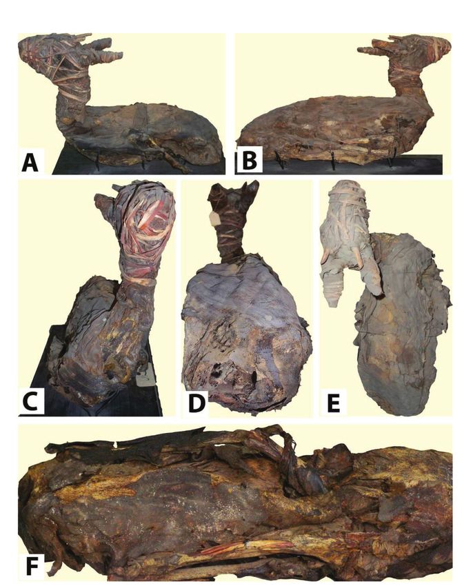
46                                                            ABDEL-MAKSOUD & EL-AMIN

1. INTRODUCTION                                     The gazelle mummy studied was found
                                                in a necropolis which included several
    The Egyptians depicted their gods not
                                                catacombs used for the burial of gazelle
only as humans but also in animal form.
                                                mummies. This necropolis is located in a
This does not mean that these gods in
                                                desert area some three kilometers south of
animal shape were mere animals. Just as the
                                                Komir, Esna, Qena, Egypt. It dates to the
Egyptians did not worship images or
                                                Late and Greco Roman Periods. Other
human beings, they did not worship
                                                animals, such as ibises and baboons, were
animals, but gods.
                                                kept in large numbers as cult animals and
    In prehistoric times, animals were
                                                could be purchased to serve as mummified
sometimes ritually buried: predynastic
                                                messengers to the deity. This suggests that
graves have been found with gazelles
                                                gazelles may have been kept for a similar
wrapped in mats and provided with
                                                purpose at a temple complex as sacred
funerary gifts such as pots of food and
                                                animals. The mummified gazelles found in
drink. This indicates that people were
                                                the Komir necropolis were wrapped with
concerned that these creatures be sustained     the limbs tucked under their bellies in a
in the afterlife. Scattered data from the Old   pose reminiscent of the recumbent gazelles
Kingdom to the New Kingdom show that            in the two and three dimensional depictions
people attempted to bring animals with          of them in Egyptian art. Many of these
them to the afterlife that they were attached   gazelle mummies were reportedly female
to, such as dogs, cats, monkeys, even a         (Strandberg, 2009).The gazelle mummy
gazelle, and a horse (TeVelde, 1980).           studied belonged to the species Dorcas Sp.
    Animal mummies are a rich and unique        (Meier, 2001).
source of information for understanding the         Brier (1994) reported that the majority of
environmental as well as the religious and      mummification techniques used on animals
cultural history of ancient Egypt. Ikram        were poor. He also said that this may be due
(2005) mentioned that there are four types      to the fact that Egyptologists Belzoni, Davis,
of animal mummies:                              and Ayrton had no interest in
    - Pets were mummified and buried in          mummification; therefore they left no
their owners’ tombs, or buried outside of       records of the processes used to preserve
them. Sometimes they had their own              the animal mummies that they unearthed.
sarcophagus or coffin, as well as their own       Fortunately, there are several Egyptologists
food offerings.                                  that have performed autopsies and
    - Victual mummies consisted of funerary     investigations on animals mummies to
food offerings for humans. Meat and              determine the methods used to embalm
poultry, prepared to be consumed, was           them (Brier, 1994). Visual examination and
wrapped up and sometimes placed in              radiography conducted on specimens from
individual coffinets or large baskets, and        the Cairo Museum by Ikram (2005) have
interred with the deceased.                     provided a great deal of information. She
    - Sacred animals, believed to possess an    proved that some of the animals she studied
aspect or essence of a deity, were              were mummified in the same manner as
worshipped during their lifetime and            upper class humans of the time. Human
mummified with pomp upon their deaths.           mummification, in its classic phase, began
    - Votive mummies were dedicated as          with extracting the brain from the nose and
offerings at the shrines of specific gods to      then filling the cranial cavity with resin.
whom these animals were sacred.                 Then a cut was made in the left side of the
    The gazelle mummy in this study was         torso and the lungs, liver, stomach, and
likely a sacred animal mummy (Lurker, 1984).    intestines were removed by the embalmer.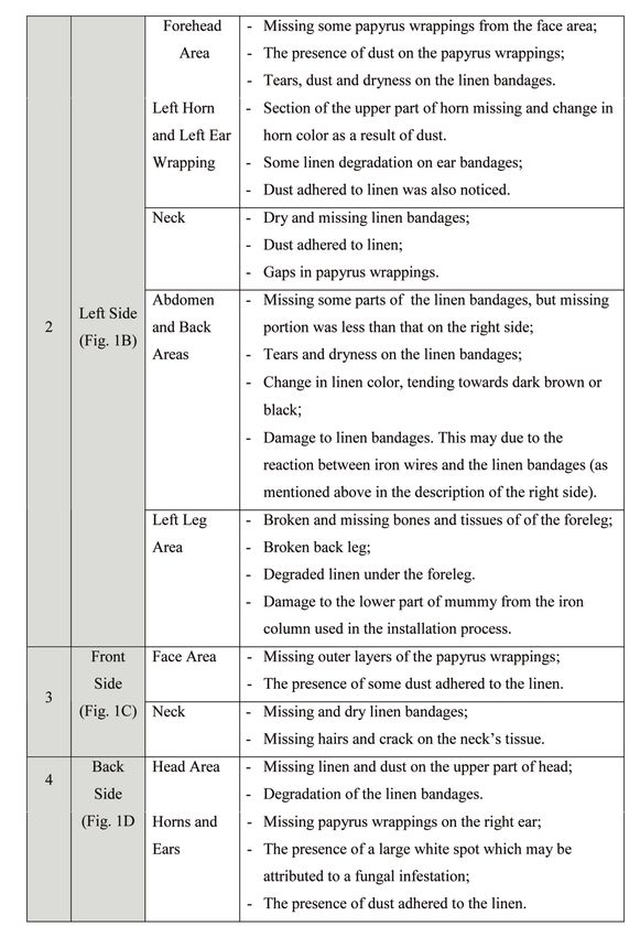
THE INVESTIGATION AND CONSERVATION OF A GAZELLE MUMMY                                     47

The body cavity was then filled with natron,     that it is difficult to assess the part played
incense, and spices, and allowed to             by each one, except under controlled
desiccate. Once dry, the mummy would be         condition, as Elton has remarked (Munro,
wrapped in bandages and prepared for            1966).
burial. Other animals were mummified                 The growth of microorganisms in
with secondary method of mummification.          organic materials such as mummies is
    The conservation of mummies is a            dependent on the presence of moisture,
politically and socially sensitive issue        although, other factors such as temperature
(Cassman & Odegaard, 2004). The                 should be taken into consideration to
preservation of a mummy after study             understand the biodeterioration mechanism
involves: (1) Identifying the type of           (Valentin, 1996). Many fungal and bacterial
mummification process used and the state         species require available moisture for their
of deterioration; (2) Selecting an              development on the surface of an object. In
appropriate method of study; (3)                this context, scant research has been done to
Individualized restoration; and (4)             determine the effect of moisture content in
Recommending          storage     conditions,   a material on the germination of microbial
including environmental and pest                spores to indicate the risk of microbial
protection. Finally, storage conditions         contamination        (Valentín,   2001).     A
should show the respect that each human         temperature range for the growth of
body deserves, taking into account the laws     microorganisms is 30°C (Valentin, 2002),
and beliefs of each nation (Lombardi, 2001).    this temperature keeps reaction rates in
    Deterioration in mummies is caused by       check, specifically the denaturing of
several factors, including environmental        collagen, the major constituent of mummy
conditions, physical damage, biological         skin (Maekawa, 1998), as high relative
damage or damage caused by previous             humidity (RH) that arrives at 65℅ or higher
conservation attempts. Sometimes, these         aids in decomposition of mummies,
factors occur in isolation, but they can also   although there are species of fungi that
be present in combination (David, 2001).        depend on a temperature range between
    Insects are considered one of the most      4°C to 35°C without need of moisture
serious factors causing damage to Egyptian      (Valentin, 2002). We can find other factors
mummies.         Panagiotakopulu       (2001)   such as pH value which aid in the growth
reported that mummies, both human and           of fungi and bacteria, whereas the fungi
animal, were highly susceptible to insect       prefer acidic environments pH 6 is suitable
attack. The presence of insects depends on      for growth (Abdel-Maksoud, 1995). High
three factors (climate, food and competition    temperatures from internal case lighting
with other living organisms) (Hill, 1985). Of   and windows can also cause the
the direct effects of climate, perhaps the       mummified skin to stiffen and become
most important, is that it governs the          more susceptible to cracking and chemical
geographical distribution of insects. Some      breakdown. The chemical destruction of
insects thrive in a temperate climate, others   mummified tissue can also occur through
in a tropical one. For most insects there is    exposure to air pollution, often high in large
an optimum climate in which they grow           cities and industrial areas. Sulfuric acid can
and increase most rapidly. The element or       result from sulfur emissions combined with
factor in that optimum climate which we         fog. Potentially, sulfuric acid produced in
can most easily measure is temperature.         the right atmospheric conditions can break
Humidity and variation of light intensity or    down the proteins of the mummified tissue
photo-periodicity can also be measured, but     (Maekawa, 1998 & Meier, 2001).
all the three are so closely bound together         Progress in research methodology has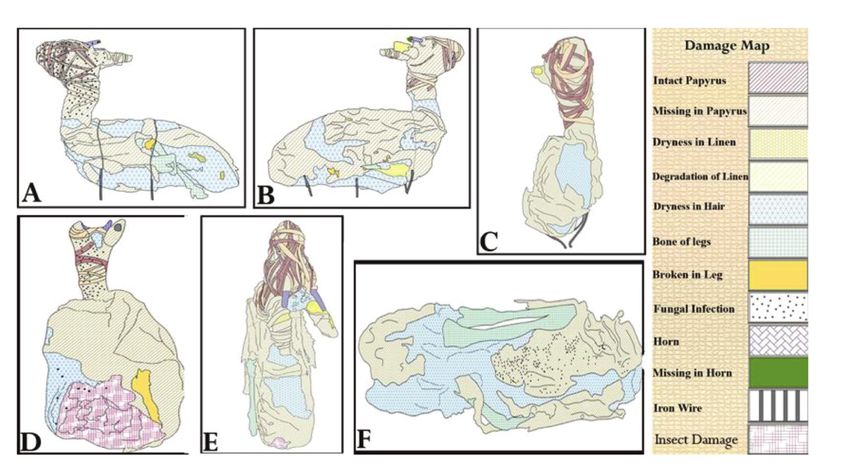
48                                                            ABDEL-MAKSOUD & EL-AMIN

produced new examination procedures             over 100 years ago, and thought to be 65
which result in greater detail in the           million years old, is shown by scanning
description of the mummys’ external             electron microscopy to have intact,
features and in dissections that use highly     mummified microscopic collagen fibers and
technical methodology, i.e. historical,         other ultra-structural features within the
pathohistological, and chemical analysis,       compact bone (Armitage, 2001). X-ray
and sophisticated radiographic techniques.      diffraction has been used to determine
Such investigations make possible a             components and crystallinity of bone and
detailed     study     of    mummification       other associated materials (Robles 2002;
techniques and facilitate finding changes in     Meneghini et al., 2003; Reiche et al., 2003;
bodies structure (Klys et al., 2001). Some of   Fantner et al., 2004; Abdel-Maksoud, 2010).
the identification methods for macrob-              The conservation of mummified remains
otanical remains include morphology using       involves two procedures. First, it is
light microscopes (Hastorf, 1999). Infrared     necessary to store the mummies in a
spectroscopy is also commonly used in           suitable environment, ideally with a relative
archaeological studies. Cotte et al., (2005)    humidity, of 40-55 percent and a constant
listed the applications of infrared             temperature of 18-22° C. Secondly, if
spectroscopy for the study of mummies:          environmental deterioration has already
    - Analyses with FTIR were performed to      occurred, then the damage cannot be
identify the chemical composition of some       eradicated thorough environmental control
fragments taken from the body, bandages         alone and it is necessary to apply other
and cartonnage wrapping of Egyptian             methods to arrest the damage (David, 2001).
mummies;                                        This study aims to:
    - Attenuated total reflection (ATR-FTIR)        1. Describe the deterioration aspects
was used to study thick samples, for            found on the mummy studied;
example in the analysis of the outer and           2. Explain the deterioration mechanisms
inner surfaces of pieces of skin taken from     of the studied mummy resulting from our
the well-known Iceman. Žemaitytė et al.,        investigations;
(2006) mentioned that Fourier Transform            3. Apply some conservation techniques
Infrared Spectroscopy (FTIR) allows one to      for the treatment and restoration of
determine the constitution of wrappings.        deteriorated mummy.
FTIR was also used in the investigation of
archaeological hair from Gravesites at the      2. MATERIALS AND METHODS
Home of Samuel Washington (Rowe, 2010).         2.1 Historical background of the gazelle
The scan can determine the morphology of        mummy studied
fibre and fabric surfaces. Titlbachova &
                                                    The mummy studied is located in the
Titlbach (1977) studied Egyptian mummies
                                                Agriculture Museum, Giza, Egypt. It is
in Czechoslovakian collections and they
                                                exhibited in the section of wild animals and
found generally good preservation, with the
                                                domesticated birds. Its Museum Number is
samples resembling modern European
                                                786. The mummy dates back to the Late
populations with significant African
                                                Period (525-343 B.C). It came from Komir, a
admixture. Hrdy (1978) mentioned that
                                                village in south Esna, Upper Egypt.
ancient Egyptian samples were studied
with scanning electron microscopy by
                                                2.2 Visual assessment by digital camera
Chiarelli et al. (1970/1971), finding
                                                and AutoCAD
significant loss of cuticular scale edges. A
specimen of hip bone from a Tyrannosaurus          To show the changes associated with
rex, excavated from a ranch in Wyoming          gazelle mummy, a high-resolution digital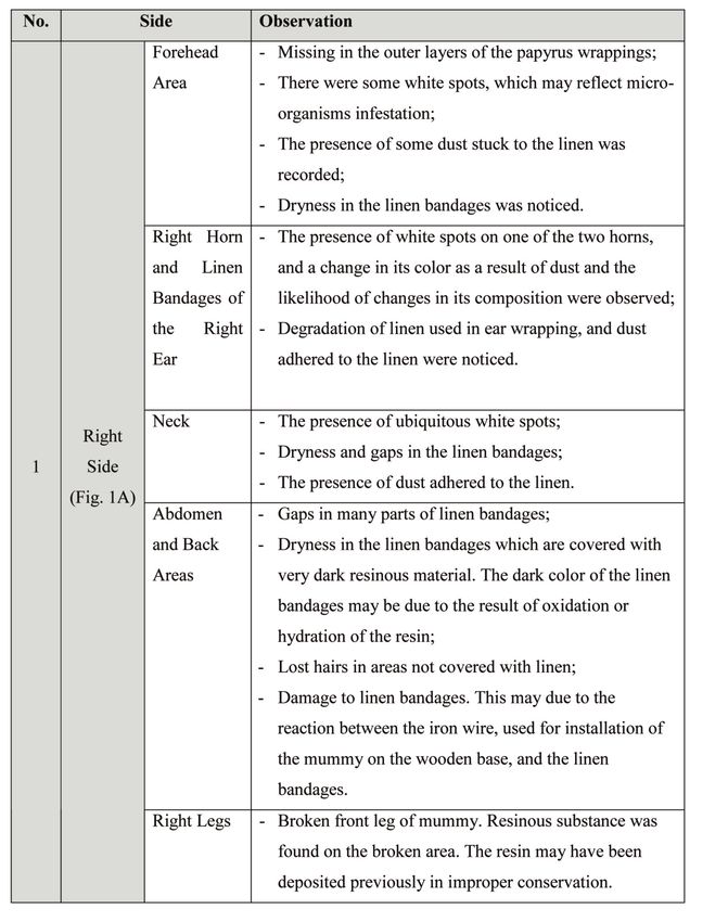
THE INVESTIGATION AND CONSERVATION OF A GAZELLE MUMMY                                     49

camera image (Kodak Easy Share M1033,           2.5 Investigation of the surface morphology
10mp, 3×Optical zoom) was used to create        by SEM
realistic photographic documentation of the
                                                   A scanning electron microscope JEOL-
aspects of deterioration. The visual
                                                JSM-5400LV was used to observe the
observation was used to follow the changes
                                                surface morphology. The fine gold coating
and to explain the deterioration forms.
                                                (JEOL-JFC-1100E) was used. Lenin,
   To show aspects of deterioration for the
                                                bandages and hair of the mummy studied
gazelle mummy, computer graphic
                                                were investigated.
documentation was done using (AutoCAD
2007). With CAD, a map of the damage was
                                                2.6 Fourier transform infrared spectroscopy
made, and each face of the mummy was
                                                 (FTIR)
documented.
                                                    Fourier transform infrared attenuated
2.3 Light microscope for the identification      total reflection (FTIR-ATR) has been
of mummy wrappings                              extensively used on linen and hair samples
                                                to investigate absorption and reactions on
   The wrappings used with mummy were
                                                surfaces. This method of analysis has been
from plant fibers. For plant identification, a
                                                used in accordance with Jadoul et al., 1996;
separate sample from the gazelle mummy
                                                Pouliot et al., 1999; Xie et al, 2002; Velkova
was taken. A thin section (30-50mµ), which
                                                & Lafleur, 2002; Liao et al, 2006; Dias et al.,
had been prepared at the botany
                                                2008; Russeau et al., 2009.
department at Ain Shams University, was
                                                    A significant advantage of ATR
examined by light microscopy for details of
                                                technique is that the archaeological samples
internal structure and compared with the
                                                require no preparation, thereby minimizing
reference    collection     kept   in    the
                                                possible damage (Bernard, 2007). Infra-red
Archarobotany Laboratory, Department
                                                spectra were obtained using a FTIR
Botany, Faculty of Science, Cairo University.
                                                spectroscopy (JASCO-ATR-FT/IR-6100).
                                                    For the archaeological bone sample, it
2.4 Isolation and identification of fungi
                                                was ground to a fine powder with an agate
   To identify fungi on the gazelle mummy,      mortar and pestle. FTIR grade potassium
some sterilized cotton swab were used to        bromide (97-99 mg) was ground to a fine
swab different areas where biological            powder in a separate agate mortar and
damage was visible on the bandages and          pestle. The two powders (100 mg total)
wrappings of the mummy studied. Czapek-         were then combined and mixed with a
Dox agar medium was used for the isolation      spatula. An additional 100 mg of KBr was
of fungi. This medium consists of 3 g           ground into a fine powder, and then used
NaNo3, 0.5 g KCL, 0.5 g MgSo4, 1 g K2HPo4,      to obtain background spectra. The sample
30 g sucrose, 17 g Agal, 1000 ml distilled      was transferred into a sample cup to
water, and pH was 5.5. A pure culture from      overflowing, and a cover slip was dragged
Czapek-Dox agar medium was made.                across the top of the cup to remove excess
Fungus colonies were identified according        powder and smoothed the sample surface
to Raper & Fennell, 1965; Barnett & Hunter,     in order to maintain uniform distribution of
1972; Domsch et al., 1980; Stevens, 1981. The   particle size. Each sample was then mixed
isolation and identification of fungi were       with KBr and placed in a DRIFT cell. This
achieved at the Micro Analytical Center,        method of analysis gives information on the
Faculty of Science, Cairo University, Egypt.    composition and crystalline of the bone
                                                mineral, and at the same time gives an
                                                indication of the behavior of the protein
50                                                              ABDEL-MAKSOUD & EL-AMIN

materials in bone (Abdel-Maksoud, 2010).         collected on an X-ray diffract meter 6000
Spectra were assigned for new and archae-        (Shimadzu, Japan) using Cu Kα radiation
ological samples. Infrared spectra were          from a tube operated at 45kv and 35mA.
obtained using a Fourier transform infrared      The two samples studied (new and archae-
spectroscopy (JASCO-FT/IR-6100).                 ological samples) were measured from 0° to
                                                 65° 2θ to obtain a diffraction pattern. The
2.7 Amino acid analysis                          crystalline index of new and archaeological
                                                 samples was determined using x-ray
   Two samples weighing 0.8 mg were
                                                 diffraction (XRD) on the basis of the full
taken from the gazelle mummy’s bone and
from a new Gazelle bone, which was used          width of half maximum (FWHM) of the
as a reference. They were both placed into       apatite diffraction 002 (Abdel-Maksoud,
a hydrolysis tube. 1ml of 6NHCl was added        2010).
(HCl Supra pure from Merck). The solution
was frozen using a mixture of dry                3. CONSERVATION TECHNIQUES
ice/ethanol and the tube was evacuated            USED
using a vacuum pump [6.5Pa (0.01 mbar)].         3.1. Cleaning processes
Intermediate flushing with oxygen-free               Mechanical and chemical cleaning
nitrogen was executed. The hydrolysis tube       processes for all Components of the
was then closed by melting the glass with a      mummy (wrappings, bandages hair and
gas-burner. The hydrolysis tubes were            etc.) were applied in accordance with
placed in an oven with a uniform                 Gänsicke et al., 2003 and Farrell et al., 2006.
temperature distribution of 110oC for 12, 24,
36, or 72 hrs. In order to create reproducible   3.2. Consolidation
hydrolysis conditions, the samples were
                                                    The consolidation process by using
hydrolyzed in an air circulation oven. The
                                                 Klucel G (Hydroxypropylcellulose) 1% in
tubes were later cooled in an ice-bath. The
                                                 ethyl alcohol was applied for Linen, hair
solution was then centrifuged in order to
                                                 and papyrus in accordance with Abdel-
precipitate insoluble components. The
                                                 Kareem (2000).
resulting supernatant was evaporated at
approximately 40oC in a rotary evaporator,
                                                 3.3. Completion process
the remaining were dissolved in
approximately 1ml of dist. water and                The authors used a new paste for the
evaporated once again to remove traces of        completion of some sections on different
acid. The sample was dissolved in 1–2 ml of      parts of the mummy studied. The paste
the sample diluting buffer after which it         used consisted of beeswax, shellac,
was ready for analysis. The instrument used      sawdust, turpentine (3:2:1:1/2 respectively),
was Eppendor- Germany (LC3000 Amino              6ml of tea tree oil diluted in ethanol (1600
Acid Analyzer). The condition of the             mg/gm), and a mixture of black ( magnetite,
instrument was: flow rate: 0.2 ml/min,            Fe3O4) and red iron oxides (hematite, Fe2O3)
pressure of buffer form 0 to 50 bar, of           were added to match the natural surface
reagent to 0-150 bar and reaction                tones of the mummy.
temperature was 123oC (according to
Abdel-Maksoud, 2011).                            3.4. Reconnecting loose part of
                                                 the gazelle’s leg
2.8 X-ray diffraction for the measurement
                                                    The authors used galvanized wire for
of bone crystallinity:
                                                 reconnecting a loose part of mummy’s leg.
     X-ray powder diffraction data were           3.5. The display of the gazelle mummy
THE INVESTIGATION AND CONSERVATION OF A GAZELLE MUMMY                                 51

  Plexiglass [polymethyl methacrylate        contains the constituent materials of gazelle
(PMMA)] and wooden base were used for        body (hair, tissue, bones), while the second
making the stand of the mummy.               part contains the mummification materials
                                             (resins, wrappings of papyrus and linen).
4. RESULTS AND DISCUSSIONS                   Through the naked eye observation, it was
4.1. Visual assessment by digital camera     found that all of these items had been
and AutoCAD                                  damaged to varying degrees which can be
   The gazelle mummy (Figs 1 and 2) being    summarized in the table 1.
studied contains several materials, which    4.2. Light microscope for the identification
can be divided into two parts, first part      of mummy wrappings

                       Table 1: Visual assessment of gazelle mummy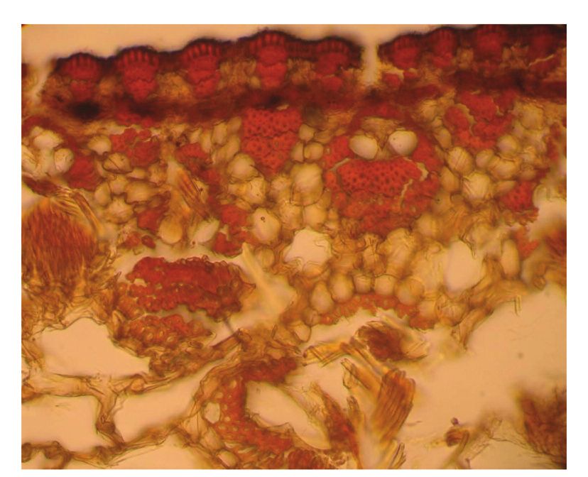
52 ABDEL-MAKSOUD & EL-AMIN
THE INVESTIGATION AND CONSERVATION OF A GAZELLE MUMMY                     53

                                                          Figure 1.Gazelle
                                                          mummy before
                                                         conservation: (A)
                                                        Right side; (B) Left
                                                        side; (C) Front side;
                                                         (D) Back side; (E)
                                                         Upper side; Lower
                                                                side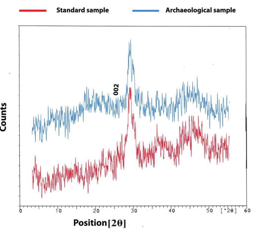
54                                                                 ABDEL-MAKSOUD & EL-AMIN

Figure 2.AutoCAD for documentation of deterioration aspects: (A) Right side; (B) Left side; (C) Front
                       side; (D) Back side; (E) Upper side; (F) Lower side.

   The mummy wrappings were identified
as Cyperus papyrus L. (Papyrus). The results
obtained in (Fig. 3) showed thatthe
epidermis is formed of ordinary thick-walled
epiderminal cells. Below are patches of
chlorenchyma tissue alternating with strands
of fibers. The ground tissue is differentiated
into a narrow peripheral zone formed of
several layers of radially elongated
chlorenchyma cells and small reduced
vascular bundles, and a large inner layer
made up of highly lacunateaerenchyma with
3-armed cells and collatral vascular bundles           Figure 3. Mummy wrappings identified as
surrounded by a sheath of 2-3 thick-walled               Cyperus papyrus L. (Papyrus) by light
                                                                     microscope.
lignified cells. The parenchyma cells are
withered especially at the middle.                 Aspergillus niger, Pencillium chrysogenum,
                                                   Rhizopus arrhizus and Rhizopus nigricans
4.3. Microbiological investigation                 (12.5%) and Aspergillus flavus (6.25%).
                                                      Biodeterioration is a significant factor in
    The most dominant fungi isolated from
                                                   the decomposition of human and animal
the mummy studied are shown in the table
                                                   bodies, because fungi grow and feed on
2. The identified fungi belong to two genera,
                                                   many of the constituent products, such as
Ascomycotina        and      Zygomycotina.
                                                   protein, fats, starch and cellulose (Elnaggar
Infestations of fungi, particularly Rhizopus
                                                   et al., 2010). The results of our study were
sp., caused the white spots found on the           paralleled by several similar studies on
linen bandages. The percentage (%) of              archaeological material. Arya et al., (2001)
identified fungi were:                              isolated twenty-two viable species of fungi
    (25%), Aspergillus fumigates (18.75%),
THE INVESTIGATION AND CONSERVATION OF A GAZELLE MUMMY 55
Table 2. Identified fungi isolated from linen sample
belonging to 17 genera in May 2000 from Zielińska-Jankiewicz et al., (2008)
the indoor air of the Egypto-Babylonian studied the species accounting for
Gallery by the gravity fall method. The air mycological contamination of the library,
and dust of the mummy chamber revealed archive and museum collections, which
six different fungi. The mycoflora was include Aspergillus, Penicillium, Geotrichum,
dominated by different Aspergilli. Isolated Alternaria, Cladosporium, Mucor, Rhizopus,
from the first toe of the right leg of an 1.54 Trichoderma, Fusarium. Arroyo (2009) found
m long Egyptian mummy was among the types ancient proteineous
Emericellanivea (Wiley & Simmons). This materials are Aspergillus and Penicillium.
fungus caused white powdery patches on Valentín (2010) stated that 16 historical
the toes of the mummy. Valentín (2001) buildings located in different climatic
stated that among the types of fungi found regions of Spain were analyzed to detect
in Spanish museums, archives and libraries cellulose and protein-degrading
are Aspergillu sniger, Aspergillus flavus, microorganisms. Non-destructive and
Aspergillus fumigates, Penicillium surface samples were taken from objects
chrysogenum, Rhizopus nigricans. Valentín made of cellulose (paper, cardboard and
(2003) reported that Penicillium and textiles) and protein materials (parchment,
Aspegilluss trains are harmful to textiles leather, mummies and silk textiles).
because they have a high level of cellulolytic Aspergillus flavus, A. fumigatus, A. niger and
activity and grow in materials with a Rhizopus nigricans were among the
moisture content of 7- 8%. David (2008)
identified fungi.
mentioned that fungi of various types are
often seen in ancient tissues as a result of
4.4. Scanning electron microscope (SEM)
poor storage of the specimen. The fungus
4.4.1. Hair sample
produces oxalic acid as another metabolic
byproduct. The acid acts in a similar way to Scanning electron microscopy was used
the acids in the decalcification process but to determine the external condition of
in this case, instead of producing a soluble individual fibers. Resin-covered fiber was
salt, the oxalic acid produces an insoluble observed (Fig. 4A and 4B) and some cracks
calcium oxalate compound that forms in the resin layer were noticed (Fig. 4C).
clusters of crystals on bone. Variable microbe degradation of the gazelle56 ABDEL-MAKSOUD & EL-AMIN
hair was observed (Fig. 4D). The presence
of tunnels within the degraded hair, similar
to those found by Wilson et al. (2007)
indicates that much of this damage was
caused by fungi. The presence of holes and
erosion of the cortex characteristic of fungal
tunneling highlighted the potentially
aggressive nature of the depositional
environment (Wilson et al., 2001).
Accumulated dusts were also observed.
Some cracks in the fiber structure and on
the surface were noticed (Fig. 4C and 4D).
4.4.2. Papyrus sample
The scanning electron microscope can be
a useful tool for papyrus investigation. The
wave pattern of cut papyrus was noticed
(Fig. 4E). The SEM showed serious tears,
holes and the separation of cellulose fibers.
The outer surface of the papyrus showed a
covering of starch and fissures along a
vascular bundle (Fig. 4F). Resin-covered
papyrus fibers were observed. Pierced cell
tissue on the inside of the papyrus was
shown (Fig. 4H). Some dust was also
observed on and inside the fibers (Fig. 4G,
H).
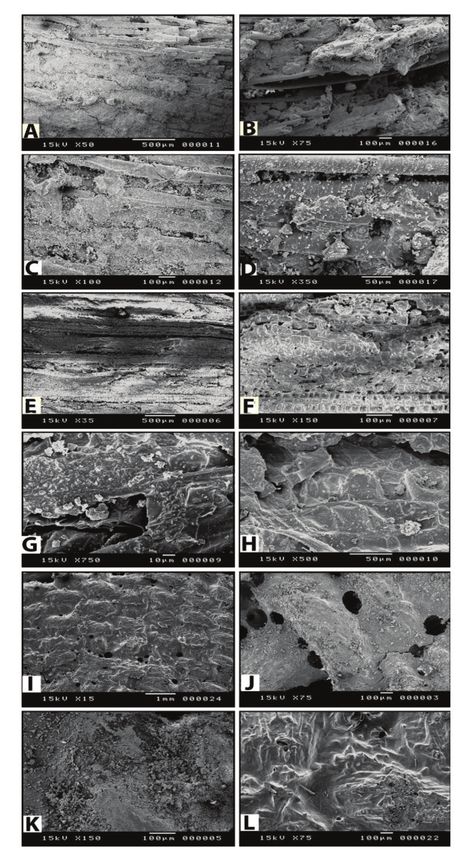
Figure 4.(A-D)SEM micrographs show damage
4.4.3. Textile sample to the mummy’s hair: (A) Dust and resin
covering the hair surface; (B) Resin within the
The scanning electron microscope is able hair fibers; (C) Accumulated dust and cracks in
to physically examine virtually any textile the fiber structure; (D) Accumulated dust, holes
material without any special preparation or and erosion of the hair; (E-H) SEM images for a
sample of the papyrus mummy wrapping; (E)
conductive coating (Wei et al., 2004).By SEM Wave pattern of the cut papyrus; (F) Starch
we can investigate the morphology of fiber covering and fissures along a vascular bundle;
and fabric surfaces. Our textile sample was (G) Dust on resin covered papyrus fibers; (H)
composed only of friable brown thread in Pierced cell tissue on the inside of the papyrus;
an open plain weave (Fig. 4I). The linen (I-L) SEM images for the sample of linen
mummy wrapping; (I) Friable thread in an open
fiber consisted of coarse material, which plain weave; (J) Holes and cracks in the resin
proves the low quality of the linen. Holes layer; (K) Thick layer of dust; (L) Resin covered
and cracks in the resin layer can be linen fiber.
observed (Fig. 4J). A thick layer of dust (Fig.
4K) and a layer of resin covered linen fibers 4.5. Fourier transform infrared
were also noticed (Fig. 4L). spectroscopy (FTIR):
4.5.1. Bone sample
The results of this section were
explained and discussed in accordance withTHE INVESTIGATION AND CONSERVATION OF A GAZELLE MUMMY 57
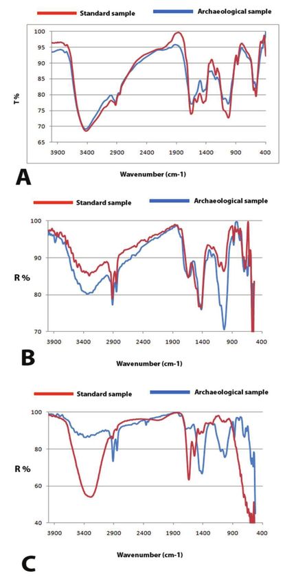
Abdel-Maksoud (2010). It is clear (Fig. 5A) 4.5.2. Hair sample
that the band at 3422.06 cm-1 in the new
The examination of these spectra
sample (control) assigned to a broad band
allowed the researcher to confirm the
represents (OH) hydroxyl stretching due to
presence of frequencies characteristic to
intermolecular hydrogen bonding of the
keratin and the changes in band intensity in
hydroxyl group. This band includes
the archaeological sample. Several
multiple bands made up of multiple N-H
important points were obtained from ATR-
groups (its primary amides), both in the
FTIR analysis of the samples (Fig. 5B):
solid state and in the presence of hydrogen
- According to Espinoza et al. (2008), the
bonding. In the archaeological sample, this
region (1200 to 1000 cm–1) is associated with
band shifted to a higher position (3429.78
vibrations of the sulphur-oxygen groups of
cm-1). The C-H stretching vibrations occur
keratin. The results proved the presence of
in the region (2924.52-2926.45) stretching of
sulphur-oxygen groups in the two samples.
aliphatic groups, and they were found in
The degradation in the archaeological
two samples that were studied. The position
sample compared to the control sample was
of the band in the archaeological sample
recorded.
was very close to that of the band of the new
- The peptide bond is the most abundant
sample. The bands between 3422.06 cm-1
bond within a keratin protein (Panayiotou,
and 2924.52 cm-1 in the samples are protein
2004). The spectra of amide I, ν(CONH), the
characteristics, and the increase or decrease
amide II, δ(CH2) and the amide III, δ(NH)
of these bands may give an indication of the
bands were examined. The spectral analysis
expansion or contraction of the protein
indicated significant, observable keratin
areas. Collagen exhibits a series of
degradation of archaeological hair sample.
absorptions from 1656.55 cm-1 to 1241.93
- Oxidation of the amino acid cystine to
cm-1. Band at 1656.55 cm-1 (C=O stretching)
cysteic acid can occur in hair, resulting in an
in the new sample is assigned to amide I
increase of the S=O stretching absorbance
and the position of this band is decreased in
(Robotham, www.thermo.com). Hair fibers
the archaeological sample (1641.13 cm-1).
analyzed by ATR-FTIR clearly show the
The increasing or decreasing of C=O is
difference between new and archaeological
dependent on the physical state of the
samples. Fig. 5B shows the region between
sample. In the solid state, the frequency of
1400 and 900 cm-1, revealing the spectral
the vibration is slightly decreased. The
differences due to the oxidation of cystine
presence of hydrogen bonding is an
to cysteic acid.
important contributing factor to this
decrease in frequency. The bands at 1562.06
4.5.3. Textile sample
cm-1 (NH, CN stretching) in the new sample
are assigned to amide II. Amide II The infrared spectra of new and archae-
disappeared in the archaeological sample. ological linen samples (Fig. 5C) were
The band at 1241.93 cm-1 is assigned to recorded from 4000-400 cm-1. The results
amide III which involves C-N stretching showed that there are changes in the IR
and N-H bending. The wave-number of spectra of the archaeological sample
these peaks depends on the secondary compared to the spectra of the control
structure of the protein (e.g., α-helix, sample. By comparing the results, it is
β-sheet, β-turn, random coil). The position evident that there are significant spectral
of this band increased in the archaeological changes in the region from 1750-1600 cm-1
sample (1243.86). for the archaeological sample. The region
from 1750-1600 cm-1 proved the most
convenient for monitoring cellulose58 ABDEL-MAKSOUD & EL-AMIN
degradation. It was confirmed that archae- its value in the reference sample was
ological linen cellulose involves carbonyl 357.83%. A similar trend was noted for the
and carboxylate group functions, which can basic amino acid histidine. Its value was
be monitored by the infrared reflection at 0.40% for the mummy bone and was 51.92%
1750-1600 cm-1 with FTIR Spectroscopy. in the reference bone.
It can be explained by the fact that the
oxidative decomposition of the side chains
of amino acids forms ammonium (NH4+).
The basic amino acids lysine and arginine
are particularly sensitive to oxidation and
our results reflected this. The archaeological
bone sample showed that ammonium
content was 73.70% and the value was
absent in the new reference bone sample.
The acidic condition of the archaeological
bone sample may lower the value of the
basic amino acids and increase the value of
ammonium (NH4+). This also indicated that
acid hydrolysis may also have occurred for
the archaeological bone sample.
In was noticed that with an increase in
NH4+, there was a clear tendency for lower
Figure 5. FTIR analysis: (A) FTIR spectra of the
modern and archaeological bone samples; (B)
FTIR spectra of the modern and archaeological
hair samples; (C) FTIR spectra of the modern
and archaeological textile samples.
4.6. Amino Acid Analysis for Bone sample
The results of the amino acid analysis of
the sample taken from both the new
reference bone sample and the archae-
ological bone sample (shown in Fig. 6A and
6B) revealed that there was less of the basic
amino acid lysine in the mummy (9.6%)
than in the reference sample (28.74%). The Figure 6. (A) Identified amino acids of bone of
value of the basic amino acid arginine in the both the standard and archaeological; (B)
archaeological sample was 153.60% while Identified proline of standard and archae-
ological samples.THE INVESTIGATION AND CONSERVATION OF A GAZELLE MUMMY 59
value of serine (7.8%) in the archaeological
sample and 37.8% in the reference sample,
and threonine, which was absent in the
archaeological sample and the value of
which was 19.4% in the new sample. a
The lower value of aspartic acid in the
archaeological sample (25.92%) than the
new sample (119.73%) indicated the
increase of hydrolysis in the archaeological
bone sample.
m
4.7. X-Ray Diffraction (XRD)
For Bone Sample
The main aim of using XRD in this study
is to measure the crystallinity index of bone Figure 7. XRD diagrams of modern (lower) and
(Fig. 7). Full Width at Half Maximum archaeological (uper) bone samples.
(FWHM) measurements were made on the
apatite d002 peak. This measurement has 5. CONSERVATION
proven useful as a measure of apatite 5.1. Cleaning processes
crystallinity. Farlow and Argast (2006) have 5.1.1. Mechanical cleaning
objected to this approach due to the
The purpose of mechanical cleaning of
possible interference of quartz. However,
the mummy studied is to:
this method of measurement is most
1. Reduce the potential for damage to the
commonly used. The samples examined
mummy by removing foreign material
generally present characteristic X-ray which may be abrasive, acidic, hygroscopic,
diffraction patterns typical of poorly or degrading;
crystalline hydroxyapatite, namely, that 2. Reveal the decorative bandages and
there is little difference between the new wrappings of the mummy by removing
sample and the archaeological sample. The surface dirt when it interferes with the
crystallinity index of the new sample was visibility of the imagery or information.
0.30 mm. The crystallinity of the peak 002 of It should be noticed that a decision must
the archaeological sample was 0.11cm. The be made to balance the probable care of
data showed that the width at half each material of the mummy against the
maximum of the peak 002 of the new possible problems related to surface
sample was more than that of the archae- cleaning.
ological sample. This means that the archae- Due to the dryness of the mummy’s
ological sample was more crystalline than materials (linen bandages, papyrus
the new sample. This also indicates that the wrappings, hair and tissue), mechanical
archaeological sample was affected by its cleaning was done carefully by using a soft
burial environment and was dryer than the brush. This was suggested by Gänsicke et
new sample. These results were confirmed al., (2003) who recommended the use of a
by Abdel-Maksoud (2010), Abdel-Maksoud soft camel-hair or similar small soft brushes
and Abdel-Hady (2011) who reported that for removing surface dust and dirt. Farrell
archaeological bones have higher et al., (2006) also used soft brushes for the
crystallinity and sharper X-ray diffraction cleaning of cartonnage mummy surface.
patterns than new samples. The cleaning process of the mummy
studied was done in several steps, every60 ABDEL-MAKSOUD & EL-AMIN
face was brushed individually (right, left, swabs. The black resinous substance on the
front, back, upper and bottom). The above-mentioned materials was not
removal of dust on the surface of the above removed. Adhesive particles and tape were
mentioned materials was done by using soft left if they were in fragile areas. After
brushes. Special care was taken with fragile cleaning, sufficient time was allowed for the
and loose fibers. Unfortunately, the cleaning mummy to dry before continuing
test results for the resinous substance, treatment. The mummy’s hair was cleaned
which was evidently from previous carefully according to a method proposed
conservation, showed that it could not be by Abdel-Maksoud (1995), who stated that
removed without damaging the mummy’s other than removal of earth and soil, fabric
bandages and hair. Therefore, the cleaning and hair did not require a great deal of
had to be stopped. cleaning. According to Gänsicke et al.
Due to the significant use of resin and (2003), horn does not need more cleaning
the bad state of preservation of gazelle than a rinse with lukewarm water, but he
mummy, dust was adhered to the materials warned against insect attack. So the horns
mentioned above, and was difficult to were cleaned using water and alcohol, then
remove by mechanical method. treated with tea tree oil as resistance
materials.
5.1.2. Chemical cleaning
5.2. Consolidation
One of the key standards of conservation
is reversibility: anything done to preserve a Consolidation is considered one of the
piece should be able to be undone with most important processes in our case study.
minimal damage to the piece itself. Conventional stitching techniques cannot
Chemical cleaning is not reversible, and so be used with linen wrapping because the
it should be used only when absolutely needle and sewing thread may break the
necessary. Before chemical cleaning several dry and fragile linen threads. For linen, hair
things should be taken into account to and papyrus consolidation, Klucel G
determine both the best treatment for that (Hydroxypropylcellulose) 1% in ethyl
particular combination of object and soil, alcohol was used. Cosolidant was applied
and to ascertain whether the object is able using a brush. Klucel G was recommended
to be cleaned, or may sustain damage by Abdel-Kareem (2000), who mentioned
during the process: chemical composition of that linen samples treated with Klucel G
the object, degree of deterioration, type of showed the best reduction in growth of
materials to be removed and the type of Aspergillus nidulans, Aspergillus
cleaner used. terrus,Penicillium asperum, Trichoderma viride
Chemical treatment was needed for and Penicillium funiculosum. It has been used
extremely dirty linen, hair and papyrus. successfully in water/alcohol solutions to
Chemical cleaning was performed by using consolidate ethnographic materials which
alcohol and distilled water (1:1/2) followed have a matte surface. Klucel G varies in
by using alcohol alone for evaporation of strength, according to grade and
water residue. concentration. Klucel G in low percentage
According to Gänsicke et al. (2003) more solutions can be brushed on local areas
adherent dirt should be removed using needing consolidation.
water or alcohol and a soft brush. Cleaning
5.3. Completion process
was performed by gently rolling swabs
across the surface. The surface tolerated There are some missing sections on
aqueous cleaning with moderately damp different parts of the mummy studied.THE INVESTIGATION AND CONSERVATION OF A GAZELLE MUMMY 61
There was a hole on the mummy’s back 5.4. Reconnecting loose part of the gazelle’s
(Fig. 8A), pits in the backbone and neck, and leg
a crack in the neck. It should be noted that
A loose part of mummy’s leg was
largest missing section was on the back of
reconnected using galvanized wire. This
the mummy. Insects had played the
was done without making any holes in the
primary role in this deterioration. The
leg. The repair was needed due to a severe
missing sections were weakened areas and
weakness that appeared through analysis.
had to be treated.
Before the completion of missing area,
5.5. The display of the gazelle mummy
disinfection by the use of Tea tree oil
(Melaleuca alternifolia) diluted in ethanol (Fig. There is no absolute rule for the display
8B). Completion is the proper process for the of mummies. There are guidelines, but also
treatment of this type of deterioration. The an ongoing debate. It was indicated that
small missing areas were filled with a paste many museums now have policies on the
to stop the holes from growing. The paste display of mummies:
consisted of beeswax, shellac, sawdust, • All such displays should be designed
turpentine (3:2:1:1/2 respectively), 6ml of tea so that the mummies are accompanied by
tree oil diluted in ethanol (1600 mg/gm), and an explanatory interpretation that places
a mixture of black ( magnetite, Fe3O4) and them in an historic context. Display of
red iron oxides (hematite, Fe2O3). On the archaeological remains for aesthetic or
back, the missing parts were large and could artistic purposes alone will not be
not be filled with the paste because it would permitted.
be too heavy. The authors used linen • Where human remains are displayed
bandages soaked in gum Arabic and tea tree in the museum, there will be a notice
oil to fill the inner part of the hole (Fig. 8C) outside the relevant display space alerting
and coated the outside with the paste (Fig. visitors to the presence of human remains.
8DـF). The following steps were done for the
It should be noted that tea tree oil was display of the gazelle mummy:
used in the paste because of its effectiveness - Plexiglass [polymethyl methacrylate
against larvae of leather beetle (Elamin (PMMA)] was the material used for making
2011). Tea tree oil (Melaleuca alternifolia) the stand. It was preferred because of its
ranges from colorless to pale yellow. The easy handling and processing, and low cost.
most active compounds in this oil are - Measurements were taken of the three
terpinen-4-ol, terpinene, 1,8-cineole and different lower parts of the mummy (front,
terpinolene. The International Standards center and back);
Organization, ISO 4730, mandates a - Three semi-circular pieces were made
minimum concentration of 30% for to fit these measurements;
terpinen-4-ol and a maximum - Support from the same material
concentration of 1,8- cineole of 15% in the (Plexiglas) was made for each piece;
oil. It is considered to have some of the - Each support was integrated thermally
strongest natural antiseptic / antifungal with the semi-circular pieces;
properties in the world (Joshi et al., 2009; - Part of the plexiglas was made into a
Tripathi et al., 2009). semi-circular shape and installed on the
The patched areas were then painted front support.
with pigments that matched the natural - Three compact pieces with supports
surface tones of the mummy. The paste was were installed on a single wooden base;
applied using different sizeed metallic - The gazelle mummy was carried
rifflers and cone tools. carefully and put on the stand (Fig. 8G).62 ABDEL-MAKSOUD & EL-AMIN
(hair, papyrus and textile).
The amino acid analysis showed
decreasing levels of lysine, arginine and
histidine in the archaeological sample,
which indicated oxidation breakdown. The
high value of NH4+ and decrease in the
aspartic acid of the archaeological sample
indicated the presence of hydrolysis
breakdown.
The results of the bone analysis by FTIR
showed that there was degradation in the
collagen, since the loss of amides was
noticed in the archaeological sample. For
hair samples, the presence of sulphur-
oxygen groups in the new and archae-
ological samples was found. Degradation
of amides in the archaeological sample
compared to the new sample was observed.
By recording the amides (I, II and III), the
degradation of the archaeological sample
compared to the new sample was recorded.
The spectral analysis of amide I, ν(CONH),
the amide II, δ(CH2) and the amide III,
Figure 8.Completion process for the back area: δ(NH) band showed significant, observable
(A) Before treatment; (B) Sterilization by tea tree keratin degradation of the archaeological
oil; (C) After placement of linen soaked in a
mixture of gum Arabic and tea tree oil; (D-E) hair sample. The region between 1400 and
During completion; (F) After completion; (G) 900 cm-1 revealed the spectral differences
Gazelle mummy on display. which may be due to the oxidation of
cystine to cysteic acid. FTIR results showed
6. CONCLUSIONS that there are significant spectral changes in
Several aspects of deterioration were carbonyl and carboxylate group functions
identified through visual assessment, (1750-1600 cm-1) for the archaeological linen
photographic and AutoCAD sample.
documentation. All aspects of deterioration The results of XRD analysis of the bone
indicated that the mummy suffers from proved that there is a little difference
deterioration. Papyrus wrappings were between the new sample and the archae-
identified by light microscope as Cyperus ological sample. The data showed that the
papyrus L. Papyrus wrappings showed that width at half maximum of the peak 002 of
the parenchyma cells were withered, the new sample was more than that for the
especially at their centers. The dominant archaeological sample. This means that the
fungi isolated from the mummy and archaeological sample was more crystalline
identified were different types from than the new sample. This also indicates
Aspergillus sp., Pencillium sp. and that the archaeological sample was affected
Rhizopus sp. by its burial environment and was dryer
An investigation using a scanning than the new sample.
electron microscope revealed several The treatment and conservation
aspects of deterioration (e.g. cracks, tears, processes of the gazelle mummy proved the
holes and etc) on the investigated samples effectiveness of soft brushes for removingTHE INVESTIGATION AND CONSERVATION OF A GAZELLE MUMMY 63
surface dust and dirt. Chemical cleaning completion of missed parts in the gazelle
was performed using alcohol and distilled mummy studied consisting of beeswax,
water (1:1/2). These materials yielded shellac, sawdust, turpentine (3:2:1:1/2
significant results in the cleaning process. respectively), 6ml of tea tree oil diluted in
Consolidation is considered one of the most ethanol (1600 mg/gm), and a pigment was
important processes in our case study. also added to give the paste tone of the
Klucel G (Hydroxypropylcellulose) 1% in mummy color, yielded significant results in
Ethyl alcohol was recommended for its filling cracks and the missing part on the
ability to consolidate materials which have mummy’s back. It can be also added that all
a matte surface and its effectiveness against the conservation techniques applied reveal
fungal infection. The paste used in the the aesthetic value of the mummy studied.
REFERENCES
Abdel-Kareem, O.M.A., (2000) Microbiological Studies to Evaluate Polymers and Resins
Used in Consolidation of Ancient Egyptian Linen Textiles, Czasopismo Techniczne
1A, 202-211.
Abdel-Maksoud, G., Abdel-Hady, M., (2011) Effect of Burial Environment on Crocodile
Bones from Hawara Excavation, Fayoum, Egypt, Journal of Cultural Heritage 12/
2, 180-189.
Abdel-Maksoud, G., (2010) Comparison between the Properties of “Accelerated-Aged”
Bones and Archaeological Bones, Mediterranean Archaeology and Archaeometry 10/1,
89-112.
Abdel-Maksoud, G., (2011) Analysis and Conservation of an Eighteenth/Nineteenth
Century Vegetable-Tanned Parchment Manuscript, Journal of Leather Technologists
and Chemists 95/2, 47-58.
Abdel-Maksoud, G., (1995) Experimental and applied study for treatment and conservation of
mummies with application on an ancient mummy, Master Thesis, Conservation
Department, Faculty of Archaeology, Cairo, 77.
Armitage, K., (2001) Scanning Electron Microscope Study of Mummified Collagen Fibers
in Fossil Tyrannosaurus rex Bone, CRSQ 38/ 2, 61-66.
Arroyo, I., (2009) The Role of Fungi in the Deterioration of Movable and Immovable
Cultural Heritage, e-conservation magazine 9, 40-50.
Arya, A., Shah, A.R., Sadasivan, S., (2001) Indoor Aeromycoflora of Baroda Museum and
Deterioration of Egyptian Mummy, Current Science 81/7, 793-799.
Barnett, H.L., Hunter, B.B., (1972) Illustrated Genera of Imperfect Fungi, 3rd Ed, Burgess
puplishing Co., Minneopolis, Minnesota.
Bernard, G., Auger, M., Soucy, J., Pouliot, R., (2007) Physical Characterization of the
Stratum Corneum of an In Vitro Psoriatic Skin Model by ATR-FTIR and Raman
Spectroscopies, Biochimica et Biophysica Acta 1770, 1317–1323.
Brier, B., (1994), Egyptian Mummies, Unraveling the Secrets of an Ancient Art, William
Marrow and Company,Inc., New Yourk , 208-239.
Cassman V., Odegaard N., (2004) Human Remains and the Conservator's Role, Studies in
Conservation 49/4, 271-282.
Cotte, M., Walter, Ph., Tsoucaris, G., Dumas P., (2005) Studying skin of an Egyptian
mummy by infrared microscopy, Vibrational Spectroscopy 38, 159–16764 ABDEL-MAKSOUD & EL-AMIN
David, A.R., (2001) Benefits and Disadvantages of Some Conservation Treatments for
Egyptian Mummies, Chungará (Arica) 33/1 Arica ene. versión On-line.
David, AR., (2008) Egyptian Mummies and Modern Science, Cambridge University Press,
London, 77, 78.
Dias, M., Naik, A., Guy, R.H., Hadgraft, J., Lane, M.E., (2008) In Vivo Infrared Spectroscopy
Studies of Alkanol Effects on Human Skin, European Journal of Pharmaceutics and
Biopharmaceutics 69, 1171–1175.
Domsch, K.H., Gams W., Anderson T.H. (1980) Compendium of Soil Fungi, Academic Press,
Inc. London.
El-Amin, A.M., (2011) Experimental Study on the Effectiveness of Natural Materials Used For
the Protection of Mummies against Deterioration Caused By Insects with Application
to an Ancient Mummy, Cairo University, Faculty of Archaeology, Conservation
Department, Thesis Submitted in Partial Fulfillments of the Requirements for
the Degree of Master of Art in Conservation Sciences, Cairo, 39-43.
Elnaggar, A., Sahab, A., Ismail, S., Mahgoub, G., Abdelhady, M., (2010) Microbial Study of
Egyptian Mummies: an Assessment of Enzyme Activity, Fungicides and Some
Mummification Materials for the Inhibition of Microbial Deterioration, e-
conservation magazine16, 39-49.
Espinoza, E.O., Baker, B.W., Moores, T.D., Voin, D., (2008) Forensic Identification of
Elephant and Giraffe Hair Artifacts Using HATR FTIR Spectroscopy and
Discriminant Analysis, Endang Species Res., Published online September 23,
2008, Inter-Research 2008, www.int-res.com
Estes, R.D., (1992) The Behaviour Guide to African Mammals. Including Hoofed Mammals,
Carnivores, Primates, University of California Press, London, 63-115.
Fantner, G.E., Birkedal, H., Kindt, J.H., Hassenkam, T., Weaver, J.C., Cutroni, J.A., Bosma,
B.L., Bawazer, L., Finch, M.M., Cidade, G.A.G., Morse, D.E., Stucky, G.D.,
Hansma, P.K., (2004) Influence of the degradation of the organic matrix on the
microscopic fracture behavior of trabecular bone, J. Bone 35, 1013-1022.
Farlow, O., Argast, A., (2006) Preservation of Fossil Bone from the Pipe Creek Sinkhole
(Late Neogene, Grant County, Indiana, U.S.A), J. Paleont. Soc. Korea 22/ 1, 51-75.
Farrell, E.F., Snow, C., Vinogradskaya, N., (2006) The Study and Treatment of Pa-Di-Mut's
Cartonnage Mummy Case, Journal of the American Institute for Conservation 45/ 1,
1-15.
Gänsicke, S., Hatchfield, P., Hykin, A., Svoboda, M., Tsu, CM-A., (2003) The Ancient
Egyptian Collection at the Museum of Fine Arts, Boston, Part 1, a Review of
Treatments in the Field and Their Consequences, Journal of the American Institute
for Conservation 42/ 2, Objects Issue, 167-192.
Hastorf, C.A., (1999) Recent Research in Paleoethnobotany, Journal of Archaeological Research
7/1, 55-103.
Hill, D.S., (1985) Agricultural Insect Pests of Temperate Regions and Their Control, London, 35.
Hrdy, D.B., (1978) Analysis of Hair Samples of Mummies from Semna South (Sudanese
Nubia), Am. J. Phys. Anthrop. 49, 277-262.
Ikram, S., (2005) The Loved Ones: Egyptian Animal Mummies as Cultural and
Environmental Indicators, Proceedings of The Sixth International Symposium on The
Archaeozoology of Southwestern Asia and Adjacent Areas, Buitenhuis, H., Choyke,
A.M., Martin, L., Bartosiewicz, L., Mashkour, M. (Eds), Arc-Publicaties 123
Groningen, Netherlands, 240.
Jadoul, A., Doucet, J., Durand, D., Preata, V., (1996) Modifications Induced on StratumTHE INVESTIGATION AND CONSERVATION OF A GAZELLE MUMMY 65
Corneum Structure after in Vitro Iontophoresis: ATR-FTIR and X-ray Scattering
Studies, Journal of Controlled Release 42, 165-173.
Johnson, J.S., (1994) Consolidation of Archaeological Bone: A Conservation Perspective,
Journal of Field Archaeology 21/ 2, 221-233.
Joshi, M., Ali, SW., Purwar, R., (2009) Ecofriendly Antimicrobial Finishing of Textiles Using
Bioactive Agents Based on Natural Products, Indian Journal of Fibre & Textile
Research 34, 295-304.
Klys, M., Lech, T., Zieba-Palus, J., Biatka, J., (2001) A chemical and physiochemical
investigations, Mummy Result of Interdisciplinary Examination of the Egyptian Mummies
of Aset-iri-khet-es from the Archaeological Museum in Cracow, Cracow, 95-105.
Liao, W., Wei, F., Liu, D., Qian, M.X., Yuan, G., Zhao, X.Sh., (2006) FTIR-ATR Detection of
Proteins and Small Molecules through DNA Conjugation, Sensors and Actuators
B 114, 445-450.
Lombardi, G.P., (2001) Mummy Conservation and Paleopathology, Chungará (Arica) 33/1
Arica ene. versión On-line.
Lurker, M., (1984) An Illustrated Dictionary of the Gods and Symbols of Ancient Egypt, Thames
and Hudson, London, 53, 54.
Maekawa, S., (1998) Oxygen-Free Museum Case, Chapter 1 Conservation of the Royal Mummy
Collection at the Egyptian Museum, research in conservation, The Getty
Conservation Institute, 3.
Meier, D., (2001) Mummies on Display: Conservation Considerations, Chungara: Revista
de Antropología Chilena 33/1, 83-85.
Meneghini, C., Dalconi, M.C., Nuzzo, S., Mobilio, S., Wenk, R.H., (2003) Rietveld
Refinement on X-Ray Diffraction Patterns of Bioapatite in Human Fetal Bones,
J. Biophysics 84, 2021-2029.
Munro, J.W., (1966) Pests of Stored Products, London, 48.
Panagiotakopulu, E., (2001) New Records for Ancient Pests: Archaeoentomology in Egypt,
Journal of Archaeological Science 28, 1235–1246.
Panayiotou, H., (2004) Vibrational Spectroscopy of Keratin Fibres, School of physical and Chemical
Science, Queensland University of Technology in Partial Fulfilment of the
Requirement of the Degree of PhD., 123.
Pouliot, R., Germain, L., Auger, F.A., Tremblay, N., Juhasz, J., (1999) Physical Charac-
terization of the Stratum Corneum of an In Vitro Human Skin Equivalent
Produced by Tissue Engineering and Its Comparison with Normal Human Skin
by ATR-FTIR Spectroscopy and Thermal Analysis (DSC), Biochimica et Biophysica
Acta 1439, 341-352.
Raper, K.B., Fennel, D.L., (1965) The genus Aspergillus, Williams and Wilkins, Baltinare,
U.S.A.
Reiche, I., Favre-quattropani, L., Vignaud, C., Bocherens, H., Charlet, L., Menu, M., (2003)
A multi-analytical study of bone diagenesis: the Neolithic Site of Bercy (Paris,
France), J. Measurement Science and Technology 14, 1608-1619.
Robles, J., (2002) Blue bone analysis as a contribution to the study of bone taphonomy in
San Josecito Cave, Nuevoleon, Mexico, J. Cave and Karst Studies 64, 145-149.
Robotham, C., FT-IR Microspectroscopy in Forensic and Crime Lab Analysis,
www.thermo.com.
Rowe, W.F., (2010) Forensic Hair and Fiber Examinations in Archaeology: Analysis of
Materials from Gravesites at the Home of Samuel Washington, Technical Briefs In
historical archaeology 5, 43–51.66 ABDEL-MAKSOUD & EL-AMIN
Russeau, W., Mitchell, J., Tetteh, J., Lane, M.E., Hadgraft, J., (2009) Investigation of the
Permeation of Model Formulations and a Commercial Ibuprofen Formulation in
Carbosil® and Human Skin Using ATR-FTIR and Multivariate Spectral Analysis,
International Journal of Pharmaceutics 374, 17–25.
Schaffer E., (1971) Consolidation of Softwood Artifacts, Studies in Conservation 16/ 3, 110-
113.
Stevens, R.B., (1981) Mycology guide book committee, Mycology Society of American
University of Washington Press, Seattle, U.S.A.
Strandberg, Å., (2009) The Gazelle in Ancient Egyptian Art. Image and Meaning. Uppsala.
Te Velde, H., (1980) A Few Remarks upon the Religious Significance of Animals in Ancient
Egypt, Numen 27, Fasc. 1, 76-82.
Titlbachova, S., Titlbach, Z., (1977) Hair of Egyptian mummies, In Multidisciplinary
Research on Egyptian Mummies, Zeitschrift fur Agyptische Sprache und
Altertumskunde 104, Strouhal, C.E. (Ed.), 79-85.
Tripathi, A.K., Upadhyay, Sh., Bhuiyan, M., Bhattacharya, P.R., (2009) A Review on
Prospects of Essential Oils as Biopesticide in Insect-Pest Management, Journal of
Pharmacognosy and Phytotherapy 1/5, 52-63.
Valentin, N., ( 2003) Microbial Contamination and Insect Infestation in Organic Materials,
Coalition 6/1, 2-5.
Valentin, N., (1996) Assessment of Biodeterioration Process in Organic Materials, Control
method, International Conference in Conservation and Restoration in Archive and
Library Materials, 22nd -29th Vol. 1, Rome, 195.
Valentin, N., (2001) Microbial Contamination and Insect Infestation in Spanish Museums,
Archives and Libraries, Coalition 3, 5-7.
Valentin, N., (2002) Non- Toxic Methods and Systems to Control Biodetreioration in
Historic Object of Large Size, Cultural Heritage Research: A Pan-European
Challenge, Proceedings of the 5th EC Conference, May 16-18, Cracow, Poland, 52-56.
Valentin, N., (2010) Microorganisms in Museum Collections, Coalition 19, 2-5.
Velkova, V., Lafleur, M., (2002) Influence of the Lipid Composition on the Organization of
Skin Lipid Model Mixtures: An Infrared Spectroscopy Investigation, Chemistry
and Physics of Lipids 117, 63–74.
Wei, Q.F., Wang, X.Q., Mather, R.R., Fotheringham, A.F., (2004) New Approaches to
Characterisation of Textile Materials Using Environmental Scanning Electron
Microscope, Fibres & Textiles in Eastern Europe 12/ 2 (46), 79-83.
Wilson, A.S., Dodson, H.I., Janaway, R.C., Pollard, A.M., Tobin, D.J., (2007) Selective
Biodegradation in Hair Shafts Derived from Archaeological, Forensic and
Experimental Context, British Journal of Dermatology 157, 450–457.
Wilson, S., Dixon, R.A., Edwards, H.G.M., Farwell, D.W., Janaway, R.C., Pollard, A.M.,
Tobin, D.J., (2001) Towards an Understanding of the Interaction of Hair with the
Depositional Environment, Chungará (Arica) 33/2 Arica jul.
Xie, J., Riley, Cl., Kumar, M., Chittur, Kr. (2002) FTIR/ATR Study of Protein Adsorption
and Brushite, Transformation to Hydroxyapatit, Biomaterials 23, 3609–3616.
Žemaitytė, R., Jonaitienė, V., Milašius, R., Stanys, S., Ulozaitė, R., (2006, Analysis and
Identification of Fibre Constitution of Archaeological Textile, Materials Science
(Medžiagotyra) 12/ 3, 258-261.
Zankiewicz, K., Kozajda, A., Piotrowska, M., S-Stańczyk, I., (2008) Microbiological
Contamination with Moulds in Work Environment in Libraries and Archive
Storage Facilities, Ann Agric. Environ. Med. 15, 71–78.You can also read