Neurodegeneration: 2021 update
←
→
Page content transcription
If your browser does not render page correctly, please read the page content below
Free Neuropathology 2:9 (2021)                                                                                                                                                         John F. Crary
doi: https://doi.org/10.17879/freeneuropathology-2021-3317                                                                                                                              page 1 of 12

                                                                                                                                                                                       Review

Neurodegeneration: 2021 update
John F. Crary
Neuropathology Brain Bank & Research CoRE, Department of Pathology, Nash Family Department of Neurosci-
ence, Ronald M. Loeb Center for Alzheimer's Disease, Friedman Brain Institute, Icahn School of Medicine at Mount
Sinai, New York, NY, USA

Corresponding author:
John F. Crary , MD-PhD · Friedman Brain Institute · Ronald M. Loeb Center for Alzheimer’s Disease · Icahn School of Medicine at Mount
Sinai · 1 Gustave L. Levy Place Box 1194 · New York, NY 10029 · USA
john.crary@mountsinai.org

Submitted: 28 March 2021                     ·        Accepted: 15 April 2021                    ·         Copyedited by: Jeffrey Nirschl                     ·       Published: 21 April 2021

Abstract
This article reviews a collection of manuscripts in the field of neurodegenerative disease chosen from what are
considered by the author to be among the 10 most important and potentially impactful topics or research trends
of 2020 relevant to the field of experimental and diagnostic neuropathology. A deliberate effort was made to
provide balance among disease categories covered. The result is a varied selection that includes not just individ-
ual papers but also research topics and trends. The association of COVID-19 with longer-term neurological symp-
toms has launched a research trend fueled by speculation that the SARS-CoV-2 might trigger neurodegenerative
changes. The onslaught of transcriptomic studies has begun to give way to proteomics, with three transformative
studies published examining glial contributions to Alzheimer disease, cerebral atherosclerosis in cognitive de-
cline, and the complex sequence of post-translational modifications of the tau protein. Plasma biomarkers for
Alzheimer disease have continued to make rapid advances, especially around highly sensitive assays capable of
detecting different forms of abnormal hyperphosphorylated tau in peripheral blood. Two studies using cryo-
electron microscopy showed the power of the approach by continuing to elucidate the diversity of filamentous
tau inclusions, and a third study gave the first glimpse of α-synuclein aggregates at near atomic resolution. An-
other study continued to delineate how different α-synuclein conformers (“strains”) target specific brain regions
and lead to neurodegeneration. In Huntington’s disease, we saw compelling molecular data showing how cells
adapt to endoplasmic reticulum stress through the unfolded protein response. Finally, the role of astrocytes in
chronic traumatic encephalopathy has emerged as a critical area of interest.

Keywords: Neurodegeneration, Neuropathology, Alzheimer disease, Tauopathy, α-synucleinopathy, Chronic traumatic encephalopa-
thy, Cryogenic electron microscopy, Proteomics

Introduction                                                                                                home, supply shortages, the loss of loved ones and
                                                                                                            a myriad of other challenges. Our laboratories
     In a year like no other, experimental neuropa-                                                         demonstrated inspiring resilience, adapting to the
thology research in neurodegeneration persevered.                                                           new landscape and soldiering on. Many rose to the
Confronted with a devastating pandemic, we en-                                                              challenge, finding purpose and inspiration in adver-
dured despite shuttered laboratories, working from                                                          sity. Dr. Rita Levy Montalcini once said, “I should

Copyright: © 2021 The author(s). This is an open access article distributed under the terms of the Creative Commons Attribution 4.0 International License (https://creativecommons.org/licenses/by/4.0/),
which permits unrestricted use, distribution, and reproduction in any medium, provided the original author and source are credited, a link to the Creative Commons license is provided, and any changes are
indicated. The Creative Commons Public Domain Dedication waiver (https://creativecommons.org/publicdomain/zero/1.0/) applies to the data made available in this article, unless otherwise stated.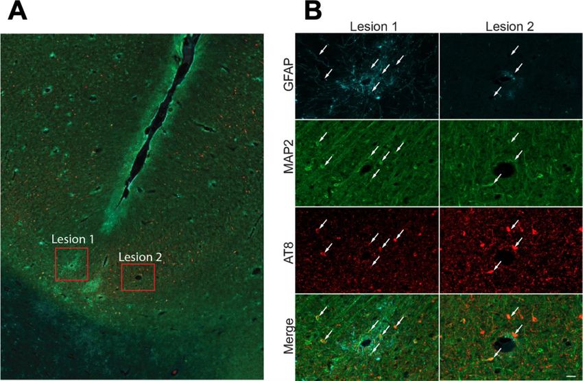
Free Neuropathology 2:9 (2021)                                                                          John F. Crary
doi: https://doi.org/10.17879/freeneuropathology-2021-3317                                               page 2 of 12

thank Mussolini for having declared me to be of an           2020). Nevertheless, researchers began to speculate
inferior race. This led me to the joy of working, not        that there may be long-term neurological sequelae
any more unfortunately in university institutes, but         that follow COVID-19 as examples of post-viral
in a bedroom.” Upon completing this review of just           tauopathy and neurodegeneration are well docu-
a few of the accomplishments of the past year, it            mented and now there is increasing alarm that there
was clear that this spirit is alive, the same that com-      might be an incipient rise in neurological complica-
pelled Levy-Montalcini to work tirelessly at home to         tions, including dementia and neurodegeneration,
study chick embryos using scalpels fashioned from            that might follow the pandemic (Badrfam & Zan-
sewing needles. The spirit could be felt emanating           difar, 2020).
from our colleagues sheltering at home, many also
literally working in their bedrooms! So, it is with                 Neurologists and other clinicians have been de-
great pride and appreciation that we take this look          scribing persistent neurological symptomatology in
back and celebrate the accomplishments of our col-           COVID-19 survivors, including inattentiveness,
leagues and recognize just a few of the many trans-          memory impairment and a constellation of other
formative advances in 2020. The pandemic is a re-            cognitive symptoms (i.e., “brain fog”). These are
minder that substantive scientific progress comes            part of an entity now termed the post-acute COVID-
with great human effort and rarely a “eureka mo-             19 syndrome (PACS). The underlying neuropatholog-
ment.” Here, we also highlight collaborative efforts,        ical substrate for these findings remains unclear, but
research trends, groundbreaking methodologies,               the effects of hypoxic-ischemic injury, chronic neu-
and tools. A concerted effort was made to seek out           roinflammation, toxic encephalopathy from break-
innovations across all neurodegenerative disease             down of the blood brain barrier are candidates. Di-
categories. Given the readership and mission of this         rect effects of the virus are not completely ruled out,
journal, we emphasized pathoanatomical studies               and the potential for neurotropic strains to emerge
most relevant to neuropathologists and clinical neu-         remains a possibility. Given the existence of enceph-
roscientists.                                                alitis lethargica and measles related subacute scle-
                                                             rosing panencephalitis, both virus related and asso-
                                                             ciated with neurofibrillary tangles, it is not unrea-
1. The long-term neuropathological se-                       sonable to speculate that a degenerative compo-
quelae of COVID-19                                           nent of PACS might exist. Critically, it is imperative
                                                             to understand whether these PACS neurological
      Over the past year, the SARS-CoV-2/COVID-19            symptoms are progressive and we expect a surge in
pandemic has permeated essentially every aspect of           translational studies in the coming years focused on
our lives, and research in neurodegenerative disease         the long-term changes that follow COVID-19.
is no exception, launching a new, albeit speculative,
research trend. Early in the pandemic, neuropa-
thologists began to wonder about the extent to
                                                             2. Proteomic analysis: spotlight on mi-
which COVID-19 might impact brain health. Theoret-           croglial and astrocyte inflammation in
ically, this could be through direct neurotropism as         Alzheimer disease
some coronaviruses are known to cause encephali-
tis, or perhaps indirectly through complications re-              In the previous decades, transformative tech-
lated to critical illness and systemic disease (Mor-         nologies have enabled large-scale genomic and tran-
gello, 2020). Early reports documented encephalitis          scriptomic studies of post-mortem brain tissues that
in the setting of COVID-19 (Reichard et al., 2020),          have been directed towards neurodegenerative dis-
but SARS-CoV-2 encephalitis was considered a rare            eases, yielding a flood of data out of which has
event predominantly restricted to susceptible popu-          emerged a rich and complex picture of Alzheimer
lations and of modest public health concern. Larger          disease (AD). But what about the proteins? Owning
autopsy series emerged, highlighting infarction and          to their relatively more complex chemical struc-
microthrombi in the brain in the acute setting (Fig-         tures, measuring structural changes on the protein
ure 1), more commonly in hospitalized patients, but          level in a high throughput manner is much more
this too was thought to be uncommon (Bryce et al.,           challenging. But now, proteomic technologies have
Free Neuropathology 2:9 (2021)                                                                                             John F. Crary
doi: https://doi.org/10.17879/freeneuropathology-2021-3317                                                                  page 3 of 12

Figure 1. Post-mortem findings in a patient with SARS-CoV-2/COVID-19. A. Fresh gross image of a right hemibrain from a 68 male with
COVID-19 showing diffuse swelling and leptomeningeal hemorrhage. B. Fixed right hemibrain from the same patient showing acute and
subacute infarction in the distribution of the middle cerebral artery territory. C. Luxol fast blue counterstained hematoxylin and eosin
(LH&E) section showing subacute hemorrhagic infarction. D. LH&E-stained section from the neocortical white matter in a 29-year-old
male COVID-19 patient with acute disseminated encephalomyelitis (ADEM).

advanced sufficiently, and we are beginning to see                      cluding controls, people with asymptomatic AD neu-
them take center stage. Here and in the next two                        ropathology and those with dementia due to AD
sections we highlight three such studies.                               (Johnson et al., 2020). Cases were derived from a to-
                                                                        tal of eight collections, including the Religious Or-
     The first study, published in Nature Medicine,                     ders Study and Memory and Aging Project (ROS-
led by Nicholas Seyfried and Allan Levey at Emory                       MAP), Mayo Clinic brain bank, Baltimore Longitudi-
University, profiled 2,000 brain samples, comparing                     nal Study of Aging (BLSA), Baltimore Coroner’s Of-
more than 3,000 proteins from multiple cohorts, in-                     fice, Banner Sun Health Research Institute, Mount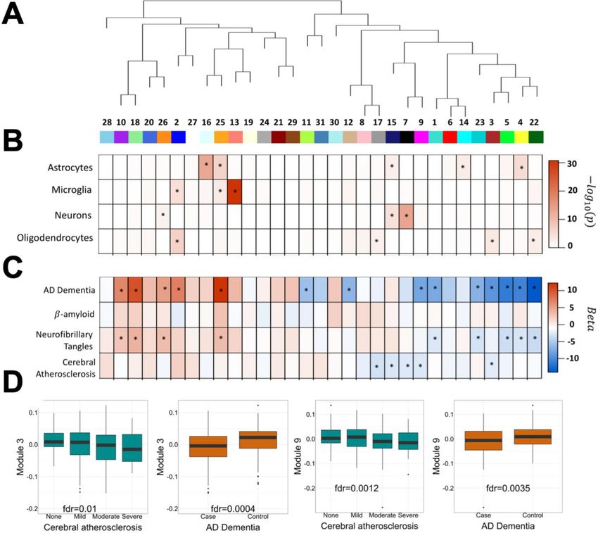
Free Neuropathology 2:9 (2021)                                                                                              John F. Crary
doi: https://doi.org/10.17879/freeneuropathology-2021-3317                                                                   page 4 of 12

Sinai School of Medicine Brain Bank, Adult Changes                       3. Proteomic analysis: the contribution
in Thought Study (ACT), and University of Pennsylva-
nia School of Medicine Brain Bank. Researchers
                                                                         of cerebral atherosclerosis to cognitive
identified 13 different gene sets (“modules”) based                      decline
on co-expression patterns (Figure 2). Of these, six
were correlated with AD neuropathological features                              Another proteomics paper published in Nature
(i.e., amyloid plaques, neurofibrillary tangles) or                      Neuroscience, also from Emory University led by
cognitive impairment. Of the three that were most                        Nicholas Seyfried, Allan Levey, and Thomas S.
closely correlated with AD features, two were down-                      Wingo, examined cerebral atherosclerosis as it re-
regulated, including the M1 that was composed of                         lates to cognitive decline and AD (Wingo et al.,
proteins involved in synaptic function and the M3                        2020). Here, the investigators performed a prote-
that tracked with mitochondrial proteins. M4 had                         ome-wide association study (PWAS), which is similar
the strongest correlation with disease traits and                        to a genome-wide association study (GWAS), to look
contained astrocyte and microglial proteins involved                     more broadly at relevant neuropathological
in sugar metabolism. Intriguingly, many of the pro-                      changes, including amyloid-beta deposition, neuro-
teins in the M4 module were encoded by genes that                        fibrillary tangles, infarcts (both macro and microin-
are AD risk loci. Importantly, regardless of their pre-                  farcts), cerebral amyloid angiopathy, TAR DNA-bind-
cise function and role in disease, 27 of them were                       ing protein 43 (TDP-43, transactive response DNA
detected in CSF, underscoring their potential utility                    binding protein 43 kDa), Lewy body pathology, hip-
as biomarkers. Because this was funded by the ac-                        pocampal sclerosis, and atherosclerosis. Out of this
celerating medicine partnership - AD (AMP-AD) pro-                       analysis, 114 proteins emerged, independently of
ject, all of these data are publicly available for the                   cerebrovascular risk factors (e.g., hypertension, dia-
research community and will surely catalyze contin-                      betes, smoking; Figure 3). Strikingly, many were
ued proteomic studies.

Figure 2. Protein network analysis in post-mortem Alzheimer disease (AD) brain. A. Brain tissue from the dorsolateral prefrontal cortex
from AD patients and compared to cognitively normal subjects with age-related pathology and pathologically negative controls from four
cohorts. B. A correlation network consisting of 13 modules using 3334 proteins was created and correlated with neuropathological as-
sessments, cell type markers, and the associated biological processes identified using gene ontology (GO) analysis. Images courtesy of Dr.
Erik C. Johnson, Allan I. Levy, Nicholas T. Seyfried, reproduced with permission under the creative commons license.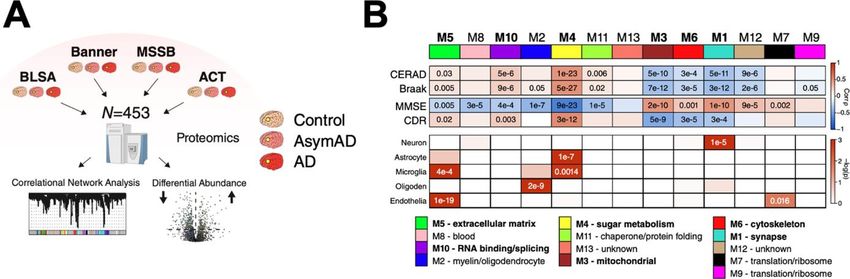
Free Neuropathology 2:9 (2021)                                                                                         John F. Crary
doi: https://doi.org/10.17879/freeneuropathology-2021-3317                                                              page 5 of 12

Figure 3. Co-expression network analysis of protein modules in post-mortem human brain. A, B. A total of 31 modules were identified
and protein signatures mapped to cell type. C. These modules were then associated with neuropathological outcomes. D. Differences in
module eigen proteins were seen modules 3 and 9 for atherosclerosis and Alzheimer disease-related dementia. Images curtesy of Drs.
Thomas Wingo, Aliza Wingo, Nicholas Seyfried, Allan I. Levey (Emory University School of Medicine).

from oligodendrocytes involved in myelination.                        functioning in brains of individuals with cerebral ath-
Other genes were involved in mRNA processing and                      erosclerosis. This may be related to another finding
splicing. They performed co-expression network                        of higher levels of neurofilament light (NfL) and me-
analysis and 31 modules were identified, with five                    dium (NfM) which were higher in cases with athero-
being linked independently to cerebral atheroscle-                    sclerosis, perhaps reflecting axonal injury. This study
rosis. This analysis pointed to oligodendrocyte func-                 will drive continued research into the effects of cer-
tion alongside downregulation of mRNA processing                      ebral atherosclerosis on the brain proteome and
in neurons and astrocytes with impaired synaptic                      their contribution to AD.
Free Neuropathology 2:9 (2021)                                                                          John F. Crary
doi: https://doi.org/10.17879/freeneuropathology-2021-3317                                               page 6 of 12

4. Proteomic analysis: A high-resolu-                        N-terminal domain inserts (0N). The 0N4R isoform is
                                                             highly overrepresented in aggregates isolated from
tion quantitative map of post-transla-                       early disease stages alongside modifications associ-
tional modifications of tau pro-                             ated with increased negative charge in the proline-
teoforms                                                     rich region and decreased positive charge in the mi-
                                                             crotubule binding domain. These findings are prom-
      Neurofibrillary tangles exist in a spectrum of         ising because they give us the most comprehensive
morphological forms that are immediately recog-              picture of tau secondary structure and provide a
nizable using immunohistochemistry with antisera             pathway towards continuing to advance the bi-
targeting hyperphosphorylated tau (p-tau). The ear-          omarkers that are rapidly evolving and identify the
liest detectable neurofibrillary change is the pres-         critical modifications that are responsible for the
ence faint granular p-tau labeling in pre-tangles; this      pathogenicity of tau paving the way towards new
staining accumulates and coalesces into intracellular        therapeutics.
tangles that eventually become extracellular
(“tombstone” or “ghost”) tangles following neuronal
                                                             5. Plasma biomarkers for Alzheimer
death. Understanding the molecular events that cor-
respond to this sequence would be extremely help-            disease
ful for developing the next generation of diagnostics
(see below) and therapeutics. The pathobiology of                  Obtaining a definitive diagnosis of a neuro-
tau pathology is complex, with multiple tau isoforms         degenerative disease continues to require an au-
undergoing innumerable secondary structural mod-             topsy, the perennial gold standard, but continued
ifications in disease states beyond just hyperphos-          advances in blood-based biomarkers are continuing
phorylation, including truncation, acetylation, ubiq-        to challenge this and offer a low-cost, convenient al-
uitination, glycosylation and methylation. It has            ternative. Over the past decade, substantial pro-
been challenging to temporally map the order of              gress has been made in our ability to assess molecu-
these events using traditional approaches (e.g.,             lar changes in the central nervous system using non-
phospho-specific tau antisera-based methods)                 invasive amyloid positron emission tomography
which lack the ability to synchronously and compre-          (PET) scans and cerebrospinal fluid (CSF) markers.
hensively detect these critical changes.                     Yet these modalities are expensive, inconvenient
                                                             and come with a mild degree of risk. The long-sought
      In the last proteomics paper, published in Cell        development of a blood-based biomarker for Alz-
in November of 2020, a powerful study, led by Dr.            heimer disease (AD), had been considered unrealis-
Judith Steene at Boston Children’s Hospital, detailed        tic because the levels of brain proteins in the blood
their focused analysis that was entirely directed to-        had been thought to be simply too low to be reliably
wards the tau protein (Wesseling et al., 2020). They         and reproducibly detected. Further, there was con-
deployed a previously published mass spectrome-              cern that the extent to which blood levels of various
try-based assay, that they term “FLEXI-tau” (Mair et         factors reflect ongoing disease processes might be
al., 2016), which allowed them to generate a high-           too disconnected or tangential to be clinically useful.
resolution quantitative proteomic map of 95 post-            So, it comes as no surprise that the research com-
translational modifications on multiple tau isoforms         munity has been stunned by the rapid progress in
from 91 human post-mortem brains. They then used             blood-based biomarkers in neurodegeneration. This
unsupervised analyses to predict sequential addi-            year a milestone was achieved, with the “Preciv-
tion of modifications. They found that while there is        ityAD™” mass-spec amyloid-β assay, which was de-
a great degree of heterogeneity, there appears to be         veloped at Washington University Saint Louis in the
a minimal set of modifications that develop in a pro-        laboratory directed by Dr. Randall Bateman, receiv-
cessive fashion and are associated with tissue frac-         ing approval under the Clinical Laboratory Improve-
tions associated with disease stage as well as tau           ment Amendments (CLIA) as well as an FDA break-
seeding activity. The analysis also highlighted a spe-       through designation. However, this is only half of
cific alternatively spliced tau isoform with four mi-        the story. The other essential neuropathological
crotubule binding domain repeats (4R), but lacking           hallmark of AD, the neurofibrillary tangle composed
Free Neuropathology 2:9 (2021)                                                                           John F. Crary
doi: https://doi.org/10.17879/freeneuropathology-2021-3317                                                page 7 of 12

of abnormal tau, is also required for a diagnosis. Re-       in development since the 1970s, but recent tech-
markably, similar rapid progress is being made on            nical advances in detectors and software algorithms
this front with a series of groundbreaking studies           have enabled solving molecular structures at near-
that were published over that past year, continuing          atomic resolution. The power of cryo-EM was first
the momentum.                                                demonstrated that year to the neurodegenerative
                                                             disease research community when the approach
      In previous years, the first papers emerged de-        was applied to paired-helical filament tau fibrils in
scribing highly sensitive immunoassays identifying           Alzheimer disease, illuminating at near atomic reso-
p-tau phosphorylated at threonines 181 or 217 as             lution the C-shaped conformation of the core (Fitz-
early promising biomarkers. In 2020, phospho-thre-           patrick et al., 2017). This report was quickly followed
onine 231 (p-tau231) was reported to be an excel-            by the solving of the structure for the J-shaped tau
lent early marker for tau pathology in CSF (Suárez-          filament core in Pick disease which contains addi-
Calvet et al., 2020). This was followed up with results      tional residues making it slightly longer than in AD
from a study using an ultrasensitive Single molecule         (Falcon et al., 2018). Next came chronic traumatic
array (SIMOA) for the quantification of p-tau231 in          encephalopathy, which has a similar C-shape to that
blood plasma (Ashton et al., 2021). The study in-            in Alzheimer disease (Falcon et al., 2019). Also de-
cluded a total of 588 subjects and successfully differ-      scribed was an additional non-proteinaceous hydro-
entiated AD from amyloid-β negative cognitively              phobic density that has yet to be defined. These
normal individuals. Plasma p-tau231 also differenti-         findings support the notion that there exists a spec-
ated AD patients from non-AD neurodegenerative               trum of hitherto unrecognized tau structural fea-
disorders and amyloid-β negative MCI patients. In            tures and conformations, often termed “strains” in
samples taken from patients with post-mortem au-             a nod to the prion literature that has informed much
topsy confirmation, plasma p-tau231 was extremely            of the research on protein misfolding, that may be
accurate in identifying AD neuropathology in com-            distinct in different disease states. Now, we are con-
parison to non-AD neurodegenerative disorders                tinuing to see additional insights with two papers
(AUC = 0.99). Plasma p-tau231 was highly correlated          describing the structure of the tau filament core in
with other AD biomarkers, i.e., CSF p-tau231, tau            corticobasal degeneration as well as another which
PET ([18F]MK-6240) and amyloid-β PET ([18F]AZD).             is the first cryo-EM structure of α-synuclein (see next
The elevations in p-tau231 were a very early change,
                                                             section).
preceding amyloid-β PET and plasma p-tau181.
Plasma p-tau231 had the power to resolve even sub-                 In a paper published in Cell in February 2020, a
tle differences, differentiating subjects across even        team led by Anthony Fitzpatrick at Columbia Univer-
early Braak stages. Together, we are witnessing im-          sity detailed their study showing the cryo-EM struc-
pressive progress in blood-based AD biomarkers               ture of filaments from corticobasal degeneration
with the potential to detect the earliest disease            (Arakhamia et al., 2020). In additional analyses, they
stages, prior to significant irreversible brain destruc-     further integrated these cryo-EM structures and
tion, and enable the next generation of clinical trials.     those from AD with mass spectrometry data to lo-
                                                             calize post-translational secondary modifications. A
                                                             separate study, published in Nature, led by Sjors
6. Cryo-electron microscopy: ultrahigh                       Scheres and Michel Goedert at the MRC Laboratory
                                                             of Molecular Biology in Cambridge, England, U.K.
resolution structure of tau in cortico-                      (Zhang et al., 2020). This team also showed a similar
basal degeneration                                           structure in CBD (Figure 4). Together, in addition to
                                                             providing a validated atomic level resolution C-
     In 2017, the Nobel Prize in Chemistry was               shaped structure of the tau filament in CBD, these
awarded to Jacques Dubochet, Joachim Frank and               studies both revealed a density buried in the struc-
Richard Henderson for cryo-electron microscopy               ture that represents a molecule of unknown iden-
(cryo-EM). The approach, which involves cooling              tity, akin to what was identified in CTE. The fibrils in
samples to cryogenic temperatures and embedding              CBD are composed entirely of tau with 4 microtu-
them in an environment of vitreous water, has been           bule binding domain repeats (4R), which differs from
Free Neuropathology 2:9 (2021) John F. Crary doi: https://doi.org/10.17879/freeneuropathology-2021-3317 page 8 of 12 Figure 4. Cryo-EM of tau filaments in corticobasal degeneration. A. Negative-stain electron micrographs of type I and type II tau filaments extracted from the frontal cortex of CBD. B. Cryo-EM maps of type I and type II tau filaments from the frontal cortex of CBD. C. Top, the microtubule-binding repeats (R1–R4) of tau and the sequence after R4 that is present in the core of CBD filaments (all shown in different colors). Bottom, atomic model of the CBD type II tau filament. The extra density is shown in light blue, with K290, K294 and K370 indicated. Images courtesy of Drs. Wenjuan Zhang, Sjors Scheres and Michel Goedert MRC Laboratory of Molecular Biology, Cambridge). Pick disease which is composed of 3R tau as well as tential to pave the way towards improved diagnos- AD and CTE which are mixed 3R and 4R. Additional tics and advancing our understanding of the diver- future studies directed towards further delineation sity of conformers in tauopathy that may be associ- of the diversity of tau filament structures has the po- ated with different clinical symptomatology.
Free Neuropathology 2:9 (2021) John F. Crary
doi: https://doi.org/10.17879/freeneuropathology-2021-3317 page 9 of 12
7. Cryo-electron microscopy: ultrahigh using recombinant proteins by altering buffer condi-
tions (Lau et al., 2020). They generated a host of fi-
resolution structures of α-synuclein brils that varied in their biophysical properties and
conformers compared them with those derived from human
post-mortem brain samples. Then, the fibrils were
This year we also saw reports of the first ultra- inoculated into a transgenic mouse line that overex-
high resolution cryo-EM structures of α-synuclein fi- presses human mutant α-synuclein and observed
brils published in Nature also from Sjors Scheres and them. Over time, the mice developed a range of
Michel Goedert at the MRC Laboratory of Molecular traits that were strain specific, including aggregate
Biology (Schweighauser et al., 2020). The synucle- morphology, incubation periods and behavioral
inopathies are a group of neurodegenerative disor- changes, that could be propagated serially (Figure
ders that include Parkinson’s disease (PD), PD de- 5). In the brains of these animals, distinct neuroana-
mentia, diffuse Lewy body disease (DLBD) and mul- tomical vulnerability was observed that was dictated
tiple system atrophy (MSA). Co-first authors by strain type. These findings give us an additional
Schweighauser and Shi et al., studied three cases of model for testing questions related to how these dif-
DLBD and five with MSA. In MSA, they found two ferent α-synuclein conformers target specific cells
types of filament, that they termed type 1 and type and brain regions leading to neurodegeneration.
2, each with filaments with different protofibrils that
make up the core. The conformation of α-synuclein
filaments derived from DLBD brains by cryo-EM pre-
cluded 3D imaging, so 2D class averaging was used
to show that there were differences in the structure
from MSA. Overall, these structural differences are
of great interest as accumulating evidence suggests
that α-synuclein might behave in a prion-like man-
ner, with templating and propagation of abnormal
structures/conformers. This knowledge may be
helpful in understanding the pathogenesis and nat-
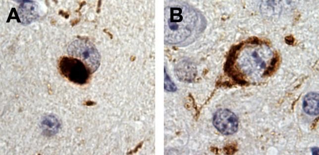
ural history of the synucleinopathies, and especially Figure 5. Distinct midbrain pathology in transgenic α-synuclein
in the development of specific α-synuclein PET trac- (M83) mice inoculated with salt (S) fibrils or no salt (NS) fibrils.
ers, which have been so far elusive. A. The NS fibril-injected mice show the “Lewy body-like” α-synu-
clein pathology. B. The S fibril-injected mice exhibit the “ring-
like” α-synuclein pathology. Both images show stains with the
8. Characterizing α-synuclein strains EP1536Y antibody that recognizes synuclein phosphorylated at
serine 129. Images courtesy of Dr. Joel Watts (University of To-
The α-synucleinopathies, including Parkinson ronto).
disease, diffuse Lewy body disease, and dementia
with Lewy bodies, while linked by aggregation of α-
synuclein, have been hypothesized to diverge based 9. Huntington’s disease and chorea
on the prion-like properties unique disease confor-
mational structural folding that templates replica-
Abnormalities in proteostasis are a key feature
tion of the abnormality. These strains are hypothe-
of essentially all neurodegenerative disorders. This
sized to have different properties and may influence
change is associated with endoplasmic reticulum
neuropathological features and symptomatology.
(ER) stress, which then triggers the unfolded protein
This year, a study was published that took us response (UPR). XBP1 is a key mediator of the ER
one step closer to understanding the pathobiology stress response thought to be a master regulator of
and diversity of α-synuclein strains. In an elegant set the UPR that drives the adaptation response to re-
of experiments, Lau et al. recreated an array of α- cover proteostasis through a number of mecha-
synuclein fibrils with different confirmations in vitro nisms.Free Neuropathology 2:9 (2021) John F. Crary
doi: https://doi.org/10.17879/freeneuropathology-2021-3317 page 10 of 12
In a series of compelling experiments, a team ingly found that these pathways were not responsi-
from the University of Chile showed that treatment ble. Remarkably, they found IGF2 signaling en-
with IGF2, a factor that they previously implicated as hanced secretion of soluble mutant huntingtin into
a downstream effector of XBP1 in animal models de- exosomes/microvesicles. These findings were not
ficient in XBP1, may be an important contributor to limited to cell culture but were recapitulated when
this finding (García-Huerta et al., 2020). Treatment IGF2 was infused into the brain of HD mice. Finally,
with IGF1 led to a marked reduction in the burden of these findings were validated in human tissue sam-
intracellular aggregates of mutant huntingtin in cel- ples, with a reduction in IGF2 in post-mortem HD
lular models, including an induced pluripotent stem brain and blood. These findings take us one step
cell (iPSC)-derived model of medium spiny neurons closer to understanding the role of the UPR in HD
from HD patients. When the autophagy and the and suggest a mechanism that could be targeted for
ubiquitin proteasome were assessed, they surpris- therapeutics.
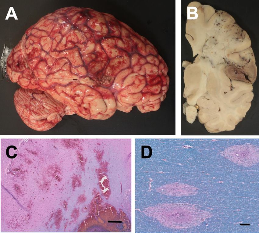
Figure 6. Immunofluorescence microscopy in a sulcal region from an individual who passed away in their 20s with chronic traumatic
encephalopathy (CTE). A. Low power image highlighting two perivascular lesions. B. High power image showing staining with markers for
astroglia (GFAP, blue), neurons (MAP2, green) and abnormal hyperphosphorylated p-tau (AT8; green). White arrows mark colocalization.
In these lesions, there is only MAP2 overlap with p-tau. No GFAP/AT8 colocalization was seen in this case. Scale bar is 50 um. Images
courtesy of Dr. Jonathan Cherry and Ann McKee (Boston University and Boston VA).Free Neuropathology 2:9 (2021) John F. Crary
doi: https://doi.org/10.17879/freeneuropathology-2021-3317 page 11 of 12
10. Chronic traumatic encephalopathy: sulcal depths, the precise region that is biomechani-
cally vulnerable in CTE and where tau pathology first
what about the astrocytes? emerges (Arena et al., 2020). This is in contrast to
neuronal tau pathology which they found was more
Slightly over ten years ago, chronic traumatic
diffuse. This seems to suggest that astrocyte pathol-
encephalopathy (CTE) was propelled to the fore of
ogy is an early event. In another study published in
neurodegenerative disease research where it has re-
Brain Pathology led by Ann McKee and John Crary
mained. Since that time, broad consensus has
(writer of this review), a spectrum of cases that also
emerged among neuropathologists that CTE has a
included very early CTE lesions was able to pinpoint
distinct neuropathological presentation that is dis-
abnormal tau in early pathognomonic lesions to
tinguishable from other tauopathies, with a unique
perivascular neurons (Figure 6), but not astrocytes,
pathognomonic perivascular lesion that was codi-
which appeared to accumulate later in the disease
fied in consensus criteria (McKee et al., 2016). In this
progression (Cherry et al., 2020). This tau pathology
report, the pathognomonic lesion was described as
consisted predominantly of tau isoforms containing
consisting of “p-tau aggregates in neurons, astro-
four microtubule binding domain repeats (4R tau),
cytes, and cell processes around small vessels in an
an isoform that has been proposed to be especially
irregular pattern at the depths of the cortical sulci”.
toxic given its increased propensity towards aggre-
While this working definition was considered suffi-
gation. Together, these findings point to a complex
ciently precise, confusion emerged and persisted in
picture of evolving tau pathology in neurons and as-
the literature around the perivascular astrocytes in
trocytes early in CTE and we expect that research in
aging related tau astrogliopathy (ARTAG) (Kovacs et
the coming years around the role of astrocytes in
al., 2016) that can generally be differentiated based
early CTE will intensify.
on the absence of neurons and localization in the
white matter or subpial compartments. Clarification
was not sufficient to put the astrocyte question to Acknowledgement
rest (McKee et al., 2020).
Dr. Crary receives research funding from the
Setting aside ARTAG, there is a tremendous NIH (P30AG066514, R01AG054008, R01NS095252,
burden of p-tau positive gray matter astrocytes in R01AG062348, R01NS086736, U54NS115266,
CTE. A study led by John Trojanowski at the Univer- U54NS115322). We further acknowledge the Rain-
sity of Pennsylvania and William Stewart at the Uni- water Charitable Trust/Tau Consortium, David and
versity of Glasgow published in Brain Communica- Elsie Werber, Alexander Saint Amand Scholar Award
tions sought to neuroanatomically map these astro- and Karen Strauss Cook Research Scholar Award.
cytes and found a preferential accumulation in the
References
Arakhamia, T., Lee, C. E., Carlomagno, Y., Duong, D. M., Kundinger, S. R., Badrfam, R., & Zandifar, A. (2020). From encephalitis lethargica to
Wang, K., Williams, D., DeTure, M., Dickson, D. W., Cook, C. N., Seyfried, COVID-19: Is there another epidemic ahead? Clinical Neurology and
N. T., Petrucelli, L., & Fitzpatrick, A. W. P. (2020). Posttranslational Neurosurgery, 196, 106065.
Modifications Mediate the Structural Diversity of Tauopathy Strains. https://doi.org/10.1016/j.clineuro.2020.106065
Cell, 180(4), 633-644.e12. https://doi.org/10.1016/j.cell.2020.01.027
Bryce, C., Grimes, Z., Pujadas, E., Ahuja, S., Beasley, M. B., Albrecht, R.,
Arena, J. D., Johnson, V. E., Lee, E. B., Gibbons, G. S., Smith, D. H., Hernandez, T., Stock, A., Zhao, Z., Rasheed, M. A., Chen, J., Li, L., Wang,
Trojanowski, J. Q., & Stewart, W. (2020). Astroglial tau pathology alone D., Corben, A., Haines, K., Westra, W., Umphlett, M., Gordon, R. E.,
preferentially concentrates at sulcal depths in chronic traumatic Reidy, J., … Fowkes, M. (2020). Pathophysiology of SARS-CoV-2:
encephalopathy neuropathologic change. Brain Communications, 2(2), Targeting of endothelial cells renders a complex disease with
fcaa210. https://doi.org/10.1093/braincomms/fcaa210 thrombotic microangiopathy and aberrant immune response. The
Mount Sinai COVID-19 autopsy experience. MedRxiv,
Ashton, N. J., Pascoal, T. A., Karikari, T. K., Benedet, A. L., Lantero- 2020.05.18.20099960. https://doi.org/10.1101/2020.05.18.20099960
Rodriguez, J., Brinkmalm, G., Snellman, A., Schöll, M., Troakes, C., Hye,
A., Gauthier, S., Vanmechelen, E., Zetterberg, H., Rosa-Neto, P., & Cherry, J. D., Kim, S. H., Stein, T. D., Pothast, M. J., Nicks, R., Meng, G.,
Blennow, K. (2021). Plasma p-tau231: A new biomarker for incipient Huber, B. R., Mez, J., Alosco, M. L., Tripodis, Y., Farrell, K., Alvarez, V. E.,
Alzheimer’s disease pathology. Acta Neuropathologica. McKee, A. C., & Crary, J. F. (2020). Evolution of neuronal and glial tau
https://doi.org/10.1007/s00401-021-02275-6 isoforms in chronic traumatic encephalopathy. Brain Pathology (Zurich,
Switzerland). https://doi.org/10.1111/bpa.12867Free Neuropathology 2:9 (2021) John F. Crary
doi: https://doi.org/10.17879/freeneuropathology-2021-3317 page 12 of 12
Falcon, B., Zhang, W., Murzin, A. G., Murshudov, G., Garringer, H. J., McKee, A. C., Stein, T. D., Crary, J. F., Bieniek, K. F., Cantu, R. C., & Kovacs,
Vidal, R., Crowther, R. A., Ghetti, B., Scheres, S. H. W., & Goedert, M. G. G. (2020). Practical Considerations in the Diagnosis of Mild Chronic
(2018). Structures of filaments from Pick’s disease reveal a novel tau Traumatic Encephalopathy and Distinction From Age-Related Tau
protein fold. Nature, 561(7721), 137–140. Astrogliopathy. Journal of Neuropathology and Experimental Neurology,
https://doi.org/10.1038/s41586-018-0454-y 79(8), 921–924. https://doi.org/10.1093/jnen/nlaa047
Falcon, B., Zivanov, J., Zhang, W., Murzin, A. G., Garringer, H. J., Vidal, Morgello, S. (2020, August). Coronaviruses and the central nervous
R., Crowther, R. A., Newell, K. L., Ghetti, B., Goedert, M., & Scheres, S. system. Journal of Neurovirology; J Neurovirol.
H. W. (2019). Novel tau filament fold in chronic traumatic https://doi.org/10.1007/s13365-020-00868-7
encephalopathy encloses hydrophobic molecules. Nature, 568(7752),
420–423. https://doi.org/10.1038/s41586-019-1026-5 Reichard, R. R., Kashani, K. B., Boire, N. A., Constantopoulos, E., Guo, Y.,
& Lucchinetti, C. F. (2020). Neuropathology of COVID-19: A spectrum of
Fitzpatrick, A. W. P., Falcon, B., He, S., Murzin, A. G., Murshudov, G., vascular and acute disseminated encephalomyelitis (ADEM)-like
Garringer, H. J., Crowther, R. A., Ghetti, B., Goedert, M., & Scheres, S. H. pathology. Acta Neuropathologica, 140(1), 1–6.
W. (2017). Cryo-EM structures of tau filaments from Alzheimer’s https://doi.org/10.1007/s00401-020-02166-2
disease. Nature, 547(7662), 185–190.
https://doi.org/10.1038/nature23002 Schweighauser, M., Shi, Y., Tarutani, A., Kametani, F., Murzin, A. G.,
Ghetti, B., Matsubara, T., Tomita, T., Ando, T., Hasegawa, K., Murayama,
García-Huerta, P., Troncoso-Escudero, P., Wu, D., Thiruvalluvan, A., S., Yoshida, M., Hasegawa, M., Scheres, S. H. W., & Goedert, M. (2020).
Cisternas-Olmedo, M., Henríquez, D. R., Plate, L., Chana-Cuevas, P., Structures of α-synuclein filaments from multiple system atrophy.
Saquel, C., Thielen, P., Longo, K. A., Geddes, B. J., Lederkremer, G. Z., Nature, 585(7825), 464–469.
Sharma, N., Shenkman, M., Naphade, S., Sardi, S. P., Spichiger, C., https://doi.org/10.1038/s41586-020-2317-6
Richter, H. G., … Hetz, C. (2020). Insulin-like growth factor 2 (IGF2)
protects against Huntington’s disease through the extracellular disposal Suárez-Calvet, M., Karikari, T. K., Ashton, N. J., Lantero Rodríguez, J.,
of protein aggregates. Acta Neuropathologica, 140(5), 737–764. Milà-Alomà, M., Gispert, J. D., Salvadó, G., Minguillon, C., Fauria, K.,
https://doi.org/10.1007/s00401-020-02183-1 Shekari, M., Grau-Rivera, O., Arenaza-Urquijo, E. M., Sala-Vila, A.,
Sánchez-Benavides, G., González-de-Echávarri, J. M., Kollmorgen, G.,
Johnson, E. C. B., Dammer, E. B., Duong, D. M., Ping, L., Zhou, M., Yin, L., Stoops, E., Vanmechelen, E., Zetterberg, H., … ALFA Study. (2020). Novel
Higginbotham, L. A., Guajardo, A., White, B., Troncoso, J. C., tau biomarkers phosphorylated at T181, T217 or T231 rise in the initial
Thambisetty, M., Montine, T. J., Lee, E. B., Trojanowski, J. Q., Beach, T. stages of the preclinical Alzheimer’s continuum when only subtle
G., Reiman, E. M., Haroutunian, V., Wang, M., Schadt, E., … Seyfried, N. changes in Aβ pathology are detected. EMBO Molecular Medicine,
T. (2020). Large-scale proteomic analysis of Alzheimer’s disease brain 12(12), e12921. https://doi.org/10.15252/emmm.202012921
and cerebrospinal fluid reveals early changes in energy metabolism
associated with microglia and astrocyte activation. Nature Medicine, Wesseling, H., Mair, W., Kumar, M., Schlaffner, C. N., Tang, S.,
26(5), 769–780. https://doi.org/10.1038/s41591-020-0815-6 Beerepoot, P., Fatou, B., Guise, A. J., Cheng, L., Takeda, S., Muntel, J.,
Rotunno, M. S., Dujardin, S., Davies, P., Kosik, K. S., Miller, B. L., Berretta,
Kovacs, G. G., Ferrer, I., Grinberg, L. T., Alafuzoff, I., Attems, J., Budka, S., Hedreen, J. C., Grinberg, L. T., … Steen, J. A. (2020). Tau PTM Profiles
H., Cairns, N. J., Crary, J. F., Duyckaerts, C., Ghetti, B., Halliday, G. M., Identify Patient Heterogeneity and Stages of Alzheimer’s Disease. Cell,
Ironside, J. W., Love, S., Mackenzie, I. R., Munoz, D. G., Murray, M. E., 183(6), 1699-1713.e13. https://doi.org/10.1016/j.cell.2020.10.029
Nelson, P. T., Takahashi, H., Trojanowski, J. Q., … Dickson, D. W. (2016).
Aging-related tau astrogliopathy (ARTAG): Harmonized evaluation Wingo, A. P., Fan, W., Duong, D. M., Gerasimov, E. S., Dammer, E. B., Liu,
strategy. Acta Neuropathologica, 131(1), 87–102. Y., Harerimana, N. V., White, B., Thambisetty, M., Troncoso, J. C., Kim,
https://doi.org/10.1007/s00401-015-1509-x N., Schneider, J. A., Hajjar, I. M., Lah, J. J., Bennett, D. A., Seyfried, N. T.,
Levey, A. I., & Wingo, T. S. (2020). Shared proteomic effects of cerebral
Lau, A., So, R. W. L., Lau, H. H. C., Sang, J. C., Ruiz-Riquelme, A., Fleck, S. atherosclerosis and Alzheimer’s disease on the human brain. Nature
C., Stuart, E., Menon, S., Visanji, N. P., Meisl, G., Faidi, R., Marano, M. Neuroscience, 23(6), 696–700.
M., Schmitt-Ulms, C., Wang, Z., Fraser, P. E., Tandon, A., Hyman, B. T., https://doi.org/10.1038/s41593-020-0635-5
Wille, H., Ingelsson, M., … Watts, J. C. (2020). α-Synuclein strains target
distinct brain regions and cell types. Nature Neuroscience, 23(1), 21–31. Zhang, W., Tarutani, A., Newell, K. L., Murzin, A. G., Matsubara, T.,
https://doi.org/10.1038/s41593-019-0541-x Falcon, B., Vidal, R., Garringer, H. J., Shi, Y., Ikeuchi, T., Murayama, S.,
Ghetti, B., Hasegawa, M., Goedert, M., & Scheres, S. H. W. (2020). Novel
Mair, W., Muntel, J., Tepper, K., Tang, S., Biernat, J., Seeley, W. W., Kosik, tau filament fold in corticobasal degeneration. Nature, 580(7802), 283–
K. S., Mandelkow, E., Steen, H., & Steen, J. A. (2016). FLEXITau: 287. https://doi.org/10.1038/s41586-020-2043-0
Quantifying Post-translational Modifications of Tau Protein in Vitro and
in Human Disease. Analytical Chemistry, 88(7), 3704–3714.
https://doi.org/10.1021/acs.analchem.5b04509
McKee, A. C., Cairns, N. J., Dickson, D. W., Folkerth, R. D., Keene, C. D.,
Litvan, I., Perl, D. P., Stein, T. D., Vonsattel, J.-P., Stewart, W., Tripodis,
Y., Crary, J. F., Bieniek, K. F., Dams-O’Connor, K., Alvarez, V. E., Gordon,
W. A., & TBI/CTE group. (2016). The first NINDS/NIBIB consensus
meeting to define neuropathological criteria for the diagnosis of chronic
traumatic encephalopathy. Acta Neuropathologica, 131(1), 75–86.
https://doi.org/10.1007/s00401-015-1515-zYou can also read