In vivo Evaluation of a Newly Synthesized Acetylcholinesterase Inhibitor in a Transgenic Drosophila Model of Alzheimer's Disease - Frontiers
←
→
Page content transcription
If your browser does not render page correctly, please read the page content below
ORIGINAL RESEARCH
                                                                                                                                               published: 30 June 2021
                                                                                                                                       doi: 10.3389/fnins.2021.691222

                                            In vivo Evaluation of a Newly
                                            Synthesized Acetylcholinesterase
                                            Inhibitor in a Transgenic Drosophila
                                            Model of Alzheimer’s Disease
                                            Giuseppe Uras 1 , Alessia Manca 2 , Pengfei Zhang 3 , Zsuzsa Markus 4 , Natalie Mack 5 ,
                                            Stephanie Allen 1 , Marco Bo 2 , Shengtao Xu 3 , Jinyi Xu 3* , Marios Georgiou 4* and
                                            Zheying Zhu 1*
                                            1
                                              Division of Molecular Therapeutics and Formulation, School of Pharmacy, The University of Nottingham, University Park,
                                            Nottingham, United Kingdom, 2 Department of Biomedical Sciences, University of Sassari, Sassari, Italy, 3 State Key
                                            Laboratory of Natural Medicines, Department of Medicinal Chemistry, China Pharmaceutical University, Nanjing, China,
                                            4
                                              Queens Medical Centre, School of Life Sciences, The University of Nottingham, Nottingham, United Kingdom, 5 School
                           Edited by:       of Biosciences, University of Nottingham, Nottingham, United Kingdom
              David Baglietto-Vargas,
           University of Malaga, Spain
                                            Alzheimer’s disease is a neurodegenerative disease characterized by disrupted memory,
                         Reviewed by:
                              Hou Lina,
                                            learning functions, reduced life expectancy, and locomotor dysfunction, as a result of
 Shanghai Jiao Tong University, China       the accumulation and aggregation of amyloid peptides that cause neuronal damage
                            Edgar Buhl,
                                            in neuronal circuits. In the current study, we exploited a transgenic Drosophila
 University of Bristol, United Kingdom
                                            melanogaster line, expressing amyloid-β peptides to investigate the efficacy of a newly
                *Correspondence:
                             Jinyi Xu       synthesized acetylcholinesterase inhibitor, named XJP-1, as a potential AD therapy.
                  jinyixu@china.com         Behavioral assays and confocal microscopy were used to characterize the drug effect
                     Marios Georgiou
  marios.georgiou@nottingham.ac.uk
                                            on AD symptomatology and amyloid peptide deposition. The symptomatology induced
                         Zheying Zhu        in this particular transgenic model recapitulates the scenario observed in human AD
      zheying.zhu@nottingham.ac.uk
                                            patients, showing a shortened lifespan and reduced locomotor functions, along with
                   Specialty section:
                                            a significant accumulation of amyloid plaques in the brain. XJP-1 treatment resulted
         This article was submitted to      in a significant improvement of AD symptoms and a reduction of amyloid plaques
                   Neurodegeneration,
                                            by diminishing the amyloid aggregation rate. In comparison with clinically effective AD
               a section of the journal
            Frontiers in Neuroscience       drugs, our results demonstrated that XJP-1 has similar effects on AD symptomatology,
             Received: 05 April 2021        but at 10 times lower drug concentration than donepezil. It also showed an earlier
             Accepted: 11 May 2021          beneficial effect on the reduction of amyloid plaques at 10 days after drug treatment,
            Published: 30 June 2021
                                            as observed for donepezil at 20 days, while the other drugs tested have no such
                              Citation:
            Uras G, Manca A, Zhang P,
                                            effect. As a novel and potent AChE inhibitor, our study demonstrates that inhibition of
    Markus Z, Mack N, Allen S, Bo M,        the enzyme AChE by XJP-1 treatment improves the amyloid-induced symptomatology
    Xu S, Xu J, Georgiou M and Zhu Z
                                            in Drosophila, by reducing the number of amyloid plaques within the fruit fly CNS.
  (2021) In vivo Evaluation of a Newly
     Synthesized Acetylcholinesterase       Thus, compound XJP-1 has the therapeutic potential to be further investigated for the
   Inhibitor in a Transgenic Drosophila     treatment of AD.
        Model of Alzheimer’s Disease.
           Front. Neurosci. 15:691222.      Keywords: Alzheimer’s disease, Aβ42, acetylcholinesterase, acetylcholinesterase inhibitors, Drosophila
      doi: 10.3389/fnins.2021.691222        melanogaster, amyloid aggregation 3

Frontiers in Neuroscience | www.frontiersin.org                                    1                                            June 2021 | Volume 15 | Article 691222
Uras et al.                                                                                                   Amyloid Clearance in Alzheimer’s Model

INTRODUCTION                                                              potentially prevented or at least delayed the formation of
                                                                          larger plaques (Shrivastava et al., 2019). Thus, development
Alzheimer’s disease (AD) is recognized as the worldwide                   of novel AChE inhibitors that would be able to increase the
leading cause of dementia, with 5.8 million patients currently            level of acetylcholine, and therefore improve the cognitive
affected in the US, with this number expected to double by                symptomatology and also reduce the available AChE surface
2050 (Alzheimer’s Association, 2019). Alzheimer’s disease is a            for amyloid interaction and subsequent aggregation, remains a
progressive neurodegenerative disease with sufferers presenting           promising therapeutic strategy for AD patients.
an array of characteristic phenotypes including amyloid plaques,              The FDA has also approved an n-methyl D-aspartate
neurofibrillary tangles, oxidative stress, neuroinflammation, and         (NMDA) receptor antagonist, named memantine, as an AD
reduction of cholinergic marker levels, which lead to neuronal            therapy. Several reports have highlighted the involvement of the
death and brain atrophy (Hampel et al., 2018). Amyloid                    glutamatergic system in AD disease progression (Tayeb et al.,
plaques are formed of amyloid-β peptides, secreted by neurons,            2012). Memantine is currently administrated either alone, or in
which form insoluble toxic aggregates that lead to local                  combination with donepezil, in patients with severe cases of AD,
neuroinflammatory and neurodegenerative responses (Sharma                 despite some disagreeing reports on the further beneficial effects
et al., 2019). The amyloidogenic pathway can generate amyloid-            of this combined therapy (Parsons et al., 2013).
β peptides of various lengths, ranging from 17 to 42 amino                    Testing the efficacy of AD drugs is particularly complicated by
acids (Wang et al., 2015). Sequential cleavages of the amyloid            the complex and not fully understood part of the disease. Several
precursor protein (APP) performed by β- and γ-secretase is                models, such as mice, zebrafish, Drosophila melanogaster, and
required to generate neurotoxic amyloid-β peptides, which is              cell culture, have been explored to study both the mechanistic
primarily 40 or 42 amino acids in length (O’Brien and Wong,               biology behind AD pathogenesis and for drug screening purposes
2011). On the other hand, neurofibrillary tangles are composed            (Drummond and Wisniewski, 2017). However, the progress of
of a number of fused Tau protein, due to the presence of                  new drug discovery and development has been slow due to the
a hyperphosphorylated form of Tau (Zhang et al., 2016a). In               limited availability of in vitro and in vivo models that recapitulate
normal physiological conditions, Tau is required to stabilize             AD pathologies, together with neuronal death. This fundamental
and promote microtubule polymerization, while in AD, Tau is               limit often results in a failure during clinical trials of potential
hyperphosphorylated, leading to a disruption of microtubule               AD therapies, since the results in pre-clinical studies are not
architecture and stability (Sharma et al., 2019).                         confirmed in AD patients. As a result, since 2003, 100% of
    To date, the AD triggering mechanism is not fully understood,         drug candidates for AD treatment have failed in clinical trials
with the exception of genetic mutations causing familial AD               (Cummings et al., 2019). In recognition of the fact that the AD
where the E693G mutation (hereafter referred to as Arctic                 animal models have only limited predictive value for how drugs
mutation) of the APP protein is one of the most severe, due               behave in AD humans, pathologically and genetically relevant
to the rapid aggregation of mutant amyloid-β peptides in the              models are therefore required for informing the design of more
brain (Nilsberth et al., 2001; Norlin et al., 2012; Mendez, 2017).        effective therapies.
However, according to the cholinergic hypothesis, a drop in the               The common fruit fly D. melanogaster has been explored
level of cholinergic markers, acetylcholine and acetyl-transferase,       as an AD model, as first reported in 2005 (Crowther et al.,
is the starting point of AD pathogenesis in the brain (Ramos-             2005). Fruit fly AD models have been exploited to study both
Rodriguez et al., 2013; Hampel et al., 2018). Acetylcholine plays         pathways involved in AD pathogenesis and to test several
a crucial role in memory and learning circuits, which undergo             potential AD therapies, such as peptides, radiations, natural-
a severe neurodegeneration during AD progression (Mesulam,                derived compounds, and AChE inhibitors (Luo et al., 1992;
2013; Whitehouse et al., 1981). Furthermore, depletion of                 Zhang et al., 2016b; Pham et al., 2018; Ali et al., 2019; Hwang et al.,
acetylcholine is linked with an aberrant vasomotor control of the         2019; Kizhakke et al., 2019; Miyazaki et al., 2019; Zhong et al.,
blood–brain barrier (hereafter referred to as BBB), leading to a          2019; Ogunsuyi et al., 2020). In addition to this, the models offer
defective clearance of the amyloid peptides (Hunter et al., 2012).        the possibility to study some human AD patient features, such as
    The Food and Drug Administration (FDA) has currently                  reduced lifespan, locomotor defects, and cognitive impairment.
approved three acetylcholinesterase (AChE) inhibitors for AD              The exploitation of the lifespan assay has been extensively used
treatment; these therapies increase the acetylcholine levels in an        to define the effects of compounds on the reduced survival time
attempt to improve the patient’s cognitive functions (Ramos-              caused by the expression of toxic AD proteins. Moreover, the
Rodriguez et al., 2013; Shrivastava et al., 2019). Therapy with           climbing assay allows one to define any locomotor defects caused
AChE inhibitors has clinically resulted in modest disease-                by AD gene expression and the possibility to detect amelioration
modifying effects and beneficial effects in the psychiatric               following treatment (Kohlhoff et al., 2011; Nichols et al., 2012;
symptomatology (Tayeb et al., 2012). Such therapies also delayed          Pham et al., 2018; Ali et al., 2019).
by at least 1 year nursing home placement of AD patients,                     However, it has to be mentioned that the role of acetylcholine
resulting in a significant reduction of the economic burden               in invertebrates has some peculiar differences to that observed
experienced by patients’ families (Wattmo et al., 2011). It               in the vertebrate organisms. In the fruit fly, acetylcholine is the
has been suggested that AChE enzyme facilitates amyloid                   primary excitatory neurotransmitter in the CNS, and the primary
aggregation by acting as a nucleation point, while treatment              transmitter of sensory neurons (Lee and O’Dowd, 1999). In
with inhibitors reportedly reduced the aggregation rates and              addition to this, acetylcholine is not present in the neuromuscular

Frontiers in Neuroscience | www.frontiersin.org                       2                                       June 2021 | Volume 15 | Article 691222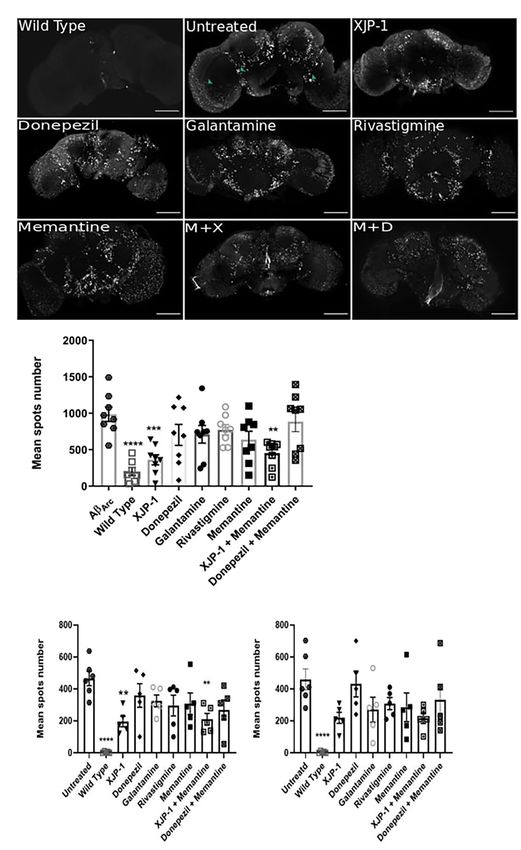
Uras et al.                                                                                                               Amyloid Clearance in Alzheimer’s Model

junction (hereafter referred to as NMJ) as it is in the vertebrates.                   MATERIALS AND METHODS
Instead, the NMJ of fruit flies or invertebrates is regulated
by glutamate. Despite this difference in the NMJ physiology,                           Reagents
acetylcholine in the fruit fly regulates jumping, geotaxis, and                        AChE inhibitors donepezil, galantamine, and rivastigmine
motion detection stimuli (Hou et al., 2003; Takemura et al.,                           were purchased from Sigma-Aldrich Ltd. NMDAR antagonist
2013). The fruit fly AChE folding structure is also similar to                         memantine was purchased from Sigma-Aldrich Ltd. Drug
the vertebrate enzyme, with variations observed mainly in the                          candidate XJP-1 was gifted by Prof. Jinyi Xu (China
peripheral anionic site (PAS) but not in the catalytic active site                     Pharmaceutical University).
(CAS) (Wiesner et al., 2007).
    Drosophila melanogaster offers several advantages as a model
organism, such as a well-defined genome, the possibility to                            Fly Strains
study hundreds of animals simultaneously, and low cost of                              The UAS-APPE693G (#33774) and elav-Gal4 (#458) flies were
maintenance (Sivanantharajah et al., 2019; Tue et al., 2020).                          purchased from Bloomington Drosophila Stock Centre Indiana.
Moreover, the easily accessible Drosophila brain allows one                            Flies were kept at 25◦ C in 25-ml plastic vials containing 5 ml
to image and quantify amyloid plaque deposition in amyloid                             of standard fly food. elav-Gal4 virgin females were crossed with
transgenic models, along with the possibility to investigate                           UAS-APPE693G males, all stocks were kept at 25◦ C, and the
selected areas of the brain involved in cognitive processes, such                      female progeny was used for all the assays. Elav-Gal4 flies were
as the mushroom bodies (Hwang et al., 2019; Zhong et al., 2019).                       used as a wild-type (WT) control.
    In the work presented here, we drive the expression of the
APP gene carrying the Arctic mutation in D. melanogaster                               Fly Food Preparation
(hereafter referred to as Aβarc flies) as previously reported                          Flies were fed using the standard fly food recipe containing
(Sandin et al., 2016). The Arctic mutation was initially genotyped                     (quantity for 1 L): 70.62 ml of golden syrup, 15.87 g of yeast,
from a Swedish family with a history of early-onset familial AD                        9.12 g of soya flour, 67 g of cornmeal, 5.25 g of agar, and
(Nilsberth et al., 2001), causing the production of toxic amyloid-                     711.25 ml of water. To add the drug of interest into the food,
β peptides (Norlin et al., 2012). The Arctic mutation has been                         an aliquot of the drug stock solution was dissolved in a volume
linked to a faster amyloid aggregation rate both in vitro and                          of water to 1/5th of the total food volume and mixed on the
in vivo (Dahlgren et al., 2002; Englund et al., 2007). In addition,                    magnetic stirrer for 2 min. Subsequently, once the temperature
amyloid-β peptides carrying the Arctic mutation form fibrils                           has cooled to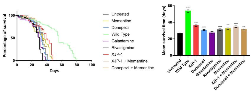
Uras et al.                                                                                                  Amyloid Clearance in Alzheimer’s Model

    + (percentage of total flies between the 30-ml and 40-ml              function to the original image; the particle analysis was carried
lines) × 0.75                                                             out to generate size, intensity, and shape measurements of the
    + (percentage of total flies between the 20-ml and 30-ml              plaques; the number of particles (amyloid spots) was used to
lines) × 0.50.                                                            characterize the effect of each treatment.

Immunohistochemistry                                                      Western Blot
Adult flies were firstly anesthetized with CO2 and subsequently           A total of 50 fly heads were dissected and frozen overnight
killed in pure ethanol (100%). Brains were dissected in phosphate         at -80◦ C. Samples were then homogenized in 150 µl of RIPA
buffered saline (PBS) and then fixed in 4% paraformaldehyde               Lysis and Extraction Buffer (#89901, ThermoFisher Scientific).
(PFA) for 20 min at room temperature (RT). Tissues were then              Subsequently, each sample was centrifuged at 14,000g at 4◦ C for
washed three times in 1% PBS-Tween 20 (PBS-T) for 2 min, and              10 min, and the supernatant was then transferred into a fresh
subsequently placed in 5% normal goat serum (NGS) in PBS-                 new vial. Protein concentrations were quantified using Bradford
T for 2 h at room temperature. After removing the 5% NGS-T,               Assay Kit (#ab102535, Abcam, United Kingdom). According
the rabbit anti-Aβ42 primary antibody (#ab2539, Abcam plc.)               to protein concentrations, samples were then dissolved in
was added at a final concentration of 1:200 in 5% NGS-T and               4 × NuPage LDS Sample Buffer (#NP0007, Invitrogen) and
left to incubate overnight at +4◦ C. The primary antibody was             loaded into 4%–12% Bis-Tris Gel (#NP0322PK2, Invitrogen).
then washed three times with 1% PBS-T and subsequently the                Following separation, proteins were transferred onto a 0.2-µm
goat anti-rabbit secondary antibody was added to a final 1:250            nitrocellulose membrane (#1620150, Bio-Rad) using semi-dry
concentration. The sample was allowed to incubate for 1 h at              apparatus TransBlot Turbo (#1704150, Bio-Rad). Membranes
room temperature. Goat anti-rabbit 488 Alexa Fluor secondary              were then blocked in 5% bovine serum albumin (BSA) for 1 h at
antibody (#A-11034; ThermoFisher Scientific) was then washed              room temperature and subsequently incubated overnight at 4◦ C
three times with 1% PBS-T. Brain samples were then mounted                with rabbit anti-Aβ42 primary antibody (#ab2539, Abcam plc)
for confocal imaging exploiting a “bridge” structure to avoid             at final 1:1000 concentration. Monoclonal Anti-β actin antibody
any sample damage caused by the coverslip. Two 0.1-mm thin                was used as loading control in Western Blot experiments
coverslips were attached to the glass slide at approximately 5 mm         (#ab1801, AbCam plc.). Each membrane was then washed three
distance to each other. Subsequently, 20 µl of mounting media             times in 5% TBS-T for 5 min and subsequently incubated
(90% PBS and 10% glycerol) was added in the gap between the               for 1 h at room temperature with goat-anti rabbit IgG H&L
two coverslips. Samples were then placed into the mounting                (HRP) (#ab6721, AbCam plc.) secondary antibody at a final
media, and an additional cover slip to bridge the gap between             concentration of 1:5000. Excess secondary antibody was then
the two “pylon” coverslips. Samples were analyzed by confocal             discarded and membrane was washed three times using 5% TBS-
microscopy within 24 h from mounting.                                     T. Each membrane was then imaged using Fujifilm LAS-4000
                                                                          (Fujifilm). Results were then analyzed using FIJI.
Confocal Imaging
All brain images were acquired using the Zeiss880 confocal
microscope. Laser power was set at 2.5%, gain at 520. Digital             Quantification of AChE-Induced Aβ42
offset was set at 200. The imaging acquisition speed was 1.03 s,          Aggregation
averaging two times per slice. For visualization purposes, the            Measurements of AChE-induced amyloid aggregation were
figures presented have been made by setting the brightness value          taken following the protocol previously reported (Jiang et al.,
to 30,000 and subsequently converting into 8-bit images.                  2019). Briefly, hexafluoroisopropanol (HFIP)-treated E22G Aβ
                                                                          peptides (#SP-Ab-11_0.1, JPT–Innovative Peptide Solution) were
Image Analysis                                                            dissolved in DMSO to reach a final 200 µM stock. The dissolved
A macro was created in FIJI to identify and measure amyloid               peptides were subsequently centrifuged at 13,500g for 10 min,
spots in the images scanned with the confocal microscope. The             and the supernatant was then transferred into a fresh vial and
processing analysis was carried out using the following steps:            used for the following experiments. To evaluate the aggregation
raw 16 bit confocal images were imported into FIJI; maximum               rate in the presence of AChE inhibitors, 2 µl of the compound of
projection (focus stack flattening) was applied and a copy of this        interest (at the appropriate concentration) was added into each
image was created; the copy was used for object identification            vial, followed by 2 µl of 200 µM Aβ peptides stock, 20 µl of
and the creation of the mask; a Gaussian blur filter was applied          AChE (#C3389, Sigma-Aldrich Ltd) (2 U/ml, in 1 × PBS at pH
to the image to smooth the noise pixels; a threshold of 14,850–           8.0), and 76 µl 1 × PBS, pH 8.0. The reaction was then incubated
61,890 was used to separate the plaque signals from the rest of the       at room temperature (RT) for 24 h. Subsequently, 100 µl of 5 µM
image and from the background; the lower value of thresholding            Thioflavin T (#ab120751, ThT) was added into each vial. After 1-h
14,850 was determined by using negative control and non-stained           incubation at RT, fluorescence emission was recorded at 490 nm
areas of the brain. Top value was set just below the maximum              with an excitation wavelength of 450 nm using a Tecan Spark
to avoid occasionally appearing large clumps of overexposed               microplate reader. Results were then processed as previously
areas; a mask was created based on the threshold areas; particle          described by Jiang et al. (Jiang et al., 2019) using the subsequent
analysis was carried out on the mask while fluorescence intensity         formula: (Fi - Fb )/(Fo - Fb ) - 100, where Fi corresponds to amyloid
measurements were performed by redirecting the measure                    aggregation in the presence of peptides, AChE, AChE inhibitors,

Frontiers in Neuroscience | www.frontiersin.org                       4                                      June 2021 | Volume 15 | Article 691222
Uras et al. Amyloid Clearance in Alzheimer’s Model
and ThT; Fo represents the amyloid aggregation in presence of Locomotor Functions Improve Following
peptides, AChE, and ThT; and Fb corresponds to blank control XJP-1 Treatment
containing ThT only.
The climbing assay is a behavioral test, based on negative geotaxis
against gravity, which is used to assess the locomotor function
Statistical Analysis in Drosophila (Nichols et al., 2012). With this assay, Aβarc
All statistical analyses were performed using GraphPad Prism 9 flies recorded a time-dependent worsening of climbing ability,
software. Data obtained were firstly tested for normality using the thereby faithfully recapitulating symptoms observed in human
Shapiro–Wilk test. The Kaplan–Meier test was used to compare patients (Figure 2A). Treatment with XJP-1 improved locomotor
different survival curves. The Kruskal–Wallis test, followed by function in Aβarc flies over 20 days, with the exception of days
Dunn’s post hoc, was used to compare differences between 5 and 21 of analysis (Figure 2B). The climbing performance
three or more groups in non-normally distributed data. The recorded in flies treated with XJP-1 were constantly twofold
ANOVA test was used to compare differences between three better than the untreated Aβarc flies, with peaks at day 16 of
or more groups of normally distributed samples. The Friedman analysis when the treated group recorded a climbing score of
test, followed by Dunn’s post hoc test, was used to analyze 67.29 against 3.62 for the untreated groups (Table 1).
differences between three or more groups in the climbing assay. On the other hand, among the FDA-approved therapies,
Each experiment was performed in triplicate, and all results are only donepezil treatment showed results comparable to XJP-1,
presented as mean ± standard error of the mean (SEM) or recording a homogeneous climbing performance over the first
mean ± standard deviation (SD). Results with a P-value < 0.05 19 days of analysis, with a minimum of twofold improvement
were considered significant. of the locomotor performance, except for day 9 of analysis
(Figure 2C; Table 1).
Rivastigmine, galantamine, and memantine had a beneficial
RESULTS effect that was limited only to the first 10 days of analysis
(Figures 2D–F). Aβarc flies treated with combined therapies
XJP-1 Treatment Improves Life showed a significant improvement in locomotor function only
throughout the first 10 days of analysis, and at day 14 for XJP-
Expectancy in Aβarc Flies 1 and memantine, therefore having a worse effect on locomotor
In order to investigate the effect of the Aβarc mutation on
functions than donepezil and XJP-1 alone (Figures 2G,H,
life expectancy, flies were firstly grown on food without
Table 1).
any therapy. Expression of amyloid peptides resulted in a
dramatic drop of mean survival time when compared to
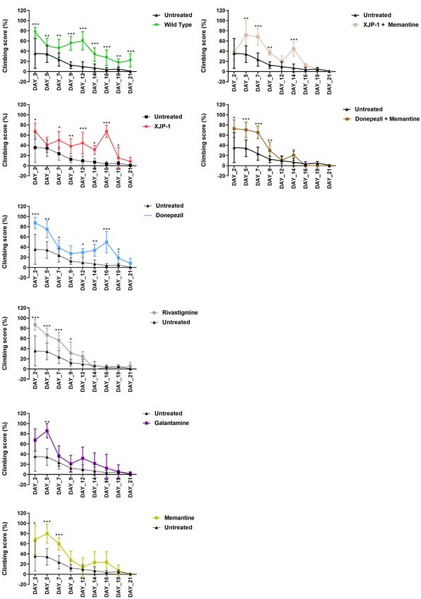
WT flies (Figures 1A,B). To investigate the effects of XJP- XJP-1 Reduces the Number of Amyloid
1 treatment on life expectancy, Aβarc flies were treated with Plaques in the Brain
40 µM (as the minimum effective concentration) of the new The deposition, accumulation, and aggregation of amyloid
AChE inhibitor after testing a range of concentrations (10– peptides in the central nervous system (CNS) is a crucial point
40 µM); 10 to 30 µM did not produce any significant in the development of Alzheimer’s symptomatology. Using HFIP-
amelioration in pilot lifespan assay (data not shown). XJP-1 treated Aβ peptides, together with confocal microscopy, we found
therapy resulted in a significant increase in the mean survival Aβarc fly brains to possess large and extensively distributed
time, 26. 74 days vs. 36.38 days (Table 1), of Aβarc flies amyloid aggregates (hereafter referred to as amyloid spots) at
with around 40% of the entire population surviving after day 10 days post-eclosion (Figure 3A). To investigate whether flies
40 (Figures 1A,B). Comparable results were also recorded treated with XJP-1, or clinically available therapies, would present
with other FDA-approved AD drugs, including the AChE any difference in amyloid pathology in the CNS, we imaged
inhibitors donepezil (30.76 days) (0.5 mM as the minimum Aβarc fly brains following 10 days of treatment (Figure 3A).
effective concentration), rivastigmine (31.49 days) (0.1 mM), Subsequent quantification showed a significant reduction in
and the NMDAR antagonist memantine (32.61 days) (0.5 mM). amyloid spot counts in flies treated with either XJP-1 or XJP-1 and
Galantamine (0.5 mM), however, had no significant effect memantine combined. None of the clinically approved therapies
(Table 1). Among all the tested compounds, XJP-1 showed most had similar results to the new AChE inhibitor (Figure 3B). To
potent efficacy at a concentration of 40 µM, more than 10 further investigate whether there was a remarkable difference
times lower than observed for donepezil, on Aβarc survival time in amyloid spot counts in those brain areas rich in cholinergic
(Figures 1A,B). neurons, we exploited the Fly Brain Observatory software to
Since the FDA currently approves a combined therapy of determine areas of the brain that are rich in cholinergic neurons.
memantine and donepezil, we investigated whether XJP-1 and We found that mushroom bodies (Supplementary Figure 1A),
memantine treatment would further enhance the results obtained fan-shaped bodies (Supplementary Figure 1B), the medulla
by XJP-1 alone. Both a combination of FDA-approved therapies (Supplementary Figure 1C), and optic lobes (Supplementary
and XJP-1 plus memantine showed a significant improvement Figure 1D), were rich in cholinergic neurons compared to other
of the Aβarc Drosophila lifespan (Figures 1A,B). However, no areas, such as the antennal lobes (Supplementary Figure 1E)
significant difference was recorded when the combined therapies and suboesophageal ganglion (Supplementary Figure 1F) (Xu
were compared to the corresponding monotherapies (Table 1). et al., 2020). Analysis of mushroom body and fan-shaped
Frontiers in Neuroscience | www.frontiersin.org 5 June 2021 | Volume 15 | Article 691222Frontiers in Neuroscience | www.frontiersin.org
Uras et al.
TABLE 1 | Data table.
Aβarc XJP-1 Donepezil Rivastigmine Galantamine Memantine XJP-1 Donepezil
(Memantine) (Memantine)
Mean Mean P value vs. Mean P value vs. Mean P value vs. Mean P value vs. Mean P value vs. Mean P value vs. Mean P value vs.
Aβarc Aβarc Aβarc Aβarc Aβarc Aβarc Aβarc
Lifespan assay 26.74 36.38 < 0.0001 30.76 < 0.0001 31.49 < 0.0001 27.52 0.9994 32.61 0.0008 34.68 < 0.0001 31.98 0.0005
Climbing assay Day-2: 35.625 67.81 0.016 87.5 0.0003 87.083 0.0005 67.75 0.0511 67.868 0.041 37 0.932 72.6375 0.032
Day-5: 34.3329 40.86 0.0724 75 0.006 66.75 0.0003 86.008 0.0088 79.86 0.0005 71.25 0.0088 70.5145 0.0003
Day-7: 23.6085 50.08 0.04 38.5 0.028 56.223 0.0005 36.174 0.58 59.903 0.0006 68.26 0.0007 65.3487 0.0004
Day-9: 12.5175 38.278 0.0014 27.1 0.77 31.25 0.042 21.166 0.61 27.487 0.059 36.93 0.0056 29.8625 0.0071
Day-12: 9.5967 44.4 0.0008 29.5 0.019 24.438 0.58 31.832 0.0532 14.25 0.66 19.68 0.0839 10.685 0.7643
Day-14: 6.925 31.05 0.022 33.6 0.0047 0 0.865 21.583 0.0799 23.369 0.062 44.35 0.0006 20.7625 0.0664
Day-16: 3.6242 67.29 0.0002 49.7 0.0003 3.261 0.832 12.5 0.0527 23.369 0.052 11.84 0.054 0.06273 0.71
Day-19: 4.886 15.418 0.032 18.7 0.012 2 0.75453 5.556 0.93 7.293 0.057 5.313 0.513 0.24573 0.361
Day-21: 0.832 8.369 0.1734 8.13 0.0615 5.716 0.566 1.667 0.91 0 0.65 0 0.66 0 0.982
6
Amyloid spots (10 days) 987.5 362.3 0.0005 703.9 0.2691 773.1 0.5659 711.6 0.2961 639 0.1095 453 0.0035 886.3 0.984
Amyloid spots (20 days) 1137 383.5 < 0.0001 390.5 < 0.0001 525.7 0.0582 673.4 0.0657 373.5 < 0.0001 520.2 0.0002 528 0.0096
MB + FBS amyloid spots (10 days) 465.3 196 0.0034 358.8 0.5605 325.4 0.2709 296 0.1204 307.6 0.1686 210.4 0.006 269.4 0.519
MB + FBS amyloid spots (20 days) 324.6 154.2 0.0029 133.2 0.0008 191.2 0.27 230.4 0.1934 133.2 0.0008 113.6 0.0002 164.4 0.0055
Medulla + OL (10 days) 459.7 219.2 0.0675 431 0.9996 308.4 0.2195 270.2 0.4465 285.2 0.2961 216.8 0.0635 331.8 0.6279
Medulla ( OL (20 days) 607 321.2 0.0126 304.2 0.0075 419.4 0.1776 175 0.0014 253.8 < 0.0001 202.4 0.0003 225 0.0006
Amyloid Clearance in Alzheimer’s Model
June 2021 | Volume 15 | Article 691222
Amyloid aggregation 93.85 23.52 0.0001 24.6 0.0002 56.12 0.0354 44.61 0.005 N/A N/A 26.72 0.0002 33.72 0.0008
Comparison of mean and P-values of different treatment groups against the untreated Aβarc flies.Uras et al. Amyloid Clearance in Alzheimer’s Model
FIGURE 1 | Aβarc flies’ lifespan. (A) Kaplan–Meier survival trajectories of AβArc flies under different drug treatments. (B) Mean survival time of AβArc flies on different
treatments. Kruskal–Wallis test followed by Dunn’s post hoc was used to compare the differences between different groups. Data are expressed as mean ± SEM,
n = 3 (number of independent experiments with a minimum of 100 flies per genotype). P < 0.05 was considered as significant. *P < 0.05; **P < 0.01; ***P < 0.001;
****P < 0.0001. Genotypes: Untreated = Aβarc -expressing flies; Wild Type = elav-Gal4 line; all treated groups are Aβarc -expressing flies fed with the stated drug.
body areas recorded a significant reduction of amyloid spots peptide aggregation, as previously reported (Inestrosa et al., 1996;
in flies treated with both XJP-1 monotherapy and XJP-1 and Rees et al., 2003; Lushchekina et al., 2017). We therefore used an
memantine combined therapy (Figure 3C). Conversely, none of in vitro enzymatic assay to evaluate amyloid peptide aggregation
the treatments tested had a beneficial impact on the medulla and rate, where amyloid-β peptides carrying the Arctic mutation were
optic lobe amyloid spot count (Figure 3D). co-incubated with AChE enzyme in the presence, or not, of all
To assess whether AD progression alters these amyloid spot the AChE inhibitors tested. We found a significant drop in all
count results, we also analyzed the Aβarc fly brains 20 days treatments studied, with the greatest reductions observed for
post-eclosion (Figure 4A). The whole brain analysis showed XJP-1 and donepezil, both recording an almost 80% decrease in
a significant reduction of the amyloid spot counts, similar aggregation rate (Figure 5). Once again, the combined therapies
to that observed with XJP-1 at 10 days, in flies treated did not result in any further decrease in amyloid aggregation
with XJP-1, donepezil, and memantine, while galantamine and rate (Figure 5).
rivastigmine failed to show any significant effect (Figure 4B).
The same reduction trends were recorded in the mushroom
bodies and fan-shaped bodies (Figure 4C). Although none of DISCUSSION
the treatments tested reduced the amyloid spot counts in the
medulla and optical lobe areas after 10 days (Figure 3D), analysis Over the past 10 years, the fruit fly has emerged as a powerful
of samples treated for 20 days showed a significant decrease in vivo model for neurodegenerative diseases, including AD
in the amyloid spot levels for all treatment tested, with the (Zhang et al., 2016a; Pham et al., 2018; Ali et al., 2019; Cornelison
exception of galantamine and rivastigmine (Figure 4D). The et al., 2019; Higham et al., 2019; Hwang et al., 2019; Miyazaki
combined therapies investigated, however, did not show any et al., 2019; Sivanantharajah et al., 2019; Tue et al., 2020).
further beneficial effects than the single therapies (Table 1). Transgenic Drosophila AD models have been employed for
a number of drug testing studies involving different targets
such as AChE, GSK-3β, lysozyme, radiation, Tau protein, and
XJP-1 Reduces Amyloid Aggregation via dopaminergic receptors (Sandin et al., 2016; Zhang et al.,
AChE Inhibition 2016a; Pham et al., 2018; Ali et al., 2019; Hwang et al.,
To confirm that the observed reduction in amyloid spots was 2019; Miyazaki et al., 2019; Zhong et al., 2019). Flies use a
not a result of a reduced quantity of amyloid peptides within variety of neurotransmitters to communicate between neurons
the Aβarc fly brains, we quantified amyloid peptide expression (e.g., dopamine, GABA, glutamate, acetylcholine, serotonin);
following 10 and 20 days of treatment. At both time points, however, differences have been observed in the functions of
and for all drug treatments, amyloid peptide expression was these neurotransmitters between invertebrates and vertebrates.
statistically similar to the untreated brains (Supplementary In Drosophila, glutamate is known to be the primary excitatory
Figure 2). This result is expected since the expression of the neurotransmitter at the NMJ, whereas acetylcholine plays this
transgene is determined by the UAS/Gal4 system and is not role in mammals (Jan and Jan, 1976; Colombo and Francolini,
targeted by any of the drugs studied. We hypothesized that the 2019). Conversely, in the mammalian CNS, glutamate is the
AChE enzyme was functioning as a nucleation point for amyloid primary excitatory neurotransmitter. In flies, acetylcholine is
Frontiers in Neuroscience | www.frontiersin.org 7 June 2021 | Volume 15 | Article 691222Uras et al. Amyloid Clearance in Alzheimer’s Model FIGURE 2 | Aβarc flies climbing assay. Aβarc flies climbing assay under different treatments. Data show the climbing performance trends over a 21-day period. Data generated from the climbing index was processed as a percentage of the total. In order to compare vials with a different number of flies, repeated measures analysis of variance was used to compare climbing scores between treated and untreated groups. (A) Climbing assay of untreated Aβarc flies. (B) Climbing assay of Aβarc flies treated with XJP-1. (C) Climbing assay of Aβarc flies treated with donepezil. (D) Climbing assay of Aβarc flies treated with rivastigmine. (E) Climbing assay of Aβarc flies treated with galantamine. (F) Climbing assay of Aβarc flies treated with memantine. (G) Climbing assay of Aβarc flies treated with XJP-1 and memantine. (H) Climbing assay of Aβarc flies treated with donepezil and memantine. Data are presented in the figure as the mean ± SD; data are compared against the untreated group. n = 3 (number of independent experiments, each experiment with a minimum of 10 flies per treatment). P < 0.05 was considered as significant. *P < 0.05; **P < 0.01; ***P < 0.001; ****P < 0.0001. Frontiers in Neuroscience | www.frontiersin.org 8 June 2021 | Volume 15 | Article 691222
Uras et al.                                                                                                                    Amyloid Clearance in Alzheimer’s Model

  FIGURE 3 | Amelioration of amyloid spots in Aβarc flies CNS after 10 days of treatment. (A) Representative confocal images of WT (top left panel) or Aβarc brains;
  arrows show amyloid spots; scale bar: 100 µm. (B) Whole brain quantification of amyloid spots. ANOVA test followed by Bonferroni’s post hoc was used to
  compare the differences between three or more groups, P < 0.05 was considered as significant. Data are presented as mean ± SEM. n = 3, with a minimum of five
  flies per experiment analyzed. *P < 0.05; **P < 0.01; ***P < 0.001; ****P < 0.0001. (C) Mushroom body and fan-shaped body quantification of amyloid spots.
  ANOVA test followed by Bonferroni’s post hoc was used to compare the differences between three or more groups, P < 0.05 was considered as significant. Data
  are presented as mean ± SEM. n = 3, with a minimum of five flies per experiment analyzed. *P < 0.05; **P < 0.01; ***P < 0.001; ****P < 0.0001. (D) Medulla and
  Optic lobe quantification of amyloid spots. ANOVA test followed by Bonferroni’s post hoc was used to compare the differences between three or more groups,
  P < 0.05 was considered as significant. Data are presented as mean ± SEM. n = 3, with a minimum of five flies per experiment analyzed. *P < 0.05; **P < 0.01;
  ***P < 0.001; ****P < 0.0001.

Frontiers in Neuroscience | www.frontiersin.org                                   9                                            June 2021 | Volume 15 | Article 691222
Uras et al.                                                                                                                   Amyloid Clearance in Alzheimer’s Model

  FIGURE 4 | Amelioration of amyloid spots in Aβarc flies CNS after 20 days of treatment. (A) Representative confocal images of WT (top left panel) or Aβarc brains;
  arrows show amyloid spots; scale bar: 100 µm. (B) Whole brain quantification of amyloid spots. ANOVA test followed by Bonferroni’s post hoc was used to
  compare the differences between three or more groups, P < 0.05 was considered as significant. Data are presented as mean ± SEM. n = 3, with a minimum of 1 fly
  per experiment analyzed. *P < 0.05; **P < 0.01; ***P < 0.001; ****P < 0.0001. (C) Mushroom body and fan-shaped body quantification of amyloid spots. ANOVA
  test followed by Bonferroni’s post hoc was used to compare the differences between three or more groups, P < 0.05 was considered as significant. Data are
  presented as mean ± SEM. n = 3, with a minimum of 1 fly per experiment analyzed. *P < 0.05; **P < 0.01; ***P < 0.001; ****P < 0.0001. (D) Medulla and Optic
  lobe quantification of amyloid spots. ANOVA test followed by Bonferroni’s post hoc was used to compare the differences between three or more groups, P < 0.05
  was considered as significant. Data are presented as mean ± SEM. n = 3, with a minimum of five flies per experiment analyzed. *P < 0.05; **P < 0.01; ***P < 0.001;
  ****P < 0.0001.

Frontiers in Neuroscience | www.frontiersin.org                                  10                                            June 2021 | Volume 15 | Article 691222
Uras et al.                                                                                                           Amyloid Clearance in Alzheimer’s Model

                                                                                    effective at a concentration as low as 30 µM (Chakraborty et al.,
                                                                                    2011; Pham et al., 2018).
                                                                                        XJP-1 was able to reduce the amyloid spot counts as early as
                                                                                    10 days after commencing the treatment, in crucial areas of the
                                                                                    Drosophila brain for memory and learning functions. Among the
                                                                                    clinically available AChE inhibitors, none of them showed such
                                                                                    an early effect on amyloid spot counts, with only donepezil having
                                                                                    similar results as XJP-1, but after 20 days of administration and
                                                                                    at a concentration more than 10 times higher. This is the first
                                                                                    evidence that XJP-1 therapy results in an overall improvement of
                                                                                    AD symptomatology in vivo.
                                                                                        On the other hand, a combined treatment of XJP-1 and
                                                                                    memantine did not result in further significant improvement
                                                                                    when compared to XJP-1 treatment alone, as well as combined
                                                                                    donepezil and memantine therapy. Combined therapy is usually
                                                                                    only given to patients when they enter an advanced stage of
                                                                                    the disease, when memantine is added to patients already on
                                                                                    donepezil therapy (Tayeb et al., 2012; Parsons et al., 2013).
                                                                                    In our study, all drugs were administrated continuously from
                                                                                    24 h upon eclosion, including combined therapies, giving a
                                                                                    possible explanation on why donepezil with memantine, and
                                                                                    XJP-1 with memantine, did not show any further beneficial
                                                                                    effect when compared to single therapy on this AD model. This
                                                                                    limitation may also explain the weaker efficacy of galantamine
                                                                                    and rivastigmine compared to donepezil, as they are currently
  FIGURE 5 | Reduction of AChE-induced Aβ-peptide aggregation rates in the          prescribed to Alzheimer’s patients within mild to moderate stages
  presence of AChE inhibitors. ANOVA test followed by Bonferroni’s post hoc         of AD, while donepezil is given to AD patients at all stages of the
  was used to compare the differences between three or more groups. Data are
                                                                                    disease, from mild to severe (Haake et al., 2020). Moreover, the
  presented as mean ± SEM of n = 3. *P < 0.05; **P < 0.01; ***P < 0.001;
  ****P < 0.0001.                                                                   NMDAR antagonist memantine is also administered during all
                                                                                    stages of AD, alone or as combination with donepezil to further
                                                                                    improve the cognitive functions, as efficacy against the amyloid
                                                                                    pathology is still controversial (Folch et al., 2018).
broadly expressed and is the primary excitatory neurotransmitter                        Over the past decade, the efforts to find a cure to AD have
in the CNS, while the role of the glutamatergic system in the                       been mainly focused on anti-amyloid agents, such as beta-
Drosophila brain remains rather ambiguous (Buchner, 1991).                          secretase 1 (BACE1) inhibitors, assuming that the aggregation
In fact, there is evidence that glutamate can be inhibitory                         of amyloid-β peptides into plaques is the central triggering
within certain fly CNS systems, such as in the olfactory and                        mechanism. However, a number of clinical trials involving this
visual systems (Liu and Wilson, 2013; Molina-Obando et al.,                         type of target have failed to produce any significant improvement
2019). Although differences in the functions of neurotransmitter                    (Cummings et al., 2019). Thus, it is necessary to investigate novel
systems should be taken into account when interpreting results,                     therapeutics for which the mechanism of action involves directly,
if using the fly to model neurodegenerative diseases, it should                     or indirectly, multiple targets such as the cholinergic pathway,
be noted that it is the damaged cholinergic portion of the                          amyloid pathology, and Tau hyperphosphorylation. A number of
brain that is linked to the development of Alzheimer’s disease,                     studies have placed the cholinergic system at the center of the AD
hence the use of cholinesterase inhibitors to slow disease                          etiopathogenesis, involving the overactivation of crucial enzymes,
development. The ability to use Drosophila as a model for human                     such as BACE1 and GSK3-β (Noh et al., 2009; Yoshiyama et al.,
neurodegenerative diseases has proven to be, and will continue                      2010; Medeiros et al., 2011; Potter et al., 2011; Kalkman and
to be, a powerful tool to understand and suppress degenerative                      Feuerbach, 2016; Zhang et al., 2016a). Our study has shown a
mechanisms (Lenz et al., 2013).                                                     clear involvement of AChE inhibition on amyloid aggregation
    In this work, we have demonstrated the capacity of a new                        resulting in an amelioration of symptoms. The effect of XJP-1 on
AChE-I compound, XJP-1, to ameliorate amyloid-β deposition                          Aβarc transgenic flies were partially replicated only by donepezil
and consequent behavioral phenotypes in the Aβarc transgenic                        at a concentration 10 times higher than our drug candidate, with
Drosophila AD model. Treatment with XJP-1 resulted in an                            XJP-1 having an earlier beneficial effect in reducing the amyloid
increased survival time of Aβarc flies of more than 10 days,                        spot counts after 10 days of treatment. Other than that, the other
improving as well the locomotor functions of the transgenic fruit                   FDA-approved AChE inhibitors, galantamine and rivastigmine,
flies. Similar results were scored by the FDA therapy donepezil,                    reduced the amyloid aggregation rate of about 50%, while XJP-
despite being administrated at a concentration 10-fold higher                       1 and donepezil reduced it by about 80%, potentially explaining
than XJP-1, in contrast with previous reports of donepezil being                    the limited beneficial effects observed in Aβarc flies treated with

Frontiers in Neuroscience | www.frontiersin.org                                11                                     June 2021 | Volume 15 | Article 691222
Uras et al. Amyloid Clearance in Alzheimer’s Model
galantamine and rivastigmine. In addition to this, docking AUTHOR CONTRIBUTIONS
studies carried out on XJP-1 showed a dual-binding property
for both the PAS and CAS of the AChE enzyme (Wang et al., GU and AM equally contributed to this work. GU conceived
2015), which may further justify the low dosage needed to the study and its experimental design, contributed to samples
slow down the amyloid aggregation, since the PAS of AChE collection, carried out the samples analysis, analyzed the
has been linked to increased amyloid-β peptide aggregation results, performed research, and wrote the manuscript. AM
rate (Inestrosa et al., 1996). Despite not being tested in this and ZM contributed to sample collection and carried out the
study, XJP-1 has been predicted to have anti-inflammatory sample analysis. MB reviewed the manuscript. PZ, SX, and JX
and anti-oxidant properties due to its natural product-derived synthesized and supplied XJP-1 and reviewed the manuscript.
structure, which may have also played a role in ameliorating MG, SA, NM, and ZZ conceived the study and its experimental
the AD symptomatology in Aβarc flies (Wang et al., 2015, design and reviewed and approved the manuscript. All authors
2018). contributed to the article and approved the submitted version.
Taken together, the results of these experiments suggest
that XJP-1 ameliorates AD symptomatology by indirectly
reducing amyloid aggregation via AChE inhibition. Despite FUNDING
XJP-1 showing promising efficacy with this AD model,
further studies will be necessary to investigate whether XJP- Ph.D. studentship funded by the School of Pharmacy, The
1 results can be translated into mammalian models and University of Nottingham; National Natural Science of
whether treatment can also improve Tau-related phenotypes Foundation of China; Erasmus + European funds.
presented by AD patients.
SUPPLEMENTARY MATERIAL
CONCLUSION
The Supplementary Material for this article can be found
This work demonstrates that XJP-1 is an effective and potent AD online at: https://www.frontiersin.org/articles/10.3389/fnins.
drug candidate, which efficiently rescues AD symptoms in an 2021.691222/full#supplementary-material
Aβarc fly model, with earlier effects at a much lower concentration
Supplementary Figure 1 | Representative images of cholinergic neurons network
than observed for any of the currently approved therapies. This in Drosophila Melanogaster brain obtained from the Virtual Fly Observatory. (A)
includes a significant improvement in locomotor defects and Cholinergic neurons within the mushrooms bodies. (B) Cholinergic neurons within
lifespan. By inhibiting AChE, XJP-1 indirectly reduced amyloid the fan-shaped bodies. (C) Cholinergic neurons within the medulla. (D)
aggregation and therefore also the number of amyloid spots Cholinergic neurons within the optic lobes. (E) Cholinergic neurons within the
antennal lobes. (F) Cholinergic neurons within the subesophageal ganglion.
detected at an earlier stage in Aβarc Drosophila brains.
Supplementary Figure 2 | Amyloid peptides quantity in Aβarc flies’ heads. (A)
Representative images of WB membranes showing amyloid peptides quantity after
10 and 20 days of treatment. (B) AβArc peptide quantification by membrane image
DATA AVAILABILITY STATEMENT analysis. ANOVA test followed by Bonferroni’s post hoc was used to compare the
differences between different groups. Data are presented as mean ± SEM, n = 3
The raw data supporting the conclusions of this article will be (number of independent experiments with 50 fly heads per treatment group).
made available by the authors, without undue reservation. P < 0.05 was considered as significant. ∗ P < 0.05; ∗∗ P < 0.001; ∗∗∗ P < 0.0001.
REFERENCES Cornelison, G. L., Levy, S. A., Jenson, T., and Frost, B. (2019). Tau-induced nuclear
envelope invagination causes a toxic accumulation of mRNA in Drosophila.
Ali, F., Rahul, Jyoti, S., Naz, F., Ashafaq, M., Shahid, M., et al. (2019). Therapeutic Aging Cell 18:e12847. doi: 10.1111/acel.12847
potential of luteolin in transgenic Drosophila model of Alzheimer’s disease. Crowther, D. C., Kinghorn, K. J., Miranda, E., Page, R., Curry, J. A., Duthie,
Neurosci. Lett. 692, 90–99. doi: 10.1016/j.neulet.2018.10.053 F. A., et al. (2005). Intraneuronal Abeta, non-amyloid aggregates and
Alzheimer’s Association (2019). Available online at: www.alz.org neurodegeneration in a Drosophila model of Alzheimer’s disease. Neuroscience
Basun, H., Bogdanovic, N., Ingelsson, M., Almkvist, O., Näslund, J., Axelman, K., 132, 123–135. doi: 10.1016/j.neuroscience.2004.12.025
et al. (2008). Clinical and neuropathological features of the arctic app gene Cummings, J., Lee, G., Ritter, A., Sabbagh, M., and Zhong, K. (2019). Alzheimer’s
mutation causing early-onset Alzheimer disease. Arch. Neurol. 65, 499–505. disease drug development pipeline: 2019. Alzheimer’s Dementia 5, 272–293.
doi: 10.1001/archneur.65.4.499 Dahlgren, K. N., Manelli, A. M., Stine, W. B. Jr., Baker, L. K., Krafft, G. A., and
Buchner, E. (1991). Genes expressed in the adult brain of drosophila and effects of LaDu, M. J. (2002). Oligomeric and fibrillar species of amyloid-beta peptides
their mutations on behavior: a survey of transmitter- and second messenger- differentially affect neuronal viability. J. Biol. Chem. 277, 32046–32053. doi:
related genes. J. Neurogenet. 7, 153–192. doi: 10.3109/01677069109167432 10.1074/jbc.m201750200
Chakraborty, R., Vepuri, V., Mhatre, S. D., Paddock, B. E., Miller, S., Michelson, Drummond, E., and Wisniewski, T. (2017). Alzheimer’s disease: experimental
S. J., et al. (2011). Characterization of a Drosophila Alzheimer’s disease model: models and reality. Acta Neuropathol. 133, 155–175.
pharmacological rescue of cognitive defects. PLoS One 6:e20799. doi: 10.1371/ Englund, H., Sehlin, D., Johansson, A. S., Nilsson, L. N., Gellerfors, P., Paulie, S.,
journal.pone.0020799 et al. (2007). Sensitive ELISA detection of amyloid-beta protofibrils in biological
Colombo, M. N., and Francolini, M. (2019). Glutamate at the vertebrate samples. J. Neurochem. 103, 334–345.
neuromuscular junction: from modulation to neurotransmission. Cells 8:996. Folch, J., Busquets, O., Ettcheto, M., Sánchez-López, E., Castro-Torres, R. D.,
doi: 10.3390/cells8090996 Verdaguer, E., et al. (2018). Memantine for the treatment of dementia: a review
Frontiers in Neuroscience | www.frontiersin.org 12 June 2021 | Volume 15 | Article 691222Uras et al. Amyloid Clearance in Alzheimer’s Model
on its current and future applications. J. Alzheimer’s Dis. JAD. 62, 1223–1240. Mendez, M. F. (2017). Early-Onset Alzheimer disease. Neurol. Clin. 35, 263–281.
doi: 10.3233/jad-170672 doi: 10.1016/j.ncl.2017.01.005
Haake, A., Nguyen, K., Friedman, L., Chakkamparambil, B., and Grossberg, G. T. Mesulam, M. M. (2013). Cholinergic circuitry of the human nucleus basalis and its
(2020). An update on the utility and safety of cholinesterase inhibitors for the fate in Alzheimer’s disease. J. Comp. Neurol. 521, 4124–4144. doi: 10.1002/cne.
treatment of Alzheimer’s disease. Expert Opin. Drug Saf. 19, 147–157. doi: 23415
10.1080/14740338.2020.1721456 Miyazaki, H., Okamoto, Y., Motoi, A., Watanabe, T., Katayama, S., Kawahara,
Hampel, H., Mesulam, M. M., Cuello, A. C., Farlow, M. R., Giacobini, E., Grossberg, S. I., et al. (2019). Adzuki bean (Vigna angularis) extract reduces amyloid-
G. T., et al. (2018). The cholinergic system in the pathophysiology and treatment beta aggregation and delays cognitive impairment in Drosophila models of
of Alzheimer’s disease. Brain 141, 1917–1933. Alzheimer’s disease. Nutr. Res. Pract. 13, 64–69. doi: 10.4162/nrp.2019.13.1.64
Higham, J. P., Levy, S. A., Jenson, T., and Frost, B. (2019). Alzheimer’s disease Molina-Obando, S., Vargas-Fique, J. F., Henning, M., Gür, B., Schladt, T. M.,
associated genes ankyrin and tau cause shortened lifespan and memory loss in Akhtar, J., et al. (2019). ON selectivity in the Drosophila visual system is a
Drosophila. Front. Cell. Neurosci. 13:260. doi: 10.3389/fncel.2019.00260 multisynaptic process involving both glutamatergic and GABAergic inhibition.
Hou, D., Suzuki, K., Wolfgang, W. J., Clay, C., Forte, M., Kidokoro, Y., et al. (2003). eLife 8:e49373.
Presynaptic impairment of synaptic transmission in Drosophila embryos Nichols, C. D., Becnel, J., and Pandey, U. B. (2012). Methods to assay Drosophila
lacking Gs(alpha). J. Neurosci. 23, 5897–5905. doi: 10.1523/jneurosci.23-13- behavior. JVE 61:e3795. doi: 10.3791/3795
05897.2003 Nilsberth, C., Westlind-Danielsson, A., Eckman, C. B., Condron, M. M., Axelman,
Hunter, J. M., Kwan, J., Malek-Ahmadi, M., Maarouf, C. L., Kokjohn, T. A., K., Forsell, C., et al. (2001). The ‘Arctic’ APP mutation (E693G) causes
Belden, C., et al. (2012). Morphological and pathological evolution of the brain Alzheimer’s disease by enhanced Abeta protofibril formation. Nat. Neurosci. 4,
microcirculation in aging and Alzheimer’s disease. PLoS One 7:e36893. doi: 887–893. doi: 10.1038/nn0901-887
10.1371/journal.pone.0036893 Noh, M. Y., Koh, S. H., Kim, Y., Kim, H. Y., Cho, G. W., and Kim, S. H. (2009).
Hwang, S., Jeong, H., Hong, E. H., Joo, H. M., Cho, K. S., and Nam, S. Y. Neuroprotective effects of donepezil through inhibition of GSK-3 activity in
(2019). Low-dose ionizing radiation alleviates Abeta42-induced cell death via amyloid-beta-induced neuronal cell death. J. Neurochem. 108, 1116–1125. doi:
regulating AKT and p38 pathways in Drosophila Alzheimer’s disease models. 10.1111/j.1471-4159.2008.05837.x
Biol. Open. 8:bio036657. Norlin, N., Hellberg, M., Filippov, A., Sousa, A. A., Grobner, G., Leapman, R. D.,
Inestrosa, N. C., Alvarez, A., Perez, C. A., Moreno, R. D., Vicente, M., Linker, et al. (2012). Aggregation and fibril morphology of the Arctic mutation of
C., et al. (1996). Acetylcholinesterase accelerates assembly of amyloid-beta- Alzheimer’s Abeta peptide by CD. TEM, STEM and in situ AFM. J. Struct. Biol.
peptides into Alzheimer’s fibrils: possible role of the peripheral site of the 180, 174–189. doi: 10.1016/j.jsb.2012.06.010
enzyme. Neuron 16, 881–891. doi: 10.1016/s0896-6273(00)80108-7 O’Brien, R. J., and Wong, P. C. (2011). Amyloid precursor protein processing and
Jan, L. Y., and Jan, Y. N. (1976). L-glutamate as an excitatory transmitter at the Alzheimer’s disease. Annu. Rev. Neurosci. 34, 185–204.
Drosophila larval neuromuscular junction. J. Physiol. 262, 215–236. doi: 10. Ogunsuyi, O. B., Oboh, G., Oluokun, O. O., Ademiluyi, A. O., and Ogunruku, O. O.
1113/jphysiol.1976.sp011593 (2020). Gallic acid protects against neurochemical alterations in transgenic
Jiang, C. S., Ge, Y. X., Cheng, Z. Q., Song, J. L., Wang, Y. Y., Zhu, K., et al. (2019). Drosophila model of Alzheimer’s disease. Adv. Traditional Med. 20, 89–98.
Discovery of new multifunctional selective acetylcholinesterase inhibitors: doi: 10.1007/s13596-019-00393-x
structure-based virtual screening and biological evaluation. J. Comput. Aided Parsons, C. G., Danysz, W., Dekundy, A., and Pulte, I. (2013). Memantine and
Mol. Des. 33, 521–530. doi: 10.1007/s10822-019-00202-2 cholinesterase inhibitors: complementary mechanisms in the treatment of
Kalkman, H. O., and Feuerbach, D. (2016). Modulatory effects of alpha7 nAChRs Alzheimer’s disease. Neurotox. Res. 24, 358–369. doi: 10.1007/s12640-013-
on the immune system and its relevance for CNS disorders. Cell Mol. Life. Sci. 9398-z
73, 2511–2530. doi: 10.1007/s00018-016-2175-4 Pham, H. M., Xu, A., Schriner, S. E., Sevrioukov, E. A., and Jafari, M. (2018).
Kizhakke, P. A., Olakkaran, S., Antony, A., Tilagul, K. S., and Hunasanahally, Cinnamaldehyde improves lifespan and healthspan in Drosophila melanogaster
P. G. (2019). Convolvulus pluricaulis (Shankhapushpi) ameliorates human models for Alzheimer’s disease. Biomed. Res. Int. 2018:3570830.
microtubule-associated protein tau (hMAPτ) induced neurotoxicity in Potter, P. E., Kitazawa, M., Caccamo, A., Baglietto-Vargas, D., Estrada-Hernandez,
Alzheimer’s disease Drosophila model. J. Chem. Neuroanat. 95, 115–122. doi: T., Cribbs, D. H., et al. (2011). Pre- and post-synaptic cortical cholinergic
10.1016/j.jchemneu.2017.10.002 deficits are proportional to amyloid plaque presence and density at preclinical
Kohlhoff, K. J., Jahn, T. R., Lomas, D. A., Dobson, C. M., Crowther, D. C., stages of Alzheimer’s disease. Acta Neuropathol. 122, 49–60. doi: 10.1007/
Vendruscolo, M., et al. (2011). The iFly tracking system for an automated s00401-011-0831-1
locomotor and behavioural analysis of Drosophila melanogaster. Integr. Biol. Ramos-Rodriguez, J. J., Pacheco-Herrero, M., Thyssen, D., Murillo-Carretero,
(Camb) 3, 755–760. doi: 10.1039/c0ib00149j M. I., Berrocoso, E., Spires-Jones, T. L., et al. (2013). Rapid beta-
Lee, D., and O’Dowd, D. K. (1999). Fast excitatory synaptic transmission mediated amyloid deposition and cognitive impairment after cholinergic denervation in
by nicotinic acetylcholine receptors in Drosophila neurons. J. Neurosci. 19, APP/PS1 mice. J. Neuropathol. Exp. Neurol. 72, 272–285. doi: 10.1097/nen.
5311–5321. doi: 10.1523/jneurosci.19-13-05311.1999 0b013e318288a8dd
Lenz, S., Karsten, P., Schulz, J. B., and Voigt, A. (2013). Drosophila as a screening Rees, T., Hammond, P. I., Soreq, H., Younkin, S., and Brimijoin, S. (2003).
tool to study human neurodegenerative diseases. J. Neurochem. 127, 453–460. Acetylcholinesterase promotes beta-amyloid plaques in cerebral cortex.
doi: 10.1111/jnc.12446 Neurobiol. Aging 24, 777–787. doi: 10.1016/s0197-4580(02)00230-0
Liu, W. W., and Wilson, R. I. (2013). Glutamate is an inhibitory neurotransmitter Sandin, L., Bergkvist, L., Nath, S., Kielkopf, C., Janefjord, C., Helmfors, L., et al.
in the Drosophila olfactory system. Proc. Natl. Acad. Sci. U S A. 2013:201220560. (2016). Beneficial effects of increased lysozyme levels in Alzheimer’s disease
Luo, L., Tully, T., and White, K. (1992). Human amyloid precursor modelled in Drosophila melanogaster. FEBS J. 283, 3508–3522. doi: 10.1111/
protein ameliorates behavioral deficit of flies deleted for febs.13830
Appl gene. Neuron 9, 595–605. doi: 10.1016/0896-6273(92)90 Sharma, P., Srivastava, P., Seth, A., Tripathi, P. N., Banerjee, A. G.,
024-8 and Shrivastava, S. K. (2019). Comprehensive review of mechanisms of
Lushchekina, S. V., Kots, Novichkova, D. A., Petrov, K. A., and Masson, P. (2017). pathogenesis involved in Alzheimer’s disease and potential therapeutic
Role of acetylcholinesterase in β-Amyloid aggregation studied by accelerated strategies. Prog. Neurobiol. 174, 53–89. doi: 10.1016/j.pneurobio.2018.1
molecular dynamics. BioNanoScience 7, 396–402. doi: 10.1007/s12668-016- 2.006
0375-x Shrivastava, S. K., Sinha, S. K., Srivastava, P., Tripathi, P. N., Sharma, P., Tripathi,
Medeiros, R., Kitazawa, M., Caccamo, A., Baglietto-Vargas, D., Estrada-Hernandez, M. K., et al. (2019). Design and development of novel p-aminobenzoic
T., Cribbs, D. H., et al. (2011). Loss of muscarinic M1 receptor exacerbates acid derivatives as potential cholinesterase inhibitors for the treatment of
Alzheimer’s disease-like pathology and cognitive decline. Am. J. Pathol. 179, Alzheimer’s disease. Bioorg. Chem. 82, 211–223. doi: 10.1016/j.bioorg.2018.1
980–991. doi: 10.1016/j.ajpath.2011.04.041 0.009
Frontiers in Neuroscience | www.frontiersin.org 13 June 2021 | Volume 15 | Article 691222You can also read