Phytopythium vexans Associated with Apple and Pear Decline in the Saïss Plain of Morocco
←
→
Page content transcription
If your browser does not render page correctly, please read the page content below
Article

Phytopythium vexans Associated with Apple and Pear Decline
in the Saïss Plain of Morocco
Salma Jabiri 1,2, Chaimaa Bahra 1, Dustin MacLean 3, Nabil Radouane 1,4, Essaid Ait Barka 5,
Mohamed Bendriss Amraoui 2 and Rachid Lahlali 1,*

                                          1 Phytopathology Unit, Department of Plant Protection, Ecole Nationale d’Agriculture de Meknès, BP S/40,
                                            Meknès 50001, Morocco; jabirisalma17@gmail.com (S.J.); cbahra@enameknes.ac.ma (C.B.);
                                            nabil.radouane@usmba.ac.ma (N.R.)
                                          2 Faculty of Sciences Dhar El Mehraz, Sidi Mohamed Ben Abdellah University, Fès-Atlas, B.P. 1796,

                                            Fès 30050, Morocco; mohamed.bendrissamraoui@usmb.ac.ma
                                          3 University of British Columbia Okanagan Campus, 3333 University Way, Kelowna, BC V1V 1V7, Canada;

                                            dmacle02@uoguelph.ca
                                          4 Laboratory of Functional Ecology and Environmental Engineering, Sidi Mohamed Ben Abdellah University,

                                            P.O. Box 2202, Routed’Imouzzer, Fez 30050, Morocco
                                          5 Morocco Unité de Recherche Résistance Induite et Bio-Protection des Plantes-EA 4707, Université de Reims

                                            Champagne-Ardenne, 51100 Reims, France; ea.barka@univ-reims.fr
                                          * Correspondence: rlahlali@enameknes.ac.ma; Tel.: +212-55-30-02-39

                                          Abstract: An extensive survey conducted in the Saïss plain of Morocco during the 2017–2018 grow-
                                          ing season revealed that 35 out of 50 apple and pear orchards were infested with a pathogen that
                                          causes the decline disease. Morphological and phylogenetic tree analyses using the cox II gene al-
Citation: Jabiri, S.; Bahra, C.;          lowed us to identify the pathogen as Phytopythium vexans. Interestingly, no Phytophthora and
MacLean, D.; Radouane, N.;                Pythium species were isolated. The occurrence and prevalence of the disease varied between loca-
Baraka, E.A.; Bendriss Amraoui, M.;       tions; the most infested locations were Meknes (100%), Imouzzer (83%), and Sefrou (80%). To fulfill
Lahlali, R. Phytopythium vexans           Koch’s postulate, a greenhouse pathogenicity test was performed on the stem and collar of one-
Associated with Apple and Pear            year-old healthy seedlings of apple rootstock M115. Symptoms similar to those observed in the field
Decline in the Saïss Plain of             were reproduced in less than 4 months post-inoculation with root rot disease severity ranging from
Morocco. Microorganisms 2021, 9,          70 to 100%. The survey results evidenced that apple rootstocks, soil type, and irrigation procedure
1916. https://doi.org/10.3390/
                                          may contribute significantly to the occurrence of the disease. The disease was most prevalent in drip
microorganisms9091916
                                          water irrigation and sandy-clay soil on wild apple rootstock. Accordingly, a rational drip advanced
                                          watering system and good sanitation practices could eliminate water stagnation and help prevent
Academic Editor: Kris Audenaert
                                          the onset of this disease. It was concluded that Pp. vexans occurrence may be strongly influenced by
Received: 17 July 2021
                                          irrigation mode and type of soil. Therefore, the obtained findings of this study could help to better
Accepted: 7 September 2021                understand the recurrence of this disease and to develop a reliable integrated strategy for its man-
Published: 9 September 2021               agement.

Publisher’s Note: MDPI stays neu-         Keywords: dieback disease; oomycetes; sequencing; phylogeny
tral with regard to jurisdictional
claims in published maps and institu-
tional affiliations.

                                          1. Introduction
                                               Several microorganisms ubiquitous in the soil have been assumed to be causal agents
                                          of apple and pear decline diseases [1], with microorganisms belonging to the oomycetes
Copyright: © 2021 by the authors. Li-
                                          considered to be the main causal agents of these diseases [2]. They are divided into several
censee MDPI, Basel, Switzerland.
This article is an open access article
                                          more or less virulent genera and species; the most devastating of which are the species
distributed under the terms and con-
                                          belonging to Pythium and Phytophthora [3]. Root rot is the most common disease caused
ditions of the Creative Commons At-       by these pathogens [4]. Phytophthora and Pythium species can survive for a long time in
tribution (CC BY) license (http://crea-   the soil and in diseased plants. They grow under persistent conditions of humidity, too
tivecommons.org/licenses/by/4.0/).        frequent watering, or excessive irrigation and at temperatures around 15–16 °C. These

Microorganisms 2021, 9, 1916. https://doi.org/10.3390/microorganisms9091916                                 www.mdpi.com/journal/microorganisms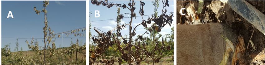
Microorganisms 2021, 9, 1916                                                                                          2 of 17

                               pathogens spread through streams, watering, certain cultural practices, and by transpor-
                               tation of agricultural products [5–7]. Symptoms begin at the roots or collar; the roots are
                               stubbed near the root collar and become rotted. In addition, the root system is reduced
                               with the appearance of sores and rot sometimes making the roots appear brown and
                               spongy, especially in the crown of some conifer species. These oomycetes are also in-
                               volved in negative plant-soil feedback due to monoculture and replanting problems of
                               apple orchards [8–10].
                                     In Morocco, weather conditions are diversified, which allows for the production of a
                               wide range of fruit during each growing season. The rosaceous fruit trees cover an area of
                               more than 300,000 ha, distributed between stone fruit 85% of total production, and 15%
                               for quince, apple pear, and almond, with the majority being almond trees [11]. The Middle
                               Atlas, the Rif, Saïss, Haouz, and Moulouya are the main production areas, with more than
                               56% of the total planted area being apple trees. Apple trees occupied the largest rosaceous
                               planted area with 32,000 ha and an estimated yield of 600,000 tons a year [12]. However,
                               rosaceous fruit crops are largely threatened by devastating and widespread diseases in
                               most parts of the world due to climate change, as well as changes in cultural practices [13].
                                     A new genus of Phytopythium, a relative of Phytophthora and Pythium has been re-
                               ported [14] as a pathogen of several fruit trees, including citrus in Tunisia [15], kiwifruit
                               in Turkey [16], avocados in the Canary Islands [17,18], and grapevines in South Africa
                               [19], and isolated from freshwater environments in Korea [20]. Similar symptoms to those
                               described previously were seen in apple and pear trees in Morocco. Recently, Jabiri et al.
                               [21] reported, for the first time, the occurrence of Phytopythium vexans on apple trees.
                               Therefore, the present study will focus on determining the occurrence and distribution of
                               these oomycete pathogens in apple and pear growing area of Saïss.
                                     In 2018, symptoms similar to decline disease were observed on rosaceous fruit trees,
                               including apple and pear, in growing areas of the Saïss region. The most prevalent symp-
                               toms were dry necrotic bark with brown lesions and yellowing foliage, wilting, and trunk
                               cankers, crown and root rot causing tree decline. The role played by oomycete species in
                               apple and pear disease in Morocco and has not yet been fully investigated. A preliminary
                               investigation evidenced the occurrence of Phytopythium vexans as the major causal agent
                               of these symptoms [21]. Therefore, an extensive survey was undertaken to better under-
                               stand the etiology of the disease. The present study focuses on the distribution of the oo-
                               mycete genus pathogenic to apple and pear trees in Morocco, in order to (i) identify and
                               characterize the oomycete microorganisms associated with the disease, (ii) investigate the
                               possible farming factors and practices allowing the occurrence of the disease, and (iii) ful-
                               fill Koch’s postulates in order to verify the role of the isolated pathogen as causative agent
                               of the disease.

                               2. Materials and Methods
                               2.1. Sampling Sites
                                    Fifty orchards of apple and pear trees in five locations of the Fez-Meknes region
                               (Meknes, El Hajeb, Sefrou, Imouzzer, and Azrou) were sampled to look for the responsible
                               agent of tree decline (Figure 1). Most orchards were selected based on tree health infor-
                               mation provided by producers and technical advisers of agricultural cooperatives. The
                               criteria by which the target areas were selected were early symptoms of tree decline such
                               as reduced growth and vigor, leaf chlorosis, or other more obvious symptoms like wilting,
                               yellowing and defoliation, and dieback of shoots and branches.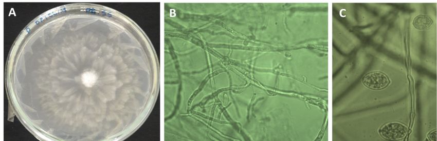
Microorganisms 2021, 9, 1916                                                                                            3 of 17

                               Figure 1. Map showing locations of the Meknes-Fes region in Morocco (Upper quadrat) and en-
                               larged views of Meknes, El-Hajeb, Imouzzer-Kander, Sefrou, and Azrou (from the shaded square).
                               The map was performed by ArcGIS software (v.10.9) and indicated the names of different districts
                               where samples were collected.

                                    The sampling method consisted of taking soil samples in triplicate from three differ-
                               ent locations around each diseased tree. Each composite sample was made up of three
                               sub-samples taken from the soil in the targeted area at a depth of 10 to 20 cm below the
                               organic surface layer, while collecting the lateral roots. Approximately 1 kg of soil was
                               collected by mixing sub-samples from all three sites. Each representative sample was
                               sieved through a 5 mm sieve. Soil samples (50) were stored in a cold room until use [22].

                               2.2. The Survey
                                    Datasets on cultural practices that may influence disease development such as soil
                               type, cultivars, rootstocks, and irrigation systems were collected during this survey and
                               the impact of these cultural practices was assessed. As the occurrence of oomycete patho-
                               gens is significantly influenced by watering, farmers were interviewed about drip irriga-
                               tion (flow rate per tree and duration) versus submersion irrigation (delivered amount of
                               water per tree and duration between two consecutive watering).

                               2.3. Isolation of the Pathogenic Fungi: Fruit and Soil Baiting
                                     According to Jabiri et al. [21], symptoms observed on apple and pear trees in orchards
                               (Figure 2) were caused by oomycete pathogens. To isolate these pathogens, two tech-
                               niques were employed and conceived in the laboratory. The first technique consisted of
                               using pear fruit as bait for the 50 samples. For each of the 50 samples, three plastic boxes
                               labeled sterile were used; each (1 L capacity) was filled with soil samples on which a dis-
                               infected green pear fruit was placed, and sterile distilled water was added until the fruit
                               was submerged. Subsequently, boxes were sealed and incubated at 4 °C in a cold chamber
                               for 7–10 days. Pear fruit presenting brown lesions were cut into smaller pieces of 1 cm and
                               four pieces were placed into Petri dishes containing modified CMA medium. All plates
                               were sealed with parafilm and incubated at 25 °C in darkness. After 7 days post-incuba-
                               tion, fungal isolates were checked, subcultured, and purified on PDA medium.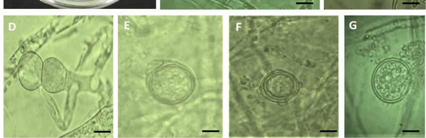
Microorganisms 2021, 9, 1916 4 of 17
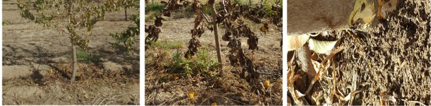
Figure 2. Decline disease symptoms such as yellowing (A), wilting (B), and brown lesions on the
crown (C) on young pear tree sampled in the Sefrou location, Louata farm.
The second technique consisted of using apple seeds as bait. Apple seeds were disin-
fected by soaking in a solution of 1% sodium hypochlorite (NaOCI) for 2 min, rinsed three
times with sterile distilled water (SDW) and dried at ambient temperature. The seeds were
shelled, cut in half, and used as live bait for oomycetes. Four cotyledons were deposited
on the soil surface already prepared and placed in Petri dishes [23]. Petri dishes were filled
with 60 mL of SDW. Three boxes were used for each representative soil sample. These
dishes were then incubated at 4 °C and checked daily for the appearance of white myce-
lium on apple cotyledons. The cotyledons were examined under the microscope 4 days
post-incubation, and those with coenocytic mycelium were removed and gently washed
with SDW to clear cotyledons of adhering soil debris. Mycelial filaments were taken from
the edge of the cotyledon using a sterile needle, then placed on Petri dishes containing
modified corn meal agar (CMA) medium. To render this medium selective, a stock solu-
tion composed of the following ingredients: penicillin G (1 g), Polymyxin B (1 g), ampicil-
lin (1 g), PCNB (pentachloronitrobenzene, 0.01 g), and hymexazol (0.5 mL) was prepared
in 100 mL of SDW. Once the temperature of the CMA medium was around 40–45 °C, 2
mL of this stock solution was added [24,25]. In cases where there were no apparent my-
celia on the seed surface but attached sporangia were visible under the microscope, each
seed was deposited into the center of each Petri dish containing modified CMA medium.
These Petri dishes were incubated at 25 °C for 2 to 6 days until the development of a fungal
colony.
2.4. Morphological Identification
The identification of the pathogenic oomycetes was initially based on visual observa-
tion of the forming colony (color and shape), mycelial growth, and sporulation. Under the
light microscope, colony and hyphal morphology, and the production and morphological
traits of sporangia, oogonium, antheridia, oospores and chlamydospores were examined
accordingly [4,26–29]. To induce the formation of sporangia, mycelial plugs of 1 cm in
diameter were cut from the edges of 5-day old growing cultures on V8 agar medium and
then placed in Petri dishes previously filled with 10 mL of non-sterile soil extract [4]. The
Petri dishes were placed at 18 °C for 24 h followed by 4 °C for 2 h [30]. The dishes were
examined daily for sporangial development and zoospore release over a period varying
from 2 to 7 days.
Microorganisms 2021, 9, 1916                                                                                          5 of 17

                               2.5. Molecular Identification
                                     Fresh mycelia were harvested by scraping the surface of 7-day old growing culture
                               onto PDA medium with a sterile spatula using the protocol described by Doyle and Doyle
                               [31] with some modifications. Approximately 50 to 100 mg of the mycelium of each fungal
                               isolate were placed in 2 mL Eppendorf tubes containing 500 μL of the extraction buffer.
                               The mixture was ground for 1 min, incubated for 30 min at 65 °C in a water bath and
                               briefly centrifuged at 13,000 rpm for 5 min. Afterward, 400 μL of the supernatant was
                               recovered and an equivalent volume (400 μL) of chloroform/isoamyl alcohol (24/1) was
                               added. The mixture was gently vortexed for 5 min and centrifuged at 14,000 rpm for 5
                               min. The supernatant (350 μL) was recovered, precipitated with isopropanol (350 μL), and
                               then centrifuged at 14,000 rpm for 10 min. The supernatant was then removed and 500 μL
                               of ethanol at 70 °C was added to the pellet, vortexed, and centrifuged for 5 min at 14,000
                               rpm. The pellet was dried in an incubator at 60 °C (30–45 min), resuspended in 50 μL of
                               SDW, and stored at −80 °C until use.
                                     Molecular identification was performed by sequencing the Cox II gene using a pair
                               of primers amplifying 563 bp of FM78 (5′-ACAAATTTCACTACATTGTCC-3′), and FM75
                               (5′-CCTTGGCAATTAGGATTTCAAGAT-3′), DNA amplification for the detection of Phy-
                               topythium species was carried as follow: one cycle at 95 °C for 2 min; followed by 35 cycles
                               of 1 min annealing at 56 °C, 2 min extension at 72 °C, and 1 min denaturation at 94 °C; and
                               finally by one extension cycle at 72 °C for 10 min [32]. The PCR amplified products were
                               separated on 1% agarose gel in 1X TBE buffer (Tris-boric acid-EDTA), and stained with
                               0.4% Ethidium bromide, and visualized under UV light. The PCR products were se-
                               quenced at STAB Vida Inc. (Caparica, Portugal) using Sanger dideoxy sequencing
                               method. The Cox II sequences of each isolate was compared to the sequences submitted
                               to GenBank. The species identity of each fungal isolate was determined using Basic Local
                               Alignment Search Tool (BLAST), where the analysis of each sequence is grouped with the
                               most closely related species at NCBI-BLAST (http://www.ncbi.nlm.nih.gov/BLAST/, ac-
                               cessed on 1 July 2021).
                                     Phylogenetic and molecular evolutionary analyses were conducted using MEGA ver-
                               sion 10.1.7 [33]. An alignment of the Cox II sequences was generated using Clustal W [34].
                               Phylogenetic analyses of Cox II were performed by the maximum likelihood method and
                               Kimura 2-parameter model [35]. The phylogenetic tree was evaluated by bootstrap anal-
                               ysis based on 1000 replicates. In this study, a closely related species of Phytopythium
                               vexans (AB468910), Pp. helicoides (MW450816), Pp. sindhum (KJ595436), Pp. litorale
                               (MT050458), Pythium mercuriale (AB920504), Phytophthora tropicalis (DQ469735), Phy-
                               tophthora capsici (DQ469734), Phytophthora lateralis (AY129207), Phytophthora palmivora
                               (AY129220), Pythium ultimum (AF196640), Pythium aphanidermatum (AB160854), Pp.
                               aichiense (AB948192), Pp. babaiiaharii (MT720670), Pp. boreale (AB690677), Pp. carbonicum
                               (AB690678), Pp. chamaehyphon (AB690674), Pp. citrinum (AB690679), Pp. delawarense
                               (AB690672), Pp. fagopyri (AB690671), Pp. iriomotense (AB690689), Pp. longitubum
                               (MT720672), Pp. megacarpum (AB690665), Pp. montanum (AB690667), Pp. nanjigens
                               (MG788317), Pp. oedochilum (AB690676), and Pythium oligandrum (EU265664), were used
                               as references.

                               2.6. Pathogenicity Test
                                    In order to fulfill Koch’s postulates and to ensure that all fungal isolates were able to
                               produce the disease, a pathogenicity test was performed on 115 one-year old apple trees
                               as previously described [4]. Fungal isolates were grown on PDA at 25 °C for 7 days. Each
                               apple plant was wounded at two sites, collar and stem, with a cork-borer at a diameter of
                               5 mm, each wound was filled with a mycelial plug (diameter 5 mm), cut from the edge of
                               a fresh, actively growing colony of each fungal isolate and then covered with parafilm.
                               Wounded plants inoculated with PDA free of fungal mycelia were used as controls. This
                               experiment was repeated twice with four replicates for each fungal isolate. Inoculated and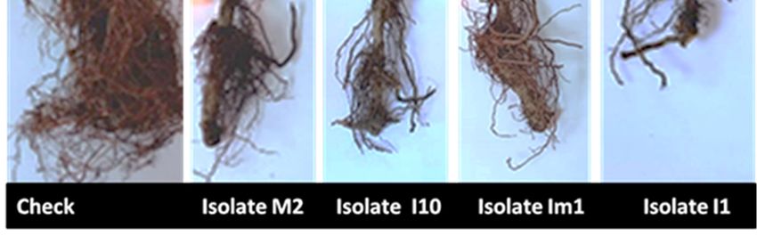
Microorganisms 2021, 9, 1916                                                                                         6 of 17

                               control plants were grown in pot cultures (20 cm diameter × 19 cm deep) containing a
                               potting soil. All pots were arranged in full randomized design and watered once a week
                               or more when needed. The water irrigation was applied directly to the soil. The green-
                               house temperature was around 28 °C and maintained with heated ventilation. Plants were
                               inspected weekly for symptoms. The onset of symptoms were recorded 4 months post-
                               inoculation. At the end of the experiment, the size and color of the necrotic lesions induced
                               by each fungal pathogen was recorded. Data collected included visual ratings of shoots
                               and roots, as previously described by Rodriguez-Padron et al. [17]. Accordingly, shoots
                               were visually inspected for disease symptoms and rated on a scale of 1 to 5 with 1 =
                               healthy leaf, 2 = early yellowing, 3 = half-yellowed leaf 4 = dry leaf, and 5 = wilting leaf.
                               Roots were cleaned, visually inspected for root rot and rated on a scale of 1 to 5 with 1 =
                               healthy white roots/ no disease recovered; 2 = 25% root rot or seemingly healthy roots +
                               onset of root rots), 3 = 50% root rot and early browning, 4 = 75% root rot (brown rot) and
                               5 = 100% dead roots-root system destroyed. Fungal isolates were re-isolated from necrotic
                               tissues to complete Koch’s postulates.

                               2.7. Statistical Analysis
                                    A chi-square analysis test (χ2) was used to assess the relationship between disease
                               prevalence and some common farming practices on the Saïss plain, such as rootstocks, soil
                               type, and type of plantation, cultivars, and irrigation mode in orchards. All tests were
                               arranged in a completely randomized design (CRD). Analysis of variance (ANOVA) was
                               performed using SPSS statistical software (IBM SPPSS Statistics 25) to assess the effect of
                               isolate inoculation. When the effect was revealed to be significant, the least square differ-
                               ence (LSD) test was employed for mean separation (p < 0.05).

                               3. Results
                               3.1. Field Symptoms
                                    Decline symptoms on trees were observed in pear and apple growing areas of the
                               Fes-Meknès region of Morocco. These symptoms included a quick dieback, necrotic
                               brown lesions in the bark, defoliation and yellowing, wilting, and root rot leading to the
                               death of the infected tree (Figure 2). Accordingly, an extensive survey of 50 fields of apple
                               and pear was performed in order to determine the causal agents of the tree decline in the
                               region. After a deep investigation into the cultural practices adopted by the farmers, in
                               particular excessive watering of trees, it was assumed that the disease was caused by oo-
                               mycetes. Therefore, laboratory analyses were performed to look for the presence of these
                               oomycete pathogens by using soil baiting.

                               3.2. Isolation, Morphological, and Molecular Identification of Fungal Isolates
                                    A total of 35 oomycetous isolates were obtained from representative samples of 50
                               surveyed fields (Table 1). The visual observation of morphological traits such as color and
                               shape of the obtained growing colony underlines a typical colony of an oomycete patho-
                               gen (Figure 3). All fungal colonies had a whitish-blooming mycelium appearance with no
                               visual sporulation at the surface, which confirmed their identity as oomycetous mycelia.
                               Under the light microscope, non-septate hyphae were observed for all 35 fungal isolates
                               with major hyphae ranging in width from 2.5 to 6.3 μm (Figure 3A). Furthermore, the
                               microscope examination detected the presence of oomycete traits in V8 medium after in-
                               cubation for 4 days, such as coenocytic hyphae (Figure 3B), globose/subglobose sporangia
                               (Figure 3C) with/out papilla, 15.9 × 26.10 μm in diameter, double oospore (Figure 3D),
                               oogonium with single lobed branched antheridium (Figure 3E), oogonium with two-lobed
                               branched antheridium (Figure 3F), and thick-walled chlamydospores (Figure 3G). Oogo-
                               nia were smooth, globose, and terminal with an average diameter of 26 μm. Zoospores
                               released from sporangia were Pythium-like.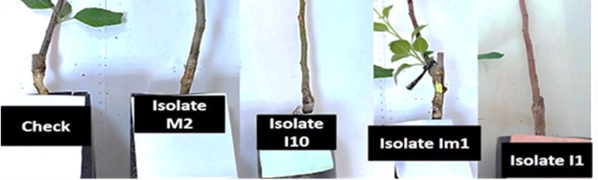
Microorganisms 2021, 9, 1916                                                                                                   7 of 17

                                     Figure 3. Morphology of Phytopythium vexans: white transparent colony on PDA medium (A), coe-
                                     nocytic hyphae (B), sporangia (C), double oospore (D), oogonium with single lobe branched anther-
                                     idium (E), oogonium with two lobes branched antheridium (F), and thick-walled chlamydospores
                                     (G). Bar scale = 15 μm.

      Table 1. Identity of Phytopythium vexans isolates based on morphology and COX II genes, and their GenBank accessions
      numbers (an.).

  Fungal                       Type of Plan- Rootstoc                                                         Genbank COX II an.
                Origin                                        Type of Soil            Identified Species
  Isolate                         tation        k                                                                        1

     E4        El-Hajeb          Pear tree    M106                Clay               Phytopythium vexans           MW815816
     E5        El-Hajeb         Apple tree    Pajam               Clay               Phytopythium vexans              -
    M1         Meknès           Apple tree    M111           Calcimagnesic           Phytopythium vexans           MW815828
    M2         Meknès           Apple tree    Pajam          Calcimagnesic           Phytopythium vexans           MW815829
     S1         Sefrou           Pear tree    Wild         Calcimagnesic-clay        Phytopythium vexans           MW815836
     S2         Sefrou           Pear tree    Wild         Calcimagnesic-clay        Phytopythium vexans           MW815837
     S3         Sefrou          Apple tree     M7              Sandy-clay            Phytopythium vexans           MW815838
     S4         Sefrou          Apple tree     M9              Sandy-clay            Phytopythium vexans           MW815839
     S5         Sefrou          Apple tree     M7              Sandy-clay            Phytopythium vexans           MW815840
     S8         Sefrou          Apple tree     M9              Sandy-clay            Phytopythium vexans           MW815841
     S9         Sefrou          Apple tree     M9              Sandy-clay            Phytopythium vexans           MW815842
    S10         Sefrou          Apple tree     M7              Sandy-clay            Phytopythium vexans           MW815843
     A1         Azrou           Apple tree     M9           Calcareous-clay          Phytopythium vexans           MW815813
     A6         Azrou           Apple tree    Wild              Clay-silty           Phytopythium vexans           MW815814
     A7         Azrou           Apple tree    Pajam             Clay-silty           Phytopythium vexans           MW815815
     I1       Imouzzer          Apple tree    Wild             Sandy-clay            Phytopythium vexans              -
    Im1       Imouzzer          Apple tree    Pajam            Sandy-clay            Phytopythium vexans           MW815817
     I2       Imouzzer           Pear tree    Wild             Sandy-clay            Phytopythium vexans              -
     I3       Imouzzer          Apple tree    Wild             Sandy-clay            Phytopythium vexans           MW815819
    Im3       Imouzzer          Apple tree    Wild             Sandy-clay            Phytopythium vexans           MW815818
     I5       Imouzzer          Apple tree    M106             Sandy-clay            Phytopythium vexans           MW815830
     I6       Imouzzer          Apple tree    M111             Sandy-clay            Phytopythium vexans           MW815831
     I7       Imouzzer          Apple tree    Wild             Sandy-clay            Phytopythium vexans           MW815832
     I8       Imouzzer          Apple tree    Wild             Sandy-clay            Phytopythium vexans           MW815833
Microorganisms 2021, 9, 1916                                                                                                 8 of 17

    I10       Imouzzer         Apple tree    Wild           Sandy-clay             Phytopythium vexans           MW815834
    I12       Imouzzer         Apple tree    Wild           Sandy-clay             Phytopythium vexans           MW815825
    I13       Imouzzer         Apple tree    Wild           Sandy-clay             Phytopythium vexans           MW815835
    I14       Imouzzer         Apple tree    Wild           Sandy-clay             Phytopythium vexans           MW815844
    I15       Imouzzer         Apple tree    Wild           Sandy-clay             Phytopythium vexans           MW815820
    I16       Imouzzer         Apple tree    Wild           Sandy-clay             Phytopythium vexans           MW815821
    I17       Imouzzer         Apple tree    Wild           Sandy-clay             Phytopythium vexans           MW815822
    I18       Imouzzer         Apple tree    Wild           Sandy-clay             Phytopythium vexans           MW815823
    I19       Imouzzer         Apple tree    Wild           Sandy-clay             Phytopythium vexans           MW815824
    I20       Imouzzer         Apple tree    Wild           Sandy-clay             Phytopythium vexans           MW815826
    I21       Imouzzer         Apple tree    Wild           Sandy-clay             Phytopythium vexans           MW815827
      1X: a total of 35 isolates of Phytopythium vexans were identified morphologically and based on phylogenetic tree analyses
      and had their Cox II gene subsequently sequenced and deposited in Genebank. (-): missed sequences.

                                        To confirm the exact identity of the obtained oomycetous isolates, specific primers
                                   aiming to identify the cytochrome subunit II (Cox II) gene were used. Interestingly, the
                                   results of sequencing showed that all isolates were 99% similar to those of Pp. vexans in
                                   the genbank. Results from the blast and phylogenetic tree analysis of Cox II showed the
                                   high similarity of the isolates to Pp. vexans; the phylogenic analysis grouped all the isolates
                                   into a single clade of Pp. vexans (Figure 4, Table 1).

                                   3.3. Distribution of the Pathogenic Oomycetes in the Surveyed Apple and Pears Orchards
                                         The prevalence of oomycetes in samples taken from each region was shown in Table
                                   2. Results indicated that positive samples 20, 8, 3, 2, and 2 were from the Imouzzer, Sefrou,
                                   Azrou, El Hajeb, and Meknes locations, respectively. Moreover, no samples from Sidi
                                   Lmakhfi district in Azrou and Chlihat district in El-Hajeb were revealed to be positive for
                                   the pathogen (Tables 1 and 2). However, the Imouzzer location was the most infected of
                                   all the surveyed regions.
                                         The survey pointed out that similar symptoms occurred on apple and pear trees, with
                                   a higher frequency on apple trees (Table 1 and Figure 5A). Results also indicated that there
                                   was a significant effect (p < 0.05) of soil type on disease prevalence (Figure 5B). A signifi-
                                   cant effect was also seen with rootstocks (p < 0.028). The wild rootstock was the most sus-
                                   ceptible to the disease in comparison with the other rootstocks (Figure 5C).
                                         Furthermore, results pointed out that drip irrigation (75%) versus submersion irriga-
                                   tion was the most used irrigation mode in the prospected region with an average flow rate
                                   of 16 L/h per tree. The duration of irrigation ranged from 1–2 h with an interval of 24 to
                                   48 h and differed among farmers and according to season. In contrast, the amount of water
                                   delivered by submersion irrigation ranged from 80 to 100 L per tree with an interval of 5
                                   days to one week between two successive watering’s. Furthermore, the impact of the wa-
                                   tering system on disease prevalence was significant (p < 0.05). The highest prevalence was
                                   observed on farms adopting drip irrigation (Figure 5D). This result was considered nor-
                                   mal as most of the farms surveyed adopted drip irrigation.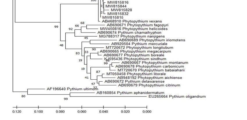
Microorganisms 2021, 9, 1916                                                                                                    9 of 17

      Figure 4. Phylogenetic tree of oomycetes fungi Pp. vexans was constructed using the maximum likelihood method on their
      cytochrone oxidase subunit II sequences, showing the position of our oomycetes isolates that were obtained in this study
      within the Phytopythium and Pythium complex. Isolates from this study are indicated in bold characters. The sequences of
      Phytopythium, Phytophthora and Pythium from Genebank; Pp. vexans (AB468910), Pp. helicoides (MW450816), Pp. sindhum
      (KJ595436), Pp. litorale (MT050458), Pythium mercuriale (AB920504), Phytophthora tropicalis (DQ469735), Phytophthora capsici
      (DQ469734), Phytophthora lateralis (AY129207), Phytophthora palmivora, (AF196640) Pythium ultimum (AF196640), Pythium
      aphanidermatum (AB160854), Pp. aichiense (AB948192), Pp. babaiiaharii (MT720670), Pp. boreale (AB690677), Pp. carbonicum
      (AB690678), Pp. chamaehyphon (AB690674), Pp. citrinum (AB690679), Pp. delawarense (AB690672), Pp. fagopyri (AB690671),
      Pp. iriomotense (AB690689), Pp. longitubum (MT720672), Pp. megacarpum (AB690665), Pp. montanum (AB690667), Pp. nan-
      jigens (MG788317), Pp. oedochilum (AB690676), and Pythium oligandrum (EU265664), were used as references. Numbers at
      the nodes of clusters represent the bootstrap values that were generated from 1000 pseudoreplicates.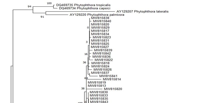
Microorganisms 2021, 9, 1916                                                                                                  10 of 17

                               Table 2. Occurrence and distribution of pathogenic oomycetes in apple and pear orchards in differ-
                               ent regions in Morocco.

                                                                                           Positive Isolations
                                   Location     District     No. Orchards a
                                                                               Phytopythium vexans Disease Prevalence (%)
                                    Azrou       Tigrigra            2                   1                      33
                                              Sidi Lmakhfi          2                   -
                                                Ain Louh            5                   2

                                   El-Hajeb Tamchachate             3                      2                          40
                                              Chlihat               2                      -

                               Imouzzer        Ain Chifa             9                    8                           83
                                               Aït Sbaà             14                    11
                                                Farha                1                    1

                                   Meknès        Majjat             2                      2                          100

                                               Aghbalou
                                    Sefrou                          2                      2
                                                Akourar                                                               80
                                               Laanoucer             8                    6
                                    Total                           50                    35                          70
                               a   Numbers of surveyed orchards.

                               Figure 5. Disease prevalence (%) according to the type of plantation apple/pear tree (A), type of soil
                               (B), apple rootstocks (C), and irrigation mode (submersion versus drip irrigation mode) in the sur-
                               veyed orchards of Saïss plain (D). Asterisks (*) indicate significant effect (p < 0.05) according to the
                               Chi square analysis test (χ2).

                               3.4. Pathogenicity Test
                                    All fungal isolates were pathogenic to apple seedlings and most showed symptoms
                               1-month post-inoculation. Accordingly, necrotic lesions on the inoculated stem were ob-
                               served, for the first time, 18 days post-inoculation, and after 30 days post-inoculation,
                               more than 95% of the inoculated apple seedlings showed symptoms most likely similar to
                               those observed in the field (Figure 6). The bark tissue turned brown and rotted, and the
Microorganisms 2021, 9, 1916 11 of 17
lesion reached the wood, while control plants were healthy and asymptomatic. Four
months later, apple seedling growth was substantially reduced compared to un-inocu-
lated seedlings, which were healthy and grew normally and the root system of inoculated
seedlings presented significant necrosis and acute decay (Figure 7). All inoculated patho-
gens were successfully re-isolated from the inoculated stems and collars of apple seed-
lings.
Figure 6. Symptoms of yellowing, wilting, and lesions on stems of apple rootstock M115 seedlings
at the end of the stem pathogenicity test: healthy control ((A), uninoculated plants); plants inocu-
lated with Phytopythium vexans M2 (B), Pp. vexans I10 (C), Pp. vexans Im1 (D), and Pp. vexans I1 (E).
Figure 7. Root rot symptoms in apple rootstock M115 seedlings at the end of the root pathogenicity
test (four months post-inoculation) under greenhouse conditions: healthy control ((A), uninoculated
plants); plants inoculated with Phytopythium vexans M2 (B), Pp. vexans I10 (C), Pp. vexans Im1 (D),
and Pp. vexans I1 (E).Microorganisms 2021, 9, 1916 12 of 17
Results of pathogenicity tests after 4 months post-inoculation are listed in Table 3.
Statistical analysis showed a highly significant effect (p < 0.0001) of fungal isolates on col-
lar and stem lesion length, leaves, and root necrosis indices relative to untreated controls
(Table 3). In addition, a significant difference was observed between Pp. vexans isolates for
all evaluated traits. Pp. vexans isolates I17, I3, I1, and I12 were revealed to be the most
aggressive with recorded lesion sizes (cm) of 5.46 ± 0.11, 4.53 ± 0.55, 6.13 ± 0.15, 6.12 ± 0.75,
respectively. These isolates induced a marked dryness on the stem. On the collar, results
underlined that isolate S5 was the most aggressive and had a lesion size of 5.93 ± 0.15
(Table 3). Furthermore, disease symptoms on leaves and roots were rated between 4 and
5 and were statistically different from the un-inoculated control plants (p < 0.0001). Signif-
icance differences were also observed between oomycete isolates for these two evaluated
parameters (Table 3). For all isolates, root rot disease severities ranged from 70% to 100%.
Table 3. Results of pathogenicity tests in apple seedlings showing the averaged collar and stem
lesion length (cm), leaves, and root necrosis indices (1–5) after 4 months post-inoculation.
Lesion Length (cm) Scale Severity Symptoms
Fungal Isolates
Collar Stem Leaves Wilting 2 Roots Necrosis 3
Pp. vexans E4 4.20 1 ± 0.1 ghij 2.93 ± 0.15 c 1.00 ± 0.00 b 4.66 ± 0.57 cd
Pp. vexans E5 3.60 ± 0.15 cd 3.06 ± 0.11 cd 1.00 ± 0.00 b 3.66 ± 0.57 b
Pp. vexans M1 5.06± 0.05 opq 3.57 ± 0.57 efgh 1.66 ± 0.57 bcd 4.00 ± 1.00 bc
Pp. vexans M2 3.60 ± 0.10 cd 3.03 ± 0.05 cd 2.00 ± 0.00 cde 4.33 ± 0.57 bcd
Pp. vexans S1 4.53 ± 0.05 jklm 3.33 ± 0.11 cdef 1.33 ± 0.57 bc 4.33 ± 0.57 bcd
Pp. vexans S2 4.56 ± 0.11 klm 3.80 ± 0.10 ghij 2.33 ± 0.57 def 4.66 ± 0.57 cd
Pp. vexans S3 3.93 ± 0.15 defg 4.10 ± 0.10 ijk 2.00 ± 0.00 cde 5.00 ± 0.00 c
Pp. vexans S4 4.8 ± 0.10 mno 3.6 0± 0.26 efgh 5.00 ± 0.00 l 4.66 ± 0.57 cd
Pp. vexans S5 5.93 ± 0.15 t 4.20 ± 0.10 jk 4.66 ± 0.57 kl 4.66 ± 0.57 cd
Pp. vexans S8 5.16 ± 0.15 pqr 3.70 ± 0.10 fghi 3.00 ± 0.00 fgh 4.66 ± 0.57 cd
Pp. vexans S9 4.40 ± 0.10 ijkl 3.40 ± 0.10 defg 2.66 ± 0.57 efg 5.00 ± 0.00 c
Pp. vexans S10 4.96 ± 0.11 nop 3.43 ± 0.57 defg 4.67 ± 0.57 kl 4.66 ± 0.57 cd
Pp. vexans A1 3.50 ± 0.52 bc 3.36 ± 0.49 cdefg 1.33 ± 0.57 bc 4.66 ± 0.57 cd
Pp. vexans A6 3.60 ± 0.10 cd 3.10 ± 0.10 cd 1.00 ± 0.00 b 4.33 ± 0.00 bcd
Pp. vexans A7 4.46 ± 0.32 ijklm 3.62 ± 0.15 efgh 1.00 ± 0.00 b 4.66 ± 0.57 cd
Pp. vexans I1 5.56 ± 0.60 s 6.13 ± 0.15 m 4.33 ± 0.57 jkl 4.33 ± 0.57 bcd
Pp. vexans Im1 4.30 ± 0.20 hijk 4.00 ± 0.10 hij 4.66 ± 0.57 kl 4.66 ± 0.57 cd
Pp. vexans I2 5.10 ± 0.20 opqr 4.53 ± 0.30 k 2 ± 1.00 cde 4.66 ± 0.57 cd
Pp. vexans I3 4.67 ± 0.15 lmn 7.23 ± 0.55 n 2.33 ± 0.57 def 5.00 ± 0.00 c
Pp. vexans Im3 5.36 ± 0.32 qrs 4.06 ± 0.11 ij 4.00 ± 0.00 ijk 5.00 ± 0.00 c
Pp. vexans I5 4.16 ± 0.11 ghi 3.80 ± 0.1 ghij 1.66 ± 0.57 bcd 4.66 ± 0.57 cd
Pp. vexans I6 3.76 ± 0.25 cdef 3.06 ± 0.11 cd 1.00 ± 0.00 b 5.00 ± 0.00 c
Pp. vexans I7 5.03 ± 0.15 opq 2.13 ± 0.15 b 4.00 ± 0.00 ijk 4.33 ± 0.57 bcd
Pp. vexans I8 4.00 ± 0.10 efgh 3.23 ± 0.25 cde 5.00 ± 0.00 l 4.33 ± 0.57 bcd
Pp. vexans I10 4.80 ± 0.20 mno 3.68 ± 0.05 fghi 3.00 ± 0.00 fgh 4.66 ± 0.57 cd
Pp. vexans I12 4.66 ± 0.41 lmn 6.20 ± 0.75 m 3.66 ± 0.57 hij 5.00 ± 0.00 c
Pp. vexans I13 3.93 ± 0.15 defg 3.70 ± 0.10 fghi 5.00 ± 0.00 l 5.00 ± 0.00 c
Pp. vexans I14 4.03 ± 0.05 fgh 3.26 ± 0.28 cdef 1.00 ± 0.00 b 4.66 ± 0.57 cd
Pp. vexans I15 5.40 ± 0.15 rs 3.20 ± 0.20 cde 4.66 ± 0.57 kl 5.00 ± 0.00 c
Pp. vexans I16 3.93 ± 0.15 defg 3.30 ± 0.20 cdef 5.00 ± 0.00 l 4.33 ± 0.57 bcd
Pp. vexans I17 3.23 ± 0.15 b 5.46 ± 0.11 l 3.33 ± 0.57 ghi 4.33 ± 0.57 bcd
Pp. vexans I18 4.73 ± 0.15 mno 3.70 ± 0.10 fghi 4.66 ± 0.57 kl 4.66 ± 0.57 cd
Pp. vexans I19 4.30 ± 0.20 hijk 3.70 ± 0.20 fghi 5.00 ± 0.00 l 4.66 ± 0.57 cd
Pp. vexans I20 3.66 ± 0.15 cde 3.40 ± 0.25 defg 3.00 ± 0.00 fgh 4.00 ± 0.00 bcMicroorganisms 2021, 9, 1916 13 of 17
Pp. vexans I21 4.16 ± 0.15 ghi 5.23 ± 0.68 l 4.00 ± 1.00 ijk 4.33 ± 0.57 bcd
Un-inoculated control 0.00 a 0.00 a 0.00 a 0.00 a
1: Data are the average of two experiments with four replicates; in each column, values having the
same letter are not significantly different according to the LSD test (p < 0.05). 2: Leaf wilting were
rated on a scale 1 to 5. 3: Root rot was rated on a scale 1 to 5, with 1: no disease recovered; 2: 25%
rot root; 3: 50% root rot; 4: 75% root rot and 5: 100% dead roots-root system destroyed.
4. Discussion
In recent years, symptoms of decline such as root rot, brown rot, and crown canker
have been observed on pear and apple fruit trees in the Saiss region, causing serious dam-
age and reducing overall yield. In this study, we confirmed through morphological and
molecular characterization, and pathogenicity tests that these symptoms of decline were
mainly due to Pp. vexans. This result is in agreement with our previous findings [21] in
which Pp. vexans was the main pathogen of apple dieback in the Saïss region. The patho-
gen Pp. vexans, which threatens apple and pear trees in particular, was found in 34 of 50
samples from different sites of commercial apple and pear orchards in the Fez-Meknes
region.
The pathogen, Phytopythium is considered to be an intermediate between Phy-
tophthora and Pythium species [36]. It is a new genus of the Pythiaceae family and the order
Peronosporales, which was described with Phytopythium sindhum as a type species by [37].
The pathogenic oomycete Pp. vexans was reported on apple trees in South Africa in 2011
[5] and on citrus in China [38] and Tunisia in 2017 [15]. It was also isolated from infected
avocado trees in the Canary Islands [17], on avocado in Mexico [39], on kiwi in Turkey
[16], on grapevines in South Africa [19], on cassava in Brazil [40], and in Vietnam on du-
rian [41]. Furthermore, the analysis of the phylogenetic tree underlined a strong similarity
of our isolates of Pp. vexans with those of different countries mentioned above. Under the
light microscope, Pp. vexans colonies appeared more similar to Pythium than Phytophthora,
while symptoms observed in the field as well as those obtained from the pathogenicity
test of this study revealed a substantial similarity to those of Phytophthora diseases. It was
concluded that both pathogens (Phytopythium and Phytophthora) cause root rot, collar and
crown canker, branching of roots, yellowing of leaves followed by wilting, necrotic bark
becoming dry, and brown lesions in the neck [42]. It was noted that all of these symptoms
were observed and recorded during the field and pathogenicity test. In addition, Phy-
tophthora and Phytopythium are able to grow at variable temperatures [43], so it will be
useful to study the influence of temperature on the growth of Pp. vexans. Such a study can
determine whether changes in environmental conditions are the cause of its occurrence in
recent years.
Many hypotheses suggest that the main cause of apple and pear decline was exces-
sive irrigation, as these kinds of pathogens require high moisture for their proliferation,
production of sporangia, and release of infectious propagules and zoospores [44]. There-
fore, it was concluded that irrigation may have induced Pp. vexans infection on apple trees,
particularly in orchards with a high planting density [45]. In addition, Moein et al. [46]
indicated that the use of higher irrigation regimes was likely the cause of the greater dis-
ease severity on inoculated apple seedlings with either P. ultimum, P. irregulare, P. sylvati-
cum, Pp. vexans, or Phytophthora cactorum. Therefore, any action aiming to prevent a humid
microclimate in the cover will constitute an element of management since this disease de-
velops more rapidly under these conditions [47]. Our results pointed out that the disease
was more frequent in drip irrigation than in submersion irrigation due to improper man-
agement of irrigation systems by farmers who used drippers with a high-rate (16 l/h/tree)
instead of those with low flow rates (8 l/h/tree). These results were in complete agreement
with those reported by Benfradj et al. [15] who found that Pp. vexans disease was more
prevalent in drip irrigation. In our study, the disease was found in drip and in submersion
irrigation as well, however, its prevalence was higher as most farmers opted for drip irri-
gation with a higher flow rate and long duration. Therefore, it is important to manage theMicroorganisms 2021, 9, 1916 14 of 17
water supply as closely as possible to the needs of the crop and to irrigate by drip rather
than by submersion. Water in its various forms is one of the vectors of oomycetes, which
could be considered the most important mode of dissemination. Indeed, they produce
motile flagellated zoospores in water, which attack the trees through the roots. Thus, irri-
gation using contaminated water infects seedlings by Phytophthora alni in the nursery
[48,49]. In addition, the reuse of collected irrigation water is a practice presenting a high
risk in the nursery [50]. Without the installation of an effective disinfection system, these
waters can cause epidemics on irrigated plants. Depending on how it works, water can
spread infectious spores over varying distances, and a single drop of rainwater is capable
of disseminating spores in a radius of one meter [45].
The importance of the different propagules of oomycete fungi in the development of
their respective diseases differs according to the mechanism of dispersion. In the case
where the dispersion is carried out by free water, it is the zoospores that are involved in
the proliferation of the oomycete [51,52]. Regardless of the dispersal mechanism, zoo-
spores are continuously important in most oomycetous species, because they must swim
and locate the roots [53]. However, some Pythium species do not produce zoospores,
which means that there is another dispersal mechanism other than irrigation water [54].
Carlile et al. [55] pointed out that zoospores represent 95% of the Phytophthora propagules
recovered from irrigation water. They explained this finding to be due to their ability to
swim, while other propagules (the mycelium, chlamydospores, etc.) tend to sink to the
bottom of the water [56]. Other methods of dispersing oomycete propagules in fields and
orchards include movement of infested soil, infested nursery stock, and infested dust par-
ticles, all containing mycelium, chlamydospores, or oospores [57]. These findings can ex-
plain the highest level of infected samples in Imouzzer compared with other locations due
to excessive watering by small farmers as most of them are not yet familiarized with drip
irrigation.
The results of this study indicated that the prevalence of decline disease varies with
respect to the soil type and cultivar. It was found that the disease was more pronounced
in sandy-clay soil than in other types of soil. This might be due to the ability of zoospores
to move within soil particles and reach their root targets in a short amount of time. Moein
et al.[47] pointed out that soil temperature and soil type are primary factors in the inter-
action of oomycetes with their plant hosts and the subsequent severity of infection was
often dependent on these factors. Therefore, high disease severities were reported in clay
soils due to favourable conditions for the dispersal of zoospores facilitated by the high
water holding capacity of this type of soil [52,58]. Furthermore, serious damages by some
Pythium species were found in warmer areas, whereas other Pythium species are more
prevalent and virulent at lower temperatures [59,60].
Managing root rot of rosaceous trees is a difficult task and requires the use of an
integrated pest management (IPM) approach, as no single control strategy will prevent or
control this disease. However, systemic chemicals such as phenylamides and fenamiphos
were shown effective for managing rot root diseases caused by the oomycete group [61].
The effectiveness of some phosphonates and phenylamides, such as mefenoxam and met-
alaxyl, was demonstrated against Phytophthora diseases under field conditions [47]. Fur-
thermore, several Pythium species associated with root rot diseases of carrot, soybean,
corn, and forest nurseries were shown to be sensitive to metalaxyl, mefenoxam, and
fosetyl-Al [62–64]. However, Matthiesen et al. [64] noticed that the aggressiveness and
fungicide sensitivity of Pythium spp. was significantly affected by the temperature. Similar
active substances were used against Phytopyhtium spp. on soybean and corn [65]. In Mo-
rocco, the control of root rot of trees caused by oomycetes relies on the use of fungicides
as trunk injections with phosphonates or in application with drip watering. Soil fungicide
applications, including subsurface drip chemigation, have recently gained interest as a
method of improving control Phytophthora crown and root rot [66].Microorganisms 2021, 9, 1916 15 of 17
5. Conclusions
This study confirmed our previous findings and highlighted that Pp. vexans was the
major cause of apple and pear decline in Morocco. It was concluded that irrigation mode,
rootstock, and type of soil significantly impacted the disease. Therefore, the epidemiolog-
ical and economic relevance of these oomycetes as a causal agent of apple and pear dis-
eases in commercial orchards in Morocco deserves further investigation. Accordingly, dif-
ferent strategies to control this oomycetous pathogen and its subsequent disease are being
evaluated and implemented. The results of this study could help to better understand the
frequent occurrence of the disease and to develop reliable integrated strategies for its man-
agement that take into account the use of rational irrigation, appropriate chemicals, and
sanitation practices.
Author Contributions: Conceptualization, R.L.; methodology, R.L., S.J. and C.B.; software, S.J., N.R.
and R.L.; validation, R.L.; data curation, S.J. and C.B., resources, R.L.; writing—original draft prep-
aration, S.J. and R.L.; writing—review and editing, R.L., N.R., E.A.B. and D.M.; supervision, R.L.
and M.B.A.; project administration, R.L. All authors have read and agreed to the published version
of the manuscript.
Funding: This research received no external funding.
Institutional Review Board Statement: Not applicable.
Informed Consent Statement: Not applicable.
Data Availability Statement: Data is contained within the article.
Acknowledgments: This research was financially supported by the Phytopathology Unit, Depart-
ment of Plant Protection, Ecole Nationale d’Agriculture de Meknes. We are grateful to farmers for
helping us by providing the necessary information during the survey.
Conflicts of Interest: The authors declare no conflict.
References
1. Mazzola, M.; Manici, L.M. Apple replant disease: Role of microbial ecology in cause and control. Annu. Rev. Phytopathol. 2012,
50, 45–65, doi:10.1146/annurev-phyto-081211-173005.
2. Derevnina, L.; Petre, B.; Kellner, R.; Dagdas, Y.F.; Sarowar, M.N.; Giannakopoulou, A.; De la Concepcion, J.C.; Chaparro-Garcia,
A.; Pennington, H.G.; Van West, P.; et al. Emerging oomycete threats to plants and animals. Philos. Trans. R. Soc. B Biol. Sci. 2016,
371, 20150459, doi:10.1098/rstb.2015.0459.
3. Ho, H.H. The taxonomy and biology of phytophthora and pythium. J. Bacteriol. Mycol. Open Access 2018, 6, 00174,
doi:10.15406/jbmoa.2018.06.00174.
4. Wheller, T.; Erwin, D.C.; Ribeiro, O.K. Phytophthora Diseases Worldwide; APS Press: Saint Paul, MN, USA, 1996.
5. Tewoldemedhin, Y.T.; Mazzola, M.; Botha, W.J.; Spies, C.F.J.; McLeod, A. Characterization of fungi (Fusarium and Rhizoctonia)
and oomycetes (Phytophthora and Pythium) associated with apple orchards in South Africa. Eur. J. Plant Pathol. 2011, 130, 215–
229, doi:10.1007/s10658-011-9747-9.
6. Porter, L.D.; Johnson, D.A. Survival of Phytophthora infestans in Surface Water. Phytopathology 2004, 94, 380–387,
doi:10.1094/phyto.2004.94.4.380.
7. Jung, T.; Orlikowski, L.; Henricot, B.; Abad-Campos, P.; Aday, A.G.; Casal, O.A.; Bakonyi, J.; Cacciola, S.O.; Cech, T.; Chavar-
riaga, D.; et al. Widespread phytophthora infestations in European nurseries put forest, semi-natural and horticultural ecosys-
tems at high risk of phytophthora diseases. For. Pathol. 2015, 46, 134–163, doi:10.1111/efp.12239.
8. Goheen, D.J.; Filip, G.M. Root pathogen complexes in Pacific Northwest forests. Plant Dis. 1980, 64, 793–794.
9. Riolo, M.; Aloi, F.; La Spada, F.; Sciandrello, S.; Moricca, S.; Santilli, E.; Pane, A.; Cacciola, S.O. Diversity of Phytophthora com-
munities across different types of mediterranean vegetation in a nature reserve area. Forests 2020, 11, 853, doi:10.3390/f11080853.
10. Santa, O.C.; Maria, L.G. Emerging and re-emerging fungus and oomycete soil-borne plant diseases in Italy. Phytopathol. Mediterr.
2019, 58, 451–472.
11. Anonymous Rosacées Fruitières. Available online: https://www.fellah-trade.com/fr/filiere-vegetale/chiffres-cles-rosacees-
fruitieres?filiere=filiere_vegetale (accessed on 1 September 2020).
12. Moinina, A.; Boulif, M.; Lahlali, R. Important pests, diseases and weather conditions affecting apple production: Current state
and perspectives. Rev. Mar. Sci. Agron. Vét. 2019, 7, 71–87.
13. Moinina, A.; Lahlali, R.; MacLean, D.; Boulif, M. Farmers’ knowledge, perception and practices in apple pest management and
climate change in the fes-meknes region, Morocco. Horticulturae 2018, 4, 42, doi:10.3390/horticulturae4040042.Microorganisms 2021, 9, 1916 16 of 17
14. De Cock, A.; Lodhi, A.; Rintoul, T.; Bala, K.; Robideau, G.; Abad, Z.G.; Coffey, M.; Shahzad, S.; Lévesque, C. Phytopythium:
Molecular phylogeny and systematics. Pers. Mol. Phylogeny Evol. Fungi 2015, 34, 25–39, doi:10.3767/003158515x685382.
15. Benfradj, N.; Migliorini, D.; Luchi, N.; Santini, A.; Boughalleb-M’Hamdi, N. Occurrence of Pythium and Phytopythium species
isolated from citrus trees infected with gummosis disease in tunisia. Arch. Phytopathol. Plant Prot. 2017, 50, 286–302,
doi:10.1080/03235408.2017.1305479.
16. Polat, Z.; Awan, Q.N.; Hussain, M.; Akgül, D.S. First report of Phytopythium vexans causing root and collar rot of kiwi fruit in
Turkey. Plant Dis. 2017, 101, 1058, doi:10.1094/pdis-11-16-1554-pdn.
17. Rodriguez-Padron, C.; Siverio, F.; Perez-Sierra, A.; Rodriguez, A. Isolation and pathogenicity of Phytophthora species and Phy-
topythium vexans recovered from avocado orchards in the Canary Islands, including Phytophthora niederhauserii as a new patho-
gen of avocado. Phytopathol. Mediterr. 2018, 57, 89–106.
18. Rodríguez-Padrón, C.; Rodríguez, A.; Siverio, F. Survey in nurseries and irrigation water reservoirs as sources of oomycetes
found in avocado orchards in the Canary Islands. Plant Dis. 2019, 103, 1264–1274, doi:10.1094/pdis-08-18-1412-re.
19. Spies, C.F.J.; Mazzola, M.; McLeod, A. Characterisation and detection of Pythium and Phytophthora species associated with
grapevines in South Africa. Eur. J. Plant Pathol. 2011, 131, 103–119, doi:10.1007/s10658-011-9791-5.
20. Nam, B.; Choi, Y.-J. Phytopythium and Pythium species (Oomycota) isolated from freshwater environments of Korea. Mycobiology
2019, 47, 261–272.
21. Jabiri, S.; Lahlali, R.; Bahra, C.; Amraoui, M.B.; Tahiri, A.; Amiri, S. First report of Phytopythium vexans associated with dieback
disease of apple trees in Morocco. J. Plant Pathol. 2020, 102, 1319, doi:10.1007/s42161-020-00606-2.
22. Weiland, J.E.; Beck, B.R.; Davis, A. Pathogenicity and virulence of Pythium species obtained from forest nursery soils on douglas-
fir seedlings. Plant Dis. 2013, 97, 744–748, doi:10.1094/pdis-09-12-0895-re.
23. Zentmyer, G.; Gilatbick, J.; Thorn, W. Methods of isolating Phytophthora cinnamomi from soil and from host tissue. Phytopathology
1960, 50, 87–95.
24. Tsao, P.H.; Ocana, G. Selective isolation of species of Phytophthora from natural soils on an improved antibiotic medium. Nature
1969, 223, 636–638.
25. Tsao, P.H.; Guy, S.O. Inhibition of Mortierella and Pythium in a Phytophthora-isolation medium containing hymexazol. Phyto-
pathology 1977, 67, 796–801.
26. Abad, Z.G.; Shew, H.D.; Lucas, L.T. Characterization and pathogenicity of Pythium species isolated from turfgrass with symp-
toms of root and crown rot in North Carolina. Phytopathology 1994, 84, 913–921.
27. Gallegly, M.E.; Hong, C.X. Phytophthora: Identifying Species with Morphology and DNA Fingerprints; The American Phytopatho-
logical Society Press: Saint Paul, MN, USA, 2008.
28. Martin, F.N.; Abad, Z.G.; Balci, Y.; Ivors, K. Identification and detection of Phytophthora: Reviewing our progress, identifying
our needs. Plant Dis. 2012, 96, 1080–1103, doi:10.1094/pdis-12-11-1036-fe.
29. Waterhouse, G.M.; Newhook, F.J.; Stamps, D.J. Present criteria for classification of Phytophthora. In Phytophthora. Its Biology,
Taxonomy, Ecology, and Pathology; Erwin, D.C., Bartnicki-Garcia, S., Tsao, P., Eds.; The American Phytopathological Society Press:
Saint Paul, MN, USA, 1983; pp 139–147.
30. Tsopmbeng, G.; Fontem, D.; Yamde, K. Evaluation of culture media for growth and sporulation of Phytophthora colocasiae racib.,
causal agent of taro leaf blight. Int. J. Biol. Chem. Sci. 2012, 6, 1566–1573, doi:10.4314/ijbcs.v6i4.16.
31. Doyle, J.J.; Doyle, J.L. Isolation of plant DNA from fresh tissue. Focus 1990, 12, 13–15.
32. Villa, N.; Kageyama, K.; Asano, T.; Suga, H. Phylogenetic relationships of Pythium and Phytophthora species based on ITS rDNA,
cytochrome oxidase II and -tubulin gene sequences. Mycologia 2006, 98, 410–422, doi:10.3852/mycologia.98.3.410.
33. Kumar, S.; Stecher, G.; Li, M.; Knyaz, C.; Tamura, K. MEGA X: Molecular evolutionary genetics analysis across computing
platforms. Mol. Biol. Evol. 2018, 35, 1547–1549, doi:10.1093/molbev/msy096.
34. Thompson, J.D.; Higgins, D.G.; Gibson, T.J. CLUSTAL W: Improving the sensitivity of progressive multiple sequence alignment
through sequence weighting, position-specific gap penalties and weight matrix choice. Nucleic Acids Res. 1994, 22, 4673–4680,
doi:10.1093/nar/22.22.4673.
35. Kimura, M. A simple method for estimating evolutionary rates of base substitutions through comparative studies of nucleotide
sequences. J. Mol. Evol. 1980, 16, 111–120, doi:10.1007/bf01731581.
36. Mitchell, D.J. Relationships of inoculum levels of several soilborne species of phytophthora and pythium to infection of several
hosts. Phytopathology 1978, 68, 1754–1759, doi:10.1094/phyto-68-1754.
37. Bala, K.; Robideau, G.; Désaulniers, N.; De Cock, A.; Lévesque, C. Taxonomy, DNA barcoding and phylogeny of three new
species of Pythium from Canada. Persoonia 2010, 25, 22–31, doi:10.3767/003158510X524754.
38. Chen, X.-R.; Liu, B.-B.; Xing, Y.-P.; Cheng, B.-P.; Liu, M.-L.; Tong, Y.-H.; Xu, J.-Y. Identification and characterization of Phytopy-
thium helicoides causing stem rot of Shatangju mandarin seedlings in China. Eur. J. Plant Pathol. 2016, 146, 715–727,
doi:10.1007/s10658-016-0952-4.
39. Hernández, P.A.; Chávez, E.C.; Ortiz, J.D.; Beache, M.B.; Vargas, L.T.; Fuentes, Y.O. First report of Phytopythium vexans causing
the “avocado sadness” in Michoacan, Mexico. Phyton Int. J. Exp. Bot. 2019, 88, 11–13.
40. Boari, A.J.; Cunha, E.M.; Quadros, A.F.F.; Barreto, R.W.; Fernandes, A.F. First report of Phytopythium sp. causing storage root
rot and foliage blight of cassava in Brazil. Plant Dis. 2018, 102, 1042, doi:10.1094/pdis-09-17-1449-pdn.
41. Thao, L.; Hien, L.; Liem, N.; Thanh, H.; Khanh, T.; Binh, V.; Trang, T.; Anh, P.; Tu, T. First report of Phytopythium vexans causing
root rot disease on durian in Vietnam. New Dis. Rep. 2020, 41, 2, doi:10.5197/j.2044-0588.2020.041.002.Microorganisms 2021, 9, 1916 17 of 17
42. Chastagner, G.A.; Hamm, P.B.; Riley, K.L. Symptoms and Phytophthora spp. associated with root rot and stem canker of Noble
fir christmas trees in the Pacific Northwest. Plant Dis. 1995, 79, 290–293.
43. Rao, V.G. Influence of temperature upon growth and sporulation in two species of Phytophthora. Mycopathology 1970, 42, 39–48,
doi:10.1007/bf02051824.
44. Ho, H.-H. The genus Pythium in Taiwan, China (1)—A synoptic review. Front. Biol. China 2009, 4, 15–28, doi:10.1007/s11515-009-
0009-6.
45. Yang, X.; Hong, C.X. Diversity and populations of Phytophthora, Phytopythium and Pythium species recovered from sediments
in an agricultural run-off sedimentation reservoir. Plant Pathol. 2016, 65, 1118–1125.
46. Moein, S.; Mazzola, M.; Ntushelo, N.S.; McLeod, A. Apple nursery trees and irrigation water as potential external inoculum
sources of apple replant disease in South Africa. Eur. J. Plant Pathol. 2019, 153, 1131–1147, doi:10.1007/s10658-018-01631-9.
47. Moein, S. Quantification of Apple Replant Pathogens from Roots, and Their Occurrence in Irrigation Water and Nursery Trees; Stellen-
bosch University: Stellenbosch, South Africa, 2016.
48. Gibbs, J.; Cech, T.; Jung, T.; Streito, J.C. Field studies on dissemination of the alder Phytophthora and disease development. In
Forestry Commission Bulletin; Forestry Commission: Edinburgh, UK, 2003; Volume 126, pp. 55–64.
49. Jung, T.; Blaschke, M. Phytophthora root and collar rot of alders in Bavaria: Distribution, modes of spread and possible manage-
ment strategies. Plant Pathol. 2004, 53, 197–208, doi:10.1111/j.0032-0862.2004.00957.x.
50. Themann, K.; Werres, S.; Luttmann, R.; Diener, H.-A. Observations of Phytophthora spp. in water recirculation systems in com-
mercial hardy ornamental nursery stock. Eur. J. Plant Pathol. 2002, 108, 337–343, doi:10.1023/a:1015614625414.
51. Hickman, C.J.; Ho, H.H. Behaviour of zoospores in plant-pathogenic Phycomycetes. Annu. Rev. Phytopathol. 1966, 4, 195–214,
doi:10.1146/annurev.py.04.090166.001211.
52. Martin, F.N.; Loper, J.E. Soilborne plant diseases caused by Pythium spp.: Ecology, epidemiology, and prospects for biological
control. Crit. Rev. Plant Sci. 1999, 18, 111–181, doi:10.1080/07352689991309216.
53. Broders, K.D.; Lipps, P.E.; Ellis, M.L.; Dorrance, A.E. Pythium delawarii—A new species isolated from soybean in Ohio. Mycologia
2009, 101, 232–238, doi:10.3852/08-133.
54. Van der Plaats-Niterink, A.J. Monograph of the genus Pythium. Stud. Mycol. 1981, 21, 1–242.
55. Carlile, M.J. Motility, taxis, and tropism in Phytophthora. In Phytophthora, It’s Biology Taxonomy, Ecology and Pathology; The Amer-
ican Phytopathological Society: Saint Paul, MN, USA, 1983; pp. 95–107.
56. Thomson, S.; Allen, R. Occurrence of Phytophthora species and other potential plant pathogens in recycled irrigation water. Plant
Dis. Rep. 1974, 58, 945–949.
57. Weste, G.; Marks, G.C. The biology of Phytophthora cinnamomi in Australasian forests. Annu. Rev. Phytopathol. 1987, 25, 207–229.
58. Workneh, F.; Yang, X.B.; Tylka, G.L. Soybean brown stem rot, Phytophthora sojae, and Heterodera glycines affected by soil texture
and tillage relations. Phytopathology 1999, 89, 844–850, doi:10.1094/phyto.1999.89.10.844.
59. Thomson, T.; Athow, K.; Laviolette, F. The effect of temperature on the pathogenicity of Pythium aphanidermatum, Pythium de-
baryanum, and Pythium ultimum on soybean. Phytopathology 1971, 61, 933–935.
60. Ingram, D.M.; Cook, R.J. Pathogenicity of four Pythium species to wheat, barley, peas and lentils. Plant Pathol. 1990, 39, 110–117,
doi:10.1111/j.1365-3059.1990.tb02481.x.
61. Nyoni, M.; Lötze, E.; Mazzola, M.; Wessels, J.P.B.; McLeod, A. Evaluating different approaches in the application of phospho-
nates for the control of apple root diseases. Australas. Plant Pathol. 2019, 48, 461–472, doi:10.1007/s13313-019-00647-x.
62. Lu, X.H.; Michael, D.R.; Livingston, S.; Nunez, J.; Hao, J.J. Fungicide sensitivity of Pythium spp. associated with cavity spot of
carrot in California and Michigan. Plant Dis. 2012, 96, 384–388.
63. Weiland, J.E.; Santamaria, L.; Grünwald, N.J. Sensitivity of Pythium irregulare, P. sylvaticum, and P. ultimum from forest nurseries
to mefenoxam and fosetyl-Al, and control of Pythium damping-off. Plant Dis. 2014, 98, 937–942.
64. Matthiesen, R.L.; Ahmad, A.A.; Robertson, A.E. Temperature affects aggressiveness and fungicide sensitivity of four Pythium
spp. that cause soybean and corn damping off in Iowa. Plant Dis. 2016, 100, 583–591, doi:10.1094/pdis-04-15-0487-re.
65. Radmer, L.; Anderson, G.; Malvick, D.; Kurle, J.E.; Rendahl, A.; Mallik, A. Pythium, Phytophthora, and Phytopythium spp. associ-
ated with soybean in Minnesota, their relative aggressiveness on soybean and corn, and their sensitivity to seed treatment
fungicides. Plant Dis. 2017, 101, 62–72, doi:10.1094/pdis-02-16-0196-re.
66. Meyer, M.D.; Hausbeck, M.K. Using soil-applied fungicides to manage Phytophthora crown and root rot on summer squash.
Plant Dis. 2013, 97, 107–112, doi:10.1094/pdis-12-11-1071-re.You can also read