MDS Study Groups - International Congress of Parkinson's ...
←
→
Page content transcription
If your browser does not render page correctly, please read the page content below

MDS Study Groups
                                     International Congress of
                Parkinson’s Disease and Movement Disorders®
                                   October 5-9, 2018
                                 HONG KONG
                                        www.mdscongress.org
MD
 S-
 08
    1 8-
         03
            9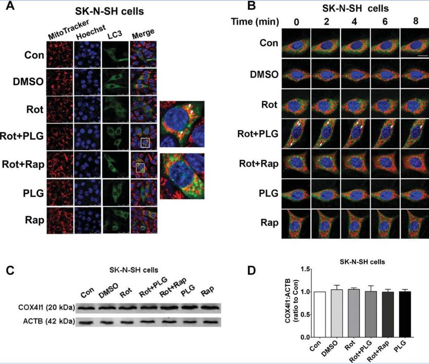
International Congress of Parkinson’s Disease and Movement Disorders®
                          MDS Study Group Abstracts
MDS Study Group:                                                 MDS Study Group:
Epidemiology                                                     Non-Motor PD Study Group (NM-PD)

624                                                              531
Genotype-phenotype correlations in 104 Uzbekish                  Beneficial nonmotor effects of subthalamic and
families with Spinocerebellar ataxias                            pallidal DBS in Parkinson's disease
F. Rakhimov, Y. Majidova, G. Rakhimbaeva (Tashkent,              H. Dafsari, M. dosSantos Ghilardi, V. Visser-
Uzbekistan)                                                      Vandewalle, A. Rizos, K. Ashkan, M. Silverdale, J.
                                                                 Evans, R. Martinez, R. Cury, M. Barbe, G. Fink, A.
MDS Study Group:                                                 Antonini, K. Ray-Chaudhuri, P. Martinez-Martin, E.
MDS Multiple System Atrophy (MSA)                                Fonoff, L. Timmermann (Cologne, Germany)

947                                                              1576
MSA Preliminary Panamerican Report                               Non-motor outcomes of subthalamic DBS in PD
M. Cesarini, E. Gatto, J. Etcheverry, G. Da Prat, N.             depend on the location of volume of activated tissue
Gonzalez Rojas, H. Teive, M. Rodriguez, J. Ziliani, M.           JN. Petry-Schmelzer, H. Dafsari, M. Krause, T.
Miranda, P. Chana, F. Cardoso, A. Cardozo, C. Uribe              Dembek, A. Keyoumars, A. Rizos, M. Silverdale, J.
Roca, I. Amorin, L. Abaroa, c. Cosentino, Y. Nuñez, A.           Evans, M. Barbe, G. Fink, P. Martinez-Martin, V.
Lescano, E. Dieguez, A. Alleva, I. Litvan, G. Wenning            Visser-Vandewalle, A. Antonini, L. Timmermann, K.
(La Plata, Argentina)                                            Ray-Chaudhuri (Cologne, Germany)

MDS Study Group:                                                 1807
Neuroimaging Study Group in Movement                             The International Parkinson and Movement
Disorders)                                                       Disorder Society Non-Motor Scale (MDS-NMS) for
                                                                 Parkinson’s Disease: Preliminary results from an
609                                                              international validation
Cortical and brainstem neurodegeneration associate               K. Ray Chaudhuri, D. Weintraub, A. Rizos, A. Schrag,
with the clinical severity in spinocerebellar ataxia             P. Martinez-Martin (London, United Kingdom)
patients
D. Tamuli, M. Kaur, S. Kumaran, A. Jaryal, A.                    MDS Study Group:
Srivastava, K. Deepak (New Delhi, India)                         Progressive Supranuclear Palsy (PSP)

696                                                              974
The clinical value of SPECT in identifying dystonic              Evolution of diagnostic certainty and PSP-
muscles of patients with cervical dystonia                       predominance types in 187 pathologically confirmed
L. Jin, L. Feng, I. Djibo, S. Chen, F. Teng, B. Li, H. Ma        PSP patients
(Shanghai, China)                                                M. Grimm, G. Respondek, I. Piot, T. Arzberger, Y.
                                                                 Compta, E. Englund, L. Ferguson, E. Gelpi, A. Giese,
1444                                                             S. Roeber, D. Irwin, W. Meissner, C. Nilsson, A.
Altered putamen and cerebellum connectivity                      Pantelyat, A. Rajput, C. Troakes, G. Höglinger
between different subtypes of Parkinson's disease                (Munich, Germany)
B. Shen, L. Zhang (Nanjing, China)
                                                                 LBA 04
1471                                                             Clinical, pathological and genetic determinants of
Semi-automated segmentation and quantification of                benign PSP
the substantia nigra depigmentation in Parkinson’s               G. Respondek, M. Jecmenica-Lucic, C. Kurz, L.
disease - a multicentre case control MRI study in                Ferguson, A. Rajput, W. Z. Chiu, J. van Swieten, E.
284 participants                                                 Englund, C. Nilsson, D. Irwin, C. Troakes, S. al Sarraj,
Y. Xing, S. Schwarz, A. Martin-Bastida, L. Parkes, H.            E. Gelpi, Y. Compta, J. Schöpe, S. Wagenpfeil, S.
Abdul-Sapuan, S. Naidu, M. Silverdale, D. Grosset, P.            Roeber, A. Giese, T. Arzberger (Munich, Germany)
Piccini, D. Burn, N. Bajaj, N. Pavese, D. Auer
(Nottingham, United Kingdom)

                                                       International Congress of Parkinson’s Disease and Movement Disorders®
                                                                  Hong Kong October 5-9, 2018 - MDS Study Groups Abstracts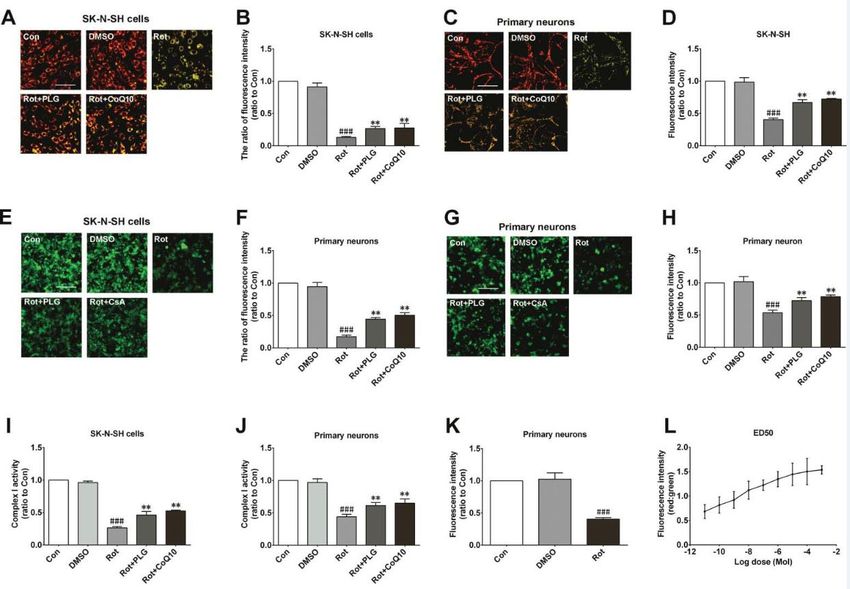
MDS Study Group:
Rare Movement Disorders (RMD)

480
An international survey of stiff person spectrum
disorders: Exploring the clinical spectrum and
unmet needs
B. Balint, E. Gatto, J. Etcheverry, M. Cesarini, V.
Virginia Parisi, M. Rodriguez Violante, P. Garcia
Ruiz, K. Bhatia (London, United Kingdom)

MDS Study Group:
Validation of Mild Cognitive Impairment in
Parkinson’s Disease (MCI)

64
The possible neuroprotective potential of
galantamine along with soya-lecithin and
hydroxychloroquine against ICV-STZ induced
cognitive dysfunction in rats
A. Singh, A. Kumar (Patiala, India)

324
After Deep Brain Stimulation Surgery for
Parkinson's Disease risk Factors for Postoperative
Delirium
F. Rakhimov, G. Rakhimbaeva (Tashkent, Uzbekistan)

405
PLG restores the balance of autophagy and
apoptosis by increasing BCL2 phosphorylation in
rotenone-induced Parkinson disease models
J. Liu, H. Yang (Beijing, China)

1417
Research on nearinfrared brain function imaging
for Freezing of Gait in Parkinson’s Disease
Y. Jingjing, L. Zhanhua, C. Ming (Dalian, China)

LBA 11
Predictive Validity of Level I PD-MCI Criteria,
using Global Cognitive Screeners, for PDD
J. Boel, G. Geurtsen, J. Hoogland, R. de Bie, J.
Goldman, B. Schmand, A. Tröster, D. Burn, I. Litvan,
G. Geurtsen (Amsterdam, Netherlands)

                                                       International Congress of Parkinson’s Disease and Movement Disorders®
                                                                  Hong Kong October 5-9, 2018 - MDS Study Groups Abstracts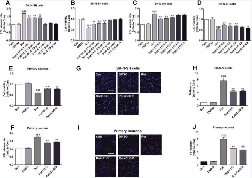
MDS Study Group:
  Epidemiology

624
Genotype-phenotype correlations in 104 Uzbekish families with Spinocerebellar ataxias
F. Rakhimov, Y. Majidova, G. Rakhimbaeva (Tashkent, Uzbekistan)
   Objective: Spinocerebellar ataxias are neurodegenerative disorders involving the cerebellum and its
connections. There are more than 30 distinct subtypes, 16 of which are associated with an identified gene.
The aim of the current study was to evaluate a large group of patients from 104 Uzbekish families with
spinocerebellar ataxias.
   Background: The background of the current study was to evaluate a large group of patients from 104
Uzbekish families with spinocerebellar ataxias.
   Methods: We studied 150 patients from 104 families with spinocerebellar ataxias who had received
molecular genetic testing for spinocerebellar ataxia types 1, 2, 3, 6, 7, 8, 10, 12, 17, and dentatorubral-
pallidoluysian atrophy. A statistical analysis of the results was performed using basic descriptive statistics
and the correlation coefficient (r), Student's t-test, chi-square test, and Yates' correction. The statistical
significance level was established for p-values
with web domain database with a restricted access for site investigators to collect de-identified information to
protect patient confidentiality.2
   Methods: Demographic and clinical data from MSA patients were collected until March 2018 and
included in the new encrypted database. Inclusion criteria required: >21 years old individuals fulfilling
clinical diagnosis of MSA on the basis of the current consensus criteria for the disease (revised criteria for
clinical diagnosis of MSA, 2008) and ancillary neuroimaging. Exclusion criteria required: patients with any
other causes of Parkinsonism.
   Results: 72 patients met criteria. The sample consisted of 61 Hispanic Latinos, 1 Amerindian and 10 non
Hispanic white. 54% probable MSA-P, 16.6% probable MSA-C, 15.7% possible MSA-P 13.8% possible
MSA-C. 55.5% were women. The mean age at start of symptoms is 58.4 years and. The mean years of
diseases duration is 4.6. The mean UMSARS II score 48.6 points, H&Y between 2.5-4 involve more than 63
patients, 93% present with orthostatic hypotension. Premotor symptoms were identified as depression 69.4%,
hyposmia 30.6%. Autonomic dysfunction occurred in 84%,(constipation 84%, orthostatism 93%, urogenital
dysfunction 91.7%) Sleep disorders: RLS 15%, insomnia 50%, RBD 56.9%. Cognitive impairment 40%, and
neuropsychiatric disorders like Hallucinations 9.7%. AS expected falls were frequent 91.7%, but were
relatively infrequent Camptocormia 8.3%and severe anterocollis 31.9%. Benefit with L-dopa was reported in
30.6%, lasting 2.3 years. Urinary incontinence was present in 46.6%. MRI is available in 69 cases. The most
frequent findings in MSA-P were Putaminal atrophy 13.8% and Putaminal rib 8.3%.
   Conclusions: Despite the larger race mixture in PAN American region, our cohort data are in accordance
with other study. On the other hand, this sample included few Amerindian representation, future studies
should include a larger representation.
   References: 1. Wenning GK, Geser F, Krismer F, Seppi K, Duerr S, Boesch S, K¨ollensperger M, Goebel
G, Pfeiffer KP, Barone P, Pellecchia MT, Quinn NP, Koukouni V, Fowler CJ, Schrag A, Mathias CJ, Giladi
N, Gurevich T, Dupont E, Ostergaard K, Nilsson CF,Widner H, Oertel W, Eggert KM, Albanese A, del
Sorbo F, Tolosa E, Cardozo A,Deuschl G, Hellriegel H, Klockgether T, Dodel R, Sampaio C, Coelho M,
Djaldetti R, Melamed E, Gasser T,KammC, Meco G, Colosimo C, Rascol O, Meissner WG,Tison F,
PoeweW&European Multiple System Atrophy Study Group (2013) The natural history of multiple system
atrophy: A prospective European cohort. Lancet Neurol, 12, 264-274. 2. Kuzdas-Wood D, Stefanova N,
Jellinger KA, Seppi K 1 Schlossmacher MG, PoeweW, & Wenning GK (2014) Towards translational
therapies for multiple system atrophy. Prog Neurobiol, 118C, 19-35 Yabe I, Soma H, Takei A, Fujiki N,
Yanagihara T, & Sasaki H (2006) MSA-C is the predominant clinical phenotype of MSA in Japan: Analysis
of 142 patients with probable MSA. J Neurol Sci, 249 115-121. 3. Wenning GK, & Stefanova N (2009)
Recent developments in multiple system atrophy. J Neurol, 256, 1791-1808. 4. Jecmenica-Lukic M, Poewe
W, Tolosa E, Wenning GK. Premotor signs and symptoms of multiple system atrophy. Lancet Neurol 2012;
11:361-8. 5. Gilman S, Wenning GK, Low PA, et al. Second consensus statement on the diagnosis of
multiple system atrophy. Neurology 2008; 71: 670-6.

 MDS Study Group:
 Neuroimaging Study Group in Movement Disorders)

609
Cortical and brainstem neurodegeneration associate with the clinical severity in spinocerebellar ataxia
patients
D. Tamuli, M. Kaur, S. Kumaran, A. Jaryal, A. Srivastava, K. Deepak (New Delhi, India)
   Objective: The aim of the study was to know the association of degree of atrophy in brain areas with
clinical severity in SCA patients.
   Background: Spinocerebellar ataxia (SCA) is a progressive neurodegenerative disorder with prominent
clinical heterogeneity amongst the subtypes. Clinical manifestations include gait imbalance, opthalmoplegia,
dysarthria, pyramidal and extrapyramidal signs. This clinical diversity is the result of unstable trinucleotide

                                                  International Congress of Parkinson’s Disease and Movement Disorders®
                                                             Hong Kong October 5-9, 2018 - MDS Study Groups Abstracts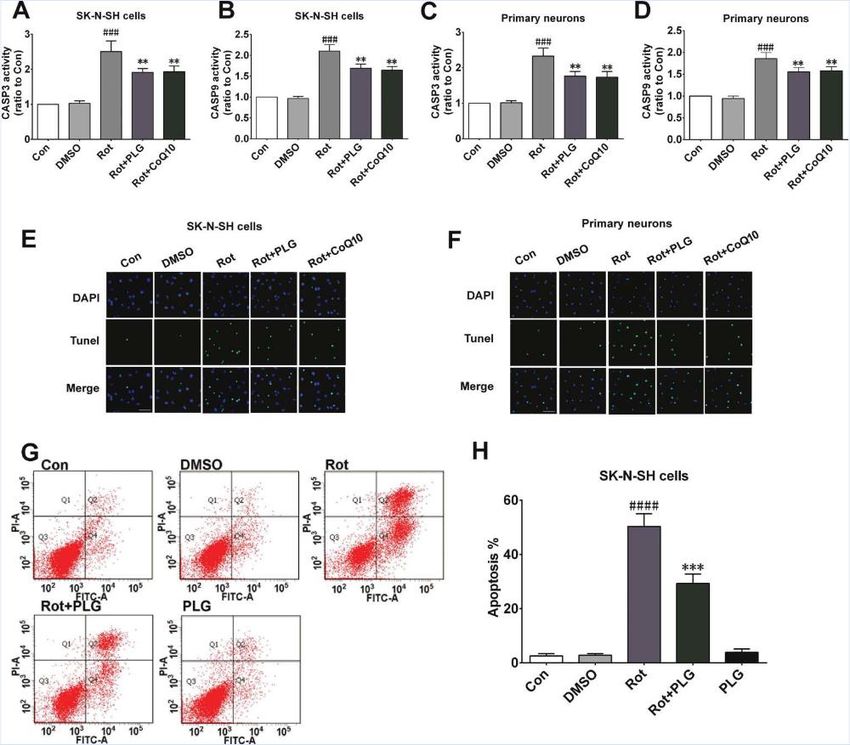
(CAG) repeat expansion especially in SCA type 1, 2 and 3 patients. This could be indicative of a differential
neuronal loss in the SCA subtypes thus culminating into a differential clinical profile.
   Methods: Clinical severity was assessed by International Cooperative Ataxia Rating Scale (ICARS) in
the genetically proven SCA (n = 49, age = 34 ± 9.4 yrs) patients. Then MRI was performed by using a 3T
scanner (Philips, Achieva) to obtain 3D T1-weighted scans of the whole brain and analysed by FreeSurfer
(version 5.3) software in the same SCA patients. MRI parameters used in T1-weighted scans were: Voxel
size = 0.6×0.6×1, FOV = 240×240×180 and flip angle = 8ᵒ. Based on the distribution of data, Pearson's
correlation (for parametric data) and Spearman's rank correlation (for nonparametric data) analyses were
done between brain areas and ICARS score of SCA patients.
   Results: In SCA patients, ICARS showed a significant inverse correlation with cortical thickness of left
rostral middle frontal (r = -0.382, p = 0.007), left caudal middle frontal (r = -0.324, p = 0.023), left medial
orbitofrontal (r = -0.351, p = 0.013), left middle temporal (r = -0.324, p = 0.023), right middle temporal (r = -
0.292, p = 0.042) and right entorhinal (r = -0.286, p = 0.046). Furthermore, the subcortical volume of
midbrain (r = -0.472, p = 0.001), pons (r = -0.523, p =
1444
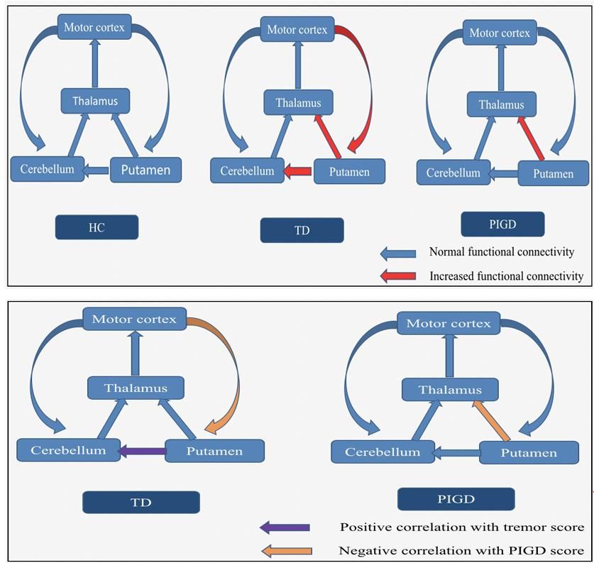
Altered putamen and cerebellum connectivity between different subtypes of Parkinson's disease
B. Shen, L. Zhang (Nanjing, China)
Objective: we investigate the functional connectivity (FC) patterns of the putamen between different
subtypes of PD) and healthy controls, and explore its clinical significance.
Background: Impairment of the basal ganglia-thalamo-cortical circuit causes motor symptoms of
Parkinson’s disease(PD).
Methods: 16 tremor dominant (TD) PD patients, 23 postural instability and gait difficulty dominant PD
patients and 31 healthy controls, were scanned with resting-state functional magnetic resonance imaging. A
voxel-wise FC analysis was performed by computing the temporal correlation between the bilateral putamen
and the other voxels within the whole brain. Correlation analysis was performed between the FC strength and
motor symptoms.
Results: Compared with PIGD dominant group, TD dominant group showed increased FC between
bilateral putamen and right cerebellum lobules VI, cerebelum crus I. While compared with healthy controls,
tremor dominant PD patients showed increased FC between left putamen and bilateral cerebellum crus I,
right cerebelum lobules VI, right thalamus, left paracentral lobule, right inferior occipital lobule, cerebellar
vermis, fusiform gyrus, left supplementary motor area. Increased FC between right putamen and right
cerebellum crus I, right cerebellum lobules VI, right thalamus, bilateral paracentral lobule, right inferior
occipital lobule, right inferior temporal gyrus, bilateral supplementary motor area and left sensorimotor
cortex were also shown in TD patients, . PIGD dominant group showed increased FC between right putamen
and right thalamus compared with healthy controls. The FC strength between the left putamen and right
cerebelum lobules VI showed positive correlation with tremor scores in TD dominant group. The FC strength
between right putamen and left sensorimotor cortex showed negative correlation with PIGD scores. While in
PIGD dominant group, the FC strength between left putamen and right thalamus showed negative correlation
with TD scores.
Conclusions: The altered connectivity of basal ganglia-cortical circuit in PD patients was related to PIGD
symptoms, Compared to PIGD patients, motor and cognitive impairment declined slowly in TD patients,
Which may be related to the increased functional connectivity between basal ganglia and cerebellum.
References: [1] O'Reilly J X, Beckmann C F, Tomassini V, Ramnani N, Johansen-Berg H. Distinct and
overlapping functional zones in the cerebellum defined by resting state functional connectivity[J]. Cereb
Cortex,2010,20(4):953-965. [2] Bostan A C, Dum R P, Strick P L. Cerebellar networks with the cerebral
cortex and basal ganglia[J]. Trends Cogn Sci,2013,17(5):241-254.
International Congress of Parkinson’s Disease and Movement Disorders®
Hong Kong October 5-9, 2018 - MDS Study Groups Abstracts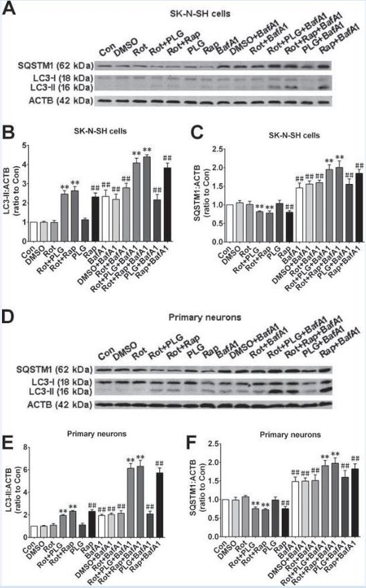
FIG. 1 (1444)

1471
Semi-automated segmentation and quantification of the substantia nigra depigmentation in
Parkinson’s disease - a multicentre case control MRI study in 284 participants
Y. Xing, S. Schwarz, A. Martin-Bastida, L. Parkes, H. Abdul-Sapuan, S. Naidu, M. Silverdale, D. Grosset, P.
Piccini, D. Burn, N. Bajaj, N. Pavese, D. Auer (Nottingham, United Kingdom)
   Objective: To provide a semi-automated segmentation and quantification method of the changes in the
substantia nigra (SN) in Parkinson’s disease (PD) using neuromelanin (NM)-sensitive MRI in a large multi-
centre dataset (Parkinson’s MR imaging repository: PaMIR).
   Background: Previous studies investigating small groups of patients (
Conclusions: Our results provide further evidence of NM volume and contrast abnormalities in PD,
reflecting pigmented cell loss in the SN and indicate the validity of the current semi-automated SN
segmentation and quantification approach. This paves the way for the assessment of using volumetric and
contrast measures of the SN as a potential multi-site diagnostic and severity marker in our large-scale NM
study. Our ongoing work focuses on quality assurance, interrater reliability and using the follow-up data to
clarify the role of NM-sensitive MRI in tracking the progression of disease.
   References: 1. Schwarz ST., Xing Y. Tomar P, Bajaj N, Auer, DP. In vivo assessment of brainstem
depigmentation in Parkinson disease: Potential as a severity marker for multicenter studies Radiology, 2017.
2. Schwarz ST., et al. Mov. Disord., 2011. 26: p.1633–8.

FIG. 1 (1471)

                                                 International Congress of Parkinson’s Disease and Movement Disorders®
                                                            Hong Kong October 5-9, 2018 - MDS Study Groups Abstracts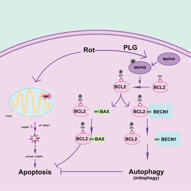
FIG. 2 (1471)

 MDS Study Group:
 Non-Motor PD Study Group (NM-PD)

531
Beneficial nonmotor effects of subthalamic and pallidal DBS in Parkinson's disease
H. Dafsari, M. dosSantos Ghilardi, V. Visser-Vandewalle, A. Rizos, K. Ashkan, M. Silverdale, J. Evans, R.
Martinez, R. Cury, M. Barbe, G. Fink, A. Antonini, K. Ray-Chaudhuri, P. Martinez-Martin, E. Fonoff, L.
Timmermann (Cologne, Germany)
   Objective: To investigate nonmotor effects of bilateral subthalamic (STN) and pallidal (GPi) deep brain
stimulation (DBS) in Parkinson’s disease (PD).
   Background: Bilateral STN-DBS as well as bilateral GPi-DBS improves quality of life and motor
symptoms in PD. Beneficial non-motor effects have been reported for STN-DBS, whereas few studies have
investigated a wide range of NMS in patients with PD undergoing GPi-DBS. Therefore, we conducted a
comparative investigation of non-motor effects for these two DBS targets. We hypothesized that (1) GPi-
DBS has beneficial effects on NMS and (2) STN-DBS and GPi-DBS have distinct non-motor effect profiles.
   Methods: In this prospective, observational, multicenter study including 60 PD patients undergoing
bilateral STN-DBS (n=40) or GPi-DBS (n=20), we examined PDQuestionnaire (PDQ), Nonmotor Symptom
Scale (NMSS), Unified PD Rating Scale-activities of daily living, -motor impairment, -complications
(UPDRS-II, -III, -IV), Hoehn&Yahr, Schwab&England Scale, and levodopa equivalent daily dose (LEDD)
preoperatively and at 6 months follow-up. Intra-group changes from baseline to follow-up were analyzed
with Wilcoxon signed-rank or paired t-test, if parametric tests were applicable, and corrected for multiple
comparisons. Differences between STN-DBS and GPi-DBS were explored with Mann-Whitney-U or
unpaired t-tests. Analyses were performed before and after propensity score matching which balanced out
demographic and preoperative clinical characteristics. Strength of clinical changes was assessed with effect
size.
   Results: In both groups, PDQ, UPDRS-II, -IV, Schwab&England Scale, and NMSS improved
significantly from baseline to follow-up. STN-DBS was significantly better for LEDD reduction, GPi-DBS

                                                 International Congress of Parkinson’s Disease and Movement Disorders®
                                                            Hong Kong October 5-9, 2018 - MDS Study Groups Abstracts
for UPDRS-IV improvement. While NMSS total score outcomes were similar, explorative NMSS domain
analyses resulted in distinct profiles. Both targets improved sleep/fatigue and mood/cognition, but only STN-
DBS improved the miscellaneous (pain/olfaction) and attention/memory and only GPi-DBS cardiovascular
and sexual function domains.
Conclusions: To our knowledge, this is the first study to report distinct patterns of beneficial nonmotor
effects of STN-DBS and GPi-DBS in PD patients. This study highlights the importance of comprehensive
assessments of NMS to tailor the choice of DBS target to patients’ individual motor and nonmotor profiles.
1576
Non-motor outcomes of subthalamic DBS in PD depend on the location of volume of activated tissue
JN. Petry-Schmelzer, H. Dafsari, M. Krause, T. Dembek, A. Keyoumars, A. Rizos, M. Silverdale, J. Evans, M.
Barbe, G. Fink, P. Martinez-Martin, V. Visser-Vandewalle, A. Antonini, L. Timmermann, K. Ray-Chaudhuri
(Cologne, Germany)
Objective: To investigate the impact of stimulation location on non-motor symptoms (NMS) in PD
patients with DBS in the subthalamic nucleus (STN) via an analysis of volumes of activated tissue (VAT).
Background: STN-DBS improves non-motor and motor symptoms. In a recent study, we showed that
NMS improvement is higher with a more anterior, ventral and medial position of the active contact, whereas
motor symptoms improve more with a more posterior and lateral position of the active contact. However,
investigating only electrode locations neglects the spatial extent of electrical stimulation, which also depends
on the stimulation parameters. We thus conducted an investigation into the spatial distribution of NMS
improvement in the STN using VATs.
Methods: Clinical data was collected from an ongoing prospective, open-label, multicenter study
(Cologne, London, Manchester) including 92 patients with bilateral STN-DBS. The following scales were
collected at preoperative baseline (MedON) and on follow-up (FU) six months after surgery
(MedON/StimON): SCOPA-motor examination (SCOPA-motor), -activities of daily living (ADL),
NMSScale (NMSS), NMSQuestionnaire (NMSQ), Hospital Anxiety and Depression Scale-anxiety and
depression subscales (HADS-A/-D). Wilcoxon signed-rank test was used to test for significant changes
between baseline and follow-up and Bonferroni-correction was applied for multiple comparisons. Individual
VATs, based on the stimulation parameters used in clinical setting, were calculated in MNI space (ICBM
2009b) as described elsewhere (1). To analyse the relationship between VATs and change scores, we
employed probabilistic stimulation maps and projected them on the DISTAL-Atlas (2).
Results: All outcomes, besides HADS-D, improved significantly at FU. Probabilistic stimulation maps
showed higher improvement of NMS for stimulation localized in the limbic and associative STN, whereas
motor symptoms improved more for stimulation localized in the motor STN.
Conclusions: Our preliminary results support the finding that the non-motor outcome after DBS may
depend on the location of neurostimulation. DBS in non-motor STN subregions was associated with bigger
improvement of NMS. The underlying mechanisms and clinical relevance of our results should be
investigated in future studies.
References: 1 Horn, A., Reich, M., Vorwerk, J., Li, N., Wenzel, G., Fang, Q.,. . . Fox, M. D. (2017).
Connectivity predicts deep brain stimulation outcome in Parkinson's disease. Ann Neurol.
doi:10.1002/ana.24974. 2 Ewert, S., Plettig, P., Li, N., Chakravarty, M. M., Collins, D. L., Herrington, T. M.,
. . . Horn, A. (2017). Toward defining deep brain stimulation targets in MNI space: A subcortical atlas based
on multimodal MRI, histology and structural connectivity. Neuroimage.
doi:10.1016/j.neuroimage.2017.05.015.
International Congress of Parkinson’s Disease and Movement Disorders®
Hong Kong October 5-9, 2018 - MDS Study Groups Abstracts1807
The International Parkinson and Movement Disorder Society Non-Motor Scale (MDS-NMS) for
Parkinson’s Disease: Preliminary results from an international validation
K. Ray Chaudhuri, D. Weintraub, A. Rizos, A. Schrag, P. Martinez-Martin (London, United Kingdom)
Objective: To provide an update on the ongoing international validation programme of an expanded,
refined and improved scale for the assessment of non-motor symptoms (NMS) in Parkinson’s disease (PD),
the MDS-NMS, sponsored by the International Parkinson and Movement Disorders Society (IPMDS).
Background: The PD NMS scale (NMSS) was developed in 2006 and remains the only dedicated
composite scale to assess NMS in PD. As it needed updating, a project to revise and update the NMSS, now
called the MDS-NMS, was commissioned by the IPMDS. The MDS-NMS has already undergone cognitive
pre-testing and a final version of MDS-NMS was developed. It includes 13 domains with 52 items total, each
item scoring for frequency (0 to 4) and severity (0 to 4), which are multiplied to obtain the item score (0 to
16). The domain scores result from sum of the item scores within each domain. Additionally, the scale
includes an optional section for evaluating non-motor fluctuations (NMFs) for 8 domains.
Methods: In this cross-sectional, open, multicentre international validation study, we report clinical data
from administration of the MDS-NMS. Acceptability, internal consistency, reliability, construct validity and
precision in 400 non-demented PD patients (MoCA score>20) is ongoing. Test-retest reliability, assessed
after two weeks (average) as well as inter-rater reliability are reported in 100 patients. NMS are also assessed
with existing measures, as are motor symptoms and global severity of PD.
Results: 347 patients, 58.8% male, mean age 68.7 yrs (35-93), median H&Y 2 (1-5), mean duration of
disease 7.2 yrs (0-30). Mean and median scores for the 13 domains and for NMFs for 8 domains are listed
[Table 1]. Standardised domain scores (% of maximum possible score), and % of patients experiencing any
problems in each domain are illustrated [Figure 1]. The most frequently reported problems were found in
domains “Others” (including loss of sense of smell) (83.3%) “Sleep/wakefulness” (79.3%), “Cognition”
(75.5%), and “Urinary” (64.8%). The greatest average severity of symptoms were found in “Urinary” (mean
13.8%), “Others” (13.3%), “Sexual” (12.9%) and “Pain” (10.2%).
Conclusions: Interim results suggest the MDS-NMS may perform well as an outcome measure in clinical
trials and epidemiological studies to assess the wide range of NMS that occur in PD.
TABLE 1 (1807)
International Congress of Parkinson’s Disease and Movement Disorders®
Hong Kong October 5-9, 2018 - MDS Study Groups AbstractsFIG. 1 (1807)
MDS Study Group:
Progressive Supranuclear Palsy (PSP)
974
Evolution of diagnostic certainty and PSP-predominance types in 187 pathologically confirmed PSP
patients
M. Grimm, G. Respondek, I. Piot, T. Arzberger, Y. Compta, E. Englund, L. Ferguson, E. Gelpi, A. Giese, S.
Roeber, D. Irwin, W. Meissner, C. Nilsson, A. Pantelyat, A. Rajput, C. Troakes, G. Höglinger (Munich,
Germany)
Objective: To examine the evolution of diagnostic certainty and clinical predominance types as defined
by the movement disorder society (MDS) criteria for the clinical diagnosis of PSP (Höglinger et al, 2017)
during the course of disease in autopsy-confirmed patients with PSP.
Background: Three degrees of diagnostic certainty ("suggestive of" = s.o., "possible", or "probable"),
and definition of clinical predominance types (PSP-RS, PSP-PGF, PSP-P, PSP-F, PSP-OM, PSP-SL, PSP-
CBS, PSP-PI) were implemented into the new clinical diagnostic criteria for PSP (MDS criteria for the
clinical diagnosis of PSP, Höglinger et al, 2017). The evolution of diagnostic certainty and predominance
types over the course of disease in PSP patients according to the MDS-criteria has not been studied so far.
However, this is relevant for further understanding the natural history and clinical spectrum of PSP.
Methods: Features relevant for the diagnosis of PSP according to the MDS-diagnostic criteria were
collected in 187 autopsy-confirmed PSP patients by chart review. Diagnostic certainty and PSP-
predominance types according to the MDS-diagnostic criteria were determined for each patient and each
year.
Results: According to the MDS-PSP diagnostic criteria, 62% (n=115) of patients had a clinical diagnosis
of PSP in the first year of disease, as opposed to 14% (n=26) according to the NINDS SPSP criteria, and
98% (n=183) at final record, as opposed to 79% (n=147) according to the NINDS/SPSP criteria. A diagnosis
of s.o. PSP was present in 44% (n=83) of patients in the first year. Of these, 77% (n=64) had a diagnosis of
"probable" PSP at final record. In the first year of disease, the variability of predominance types was greatest,
and PSP-RS represented only 11% of patients. At final record, 56% patients had a PSP-RS predominance
type.
International Congress of Parkinson’s Disease and Movement Disorders®
Hong Kong October 5-9, 2018 - MDS Study Groups AbstractsConclusions: The conversion of s.o. PSP into possible and probable PSP in 72% of the cases shows that
the concept of s.o. PSP indeed allows identification of PSP patients at a very early clinical stage.
Furthermore, our data suggest that many initially variant PSP presentations converge to a PSP-RS phenotype
during the course of the disease.
References: MDS criteria for the clinical diagnosis of PSP, Höglinger et al, 2017.
LBA 04
Clinical, pathological and genetic determinants of benign PSP
G. Respondek, M. Jecmenica-Lucic, C. Kurz, L. Ferguson, A. Rajput, W. Z. Chiu, J. van Swieten, E. Englund,
C. Nilsson, D. Irwin, C. Troakes, S. al Sarraj, E. Gelpi, Y. Compta, J. Schöpe, S. Wagenpfeil, S. Roeber, A.
Giese, T. Arzberger (Munich, Germany)
Objective: To identify clinical, pathological, and genetic determinants of survival in PSP. This study
involved 186 autopsy-confirmed PSP cases which were recruited from eight brain banks.
Background: Demographic and clinical information was retrospectively reviewed. Symptoms were
analysed with respect to their potential association with long (≥ 10 years) and medium or short survival (< 5
years and 5-10 years). Central histopathological analysis and harmonization of staining protocols has been
performed. PSP-specific brain lesions were analysed in 36 brain regions and compared between different
survival groups. In addition, the survival groups have also been stratified by known SNPs risk allele carrier
status.
Method: This study involved 186 autopsy-confirmed PSP cases which were recruited from eight brain
banks. Demographic and clinical information was retrospectively reviewed. Symptoms were analysed with
respect to their potential association with long (≥ 10 years) and medium or short survival (< 5 years and 5-10
years). Central histopathological analysis and harmonization of staining protocols has been performed. PSP-
specific brain lesions were analysed in 36 brain regions and compared between different survival groups. In
addition, the survival groups have also been stratified by known SNPs risk allele carrier status.
Results: Of 186 PSP patients with sufficient clinical data, 24 % had a long disease duration of ≥ 10 years
(mean: 13.8 [10 – 27] years), while similar percentage of patients (21.5 %) were found to have progressive
clinical course of < 5 years (mean: 3.3 [1-4] years). The analysis of timeline and evolution of main clinical
milestones revealed different pattern in long duration PSP, compared with both groups of medium and short
less survival. The latter groups presented with identical evolution of symptoms. The presence or absence of
oculomotor signs in the first 3 years from disease onset was the main predictor of short (< 5 years) or long (≥
10 years) survival, respectively. Neurodegenerative changes and neuronal tau lesions were most prominent in
infratentorial regions, while glial tau lesions showed the opposite pattern with highest density in cortical and
subcortical regions. The crucial differences between survival groups were found in the rates of glial tau
lesions in cortical, subcortical and infratentorial regions. There was no difference in SNPs risk allele carrier
status.
Conclusion: Herein, we described a benign form of PSP with distinctive clinical and pathological
patterns. The absence of supranuclear gaze palsy and abnormal saccades in the first 3 years from onset were
independent predictors of long duration PSP (≥ 10 years). Our observations suggest that glial pathology plays
a crucial role in determining survival in PSP.
International Congress of Parkinson’s Disease and Movement Disorders®
Hong Kong October 5-9, 2018 - MDS Study Groups AbstractsMDS Study Group:
Rare Movement Disorders (RMD)
480
An international survey of stiff person spectrum disorders: Exploring the clinical spectrum and unmet
needs
B. Balint, E. Gatto, J. Etcheverry, M. Cesarini, V. Virginia Parisi, M. Rodriguez Violante, P. Garcia Ruiz, K.
Bhatia (London, United Kingdom)
Objective: To build a platform in order to investigate the clinical spectrum of stiff person syndrome and
related disorders, and to explore possible gaps in management and areas for further research.
Background: Stiff person syndrome and related disorders (stiff person spectrum disorders, SPSD) are a
very rare group of autoimmune movement disorders with an estimated prevalence of 1/1 000 000. Whereas
the classical variant is associated with GAD-antibodies and manifests with predominant lumbar stiffness, the
clinical and serological spectrum has broadened to include a wider range of phenotypes and antibodies.
Methods: The survey started in 03/2018 and is conducted amongst the members of the MDS rare
movement disorders study group.
Results: The survey is ongoing and at the time of abstract submission we report the first preliminary
results. So far, 16 patients from 3 centres (UK, Argentina, Spain) were included (9 female) with a follow up
time of 10 years on average (range 2-28). Median age of onset was 52.5yrs (range 29-68). Most patients
(68,8%) had focal stiffness at onset and manifested with (painful) cramps (62.5%), axial stiffness (56,3%).
Cerebellar signs (nystagmus, ataxia) or bulbar involvement occurred in 18.8%, respectively. 12.5% of
patients displayed myoclonus at presentation, whereas prominent autonomic signs or epilepsy were rare
(6,3% and none). Most of the patients harboured GAD-antibodies (93.8%), and concomitant autoimmunity
was frequent (56.3%). Two cases were possibly paraneoplastic, as neurological symptoms occurred in close
temporal relationship with cancers (oral squamous cell carcinoma, nasopharyngeal). Treatment approaches
and outcomes were similar across the different centres. All but 1 patient received immunotherapy, most
frequently intravenous immunoglobulins (87.5%) and plasma exchange (43,8%). Other immunotherapies
used were steroids and rituximab (12.5% each), and azathioprine (6.3%). In 50%, a combination of at least
two immunotherapy approaches was required. With treatment, half of the cases improved and a third
remained stable.
Conclusions: This is the first international multicentre SPSD study with prospective data collection by
movement disorder experts. Further plans include expanding this cohort and, using a health services
approach, identifying unmet needs in SPSD and areas for further research.
MDS Study Group:
Validation of Mild Cognitive Impairment in Parkinson’s Disease (MCI)
64
The possible neuroprotective potential of galantamine along with soya-lecithin and
hydroxychloroquine against ICV-STZ induced cognitive dysfunction in rats
A. Singh, A. Kumar (Patiala, India)
Objective: The present study investigates the possible neuroprotective potential of galantamine with
soya-lecithin and HCQ against intracerebroventricular streptozotocin (ICV-STZ) induced memory
impairment in a rat model of sporadic dementia of Alzheimer’s type
International Congress of Parkinson’s Disease and Movement Disorders®
Hong Kong October 5-9, 2018 - MDS Study Groups AbstractsBackground: Galantamine an acetylcholinesterase (AChEs) inhibitor used for the symptomatic treatment
of Alzheimer’s disease. Soya-lecithin is a good source of choline improves cognitive performance.
Hydroxychloroquine (HCQ) an antimalarial drug with an anti-inflammatory property.
Methods: Animals received single bilateral ICV injections of STZ (3 mg/kg). Drugs galantamine (2
mg/kg), soya-lecithin (100 & 200 mg/kg), HCQ (25 & 50 mg/kg) and their combination was administered for
a period of 21 days. Various neurobehavioral parameters, followed by biochemical (oxidative stress
markers), AChEs level, molecular (TNF-α level), mitochondrial respiratory enzyme complexes (I-IV),
neurotransmitter levels and histopathological (H&E staining) evaluations.
Results: ICV-STZ administration significantly impaired cognitive performance indicated by MWM test,
increased oxidative stress (raised lipid peroxidation, nitrite concentration, reduced glutathione, catalase
activity), AChEs level, increased TNF-α level, decrease neurotransmitter levels, mitochondrial dysfunction
and histopathological alterations as compared to sham treatment. Chronic treatment with galantamine (2
mg/kg), soya-lecithin (100 & 200 mg/kg), HCQ (25 & 50 mg/kg) significantly improved cognitive
performance in MWM test, reduced AChEs activity, neuroinflammation, oxidative damage, TNF-α level,
restored mitochondrial respiratory enzyme complex (I-IV) activities and histopathological alterations as
compared to ICV-STZ treated animals. Further, combinations of soya-lecithin (100 & 200 mg/kg) and HCQ
(25 & 50 mg/kg) with galantamine (2 mg/kg) and soya-lecithin (100 & 200 mg/kg) and HCQ (25 & 50
mg/kg) combination suggests the modulation of the neuroprotective potential as compared to their effect
alone in ICV-STZ treated animals. Further, the present study suggests the combination potential of soya-
lecithin (100 & 200 mg/kg) and HCQ (25 & 50 mg/kg) with galantamine (2 mg/kg) and it was found that
galantamine (2 mg/kg) significantly modulate the neuroprotective potential of soya-lecithin (100 & 200
mg/kg) and HCQ (25 & 50 mg/kg) combination in ICV-STZ treated rats as compared to their effect alone.
Conclusions: The present study suggests that co-administration of galantamine with soya-lecithin and
HCQ significantly improves cognitive performance in ICV-STZ treated rats as compared to their effect
alone.
References: 1. D.S. Auld, T.J. Kornecook, S. Bastianetto, R. Quirion, Alzheimer’s disease and the basal
forebrain cholinergic system: relations to β-amyloid peptides, cognition, and treatment strategies. Prog.
Neurobiol. 68 (2002) 209-245. 2. E.X. Albuquerque, M. Alkondon, E.F. Pereira, N.G. Castro, A.
Schrattenholz, C.T. Barbosa, R. Bonfante-Cabarcas, Y. Aracava, H.M. Eisenberg, A. Maelicke, Properties of
neuronal nicotinic acetylcholine receptors: pharmacological characterization and modulation of synaptic
function. J. Pharmacol. Exp. Ther. 280 (1997) 1117-1136. 3. F. Amenta, S. Tayebati, Pathways of
acetylcholine synthesis, transport and release as targets for treatment of adult-onset cognitive dysfunction.
Curr. Med. Chem. 15 (2008) 488-498. 4. R. Anand, K.D. Gill, A.A. Mahdi, Therapeutics of Alzheimer's
disease: Past, present and future. Neuropharmacol. 76 (2014) 27-50. 5. A.S. Association, 2017, Alzheimer's
disease facts and figures. Alzheimer's Dement. 13 (2017) 325-373. 6. H. Braak, K. Del Tredici, Alzheimer’s
disease: pathogenesis and prevention. Alzheimer's Dement. 8 (2012) 227-233. 7. P.S. Aisen, K.L. Davis,
Inflammatory mechanisms in Alzheimer’s disease. Am. J. Psychiatry 151 (1994) 1105-1113. 8. D.J. Bonda,
H.P. Lee, H.G. Lee, A.L. Friedlich, G. Perry, X. Zhu, M.A. Smith, Novel therapeutics for Alzheimer's
disease: an update. Curr. Opin. Drug Discov. Devel. 13 (2010) 235. 9. H. Braak, K. Del Tredici, Alzheimer’s
disease: pathogenesis and prevention. Alzheimer's Dement. 8 (2012) 227-233. 10. P. Eikelenboom, C. Bate,
W. Van Gool, J. Hoozemans, J. Rozemuller, R. Veerhuis, A. Williams, Neuroinflammation in Alzheimer's
disease and prion disease. Glia 40 (2002) 232-239. 11. P.T. Francis, A.M. Palmer, M. Snape, G.K. Wilcock,
The cholinergic hypothesis of Alzheimer’s disease: a review of progress. J. Neurol. Neurosurg. Psychiatry 66
(1999) 137-147. 12. H. Hampel, D. Prvulovic, S. Teipel, F. Jessen, C. Luckhaus, L. Frölich, M.W.Riepe,
R.Dodel, T.Leyhe, L.Bertram, The future of Alzheimer's disease: the next 10 years. Prog. Neurobiol. 95
(2011) 718-728. 13. C. Barnes, J. Meltzer, F. Houston, G. Orr, K. McGann, G. Wenk, Chronic treatment of
old rats with donepezil or galantamine: effects on memory, hippocampal plasticity and nicotinic receptors.
Neuroscience 99 (2000) 17-23. 14. A. Kumar, A. Prakash, D. Pahwa, Galantamine potentiates the protective
effect of rofecoxib and caffeic acid against intrahippocampal Kainic acid-induced cognitive dysfunction in
rat. Brain Res. Bull. 85 (2011) 158-168. 15. N.J. Zvaifler, Update in rheumatology—focus on
hydroxychloroquine. American J. Med. 85 (1988) 68-71. 16. W.A. Van Gool, H.C. Weinstein, P.K.
Scheltens, G.J. Walstra, Effect of hydroxychloroquine on progression of dementia in early Alzheimer's
International Congress of Parkinson’s Disease and Movement Disorders®
Hong Kong October 5-9, 2018 - MDS Study Groups Abstractsdisease: an 18-month randomised, double-blind, placebo-controlled study. Lancet 358 (2001) 455-460. 17.
Y. Wu, T. Wang, Fractionation of crude soybean lecithin with aqueous ethanol. J. American Oil Chemists'
Society 81 (2004) 697-704. 18. A. Bruni, E. Bigon, E. Boarato, L. Mietto, A. Leon, G. Toffano, Interaction
between nerve growth factor and lysophosphatidylserine on rat peritoneal mast cells. FEBS letters 138
(1982) 190-192. 19. A. Bruni, G. Toffano, Lysophosphatidylserine, a short-lived intermediate with plasma
membrane regulatory properties. Pharmacol. Res. Comm. 14 (1982) 469-484. 20. F. Casamenti, P.
Mantovani, L. Amaducci, G. Pepeu, Effect of phosphatidylserine on acetylcholine output from the cerebral
cortex of the rat. J. Neurochem. 32 (1979) 529-533. 21. J. Higgins, L. Flicker, Lecithin for dementia and
cognitive impairment. The Cochrane Library. (2000) 22. J. Surh, Y.G. Jeong, G.T. Vladisavljević, On the
preparation of lecithin-stabilized oil-in-water emulsions by multi-stage premix membrane emulsification. J.
Food Eng. 89 (2008) 164-170. 23. A.A. Abdel-Hamid, A.E.-D.L. El-Firgany, Hydroxychloroquine hindering
of diabetic isletopathy carries its signature on the inflammatory cytokines. J. Mol. Histol. 47 (2016) 183-193.
24. M. Furushiro, S. Suzuki, Y. Shishido, M. Sakai, H. Yamatoya, S. Kudo, S. Hashimoto, T. Yokokura,
Effects of oral administration of soybean lecithin transphosphatidylated phosphatidylserine on impaired
learning of passive avoidance in mice. Jpn. J. Pharmacol. 75 (1997) 447-450. 25. J.M. Gutierres, F.B.
Carvalho, M.R.C.Schetinger, P.Marisco, P.Agostinho, M.Rodrigues, M.A.Rubin, R.Schmatz, C.R.da Silva,
G.D.P. Cognato, Anthocyanins restore behavioral and biochemical changes caused by streptozotocin-induced
sporadic dementia of Alzheimer's type. Life Sci. 96 (2014) 7-17. 26. A. Singh, A. Kumar, Microglial
inhibitory mechanism of coenzyme Q10 against Aβ (1-42) induced cognitive dysfunctions: possible
behavioral, biochemical, cellular, and histopathological alterations. Front. Pharmacol. 6 (2015) 268. 27. T.
Ishrat, M.B. Khan, M.N. Hoda, S. Yousuf, M. Ahmad, M.A. Ansari, A.S. Ahmad, F.Islam, Coenzyme Q10
modulates cognitive impairment against intracerebroventricular injection of streptozotocin in rats. Behav.
Brain Res. 171 (2006) 9-16. 28. G. Paxinos, C. Watson, The rat brain in stereotaxic coordinates 2nd edn.
Academic Press, New York. Rasoolijazi, H., Joghataie, MT, Roghani, M., & Nobakht, M., 2007. The
beneficial effect of (-)-epigallocatechin-3-gallate in an experimental model of Alzheimer's disease in rat: a
behavioral analysis. Iran Biomed. J. 11 (1986) 237-243. 29. A. Kumar, N. Sharma, J. Mishra, H. Kalonia,
Synergistical neuroprotection of rofecoxib and statins against malonic acid induced Huntington's disease like
symptoms and related cognitive dysfunction in rats. Eur. J. Pharmacol. 709 (2013) 1-12. 30. R. Morris,
Developments of a water-maze procedure for studying spatial learning in the rat. J. Neurosci. Methods 11
(1984) 47-60. 31. E. Wills, Mechanisms of lipid peroxide formation in animal tissues, Biochem. J. 99 (1966)
667-676. 32. M. Ellman, A spectrophotometric method for determination of reduced glutathione in tissues,
Anal. Biochem. 74 (1959) 214-226. 33. L.C. Green, D.A. Wagner, J. Glogowski, P.L. Skipper, J.S. Wishnok,
S.R. Tannenbaum, et al. Analysis of nitrate, nitrite, and [15 N] nitrate in biological fluids, Anal. Biochem.
126 (1982) 131-138. 34. Y. Kono, Generation of superoxide radical during autoxidation of hydroxylamine
and an assay for superoxide dismutase, Arch. Biochem. Biophys. 186 (1978) 189-195. 35. H. Luck, Catalase.
Methods of Enzymatic Analysis, (1965) 885-888. 36. G.L. Ellman, K.D. Courtney, V. Andres, R.M.
Featherstone. A new and rapid colorimetric determination of acetylcholinesterase activity, Biochem.
Pharmacol. 7 (1961) 88-95. 37. A.G. Gornall, C.J. Bardawill, M.M. David, Determination of serum proteins
by means of the biuret reaction, J. Biol. Chem. 177 (1949) 751-766. 38. S.B. Berman, T.G. Hastings,
Dopamine oxidation alters mitochondrial respiration and induces permeability transition in brain
mitochondria, J. Neurochem. 73 (1999) 1127-1137. 39. T.E. King, R.L. Howard, [52] Preparations and
properties of soluble NADH dehydrogenases from cardiac muscle, Method. Enzymol. 10 (1967) 275-294.
40. T.E. King, [58] Preparation of succinate dehydrogenase and reconstitution of succinate oxidase, Method.
Enzymol. 10 (1967) 322-331. 41. Y. Liu, D.A. Peterson, H.Kimura, D. Schubert, Mechanism of cellular 3‐(4,
5‐dimethylthiazol‐2‐yl)‐2, 5‐diphenyltetrazolium bromide (MTT) reduction, J. Neurochem. 69 (1997) 581-
593. 42. G. Sotocassa, E. Bokuylenstiema, L. Ernster, A. Bergstrand, An electron transport system associated
with the outer membrane of liver mitochondria, J. Cell. Biol. 32 (1967) 415-438. 43. A.Prakash, A.Kumar,
Role of nuclear receptor on regulation of BDNF and neuroinflammation in hippocampus of β-amyloid
animal model of Alzheimer’s disease. Neurotox. Res. 25 (2014b) 335-347. 44. N. Rajasekar, S. Dwivedi,
C.Nath, K.Hanif, R.Shukla, Protection of streptozotocin induced insulin receptor dysfunction,
neuroinflammation and amyloidogenesis in astrocytes by insulin. Neuropharmacol. 86 (2014) 337-352. 45.
K. Reeta, D.Singh, Y.Gupta, Chronic treatment with taurine after intracerebroventricular streptozotocin
International Congress of Parkinson’s Disease and Movement Disorders®
Hong Kong October 5-9, 2018 - MDS Study Groups Abstractsinjection improves cognitive dysfunction in rats by modulating oxidative stress, cholinergic functions and
neuroinflammation. Neurochem. Int. 108 (2017) 146-156. 46. M. Sharma, Y. Gupta, Effect of alpha lipoic
acid on intracerebroventricular streptozotocin model of cognitive impairment in rats. Eur.
Neuropsychopharmacol. 13 (2003) 241-247.
FIG. 1 (64) Experimental protocol design (ICV‐STZ: intracerebroventricular streptozotocin; MWM; Morris
water maze; IAL: initial acquired latency; RL retention latency; LPO: lipid peroxidase; GSH: reduced
glutathione; SOD: superoxide dismutase; AChEs: acetylcholinesterase; TNF: Tumor necrosis factor; H&E:
hematoxylin & eosin stain).
324
After Deep Brain Stimulation Surgery for Parkinson's Disease risk Factors for Postoperative Delirium
F. Rakhimov, G. Rakhimbaeva (Tashkent, Uzbekistan)
Objective: The aim of this study was to investigate the incidence of and risk factors for postoperative
delirium (POD) after deep brain stimulation (DBS) surgery in patients with Parkinson's disease (PD).
Background: The background of this study was to investigate the incidence of and risk factors for
postoperative delirium (POD) after deep brain stimulation (DBS) surgery in patients with Parkinson's disease
(PD)
Methods: We analyzed the preoperative T1-weigthed magnetic resonance imaging data of 71 PD patients
who underwent DBS surgery. Multiple regression analysis was done with age, l-dopa equivalent daily dose,
laterality of the surgery, target regions, number of electrode trajectories tried, grey matter (GM) volume, and
white matter (WM) volume as explanatory variables and the duration (number of days) of POD as the
response variable. In addition, regional brain atrophy associated with POD was investigated by means of
voxel-based morphometry.
Results: Excluding patients with outliers, 61 patients were included in the analyses. POD had occurred in
26 (42.6%) of the 61 patients. Age and total WM volume were shown by multiple regression analysis to
correlate significantly with the duration of POD (p < 0.05 and < 0.01, respectively). WM was significantly
reduced in the temporal stem, and the reduction in volume correlated significantly with the duration of POD
(p < 0.001). GM atrophy was not associated with POD.
International Congress of Parkinson’s Disease and Movement Disorders®
Hong Kong October 5-9, 2018 - MDS Study Groups AbstractsConclusions: We found that age and WM atrophy in the temporal stem are factors predictive of POD
after DBS surgery. In aged patients with temporal stem atrophy, surgical procedures and postoperative
management should be carefully explored to reduce the risk of postoperative delirium.
405
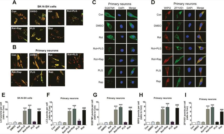
PLG restores the balance of autophagy and apoptosis by increasing BCL2 phosphorylation in
rotenone-induced Parkinson disease models
J. Liu, H. Yang (Beijing, China)
Objective: Parkinson disease (PD) is the second most common neurodegenerative disorder after
Alzheimer disease and there are few treatments currently available. The present study investigated the
protective effects of PLG in rotenone-induced PD cell and mouse models.
Background: Apoptosis and macroautophagy/autophagy play critical roles in PD pathogenesis; as such,
modulating their balance is a potential treatment strategy. BCL2 (B cell leukemia/lymphoma 2) is a key
molecule regulating this balance. PLG is an alkaloid extracted from Piper longum L. that has anti-
inflammatory and anticancer effects. Our previous study concerned PLG has protective effects in PD models
involve inhibiting mitochondrial dysfunction and apoptosis, although the underlying mechanism is unknown.
Methods: C57BL mice were orally administered rotenone and PLG, motor behavior was evaluated with
the rotarod and pole tests. The number of dopaminergic neurons was measured by immunohistochemistry. In
cell models, cell viability and cytotoxicity were measured by MTT and LDH assay, and mitochondrial
function was evaluated with JC-1 and Calcein AM assay. The interaction of BCL2 and BAX or BECN1 was
measured by co-immunoprecipitation to evaluate apoptosis or autophagy.
Results: We found that PLG administration (2 and 4 mg/kg) for 4 weeks attenuated motor deficits in mice
and prevented the loss of dopaminergic neurons in the substantia nigra induced by oral administration of
rotenone (10 mg/kg) for 6 weeks. PLG improved cell viability and enhanced mitochondrial function in
primary neurons and SK-N-SH cells. These protective effects were exerted by inducing BCL2
phosphorylation at Ser70 via MAPK8 activation, which resulted in the dissociation of BCL2 and BECN1 and
the stabilization of the BCL2 and BAX heterodimer, consequently enhancing autophagy and inhibiting
apoptosis.
Conclusions: Our results demonstrate that PLG exerts therapeutic effects in a rotenone-induced PD
models, and restoring the balance between apoptosis and autophagy by targeting BCL2 may be an effective
treatment for PD.
References: Liu J, Liu W, Lu Y, Tian H, Duan C, Lu L, Gao G, Wu X, Wang X, Yang H.
Piperlongumine restores the balance of autophagy and apoptosis by increasing BCL2 phosphorylation in
rotenone-induced Parkinson disease models. Autophagy, 2018, DOI: 10.1080/15548627. 2017. 1390636.
International Congress of Parkinson’s Disease and Movement Disorders®
Hong Kong October 5-9, 2018 - MDS Study Groups AbstractsFIG. 1 (405)
FIG. 2 (405)
International Congress of Parkinson’s Disease and Movement Disorders®
Hong Kong October 5-9, 2018 - MDS Study Groups AbstractsFIG. 3 (405)
FIG. 4 (405)
International Congress of Parkinson’s Disease and Movement Disorders®
Hong Kong October 5-9, 2018 - MDS Study Groups AbstractsFIG. 5 (405)
International Congress of Parkinson’s Disease and Movement Disorders®
Hong Kong October 5-9, 2018 - MDS Study Groups AbstractsFIG. 6 (405)
FIG. 7 (405)
International Congress of Parkinson’s Disease and Movement Disorders®
Hong Kong October 5-9, 2018 - MDS Study Groups AbstractsFIG. 8 (405)
FIG. 9 (405)
International Congress of Parkinson’s Disease and Movement Disorders®
Hong Kong October 5-9, 2018 - MDS Study Groups AbstractsFIG. 10 (405)
FIG. 11 (405)
International Congress of Parkinson’s Disease and Movement Disorders®
Hong Kong October 5-9, 2018 - MDS Study Groups AbstractsFIG. 12 (405)
FIG. 13 (405)
International Congress of Parkinson’s Disease and Movement Disorders®
Hong Kong October 5-9, 2018 - MDS Study Groups Abstracts1417
Research on nearinfrared brain function imaging for Freezing of Gait in Parkinson’s Disease
Y. Jingjing, L. Zhanhua, C. Ming (Dalian, China)
Objective: In the study ,fNIRS technique was used to study the activation level and activation mode of
prefrontal cerebral cortex for patients with FOG under cognitive processing,in order to analyze the
correlation between the severity of FOG and the prefrontal cortex function and to explore the
pathophysiology of PD with FOG.To provide a theoretical basis for the diagnosis,intervention and treatment
of PD with FOG,to help patients establish confidence,improve symptoms and improve quality of life.
Background: FNIRS is a real-time brain function monitoring,with its protability,noninvasive,high
temporal resolution,low cost and other unique advantages are widely used in the field of cognitive
neuroscience.The pathophysiology of FOG remains obscure and some research suggests that cognitive
impairment plays a key role in the occurrence of FOG.
Methods: The patients were divided into two groups:parkinson’s disease with freezing of gait(PD-FOG+)
and parkinson’s disease without freezing of gait(PD-FOG-) according to the clinical diagnostic criteria of the
United kingdom Parkinson’s disease Society Brain Bank and FOG questionnaire.Patients was assessed using
the Hoehn and Yahr scale(H&Y),motor section of the Unified Parkinson’s Disease Rating Scale(UPDRS-Ⅲ)
in the “off”(minimum of 12h without anti-parkinsonian medication).All patients underwent MMSE、
HAMD and FAB testing.The VFT was used as a stimulus task,recording group words or idioms,and the 52-
channel fNIRS system was used to perform signal acquisition when the patient performed the VFT task.The
activation of the prefrontal cortex was compared between the two groups.
Results: ①When the VFT task was performed,the number of words or idioms set out in the PD-FOG+
group was significantly less than that of the PD-FOG- group,and the FOG questionnaire was negatively
correlated with the VFT result.②For the PD-FOG+ group the activation area of the prefrontal cortex was
lesser than that of lower.③The mean oxygen hemoglobin concentrations in the left ventrolateral prefrontal
cortex of PD-FOG+ group were significantly lower than the PD-FOG- group,and the severity of the FOG
was negatively correlated with the mean oxygen hemoglobin concentration.
Conclusions: In PD-FOG+ patients,executive function is impaired,cognitive
flexibility,inhibition,memory,language use ability decreased significantly.There is a specific pattern of
structure and /or functional impairment in the left ventrolateral prefrontal cortex of PD with FOG patients,so
when the need for selection,memory,reaction inhibition and other more complex cognitive activities can not
be effectively recruited to solve the problem.Suggesting that executive dysfunction and FOG may have a
common neuropathological mechanism in the ventrolateral prefrontal cortex and the specific model brain
network damage of the wentrolateral prefrontal cortex is closely related to the occurrence of FOG.
References: 1. Maidan I, Bernad-Elazari H, Gazit E, et al. Changes in oxygenated hemoglobin link
freezing of gait to frontal activation in patients with Parkinson disease: an fNIRS study of transient motor-
cognitive failures[J]. Journal of Neurology, 2015, 262(4):899. 2.Imamura K, Okayasu N, Nagatsu T.
Cerebral blood flow and freezing of gait in Parkinson's disease[J]. Acta Neurologica Scandinavica, 2012,
126(3):210. 3.Tessitore A, Amboni M, Esposito F, et al. Resting-state brain connectivity in patients with
Parkinson's disease and freezing of gait.[J]. Parkinsonism & Related Disorders, 2012, 18(6):781.
LBA 11
Predictive Validity of Level I PD-MCI Criteria, using Global Cognitive Screeners, for PDD
J. Boel, G. Geurtsen, J. Hoogland, R. de Bie, J. Goldman, B. Schmand, A. Tröster, D. Burn, I. Litvan, G.
Geurtsen (Amsterdam, Netherlands)
Objective: PD-MCI can be used as a descriptive entity for clinical and research purposes and the PD-
MCI criteria1 need to be validated. The objective is to assess the predictive validity of Level I MDS PD-MCI
criteria, using global cognitive screeners, for PDD.
International Congress of Parkinson’s Disease and Movement Disorders®
Hong Kong October 5-9, 2018 - MDS Study Groups AbstractsYou can also read