Tracing the origin of the crayfish plague pathogen, Aphanomyces astaci, to the Southeastern United States - Nature
←
→
Page content transcription
If your browser does not render page correctly, please read the page content below
www.nature.com/scientificreports
OPEN Tracing the origin of the crayfish
plague pathogen, Aphanomyces
astaci, to the Southeastern United
States
Laura Martín‑Torrijos1*, María Martínez‑Ríos1, Gloria Casabella‑Herrero1, Susan B. Adams2,4,
Colin R. Jackson3,4 & Javier Diéguez‑Uribeondo1,4
The oomycete Aphanomyces astaci is an emerging infectious pathogen affecting freshwater crayfish
worldwide and is responsible for one of the most severe wildlife pandemics ever reported. The
pathogen has caused mass mortalities of freshwater crayfish species in Europe and Asia, and threatens
other susceptible species in Madagascar, Oceania and South America. The pathogen naturally coexists
with some North American crayfish species that are its chronic carriers. Presumptions that A. astaci
originated in North America are based on disease outbreaks that followed translocations of North
American crayfish and on the identification of the pathogen mainly in Europe. We studied A. astaci in
the southeastern US, a center of freshwater crayfish diversity. In order to decipher the origin of the
pathogen, we investigated (1) the distribution and haplotype diversity of A. astaci, and (2) whether
there are crayfish species-specificities and/or geographical restrictions for A. astaci haplotypes.
A total of 132 individuals, corresponding to 19 crayfish species and one shrimp species from 23
locations, tested positive for A. astaci. Mitochondrial rnnS and rnnL sequences indicated that A. astaci
from the southeastern US exhibited the highest genetic diversity so far described for the pathogen
(eight haplotypes, six of which we newly describe). Our findings that A. astaci is widely distributed
and genetically diverse in the region supports the hypothesis that the pathogen originated in the
southeastern US. In contrast to previous assumptions, however, the pathogen exhibited no clear
species-specificity or geographical patterns.
During the past few decades, fungal and fungal-like pathogens have caused several worldwide pandemics respon-
sible for declines in wildlife populations—even causing e xtinctions1–5. Globalization facilitates these pandem-
ics—usually consequences of the transport and introduction of exotic and invasive species6–8. Moreover, habitat
alterations due to anthropogenic activity break down natural dispersal barriers, allowing invasive species (fre-
quently carrying pathogens)9–15 to further expand their ranges. Climate change alters environmental conditions,
further benefitting some invasive species and favoring the development and spread of d isease2,4.
Fungal and fungal-like pathogenic species have impacted freshwater ecosystems particularly strongly, caus-
ing a global decline in freshwater biodiversity that is far greater than that seen in terrestrial e cosystems16,17. For
example, the panzootic chytrid fungus Batrachochytrium dendrobatidis originated in Asia and spread globally
due to amphibian trade, causing declines in more than 500 amphibian species over the past half-century18,19.
Furthermore, fungal-like pathogens, such as Saprolegnia diclina and Saprolegnia ferax (Oomycetes), are also
responsible for mass extinctions in a mphibians20,21 and may be spread by the fish t rade22. Another pathogenic
oomycete, Aphanomyces invadans, causes epizootic ulcerative syndrome (EUS), affecting more than 100 fish
species in Asia, Australia, North America and A frica23,24.
Similarly, Aphanomyces astaci causes the crayfish plague in native European, Asian and Australian crayfish
species25–28 and has decimated crayfish populations in those c ontinents5,6. This oomycete is a specialized pathogen
in freshwater c rayfish27,29,30, one third of which are threatened with extinction g lobally31. The pathogen coex-
ists naturally with North American crayfish but can efficiently colonize non-North American crayfish, almost
1
Department of Mycology, Real Jardín Botánico-CSIC, Plaza Murillo 2, 28014 Madrid, Spain. 2USDA Forest Service,
Southern Research Station, Center for Bottomland Hardwoods Research, 1000 Front Street, Oxford, MS 38655,
USA. 3Department of Biology, University of Mississippi, University, MS 38677, USA. 4These authors contributed
equally: Susan B. Adams, Colin R. Jackson and Javier Diéguez-Uribeondo. *email: lmtorrijos@rjb.csic.es
Scientific Reports | (2021) 11:9332 | https://doi.org/10.1038/s41598-021-88704-8 1
Vol.:(0123456789)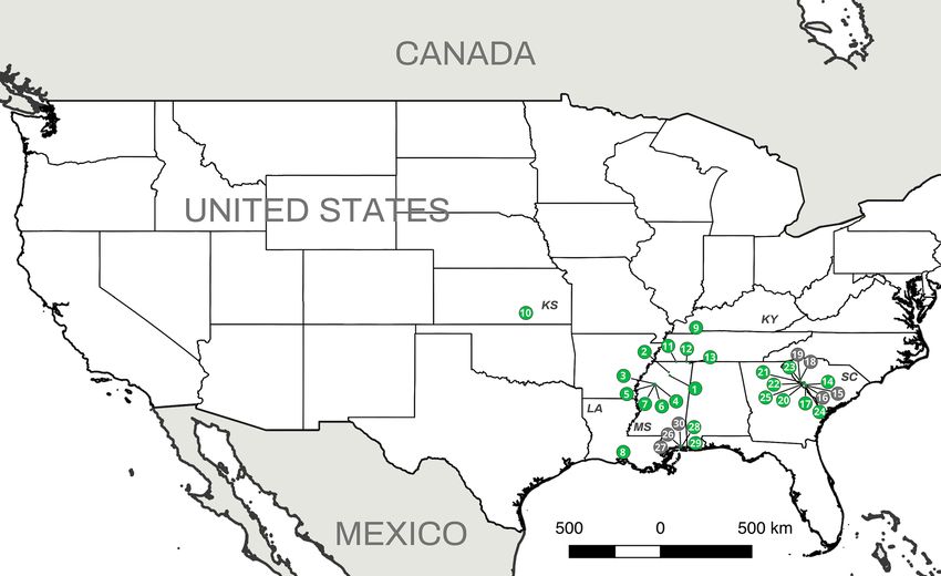
www.nature.com/scientificreports/
Figure 1. Distribution of Aphanomyces astaci haplotypes detected from locations in Europe and J apan33,52,53.
Each point and color represent the presence of an A. astaci haplotype based on concatenated mitochondrial
rnnS and rnnL regions. Colors indicate the haplotype code at each site as follows: green—a-haplotype, blue—b-
haplotype, orange—d1-haplotype, red—d2-haplotype and pink—d3-haplotype [Maps were prepared using
QGIS 2.14 (https://www.qgis.org/en/site/)].
without resistance26. In addition, A. astaci has spread rapidly throughout the world through translocations of
North American chronic c arriers25,28–40. In non-North American crayfish, crayfish plague infections typically
cause death within a few days41.
Presumptions about the origin of the crayfish plague were based on disease outbreaks that followed historical
translocations of North American crayfish species to many countries for aquaculture, sport fishing, or aquarium
pet trade42. The first known introduction of a North American crayfish and subsequent crayfish plague out-
break was recorded in Europe in the nineteenth c entury43. Later, additional large-scale introductions of North
American crayfish species were made in European and non-European countries34–37,39,40,44–49. Moreover, illegal
translocations resulted in new crayfish plague outbreaks that decimated native crayfish populations in many
countries33,46,50 (Fig. 1). Thus, A. astaci was listed among the 100 of the World’s Worst Invasive Alien S pecies51.
Knowledge about the virulence and genetic variability of A. astaci has come primarily from studies of crayfish
plague outbreaks in Europe and Asia50,52,54–60. Specifically, mitochondrial DNA regions of A. astaci have been
informative in assessing genetic diversity in both pure cultures and clinical samples of this clonally reproducing
pathogen. To date, the mitochondrial DNA variability found in crayfish plague outbreaks in Europe and Japan
has been allocated to six haplotypes (a, b, d1, d2, d3 and e-haplotypes) (Fig. 1) within two lineages33,38,52,53,61.
However, only three studies have confirmed the presence of the pathogen in North America, revealing only two
of the previously described haplotypes: a and b-haplotypes62–64.
Although evidence strongly supports a North American origin of A. astaci, our understanding of the crayfish
plague pathogen in North America is still insufficient. A clearer understanding of the diversity and distribution
of A. astaci within its native range is needed, not only to improve our comprehension of the evolution and epi-
demiology of pandemic pathogens, but also to determine future management and research directions. Similar
questions have been faced when studying other emerging pathogens. For example, despite the occurrence of
Batrachochytrium dendrobatidis in Asia, the lack of lethal outbreaks evidenced an endemic host–pathogen inter-
action in that region. Several studies have confirmed that the geographic origin of chytridiomycosis was in Asia,
explaining the survival of Asian amphibian populations and stable host–pathogen d ynamics19.
Scientific Reports | (2021) 11:9332 | https://doi.org/10.1038/s41598-021-88704-8 2
Vol:.(1234567890)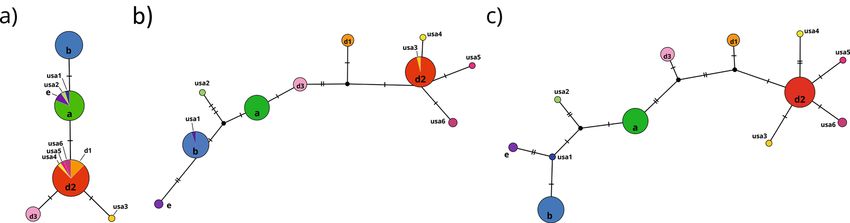
www.nature.com/scientificreports/
Figure 2. Aphanomyces astaci presence from the study locations. Aphanomyces astaci presence/absence is
represented by green (positive) or grey (negative) circles. Numbers indicate the location code at each site (as
in Supplementary Table 1). Two-letter state code is provided with the following abbreviation: KS (Kansas), KY
(Kentucky), LA (Louisiana), MS (Mississippi) and SC (South Carolina). [Maps were prepared using QGIS 2.14
(https://www.qgis.org/en/site/)].
Although more than 428 crayfish species are native to North A merica65, the diversity, distribution and preva-
lence of A. astaci there is still largely unknown. Within North America, the southeastern US harbors the highest
number of endemic crayfish species. The region represents not only a center of diversity, but also one of the two
distinct origins of freshwater c rayfish6,52,66. The presence of A. astaci has not been investigated in this crayfish-
rich region even though such knowledge would improve our understanding of the origin and diversity of the
pathogen. Thus, the main aim of this study was to evaluate the southeastern US as the possible center of origin
of the crayfish plague pathogen A. astaci. For this purpose, we tested key questions including: (1) what is the
distribution and haplotype diversity of A. astaci in the southeastern US, and (2) are A. astaci haplotypes crayfish
species-specific and/or geographically restricted. In order to perform this study, we isolated and analyzed the
pathogen from 30 distinct crayfish populations comprising a total of 21 crayfish species and one shrimp from
five states in the southeastern US.
Results
Aphanomyces astaci detection. We obtained a total of 391 crayfish from 30 locations in five states (Kan-
sas, Kentucky, Louisiana, Mississippi and South Carolina) (Fig. 2). The crayfish represented six genera and 21
species: Cambarellus shufeldtii, Cambarus latimanus, Cambarus striatus, Cambarus tenebrosus, Creaserinus fodi-
ens, Creaserinus oryktes, Faxonius etnieri species complex, Faxonius sp., Faxonius tricuspis, Faxonius wrighti,
Lacunicambarus ludovicianus, Procambarus ablusus, Procambarus acutus, Procambarus clarkii, Procambarus
hayi, Procambarus hybus, Procambarus pubescens, Procambarus raneyi, Procambarus troglodytes, Procambarus
viaeviridis and Procambarus vioscai (Supplementary Table 1). Additionally, one species of freshwater shrimp
(Palaemon kadiakensis) was sampled and analyzed for the presence of A. astaci.
From 392 individuals, 132 crayfish and one shrimp tested positive for the A. astaci ITS region, 102 tested
negative and 158 were not analyzed (i.e., the crayfish did not molt). Aphanomyces astaci-positive samples came
from 23 locations and included 19 crayfish species: C. shufeldtii, C. latimanus, C. striatus, C. fodiens, C. oryktes,
F. etnieri species complex, Faxonius sp., F. tricuspis, F. wrighti, P. ablusus, P. acutus, P. clarkii, P. hayi, P. hybus, P.
pubescens, P. raneyi, P. troglodytes, P. viaeviridis and P. vioscai) and one species of shrimp (Palaemon kadiakensis)
(Fig. 2 and Supplementary Table 1). The ITS sequences (specific primers 42 and 640) for the 132 clinical samples
were 99.82% identical to sequences of A. astaci available in GenBank (e.g., sequence FM999249-isolate SAP302)
and identical to each other.
Scientific Reports | (2021) 11:9332 | https://doi.org/10.1038/s41598-021-88704-8 3
Vol.:(0123456789)www.nature.com/scientificreports/
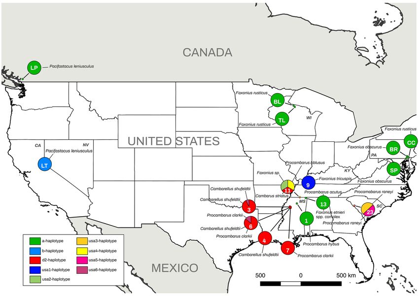
Figure 3. Aphanomyces astaci haplotypes detected from locations (numbers on pie graphs indicate locations;
see Table 1) in North America. Haplotype frequencies indicated by relative proportion of the pie graph. Each
color of the legend represents a different haplotype based on concatenated mitochondrial rnnS and rnnL
regions. Crayfish species hosting different haplotypes are indicated at each location (also in Table 1). Location
code LP corresponds to Lake P itt50, LT to Lake Tahoe63, BL to Big L
ake62, TL to Trout L
ake62, CC to Chickies
Creek64, BR to Brubaker Run64 and SP to Sunfish Pond64. Two-letter State code is provided with the following
abbreviation: CA (California) KY (Kentucky), MS (Mississipi), PA (Pennsylvania), NV (Nevada), SC (South
Carolina) and WI (Wisconsin). [Maps were prepared using QGIS 2.14 (https://www.qgis.org/en/site/)].
Sequence analyses and haplotyping of A. astaci. Twenty crayfish clinical samples (taken directly
from crayfish) and 12 pure cultures from nine locations and three states (Kentucky, Mississippi and South Caro-
lina) (Fig. 3) contained enough of the pathogen DNA for amplifying both mitochondrial regions (i.e., clinical
samples often harbor low pathogen DNA concentration).
For the phylogenetic approximations [Bayesian Inference (BI) and Maximum likelihood (ML)] and diver-
sity estimations, we included a total of 78 sequences [32 sequences from the present study, 43 obtained from
GenBank as reported from previous s tudies33,52,62 and two sequences from new A. astaci and A. fennicus isolates
(CCRJB-75 and CCRJB-76, respectively)67 with 476 and 355 bp fragments of rnnS and rnnL amplicons, respec-
tively. The phylogenetic approximations (BI and ML) supported the differentiation of the two lineages previously
described52 (Fig. 4). The genetic diversity analysis confirmed and supported the phylogenetic analysis. Although
both mitochondrial ribosomal rnnS and rnnL regions were informative, there were differences between them
(Fig. 5). We obtained five haplotypes for the rnnS subunit (Fig. 5a), represented by four segregating sites (S),
with a haplotype diversity (Hd) of 0.703, a nucleotide diversity (π) of 0.0022 and 1.018 average nucleotide dif-
ferences (k). On the other hand, we obtained ten haplotypes for the rnnL subunit (Fig. 5b), represented by13
segregating sites (S), with a haplotype diversity (Hd) of 0.786, a nucleotide diversity (π) of 0.009 and 3.16 average
nucleotide differences (k). However, concatenating rnnS and rnnL regions we confirmed a total of 12 haplotypes
represented by 17 segregating sites (S), where 13 of them were parsimony informative (Fig. 5c). The concatenated
sequences presented a haplotype diversity (Hd) of 0.801 with a nucleotide diversity (π) of 0.005 and 4.178 aver-
age nucleotide differences (k).
The phylogenetic approximations and the haplotype network confirmed the presence of eight haplotypes for
the rrnS and rrnL concatenated regions among the analyzed samples: a, d2, usa1, usa2, usa3, usa4, usa5 and usa6-
haplotypes (Figs. 4, 5) (Table 1). Six of these eight haplotypes (usa1, usa2, usa3, usa4, usa5 and usa6-haplotypes)
are described and reported here for the first time, bringing the total number of known A. astaci haplotypes to
Scientific Reports | (2021) 11:9332 | https://doi.org/10.1038/s41598-021-88704-8 4
Vol:.(1234567890)www.nature.com/scientificreports/
Figure 4. Phylogenetic analyses of Aphanomyces astaci mitochondrial regions. Bayesian phylogenetic analyses
of A. astaci mitochondrial rnnS, rnnL and concatenated rnnS + rnnL sequences obtained from infected
crayfish specimens and previous studies33,52,53,62. (a) Bayesian phylogenetic tree based on the rnnS sequences,
(b) Bayesian phylogenetic tree based on the rnnL sequences, (c) Bayesian phylogenetic tree based on the
concatenated rnnS + rnnL sequences. Values above the branches represent the Bayesian posterior probabilities
(> 0.95) and ML bootstrap support values (> 75), respectively. Scale bar for phylogenetic analysis indicates
substitutions per site. Abbreviations: Ht, haplotypes; L, lineages.
Figure 5. Haplotype network based on rnnS, rnnL and concatenated rnnS + rnnL mitochondrial DNA
sequences, generated by statistical parsimony. The area of the circles is proportional to the number of sequences.
(a) Haplotype network based on rnnS mtDNA sequences, (b) Haplotype network based on rnnL mtDNA
sequences, (c) Haplotype network based on concatenated rnnS + rnnL mtDNA sequences. Mutation steps
between haplotypes are shown as hatch marks.
12. Moreover, these results were confirmed by an independent secondary molecular test (i.e., by repeating the
amplification and sequencing of each of the haplotyped samples).
Crayfish species, A. astaci haplotype diversity and distribution. The haplotyping results for the 20
crayfish clinical samples included five haplotypes: usa1-haplotype (one Faxonius tricuspis), usa2-haplotype (one
Cambarus striatus), a-haplotype (one Procambarus hybus, one Procambarus acutus and one Faxonius etnieri),
d2-haplotype (ten Cambarellus shufeldtii, two Procambarus clarkii and one Procambarus ablusus) and usa6-
haplotype (one Cambarellus shufeldtii and one Procambarus clarkii) (GenBank accession numbers MW346503-
MW346522 for rnnS and MW346523-MW346542 for rnnL) (Table 1). The amount of infection in the remaining
samples that tested positive for A. astaci (ITS region) was too low to obtain conclusive results for both rrnS and
rrnL.
Additionally, the haplotyping results for the 12 pure cultures included four haplotypes: usa3-haplotype (one
Procambarus raneyi culture), usa4-haplotype (one Faxonius sp. culture), usa5-haplotype (one Procambarus raneyi
Scientific Reports | (2021) 11:9332 | https://doi.org/10.1038/s41598-021-88704-8 5
Vol.:(0123456789)www.nature.com/scientificreports/
Location Haplotype GenBank GenBank
code Collection code Host species State Sample type DNA extraction Species ID (rRNA) (rnnS/rnnL) rnnS rnnL
1 SA908-DEAD1 Procambarus hybus Mississippi Clinical sample CE19/01-1 Aphanomyces astaci a MW346522 MW346542
SA927-1 Cambarellus shufeldtii Mississippi Clinical sample CE19/06-12 Aphanomyces astaci usa6 MW346516 MW346536
SA927-5 Cambarellus shufeldtii Mississippi Clinical sample CE19/04-58 Aphanomyces astaci d2 MW346519 MW346539
SA927-8 Cambarellus shufeldtii Mississippi Clinical sample CE19/04-57 Aphanomyces astaci d2 MW346520 MW346540
SA927-16 Cambarellus shufeldtii Mississippi Clinical sample CE19/06-2 Aphanomyces astaci d2 MW346513 MW346533
SA927-18* Cambarellus shufeldtii Mississippi Clinical sample CE19/06-26 Aphanomyces astaci d2 MW346512 MW346532
SA927-23 Cambarellus shufeldtii Mississippi Clinical sample CE19/06-3 Aphanomyces astaci d2 MW346510 MW346530
SA927-25 Cambarellus shufeldtii Mississippi Clinical sample CE19/04-55 Aphanomyces astaci d2 MW346521 MW346541
3
SA927-27 Cambarellus shufeldtii Mississippi Clinical sample CE19/04-59 Aphanomyces astaci d2 MW346518 MW346538
SA927-18* Cambarellus shufeldtii Mississippi Culture CE19/C-136 Aphanomyces astaci d2 MW346494 MW346481
SA927-18* Cambarellus shufeldtii Mississippi Culture CE19/C-135 Aphanomyces astaci d2 MW346495 MW346482
SA927-18* Cambarellus shufeldtii Mississippi Culture CE19/C-127 Aphanomyces astaci d2 MW346498 MW346485
SA927-18* Cambarellus shufeldtii Mississippi Culture CE19/C-138 Aphanomyces astaci d2 MW346492 MW346479
SA927-14 Cambarellus shufeldtii Mississippi Culture CE19/C-140 Aphanomyces astaci d2 MW346491 MW346478
SA927-18* Cambarellus shufeldtii Mississippi Culture CE19/C-150 Aphanomyces astaci d2 MW346487 MW346474
4 SA928-7 Cambarellus shufeldtii Mississippi Clinical sample CE19/06-4 Aphanomyces astaci d2 MW346509 MW346529
SA930-2 Cambarellus shufeldtii Mississippi Clinical sample CE19/04-61 Aphanomyces astaci d2 MW346517 MW346537
SA930-4 Cambarellus shufeldtii Mississippi Clinical sample CE19/07-03 Aphanomyces astaci d2 MW346504 MW346524
SA930-6 Procambarus clarkii Mississippi Clinical sample CE19/06-15 Aphanomyces astaci usa6 MW346515 MW346535
6 SA930-8* Procambarus clarkii Mississippi Clinical sample CE19/07-08 Aphanomyces astaci d2 MW346503 MW346523
SA930-8* Procambarus clarkii Mississippi Culture CE19/C-141 Aphanomyces astaci d2 MW346490 MW346477
SA930-8* Procambarus clarkii Mississippi Culture CE19/C-128 Aphanomyces astaci d2 MW346497 MW346484
SA930-8 * Procambarus clarkii Mississippi Culture CE19/C-145 Aphanomyces astaci d2 MW346489 MW346476
7 M306-3 Procambarus clarkii Mississippi Clinical sample CE19/06-19 Aphanomyces astaci d2 MW346514 MW346534
9 SA937-5 Faxonius tricuspis Kentucky Clinical sample CE19/06-27 Aphanomyces astaci usa1 MW346511 MW346531
SA946-9 Procambarus ablusus Mississippi Clinical sample CE19/07-19 Aphanomyces astaci d2 MW346508 MW346528
11 SA946-23 Cambarus striatus Mississippi Clinical sample CE19/07-21 Aphanomyces astaci usa2 MW346507 MW346527
SA946-FAX SP JUV Faxonius sp. Mississippi Culture CE19/C-137 Aphanomyces astaci usa4 MW346493 MW346480
SA948-1 Procambarus acutus Mississippi Clinical sample CE19/07-23 Aphanomyces astaci a MW346506 MW346526
13
SA948-5 Faxonius etnieri Mississippi Clinical sample CE19/07-28 Aphanomyces astaci a MW346505 MW346525
SC32 MOLT 4* Procambarus raneyi South Carolina Culture CE19/C-130 Aphanomyces astaci usa3 MW346496 MW346483
23
SC32 MOLT 5* Procambarus raneyi South Carolina Culture CE19/C-147 Aphanomyces astaci usa5 MW346488 MW346475
Table 1. Location and samples ID of the of the crayfish species analyzed for Aphanomyces astaci haplotypes.
North American crayfish species from each of the analyzed locations (Location code:1–30), including the
corresponding collection codes, state, sample type (clinical sample/culture), DNA isolation code (CE19/),
BLAST species ID, result of the mitochondrial rnnS/rnnL haplotype and GenBank references for the rnnS and
rnnL regions. NA: not applicable. Each unique collection code refers to an individual crayfish. *Different pieces
from the same molt.
culture) and d2-haplotype (six Cambarellus shufeldtii and three Procambarus clarkii cultures) (GenBank accession
number MW346487-MW346498 for rnnS and MW346474-MW346485 for rnnL).
We found five scenarios relative to the distribution of A. astaci haplotypes among crayfish species at various
locations: (1) one haplotype from one crayfish species (Locations 1, 4, 7 and 9), (2) two haplotypes from one
crayfish species (Locations 3 and 23) (see below), (3) one haplotype from two crayfish species (Location 13), (4)
two haplotypes from two crayfish species (Location 6) (see below), and (5) three haplotypes, one from each of
three crayfish species (Location 11). At Location 23, the two haplotypes were recovered from one P. raneyi molt
(i.e., we isolated the pathogen in two different pure cultures) and at Location 6, one haplotype was recovered
from both species, and the second haplotype from only one of the species (Fig. 3) (Table 1).
Discussion
We report and describe for the first time the presence, distribution and genetic diversity of the crayfish plague
pathogen, A. astaci, in its potential center of origin, the southeastern U S68. Previous studies regarding the ori-
gin, diversity, and distribution of A. astaci have addressed different questions. In the current study, we have
explored several of them, including (1) the distribution and diversity of A. astaci in the southeastern US, and (2)
whether A. astaci haplotypes are crayfish species-specific50,54,55 and/or restricted to a narrow geographic region
of the pathogen’s native range52,62,63. Our results indicated that A. astaci is present and widely distributed in the
southeastern US (e.g., it was present in 21 out of 23 sampling sites across the nine river basins we investigated)
Scientific Reports | (2021) 11:9332 | https://doi.org/10.1038/s41598-021-88704-8 6
Vol:.(1234567890)www.nature.com/scientificreports/
and possesses the highest genetic diversity of A. astaci described from any region to date. Previously, only two
haplotypes (a and b-haplotypes) had been found in the five North American crayfish species examined from
California, Michigan and Pennsylvania (Pacifastacus leniusculus, Cambarus bartonii, Faxonius obscurus, Faxonius
rusticus and Faxonius virilis)62–64. However, in the southeastern US, we examined 21 North American crayfish
species and found a total of eight haplotypes, six of which are previously unreported. This represents almost 70%
of the A. astaci haplotype diversity known globally. The genetic diversity we describe is comparable to that of
other pathogenic oomycetes. For example, in the US, 13 Phytopthtora infestans haplotypes were recently described
using five mitochondrial l oci69. Moreover, we showed that A. astaci can chronically colonize 19 additional North
American crayfish species and also one species of freshwater shrimp (Palaemon kadiakensis). Previously, the only
known report of a wild shrimp carrying A. astaci was of a Macrobrachium lanchesteri population from Indonesia
that co-occurred with P. clarkii70. Only two (Cambarus tenebrosus and Lacunicambarus ludovicianus) of the 21
crayfish species that we sampled did not test positive for A. astaci. Thus, we confirm the presence of A. astaci
in a wider distribution (Fig. 2) than previously described in North A merica62–64, confirming the origin of this
pathogenic disease in North America.
Although the presence of the crayfish plague in North America was previously r eported62–64, those stud-
ies examined a limited number of crayfish species and found haplotypes that had been previously isolated in
Europe and Japan (i.e., a or b-haplotypes)33,62–64. Likewise, we confirmed the presence in North America of two
haplotypes (a- and d2-haplotypes) first described in Europe. In the case of the a-haplotype, it was described
as the first haplotype introduced to Europe in the nineteenth c entury43,52, yet no North American carrier has
been described in the literature for this introduction. Two recent studies described F. rusticus and F. obscurus
as the only North American crayfish known to host the a-haplotype62,64. We expanded the known range of this
haplotype to include the southeastern US and added three additional native taxa as hosts: the F. etnieri species
complex, P. acutus and P. hybus. In the case of the d2-haplotype, the only two North American crayfish described
as carriers so far in Europe are P. clarkii and Procambarus fallax virginalis. As we expected, our study shows for
the first time the presence of A. astaci in native populations of P. clarkii54, which carried the d2-haplotype and
the newly discovered usa6-haplotype.
We confirmed that at least some A. astaci haplotypes are neither host species-specific nor narrowly distributed.
Broadening and deepening our knowledge of A. astaci haplotype diversity and distribution, we recovered the
d2-haplotype from two species in different genera (Cambarellus and Procambarus) in one river basin (Locations
3, 4, 6 and 7) and from a third genus (Cambarus) in a different river basin (Location 11) (Table 1). Although
the a-haplotype was previously thought to be restricted to F. rusticus and F. obscurus62,64, we found it not only
in another congener, but also in another genus (Procambarus). In addition, we documented the a-haplotype in
two river basins in the southeastern US, even though it was previously thought to be restricted to the northern
US. We also documented the d2-haplotype from two river basins. Our results indicated that A. astaci haplotypes
tend to be neither host species-specific nor restricted to small geographic areas.
We also found instances of multiple haplotypes of A. astaci occurring in one species, in one individual, and
in one location. We isolated two pure cultures of two different, but closely related, A. astaci haplotypes (usa3 and
usa5) from a single individual (SC32 MOLT in Location 23). Also, multiple A. astaci haplotypes often co-occurred
in a location. Within Location 11, we found three haplotypes from two lineages: usa2-haplotype from Lineage
1 and d2-haplotype and usa4-haplotype from Lineage 2. We also recovered two closely related haplotypes (d2
and usa6) from Locations 3 and 6 and two others (usa3 and usa5) from Location 23. None of the phenomena
described have been documented previously. Our analysis showed no clear haplotype distributional patterns with
respect to crayfish host species or geography. Thus, the biogeographic distribution of A. astaci genetic diversity
within North America needs further investigation.
The pathogen’s genetic diversity and observed lack of species-specificity may have implications within North
America as well as on continents where A. astaci is introduced. Crayfish have been frequently translocated
within North America. For example, although P. clarkii and F. virilis are native in parts of North America, both
are invasive beyond their native range, including west of the Great Divide where all native crayfish belong to
the family A stacidae71,72. The genetic diversity of A. astaci suggests potential for intracontinental impacts from
translocations of haplotypes. Presumably all North American crayfish are resistant to all haplotypes of A. astaci,
considering that no mass mortalities have been observed after crayfish translocations. However, more subtle
effects of translocated haplotypes with differing virulence from native haplotypes would not likely have been
detected. Further understanding of the geographical distribution of A. astaci genetic diversity, its virulence, and
the immune responses of crayfish to novel haplotypes would be beneficial for managing crayfish translocations
and conserving native species in North America.
Our new approach of obtaining A. astaci from crayfish molts produced larger amounts of the pathogen for
identification and isolation in pure cultures. This resulted in detection of A. astaci in 56.17% of the analyzed clini-
cal samples. Instead of analyzing parts of the crayfish most susceptible to infection (i.e., soft abdominal cuticle,
telson or walking legs)73, we waited until the molting period. Thus, we avoided the unnecessary killing of crayfish
and maximized the amount of pathogen grown within the original host. Moreover, by incubating crayfish molts
in distilled water for three days, we allowed the pathogen to continue growing both within and outside of the
cuticle. By controlling the incubation temperature at 4 °C, we reduced bacterial blooms during the first stages
of the pathogen isolation. Bacteria commonly surround the A. astaci hyphae, and antibiotics are often added
to the PGA medium to inhibit bacterial growth. However, because bacteria rapidly develop resistance to these
antibiotics, their addition becomes less effective over time. By reducing the incubation temperature and intro-
ducing a physical b arrier74, we controlled bacterial b
looms75 and obtained many clean isolates of the pathogen.
By using this new approach, we more readily detected and isolated A. astaci. Moreover, we strongly recom-
mend isolating the pathogen in an axenic culture to assure an optimal concentration of the pathogen for analysis.
In this study, we combined the sequencing of the ITS region (for identification of the Aphanomyces species)76 and
Scientific Reports | (2021) 11:9332 | https://doi.org/10.1038/s41598-021-88704-8 7
Vol.:(0123456789)www.nature.com/scientificreports/
the mtDNA (for identification of the A. astaci haplotype)52 in both clinical samples and pure cultures. Several
studies have examined the genetic diversity of A. astaci using diverse methodologies (i.e., RAPD-PCR, the chi-
tinase gene, AFLP, microsatellites)50,52,56,58–60,77. Although the mtDNA a pproximation52 requires a large concen-
tration of the pathogen, it provides reliable r esults67 and enables detection of new diversity within A. astaci that
might go undetected with other approximations59,60. Other approaches, such as eDNA monitoring78, could also
be used to detect the presence of A. astaci in water samples and potentially to detect additional genetic diversity.
The uniquely high diversity of A. astaci haplotypes found in this study are an important step toward confirm-
ing the host–pathogen co-evolution between A. astaci and North American crayfish species. Our results suggest
that further sampling in North America will reveal additional undiscovered A. astaci haplotype diversity vital to
answering new host–pathogen co-evolutionary questions. Further, long-term monitoring might reveal emerging
A. astaci diversity, depending on the rate of pathogen evolution.
Methods
Sample collection. We sampled 25 locations in five states between February and May 2019. Additionally,
we included nine crayfish samples previously collected from five more locations and preserved in 95% etha-
nol. The specimens from Kansas were from an introduced population of Procambarus clarkii (Supplementary
Table 1). Crayfish were captured by kick-seining and trapping and then held, separated by species and collection
location, in the laboratory (US Forest Service, Southern Research Station, Center for Bottomland Hardwoods
Research, in Oxford [Mississippi, USA]) until processed. Crayfish were kept individually in labeled, individual,
plastic containers with chlorine-free water, aerators, gravel and medium size rocks. Each crayfish was kept alive
until it molted and was subsequently preserved in 95% ethanol.
Microscopic examination and Aphanomyces astaci isolation. For the microscopic examination, the
molts were carefully removed and handled individually due to the fragility of the samples. Molts were kept in
individual petri dishes with distilled water at 4 °C in order to reduce bacterial growth and optimize the growth
ycelium75. After three days, each molt was examined with an inverted microscope to check for the
of potential m
presence of growing hyphae. Each sample was divided into two parts: one for molecular identification and one
for the pathogen isolation.
Pieces of the molt intended for molecular identification were transferred into 1.5-ml tubes and were frozen
at − 80 °C until the DNA extraction was carried out in the laboratory at the Department of Biology at the Univer-
sity of Mississippi (UM). Samples were subsequently homogenized by manual mechanical disruption. A DNeasy
Blood & Tissue Kit (Qiagen, Valencia, California, USA) was used to isolate genomic DNA.
Pieces of the molt intended for culture isolation were grown in Peptone Glucose Agar (PGA) at 4 °C. A
selected agar plug was cut out from the resulting mycelia and inserted within an aluminum ring placed on a new
PGA plate to protect the growing isolate from bacterial g rowth74. Plates were incubated at 4 °C for seven days,
and each isolate was transferred into new PGA media once the hyphae spread under the metal ring. This process
was repeated until no bacterial growth was observed using an inverted microscope. Additionally, a selected agar
plug containing mycelia was placed in a 9 mm Petri dish containing 10 mL of liquid Peptone Glucose (PG-1) and
incubated at room temperature for 48 h in order to obtain material for molecular identification. The obtained
mycelium was transferred into 1.5-ml tubes and frozen at − 20 °C until the DNA extraction was carried out in
the laboratory at UM. Samples were subsequently homogenized by manual mechanical disruption, and DNA
extractions were carried out using an E.Z.N.A. Fungal DNA Mini Kit (Omega Biotek, Norcross, Atlanta, USA).
Aphanomyces astaci detection and haplotyping. To test for the presence of the A. astaci pathogen, a
fragment of the internal transcribed spacer (ITS) region was amplified using the diagnostic primers 42 (5′-GCT
TGTGCTGAGGATGTTCT-3′)73 and 640 (5′-CTATCCGACTCCGCATTCTG-3′)79 (which amplify ITS1, the
5.8S rDNA and ITS2) in a single round of amplification according to the assay described b y73. DNA extracted
from a pure culture of A. astaci was used as the positive control; sterile Milli-Q water was used as the negative
control. Amplified products (3 μL of each reaction) were analyzed by electrophoresis in 2% agarose SB gels
stained with ethidium bromide and then purified using magnetic beads. Sequencing of both strands of posi-
tive products was performed using an automated sequencer (Applied Biosystems 3730xl DNA Analyzer, DNA
Analysis Facility at Yale, USA and Applied Biosystems 3730xl DNA, Macrogen, The Netherlands). Sequences
were aligned and edited using the program Geneious 10.0.280. A BLAST search (NCBI database) was performed
to verify the identity of each sequence.
Genomic DNA samples that tested positive for the presence of A. astaci with diagnostic primers 4273 and
64079 were used to characterize the phylogenetic relationships and haplotypes. The mitochondrial ribosomal
small (rnnS) and large (rnnL) subunits were amplified using the primer pairs AphSSUF/AphSSUR (5′-AGCA CT
CCGCCTGAAGAGTA-3′ and 5′-GGGCGGTGTGTACAAAGTCT-3′) and AphLSUF/AphLSUR (5-AGGCGA
AAGC TTA CTA TGA TGG-3′ and 5′-CCAA TTC TGT GCC ACC TTC y52. Positive
T-3′), respectively, as described b
and negative controls were included. Amplified products were analyzed by electrophoresis, purified, sequenced
and aligned as described above.
Phylogenetic approximations based on Bayesian inference (BI) and maximum likelihood (ML) were used to
reconstruct relationships. The BI analysis was performed in MrBayes v.3.2.681 using the MCMC method with
10 million generations, three runs (8 chains per run) with a burn-in of 25% and a standard deviation of split fre-
quencies < 0.01. Nodes with posterior probability (pp) values ≥ 0.95 were considered supported. The ML analysis
was performed using RAxML v.882, as implemented in raxmlGUI v1.5b183, with 100 independent replicates and
1000 rapid bootstraps. Nodes with bootstrap values ≥ 75 were considered supported. The resulting trees from the
BI and ML analyses were visualized with FigTree v1.4.284. Sequences (57) corresponding to the mtDNA regions
Scientific Reports | (2021) 11:9332 | https://doi.org/10.1038/s41598-021-88704-8 8
Vol:.(1234567890)www.nature.com/scientificreports/
rnnS and rnnL of isolates analyzed in previous s tudies33,52,62,67, available in GenBank, were also included in our
analyses. Aphanomyces fennicus was used as the outgroup in both phylogenetic a pproximations67,76. Analyses
were performed with rnnS and rnnL individually, as well as with a concatenated rnnS and rnnL dataset, using
the same parameters described above.
Genetic diversity was estimated by calculating the number of polymorphic (segregating) sites (S), the number
of haplotypes, the haplotype diversity (Hd), the average number of nucleotide differences (k), and the nucleo-
tide diversity (π) utilizing the program DNAsp v.5.10.0185. Mutational changes between sequences in the most
parsimonious haplotype network were estimated using TCS v.1.2186, and the genealogical relationships were
visualized with PopArt v1.7.287.
Ethics declarations. All experimental procedures and animal manipulations, as well as field sampling,
were performed according to the US legislation.
Data availability
All data generated or analyzed during this study are included in this published article (and its Supplementary
Information files).
Received: 20 December 2020; Accepted: 5 April 2021
References
1. Fisher, M. C. et al. Emerging fungal threats to animal, plant and ecosystem health. Nature 484, 186–194 (2012).
2. Brandt, M. E. & Park, B. J. Think fungus-prevention and control of fungal infections. Emerg. Infect. Dis. 19, 1688–1689 (2013).
3. Vallabhaneni, S., Mody, R. K., Walker, T. & Chiller, T. The global burden of fungal diseases. Infect. Dis. Clin. N. Am. 30, 1–11 (2016).
4. Benedict, K., Richardson, M., Vallabhaneni, S., Jackson, B. R. & Chiller, T. Emerging issues, challenges, and changing epidemiology
of fungal disease outbreaks. Lancet Infect. Dis. 17, e403–e411 (2017).
5. Ghosh, P. N., Fisher, M. C. & Bates, K. A. Diagnosing emerging fungal threats: A one health perspective. Front. Genet. 9, 1–8 (2018).
6. Ogden, N. H. et al. Emerging infectious diseases and biological invasions: A call for a One Health collaboration in science and
management. R. Soc. Open Sci. 6, 181577 (2019).
7. Lwande, O. W. et al. Globe-trotting Aedes aegypti and Aedes albopictus: Risk factors for arbovirus pandemics. Vector Borne Zoonotic
Dis. 20, 71–81 (2020).
8. Santini, A. & Battisti, A. Complex insect-pathogen interactions in tree pandemics. Front. Physiol. 10, 1–7 (2019).
9. Crowl, T. A., Crist, T. O., Parmenter, R. R., Belovsky, G. & Lugo, A. E. The spread of invasive species and infectious disease as
drivers of ecosystem change. Front. Ecol. Environ. 6, 238–246 (2008).
10. Stout, J. C. & Morales, C. L. Ecological impacts of invasive alien species on bees. Apidologie 40, 388–409 (2009).
11. D’hondt, B. et al. Harmonia+ and Pandora+: Risk screening tools for potentially invasive plants, animals and their pathogens. Biol.
Invasions 17, 1869–1883 (2015).
12. Robinson, C. V., Uren Webster, T. M., Cable, J., James, J. & Consuegra, S. Simultaneous detection of invasive signal crayfish, endan-
gered white-clawed crayfish and the crayfish plague pathogen using environmental DNA. Biol. Conserv. 222, 241–252 (2018).
13. Blaustein, A. R. et al. Effects of invasive larval bullfrogs (Rana catesbeiana) on disease transmission, growth and survival in the
larvae of native amphibians. Biol. Invasions 22, 1771–1784 (2020).
14. Van Wilgen, B. W., Measey, J., Richardson, D. M., Wilson, J. R. & Zengeya, T. A. Biological invasions in South Africa: An overview.
In Biological Invasions in South Africa (eds van Wilgen, B. W. et al.) 3–32 (SpringerOpen, 2020).
15. Murray, A. G., Munro, L. A. & Matejusova, I. The network of farmed Pacific oyster movements in Scotland and routes for introduc-
tion and spread of invasive species and pathogens. Aquaculture 520, 734747 (2020).
16. Dudgeon, D. et al. Freshwater biodiversity: importance, threats, status and conservation challenges. Biol. Rev. Camb. Philos. Soc.
81, 163–182 (2006).
17. Strayer, D. L. & Dudgeon, D. Freshwater biodiversity conservation: recent progress and future challenges. J. N. Am. Benthol. Soc.
29, 344–358 (2010).
18. Scheele, B. C. et al. Amphibian fungal panzootic causes catastrophic and ongoing loss of biodiversity. Science 363, 1459–1463
(2019).
19. Fisher, M. C. & Garner, T. W. J. Chytrid fungi and global amphibian declines. Nat. Rev. Microbiol. 18, 332–343 (2020).
20. Kiesecker, J. M. & Blaustein, A. R. Synergism between UV-B radiation and a pathogen magnifies amphibian embryo mortality in
nature. Proc. Natl. Acad. Sci. U. S. A. 92, 11049–11052 (1995).
21. Fernandez-Beneitez, M. J., Ortiz-Santaliestra, M. E., Lizana, M. & Dieguez-Uribeondo, J. Saprolegnia diclina: another species
responsible for the emergent disease ‘Saprolegnia infections’ in amphibians. FEMS Microbiol. Lett. 279, 23–29 (2008).
22. Kiesecker, J. M., Blaustein, A. R. & Miller, C. L. Transfer of a pathogen from fish to amphibians. Conserv. Biol. 15, 1064–1070
(2001).
23. Oidtmann, B. Review of biological factors relevant to import risk assessments for epizootic ulcerative syndrome (Aphanomyces
invadans). Transbound. Emerg. Dis. 59, 26–39 (2012).
24. Kamilya, D. & Baruah, A. Epizootic ulcerative syndrome (EUS) in fish: History and current status of understanding. Rev. Fish Biol.
Fish. 24, 369–380 (2014).
25. Unestam, T. Resistance to the crayfish plague in some American, Japanese and European crayfishes. Rep. Inst. Freshw. Res. 49,
202–209 (1969).
26. Unestam, T. & Weiss, D. W. The host-parasite relationship between freshwater crayfish and the crayfish disease fungus Aphanomyces
astaci: Responses to infection by a susceptible and a resistant species. Microbiology 60, 77–90 (1970).
27. Unestam, T. On the host range and origin of the crayfish plague fungus. Rep. Inst. Freshw. Res. 52, 192–198 (1972).
28. Nyhlén, L. & Unestam, T. Ultrastructure of the penetration of the crayfish integument by the fungal parasite, Aphanomyces astaci
Oomycetes. J. Invertebr. Pathol. 26, 353–366 (1975).
29. Dieguez-Uribeondo, J. et al. Phylogenetic relationships among plant and animal parasites, and saprotrophs in Aphanomyces
(Oomycetes). Fungal Genet. Biol. 46, 365–376 (2009).
30. Rezinciuc, S., Sandoval-Sierra, J. V., Oidtmann, B. & Diéguez-Uribeondo, J. The biology of crayfish plague pathogen Aphanomyces
astaci. In Current Answers to Most Frequent Questions in Freshwater Crayfish: A Global Overview (eds Kawai, T. et al.) 182–204
(CRC Press, 2015).
31. Richman, N. I. et al. Multiple drivers of decline in the global status of freshwater crayfish (Decapoda: Astacidea). Philos. Trans. R.
Soc. Lond. B. Biol. Sci. 370, 20140060 (2015).
Scientific Reports | (2021) 11:9332 | https://doi.org/10.1038/s41598-021-88704-8 9
Vol.:(0123456789)www.nature.com/scientificreports/
32. Holdich, D. M., Reynolds, J. D., Souty-Grosset, C. & Sibley, P. J. A review of the ever increasing threat to European crayfish from
non-indigenous crayfish species. Knowl. Manag. Aquat. Ecosyst. 11, 394–395 (2010).
33. Martín-Torrijos, L. et al. Crayfish plague in Japan: A real threat to the endemic Cambaroides japonicus. PLoS ONE 13 (2018).
34. Baran, I. & Soylu, E. Crayfish plague in Turkey. J. Fish Dis. 12, 193–197 (1989).
35. Loureiro, T. G., Anastácio, P. M. S. G., Araujo, P. B., Souty-Grosset, C. & Almerão, M. P. Red swamp crayfish: Biology, ecology and
invasion—An overview. Nauplius 23, 1–19 (2015).
36. Hsieh, C. Y., Huang, C. W. & Pan, Y. C. Crayfish plague Aphanomyces astaci detected in redclaw crayfish, Cherax quadricarinatus
in Taiwan. J. Invertebr. Pathol. 136, 117–123 (2016).
37. Peiró, D. F. et al. First detection of the crayfish plague pathogen Aphanomyces astaci in South America: A high potential risk to
native crayfish. Hydrobiologia 781, 181–190 (2016).
38. Martín-Torrijos, L., Kokko, H., Makkonen, J., Jussila, J. & Diéguez-Uribeondo, J. Mapping 15 years of crayfish plague in the Iberian
Peninsula: The impact of two invasive species on the endangered native crayfish. PLoS ONE 14, (2019).
39. Matthews, M. & Reynolds, J. D. Ecological impact of crayfish plague in Ireland. Hydrobiologia 234, 1–6 (1992).
40. Alderman, D. J. Crayfish plague in Britain, the first twelve years. Freshw. Crayfish 9, 266–272 (1993).
41. Cerenius, L., Bangyeekhun, E., Keyser, P., Soderhall, I. & Soderhall, K. Host prophenoloxidase expression in freshwater crayfish
is linked to increased resistance to the crayfish plague fungus Aphanomyces astaci. Cell. Microbiol. 5, 353–357 (2003).
42. Patoka, J. et al. Aquarium hitchhikers: attached commensals imported with freshwater shrimps via the pet trade. Biol. Invasions
18, 457–461 (2015).
43. Cornalia, E. Sulla malattia dei gamberi. Atti della Soc. Ital. Sci. Nat. 2, 334–336 (1860).
44. Taugbøl, T., Skurdal, J. & Håstein, T. Crayfish plague and management strategies in Norway. Biol. Conserv. 63, 75–82 (1993).
45. Alderman, D. J. History of the spread of crayfish plague in Europe, in Crustaceans: bacterial and fungal diseases. OIE Sci. Tech.
Rev. 15, 15–23 (1997).
46. Diéguez-Uribeondo, J., Temiño, C. & Múzquiz, J. L. The crayfish plague fungus (Aphanomyces astaci) in Spain. Bull. Fr. Pêche Piscic.
753–763 (1997).
47. Kawai, T., Mitamura, T. & Ohtaka, A. The taxonomic status of the introduced North American signal crayfish, Pacifastacus lenius-
culus (Dana, 1852) in Japan, and the source of specimens in the newly reported population in Fukushima prefecture. Crustaceana
77, 861–870 (2004).
48. Kawai, T. et al. Parthenogenetic alien crayfish (Decapoda: Cambaridae) spreading in Madagascar. J. Crustac. Biol. 29, 562–567
(2009).
49. Kawai, T. & Kobayashi, Y. Origin of the red swamp crayfish Procambarus clarkii in Kamakura, Kanagawa prefecture, Japan. Nat.
Hist. Rep. Kanagawa 32 (2011).
50. Huang, T., Cerenius, L. & Söderhäll, K. Analysis of genetic diversity in the crayfish plague fungus, Aphanomyces astaci, by random
amplification of polymorphic DNA. Aquaculture 126, 1–9 (1994).
51. Lowe, S., Browne, M., Boudjelas, S. & De Poorter, M. 100 of the World’s Worst Invasive Alien Species. A selection from the Global
Invasive Species Database. ISSG. 12 (2000).
52. Makkonen, J. et al. MtDNA allows the sensitive detection and haplotyping of the crayfish plague disease agent Aphanomyces astaci
showing clues about its origin and migration. Parasitology 145, 1210–1218 (2018).
53. Panteleit, J. et al. Hidden sites in the distribution of the crayfish plague pathogen Aphanomyces astaci in Eastern Europe: Relicts
of genetic groups from older outbreaks?. J. Invertebr. Pathol. 157, 117–124 (2011).
54. Diéguez-Uribeondo, J., Huang, T.-S., Cerenius, L. & Söderhäll, K. Physiological adaptation of an Aphanomyces astaci strain isolated
from the freshwater crayfish Procambarus clarkii. Mycol. Res. 99, 574–578 (1995).
55. Kozubikova, E., Viljamaa-Dirks, S., Heinikainen, S. & Petrusek, A. Spiny-cheek crayfish Orconectes limosus carry a novel genotype
of the crayfish plague pathogen Aphanomyces astaci. J. Invertebr. Pathol. 108, 214–216 (2011).
56. Grandjean, F. et al. Microsatellite markers for direct genotyping of the crayfish plague pathogen Aphanomyces astaci (Oomycetes)
from infected host tissues. Vet. Microbiol. 170, 317–324 (2014).
57. Makkonen, J., Jussila, J., Kortet, R., Vainikka, A. & Kokko, H. Differing virulence of Aphanomyces astaci isolates and elevated
resistance of noble crayfish Astacus astacus against crayfish plague. Dis. Aquat. Organ. 102, 129–136 (2012).
58. Makkonen, J., Jussila, J. & Kokko, H. The diversity of the pathogenic Oomycete (Aphanomyces astaci) chitinase genes within the
genotypes indicate adaptation to its hosts. Fungal Genet. Biol. 49, 635–642 (2012).
59. Minardi, D., Studholme, D. J., van der Giezen, M., Pretto, T. & Oidtmann, B. New genotyping method for the causative agent of
crayfish plague (Aphanomyces astaci) based on whole genome data. J. Invertebr. Pathol. 156, 6–13 (2018).
60. Minardi, D., Studholme, D. J., Oidtmann, B., Pretto, T. & Van Der Giezen, M. Improved method for genotyping the causative agent
of crayfish plague (Aphanomyces astaci) based on mitochondrial DNA. Parasitology 146, 1022–1029 (2019).
61. Makkonen, J. et al. Mitochondrial genomes and comparative genomics of Aphanomyces astaci and Aphanomyces invadans. Sci.
Rep. 6, 36089 (2016).
62. Panteleit, J. et al. Invasive rusty crayfish (Faxonius rusticus) populations in North America are infected with the crayfish plague
disease agent (Aphanomyces astaci). Freshw. Sci. 38, 425–433 (2019).
63. Makkonen, J. et al. The signal crayfish (Pacifastacus leniusculus) in Lake Tahoe (USA) hosts multiple Aphanomyces species. J.
Invertebr. Pathol. 166, 107218 (2019).
64. Butler, E. et al. Preliminary survey of Aphanomyces sp. associated with native and invasive crayfish in the Lower Susquehanna
watershed of South Central Pennsylvania native and invasive crayfish in the Lower Susquehanna watershed of South Central
Pennsylvania. J. Freshw. Ecol. 35, 223–233 (2020).
65. Kawai, T. & Crandall, K. A. Global diversity and conservation of freshwater crayfish (Crustacea: Decapoda: Astacoidea). In A
Global Overview of the Conservation of Freshwater Decapod Crustaceans (eds Kawai, T. & Cumberlidge, N.) 65–114 (Springer,
Berlin, 2016).
66. Thoma, R. The crayfish fauna of Canada and the United States in North America in Freshwater Crayfish: A Global Overview (eds.
Kawai, T., Zen Faulkes & Scholtz, G.) 369–403 (2015).
67. Casabella-Herrero, G., Martínez-Ríos, M., Viljamaa-Dirks, S., Martín-Torrijos, L., & Diéguez-Uribeondo, J. Aphanomyces astaci
mtDNA: insights into the pathogen´s differentiation and its genetic diversity from other closely related oomycetes. Fungal Biol.
125, 316–325 (2021).
68. Crandall, K. A. & Buhay, J. E. Global diversity of crayfish (Astacidae, Cambaridae, and Parastacidae––Decapoda) in freshwater.
Hydrobiologia 595, 295–301 (2007).
69. Martin, F. N. et al. Insights into evolving global populations of Phytophthora infestans via new complementary mtDNA haplotype
markers and nuclear SSRs. PLoS ONE 14, 1–24 (2019).
70. Putra, M. D. et al. Procambarus clarkii (Girard, 1852) and crayfish plague as new threats for biodiversity in Indonesia. Aquat.
Conserv. Mar. Freshw. Ecosyst. 28, 1434–1440 (2018).
71. Larson, E. R. & Olden, J. D. Do schools and golf courses represent emerging pathways for crayfish invasions?. Aquat. Invasions 3,
465–468 (2008).
72. Larson, E. R. & Olden, J. D. The state of crayfish in the Pacific Northwest. Fisheries 36, 60–73 (2011).
73. Oidtmann, B., Geiger, S., Steinbauer, P., Culas, A. & Hoffmann, R. W. Detection of Aphanomyces astaci in North American crayfish
by polymerase chain reaction. Dis. Aquat. Organ. 72, 53–64 (2006).
Scientific Reports | (2021) 11:9332 | https://doi.org/10.1038/s41598-021-88704-8 10
Vol:.(1234567890)www.nature.com/scientificreports/
74. Cerenius, L., Fuller, M. S., Söderhäll, K. Aphanomyces astaci and Aphanomyces spp. in Zoosporic Fungi in Teaching and Research
(ed. Fuller, M. S., Jaworoski, A.) 303 (South Eastern Publishing Corp., 1987).
75. Ratkowsky, D. A., Olley, J., McMeekin, T. A. & Ball, A. Relationship between temperature and growth rate of bacterial cultures. J.
Bacteriol. 149, 1–5 (1982).
76. Viljamaa-Dirks, S. & Heinikainen, S. A tentative new species Aphanomyces fennicus sp. nov. interferes with molecular diagnostic
methods for crayfish plague. J. Fish Dis. 42, 413–422 (2019).
77. Rezinciuc, S., Galindo, J., Montserrat, J. & Diéguez-Uribeondo, J. AFLP-PCR and RAPD-PCR evidences of the transmission of the
pathogen Aphanomyces astaci (Oomycetes) to wild populations of European crayfish from the invasive crayfish species Procambarus
clarkii. Fungal Biol. 118, 612–620 (2014).
78. Wittwer, C. et al. eDNA-based crayfish plague monitoring is superior to conventional trap-based assessments in year-round detec-
tion probability. Hydrobiologia 807, 87–97 (2018).
79. Oidtmann, B., Schaefers, N., Cerenius, L., Soderhall, K. & Hoffmann, R. W. Detection of genomic DNA of the crayfish plague
fungus Aphanomyces astaci (Oomycete) in clinical samples by PCR. Vet. Microbiol. 100, 269–282 (2004).
80. Kearse, M. et al. Geneious Basic: An integrated and extendable desktop software platform for the organization and analysis of
sequence data. Bioinformatics 28, 1647–1649 (2012).
81. Ronquist, F. et al. MrBayes 3.2: Efficient Bayesian phylogenetic inference and model choice across a large model space. Syst. Biol.
61, 539–542 (2012).
82. Stamatakis, A. RAxML version 8: A tool for phylogenetic analysis and post-analysis of large phylogenies. Bioinformatics 30,
1312–1313 (2014).
83. Silvestro, D. & Michalak, I. raxmlGUI: A graphical front-end for RAxML. Org. Divers. Evol. 12, 335–337 (2012).
84. Rambaut, A. FigTree_v1.4.0 (2012). http://tree.bio.ed.ac.uk/software/figtree/.
85. Librado, P. & Rozas, J. DnaSP v5: A software for comprenhensive analysis of DNA polymorphism data. Bioinformatics 25, 1451–
1452 (2009).
86. Clement, M., Snell, Q., Walker, P., Posada, D. & Crandall, K. TCS: Estimating gene genealogies. Parallel Distrib. Process. Symp. Int.
Proc. 2, 184 (2002).
87. Leigh, J. W. & Bryant, D. POPART: Full-feature software for haplotype network construction. Methods Ecol. Evol. 6, 1110–1116
(2015).
Acknowledgements
We thank those who assisted with field work and provided sample identifications, or contributed specimens:
Zanethia C. Barnett, Mickey Bland, William Hammond, Carl Smith, and Kenneth Sterling (US Forest Service
[FS]), Earl Choice (FS volunteer), Gianna Richardson (FS volunteer and American Fisheries Society Hutton
Scholar), Bronwyn Williams (North Carolina Museum of Natural Sciences), Gregory Meyers (West Liberty
University), Becky Rosamond (US Fish and Wildlife Service), Dan Jones and Chris Steffen (Kansas Department
of Wildlife, Parks and Tourism). LMT and SBA were supported by funding from the US Forest Service. MMR
and GCH were supported by projects IND2018/AMB-10056 and IND2019/AMB-17177, Dirección General de
Investigación e Innovación Tecnológica, Consejería de Ciencia y Universidades de Innovación, Comunidad de
Madrid, Spain. JDU was supported from Project CGL2016-80526-R of the Ministerio de Economía y Competi-
tividad, Spain.
Author contributions
L.M.T. contributed to the design, with the laboratory work and writing of this manuscript. M.M.R. and G.C.H.
contributed with the laboratory work and writing of this manuscript. S.B.A., C.R.J. and J.D.U. contributed to the
supervision and writing of this manuscript.
Competing interests
The authors declare no competing interests.
Additional information
Supplementary Information The online version contains supplementary material available at https://doi.org/
10.1038/s41598-021-88704-8.
Correspondence and requests for materials should be addressed to L.M.-T.
Reprints and permissions information is available at www.nature.com/reprints.
Publisher’s note Springer Nature remains neutral with regard to jurisdictional claims in published maps and
institutional affiliations.
Open Access This article is licensed under a Creative Commons Attribution 4.0 International
License, which permits use, sharing, adaptation, distribution and reproduction in any medium or
format, as long as you give appropriate credit to the original author(s) and the source, provide a link to the
Creative Commons licence, and indicate if changes were made. The images or other third party material in this
article are included in the article’s Creative Commons licence, unless indicated otherwise in a credit line to the
material. If material is not included in the article’s Creative Commons licence and your intended use is not
permitted by statutory regulation or exceeds the permitted use, you will need to obtain permission directly from
the copyright holder. To view a copy of this licence, visit http://creativecommons.org/licenses/by/4.0/.
© The Author(s) 2021
Scientific Reports | (2021) 11:9332 | https://doi.org/10.1038/s41598-021-88704-8 11
Vol.:(0123456789)You can also read