Novel Roles of the Ack1 Kinase in Epithelial Biology - Diva Portal
←
→
Page content transcription
If your browser does not render page correctly, please read the page content below
Digital Comprehensive Summaries of Uppsala Dissertations
                from the Faculty of Medicine 1469

                Novel Roles of the Ack1 Kinase in
                Epithelial Biology

                GIULIA PILIA

    ACTA
UNIVERSITATIS
 UPSALIENSIS    ISSN 1651-6206
                ISBN 978-91-513-0355-0
  UPPSALA       urn:nbn:se:uu:diva-349384
    2018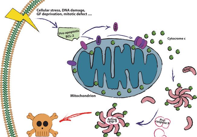
Dissertation presented at Uppsala University to be publicly examined in B42, BMC,
Husargatan 3, Uppsala, Tuesday, 21 August 2018 at 13:00 for the degree of Doctor of
Philosophy (Faculty of Medicine). The examination will be conducted in English. Faculty
examiner: Professor Bengt Hallberg (Department of Medical Biochemistry and Cell biology
at Institute of Biomedicine, University of Gothenburg).

Abstract
Pilia, G. 2018. Novel Roles of the Ack1 Kinase in Epithelial Biology. Digital Comprehensive
Summaries of Uppsala Dissertations from the Faculty of Medicine 1469. 85 pp. Uppsala: Acta
Universitatis Upsaliensis. ISBN 978-91-513-0355-0.

Epithelial homeostasis is maintained through integration of diverse signals that regulate cell
fate. A strict control of such signals is required to prevent overproliferation and, ultimately,
oncogenesis. In this thesis we identify novel roles of Activated Cdc42-associated kinase
1 (Ack1) in maintenance of epithelial homeostasis. Ack1 has been previously linked to
cytoskeletal remodeling, signal transduction and gene expression regulation. Interestingly,
our work reveals that Ack1 is also important for I) promoting extrinsic apoptosis, II)
mediating mechanically-induced inhibition of proliferation and III) attenuating mitogenic
signals, fundamental functions to prevent aberrant tissue growth.
   Apoptosis is a program of regulated cell death that can be triggered by several pathways.
Among them, the TNF-related apoptosis-inducing ligand (TRAIL)-induced apoptosis cascade
has raised interest for cancer treatment, as many cancer cell lines are susceptible to it. We found
that Ack1 increases sensitivity to TRAIL by promoting translocation of ligand-bound TRAIL-
Receptor to lipid rafts. Localisation at the lipid rafts, in turn, favors recruitment of downstream
signalling effectors, enhancing the apoptotic response.
   Yap and Taz are transcriptional co-factors that integrate mechanical and soluble cues to
regulate cell proliferation and differentiation. Yap/Taz regulation is mediated by cytoplasmic
sequestration and, particularly for Taz, proteasomal degradation via ubiquitination by the E3
ligase β-TrCP. We discovered that Ack1 is activated by mechanical signals and promotes
nuclear exclusion of Yap/Taz. Ack1 promotes Yap/Taz interaction with β-TRCP and it is
required for efficient degradation of Taz. Consequently, Ack1 limits Yap/Taz-dependent gene
expression and cell proliferation.
   The ErbB family of receptor tyrosine kinases mediates pro-survival and proliferative signals
of crucial importance in development and cancer. Among the ErbB family members, ErbB3
has significant oncogenic properties as it potently activates the PI3K/Akt signaling pathway.
We observe that Ack1 depletion increases ErbB3 total levels, but not EGFR and ErbB2, and
is required for both basal turnover of ErbB3 and its ligand-induced degradation. Consequently,
Ack1 attenuates ErbB3-dependent signalling upon Neuregulin-1β treatment. Additionally,
Ack1 reduces ErbB3 gene expression both at steady state and upon stimulation, revealing its
importance as multi-level regulator of ErbB3.
   Taken together, our data depict new roles for Ack1 in epithelial cells, highlighting its
multifaceted role in maintenance of epithelial homeostasis.

Keywords: Ack1, Epithelial homeostasis, Cell Signalling, Cancer, Apoptosis, TRAIL-R, Yap/
Taz, Mechanotransduction, ErbB3

Giulia Pilia, Department of Medical Biochemistry and Microbiology, Box 582, Uppsala
University, SE-75123 Uppsala, Sweden.

© Giulia Pilia 2018

ISSN 1651-6206
ISBN 978-91-513-0355-0
urn:nbn:se:uu:diva-349384 (http://urn.kb.se/resolve?urn=urn:nbn:se:uu:diva-349384)
To my father,

from whom I inherited the curiosity
          and the sense of humour
     needed to complete this thesis
List of Papers

This thesis is based on the following papers, which are referred to in the text
by their Roman numerals.

      I        Linderoth E.*, Pilia G.*, Mahajan N.P., Ferby I. (2013) Acti-
               vated Cdc42-associated kinase 1 (Ack1) is required for tumor ne-
               crosis factor-related apoptosis-inducing ligand (TRAIL) receptor
               recruitment to lipid rafts and induction of cell death. Journal of
               Biological Chemistry, 288(46):32922-31.

      II       Pilia G., Darai E., Ferby I. (2018) ACK1 is a mechanoresponsive
               kinase required for SCF (β-TrCP)-mediated degradation of
               YAP/TAZ. Manuscript.

      III      Pilia G.*, Sáez-Ibáñez A.R.*, Linderoth E., Ferby I. (2018)
               Ack1 is a negative regulator of ErbB3 that acts both by promot-
               ing its degradation and suppressing its expression. Manuscript.

      *Asterisks indicate equal contribution

Reprints were made with permission from the respective publishers.

The following paper, by the same author, was not included in this thesis:

          Hopkins S., Linderoth E., Hantschel O., Suarez-Henriques P., Pilia
          G., Kendrick H., Smalley M.J., Superti-Furga G., Ferby I. (2012)
          Mig6 is a sensor of EGF receptor inactivation that directly activates c-
          Abl to induce apoptosis during epithelial homeostasis. Dev Cell. 2012
          Sep 11; 23(3):547-59.
Contents
1. Introduction ............................................................................................... 13
2. Ack1 is a versatile non-receptor kinase of critical importance for
epithelial homeostasis ................................................................................... 15
2.1 The unique domain structure of Ack1 supports binding with several
interaction partners ................................................................................... 15
2.2 The activity of Ack1 is regulated by auto-inhibition.......................... 17
2.3 Ack1 is a multifunctional kinase and a signalling hub ....................... 19
2.3.1. Roles in cytoskeletal remodelling and trafficking ..................... 19
2.3.2. Roles in promoting pro-survival signals and regulation of
gene transcription ................................................................................ 20
2.3.3. Specialised functions of Ack1 in the nervous and immune
systems................................................................................................. 21
2.4 Ack1 is involved in oncogenesis and cancer progression .................. 22
3. ErbB receptors-mediated signalling is central for cell survival,
proliferation and migration ........................................................................... 24
3.1 The ErbB family of receptors is activated by asymmetric
dimerization .............................................................................................. 24
3.2 The ErbB receptors activate signalling cascades implicated in cell
survival, differentiation and migration ..................................................... 26
3.3 ErbB-dependent signalling drives development and neoplastic
transformation .......................................................................................... 28
3.4 A wide range of mechanisms modulate ErbB receptors-dependent
signalling .................................................................................................. 29
3.4.1 ErbB receptors signalling is spatiotemporally controlled via
regulated trafficking............................................................................. 30
3.4.2 ErbB signalling can be attenuated by competitive binding,
phosphatases and adaptor proteins....................................................... 31
3.5 The catalytically-impaired ErbB3 holds signalling capacity and
dedicated mechanisms of regulation ........................................................ 32
4. Mechanical regulation of Yap and Taz controls cell fate ......................... 35
4.1 Mechanical cues are crucial messengers in the regulation of
cellular functions ...................................................................................... 35
4.1.1 Cells sense mechanical signals ................................................... 36
4.2 Yap and Taz are mechano-responsive transcriptional co-factors ....... 384.2.1 Yap and Taz’s gene transcription signature directs cell fate ...... 38
4.2.2 Yap and Taz are structurally similar, yet not identical ............... 39
4.2.3 The regulatory network of Yap and Taz is dominated by
mechanotransduction ........................................................................... 40
4.2.4 Mechanical regulation of Yap and Taz governs cell fate ........... 43
5. Apoptosis: the regulated cell death program that controls tissue stability 45
5.1 Apoptosis can be triggered by both internal and external cellular
signals ....................................................................................................... 45
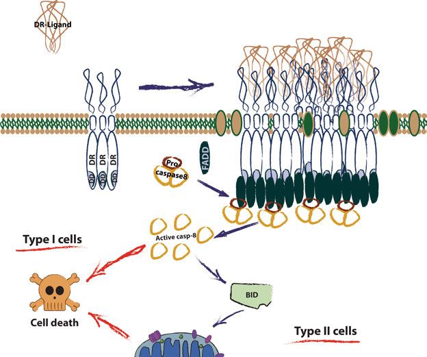
5.1.1 The intrinsic pathway of apoptosis is executed via
permeabilization of the mitochondria .................................................. 46
5.1.2 The extrinsic pathway of apoptosis is triggered by receptors at
the plasma membrane .......................................................................... 47
5.2 The regulation of TRAIL-Receptors signalling is of special interest
in cancer ................................................................................................... 50
6. Present investigations................................................................................ 52
Paper I: Activated Cdc42-associated kinase 1 (Ack1) is required for
tumour necrosis factor-related apoptosis-inducing ligand (TRAIL)
receptor recruitment to lipid rafts and induction of cell death ............. 52
Paper II: Ack1 is a mechanoresponsive kinase required for SCF (β-
TrCP)-mediated degradation of Yap/Taz ............................................ 53
Paper III: Ack1 is a negative regulator of ErbB3 that acts both by
promoting its degradation and suppressing its expression................... 54
7. Future perspectives ................................................................................... 55
Is Ack1-mediated TRAIL-R recruitment to lipid rafts an aspect of a
more general mechanism? ................................................................... 55
What are the molecular details of Ack1 regulation of Yap/Taz
downstream of mechanical stimulation?.............................................. 56
Which mechanisms underlie Ack1-dependent regulation of ErbB3? .. 56
How are Ack1 functions integrated? ................................................... 57
Acknowledgements ....................................................................................... 58
References ..................................................................................................... 63Abbreviations Ack1 Activated Cdc42-associated Kinase 1 AJ Adherens junction Akt RAC-alpha serine/threonine-protein kinase Alk Anaplastic lymphoma kinase AMOT Angiomotin aP Adipocyte fatty acid-binding protein AP-2 Adaptor protein 2 APAF Apoptotic peptidase activating factor APC Adenomatosis polyposis coli APO Apoptosis antigen AR Androgen receptor ARE Androgen responsive enhancer BAD Bcl-2-associated death promoter BAK BCL-2 homologous antagonist killer BAX BCL-2-associated X protein BCL B-cell limphoma BH BCL-2 homology BID BH3 interacting-domain death agonist BIM BCL-2 interacting mediator Cas Crk-associated substrate Casp- Caspase Cbl Casitas B-linage lymphoma proto-oncogene CD95 Cluster of differentiation 95 Cdc42 Cell division control protein 42 homolog c-Fos cellular-FBJ murine osteosarcoma viral oncogene homolog c-IAP Inhibitor of apoptosis protein CIP Contact inhibition of proliferation CK1 Casein kinase 1 CRIB Cdc42/Rack interactive binding Crk CT10 regulator of kinase DAG Diacylglycerol dATP Deoxyadenosine triphosphate Dbl Diffuse B-cell lymphoma DcR Decoy receptor DD Death domain
DISC                           Death inducing signalling complex
DNA                                         Deoxyribonucleic acid
DR                                                  Death receptor
EBD                                         EGFR binding domain
ECM                                           Extracellular matrix
EEA                                    Early endosome antigen 1
EGF                                       Epithelial growth factor
EGFR                                                EGF Receptor
EMT                          Epithelial to mesenchymal transition
ER                                              Estrogen receptor
ErbB            Erythroblastic leukemia viral oncogene homolog
ERRP                                        EGFR Related Peptide
FA                                                  Focal adhesion
FADD                    Fas-associated protein with death domain
FAK                                         Focal adhesion kinase
Fas                                 First apoptosis signal receptor
FLICE              FADD-like Interleukin-1β-converting enzyme
FLIP                                FLICE-like inhibitory protein
FOXO                                              Forkhead box O
FRS2β               Fibroblast growth factor receptor substrate 2β
GPCR                                   G-protein coupled receptor
Grb2                     Growth factor receptor-bound protein 2
GSK3                                  Glycogen synthase kinase 3
GTP                                       Guanosine tri-phosphate
H3K9                                            Histone 3 lysine 9
hnRNPU               Heterogeneous nuclear ribonuclear protein U
HOXA                                                 Homeobox A
IP3                                    Inositol 1,4,5-triphosphate
JAK                                                   Janus kinase
KDM3A                                      Lysine demethylase 3A
Lats                               Large tumour suppressor kinase
Lrig1    Leucine-rich repeats and immunoglobulin-like domains 1
LINC                    Linker of nucleoskeleton and cytoskeleton
Ltk                            Leucocyte receptor tyrosine kinase
LUBAC                    Linear ubiquitin chain assembly complex
MAPK                             Mitogen-activated protein kinase
ERK                         Extracellular signal–regulated kinase
Mdm2                             Mouse double minute 2 homolog
MHR                                        Mig6 homology region
Mig6                            Mitogen-inducible gene 6 protein
MLL2                                Myeloid lymphoid leukemia 2
MOB                                        Mps one binder protein
MOMP            Mitochondrial outer membrane permeabilization
MRTF                       Myocardin-related transcription factor
MSC                                     Mesenchymal stem cells
Mst              Mammalian Ste20-like serine/threonine kinase
mTOR                            Mammalian target of rapamycin
Nedd           Neural precursor cell expressed developmentally
               down-regulated protein
Neuregulin                                                   Nrg
NFAT5                       Nuclear factor of activated T-cells 5
NF-κB                                         Nuclear factor κ B
NGF                                          Nerve growth factor
Nrdp1                 Neuregulin receptor degradation protein 1
NSCLC                               Non-small-cell lung cancers
OPG                                             Osteoprotegerin
PDGFR                    Platelet-derived growth factor receptor
PI3K           Phosphatidylinositol-4,5-bisphosphate 3’-kinase
PIP3                    Phosphatidylinositol 3,4,5 triphosphate
PKC                                             Protein kinase C
PKD                                             Protein kinase D
PLC-γ                                         Phospholipase C-γ
PPARγ              Peroxisome proliferator-activated receptor γ
PTB                                    Phosphotyrosine binding
PUMA                    p53 upregulated modulator of apoptosis
Rac                   Ras-related C3 botulinum toxin substrate
Raf                                 Rapidly accelerated fibroma
RALT                         Receptor associated late transducer
Ras                                          Rat sarcoma protein
RCD                                          Regulated cell death
Rho                                                 Ras homolog
RING                                 Really interesting new gene
RIPK         Receptor interacting serine/threonine protein kinase
ROCK               Rho-associated coiled-coil containing kinase
RUNX                            Runt-related transcription factor
SAM                                           Sterile alpha motif
SAV1                                                   Salvador 1
SH2                                      Src homology domain 2
SH3                                      Src homology domain 3
Shc                     SH2-containing collagen-related protein
SIAH                                  Seven in absentia homolog
SLP-76       SH2-domain containing leukocyte protein of 76 kDa
SMAC          Second mitochondria derived activator of caspases
SMAD                        Sma mother against decapentaplegic
SNX9                                               Sortin nexin 9
SOCS                      Suppressors of the cytokine signalling
Sos                                             Son of sevenless
SQSTM1                                           Sequestosome 1
Src Sarcoma kinase STAT Signal transducer and activator of transcription TAD Trans-activation domain Taz Transcriptional co-activator with PDZ-binding motif TEAD TEA domain protein TGFα Transforming growth factor α TNF Tumour necrosis factor TNF-R TNF receptor TRADD TNF-R associated death domain protein TRAF TNFR associated factor TRAIL TNF-related apoptosis-inducing ligand TRAIL-R TRAIL receptor Trk Tropomyosin receptor kinase UBA Ubiquitin associated WASP Wiskott-Aldrich syndrome protein WDR5 WD repeat domain 5 WWox WW domain-containing oxidoreductase XIAP X-linked inhibitor of apoptosis Yap Yes-associated kinase ZO-2 Zonula occludens-2 β-TrCP β-Transducin repeat-containing protein
1. Introduction

The healthy functioning of a living organism requires, beside the integrity of
its parts, their ability to work together as a whole. The maintenance of tissue
homeostasis depends on the coordination of the behaviour of all the cells that
compose it. This is accomplished through a complex system of communica-
tion between cells and their environment or from cell to cell, that makes use
of signals of diverse nature. The integration of such signals can direct cell fate
towards survival, proliferation, or regulated death. How cells sense these sig-
nals and translate them into biochemical changes, a process collectively called
signal transduction, is a fascinating field of study in cell biology, with crucial
implications in both normal physiology and pathological conditions.
Persistent perturbations of the system of cell communication are indeed in-
volved in the etiology of various diseases. Cancer is a classic paradigm of the
catastrophic outcome deriving from an autonomous behaviour of cells that
evade the tissue-level control of their fate1,2. Given the fact that a large major-
ity of human malignancies originate from epithelia, the importance of ap-
proaching the study of signal transduction in epithelial cells appears compel-
ling.
    A large number of molecular events determine and sustain cancer transfor-
mation at the single-cell level. Additionally, cancer is an intrinsically hetero-
genous disease; therefore, these events vary not only in between different can-
cers, but also among the same type of cancer arising in different individuals
and in different cells in the same tumour. However, this complex view can be
simplified by conceptually framing these molecular events into functional cat-
egories, as exemplified by the now classic review of Hanahan and Weinberg
that defined them: The Hallmarks of Cancer1,2. By this classification the com-
plex series of events at the root of carcinogenesis and cancer progression are
categorised into a few groups, based on the selective advantage they confer to
the cell. The cancer properties defined in the original review comprised: eva-
sion of apoptosis, self-sufficiency in (or hypersensitivity to) growth signals,
resistance to anti-growth signals, sustained angiogenesis, limitless replicative
potential, and the ability to invade tissue and metastasize1. Later additions to
this list included deregulation of cellular energetics and the ability to avoid
immune destruction, highlighting at the same time the importance of genome
instability and tumour-promoting inflammation in enabling the occurrence of
all the aforementioned hallmarks2. It is however important to note that despite
the usefulness of this simplified view, all these cancer determinants are in

                                                                                13
strict relationship with one-another. Thus, deregulation of a single cellular process often affects more than one of them. Three of the originally presented hallmarks of cancer are of particular interest for this thesis: evasion of apop- tosis, increased sensitivity to growth signals and resistance to anti-growth sig- nals. In the work presented in this thesis we will, in fact, describe three novel and independent mechanisms by which a single protein, called Activated Cdc42-associated Kinase 1 (Ack1), prevents the emergence of these cancer traits. 14
2. Ack1 is a versatile non-receptor kinase of
critical importance for epithelial homeostasis
Ack1 is a tyrosine kinase primarily studied for its roles in remodelling of the
cytoskeleton, trafficking of receptors and regulation of gene transcription3.
The regulation of Ack1 activity downstream many different signals, and its
multifaceted roles in epithelial cells (further discussed in section 2.3), make it
a very interesting signalling hub, whose complete network of interaction is yet
to be uncovered. In the work presented in this thesis we explored novel func-
tions of Ack1, discovering an ambivalent side of this versatile kinase in the
context of epithelial biology and oncogenesis.
To set the ground for presenting our novel findings, in this chapter we will
present an overview of the current understanding of Ack1 biology.
2.1 The unique domain structure of Ack1 supports
binding with several interaction partners
Ack1 (also known as Tnk2, for tyrosine non-receptor kinase 2) belongs to the
Ack family of non-receptor tyrosine kinases. In humans, the family is formed
by two members, Ack1 and Tnk1, and it is characterized by the distinctive
presence of a SH3 (Src homology 3) domain located C-terminal to the cata-
lytic site and of a neighbouring CRIB (Cdc42/Rack interactive binding) do-
main. Additionally, Ack1 and Tnk1 both contain a SAM (Sterile Alpha Motif)
domain at the N-terminus and, at the C-terminus, a proline-rich sequence in-
cluding a clathrin-binding (CB) motif and a Ubiquitin-Associated (UBA) Do-
main. However, Ack1 is larger than Tnk1 and its 1038 amino acids-long se-
quence features a more extended proline-rich region, which includes a WW-
binding motif and a Mig6-Homology Region (MHR) (Fig. 1). The presence
of several different functional domains reflects the multiple interaction part-
ners and, consequently, physiological roles of Ack1.
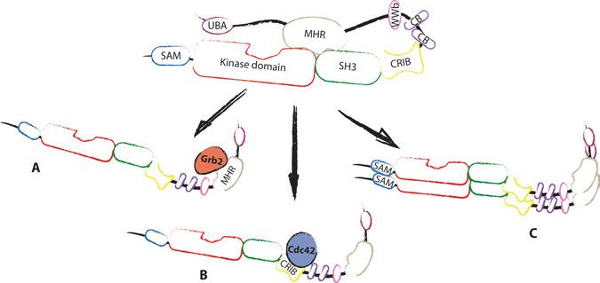
15Figure 1. Ack1 domain architecture. Representation of the main functional domains of Ack1. The yellow vertical lines indicate the position of the tyrosine auto-phosphor- ylation sites. WWb stands for WW domain binding region, see text above for other abbreviations. Walking down the Ack1 sequence from the N-terminus, the first character- ised functional motif is the SAM domain, which has been proposed to have an important role in promoting Ack1 activation. The SAM domain directs Ack1 localisation to the plasma membrane upon cell adhesion, causing an increase of its local concentration, and mediates dimerization and activation of the pro- tein4,5. In particular, a short sequence ranging from residue 10 to 14 has been found to be critical for adhesion-mediated activation6. However, it has not been clarified if its importance is dependent on the stabilisation of the alpha- 1 helix of the SAM domain or if it constitutes on its own an interacting surface crucial for homodimerization. More recently, the SAM domain of Ack1 has been proposed to mediate association with the SAM domain of another pro- tein, the SH2-domain-containing leukocyte protein of 76 kDa (SLP-76)7. The SAM domain is followed by the kinase domain (discussed in detail in section 2.2), and by a SH3 domain. The SH3 domain interacts with binding sequences in the proline-rich region, stabilising the auto-inhibited confor- mation of Ack1 (further discussed in section 2.2). Moreover, the SH3 domain mediates interaction with p130Cas to promote invasion and motility8. The CRIB domain was the earliest characterised region of Ack1. As its extended name suggests, Ack1 was first isolated as an interactor of GTP- bound Cdc429. This interaction, which engages the CRIB domain, is important for Ack1 activation and its cytoskeletal functions and it has been reported to be specific for activated Cdc42 over other GTPases such as Rac and Rho9. The second half of Ack1 encloses a large proline-rich domain that serves as a binding hub for different SH3 domain-containing binding partners, in- cluding Grb26,10, Sortin nexin 9 (SNX9)11, and cortactin12. Moreover, the pro- line-rich region comprises the MHR, two CB motifs and a WW-binding do- main. The presence of two CB domains highlights the importance of Ack1 in regulating vesicular trafficking. The two motifs are different in sequence, and mutation of both of them is required to completely abolish clathrin binding to Ack113,14. In close proximity to the CB boxes, the classical PPxY consensus sequence mediates interaction with type I WW domain-containing proteins, such as the E3 ligases Nedd 4-1 and 215,16 and the WW domain-containing oxidoreductase (WWox)17. 16
The Mig6-homology region (also known as EBD for EGFR-binding do-
main) mediates direct binding to Grb2 and EGFR. Ack1 binding to EGFR is
dependent on the kinase activity of the latter, while activation of Ack1 is dis-
pensable for the interaction18. Ack1 binding to EGFR regulates intracellular
trafficking of the receptor, the most explored and yet debated function of
Ack118–21 (discussed in section 2.4).
   Last, the UBA domain at the C-terminus confers affinity for mono- and
poly-ubiquitin chains and it is required for interaction with the receptor for
selective autophagy sequestosome 1 (p62/SQSTM1)18,21. The interaction be-
tween Ack1 and p62/SQSTM1 is suppressed by EGF stimulation and regu-
lates the sorting of EGFR-containing vesicles.

2.2 The activity of Ack1 is regulated by auto-inhibition
The crystal structure of full length Ack1 is still missing, but partial structures
have been resolved for the kinase domain, for a fragment spanning through
the kinase and SH3 domains, and for the CRIB domain in complex with
Cdc4222–24.
   The kinase domain of Ack1 displays outstanding similarities to the one of
EGFR. Not only do they share 39% sequence identity, but they also are both
stabilised in an active conformation in the absence of phosphorylation22,23.
Many kinases are relieved from auto-inhibition by means of phosphorylation
of the so-called activation loop. The activation loop of Ack1 contains one sin-
gle tyrosine at residue 284 that has been characterised as a site of auto-phos-
phorylation and as a Src substrate25,26. Although one study has reported the
importance of phosphorylation by Src for Ack1 activation26, the available
structural information and most of the experimental data suggest that the ki-
nase domain is constitutively stabilised in an active conformation. Due to the
presence of critical hydrophobic residues, the unphosphorylated activation
loop is well ordered and does not block the ATP binding pocket nor the sub-
strate recognition site22. Analogously, phosphorylation of the tyrosine in the
activation loop does not induce substantial re-organisation of the catalytic
sites, suggesting that allosteric interactions might instead be responsible for
auto-inhibition.
   The similarity of the kinase domains of EGFR and Ack1 first suggested the
possibility that an interaction in cis between the kinase domain and the MHR,
which binds the kinase domain of EGFR, could mask the active site. Experi-
mental evidence has corroborated this hypothesis by showing that the isolated
MHR interacts with the kinase domain and the association is prevented in
presence of the somatic cancer mutation E346K, which constitutively acti-
vates Ack127.

                                                                               17
A large amount of data suggests that the SH3 domain could stabilise the closed (auto-inhibited) conformation of Ack1. The MHR of Ack1 has a pro- line-rich 10 amino acids-long insertion with respect to the homologous region in Mig6 that constitutes a consensus motif for SH3 binding, and mutations in the SH3 domain enhance Ack1 activity4,6,23. The mechanism of release of the auto-inhibitory loop that leads to Ack1 activation is still debated. Self-association could be displaced by competitive binding of Grb2 to the MHR or by the conformational changes induced by binding of Cdc42 to the CRIB domain6,12,28 (Fig. 2a and b). Interestingly, three tyrosine phosphorylation sites of Ack1 recurrently detected in phosphoprote- omic studies are located in the MHR at residues Y827, Y859, Y860, suggest- ing a role of phosphorylation in maintaining the open conformation (Phos- phoSitePlus®29). Additionally, homodimerization via SAM-SAM domain in- teraction is reportedly the main mechanism of adhesion-dependent activation of Ack1 (Figure 2c)5. How exactly dimerization leads to release of the self- association loop has not been resolved in detail. However, the possibility that the formation of symmetric dimers stabilises the active conformation is sup- ported by crystallographic data23. Overall, it is possible to hypothesize that all the proposed modes of activation of Ack1 coexist in the cell and are alterna- tively mediated by different activation stimuli6. Figure 2. Ack1 activation model, according to Lin et al., 20126. Abbreviations as in Figure 1. No specific phosphatase has been described to inactivate Ack1 but both proteasomal and lysosomal degradation of active Ack1 have been re- ported15,16. Interestingly, lysosomal degradation of activated Ack1 is depend- ent on ubiquitination by Nedd4-1, which acts as a sorting signal. The main site of Ack1 ubiquitination has not been described, but deletion studies and the high concentration of lysine in the SAM domain indicate it could be the main ubiquitin acceptor. Additionally, SIAH ubiquitin ligases control the levels of 18
Ack1, promoting its proteasomal degradation independently on its activation
status30.

2.3 Ack1 is a multifunctional kinase and a signalling
hub
Ack1 is activated downstream of many different membrane receptors, such as
integrins, M3 muscarinic receptor, and a wide group of receptor tyrosine ki-
nases, namely PDGFR, EGFR, Axl and Trk4,31–33. In addition, both Ltk and
Alk have been reported to interact with Ack1 in an activation-dependent man-
ner, suggesting an additional involvement in signalling from these receptors32.
However, sensitivity to stimuli and activation kinetics have been reported to
be cell type-specific4. The vast variety of activator pathways that converge on
Ack1 is mirrored by the diversity of cellular processes in which it is involved.

2.3.1. Roles in cytoskeletal remodelling and trafficking
Ack1 has a well-established role in cytoskeletal remodelling, acting both
downstream and upstream of Cdc4228. As mentioned above, activated Cdc42
can interact with and activate Ack1. Conversely, activated Ack1 promotes ac-
tivation of Rho-GTPase family members via phosphorylation of the guanine
exchange factor Dbl28,34. Furthermore, Cdc42-dependent activation of Ack1
upon stimulation by laminin or chondroitin-sulphate proteoglycan results in
phosphorylation of p130Cas at three tyrosine residues. These phosphorylation
events promote the association of p130Cas and Crk, in turn causing the re-
modelling of the actin cytoskeleton in the context of cell migration8,35,36.
Moreover, Ack1 can promote branching of the cytoskeleton via direct phos-
phorylation of two additional substrates: WASP and cortactin. Surprisingly,
Ack1 can phosphorylate WASP at both tyrosine and serine residues, promot-
ing actin polymerisation in vitro17. This uncommon double-specificity has
been explained as the result of the high flexibility of the Ack1 kinase domain.
However, it is not clear if Ack1-dependent phosphorylation of serine residues
could happen in other contexts, as no other serine substrate has been described
to date. Upon EGF stimulation, Ack1 phosphorylates cortactin at three tyro-
sine residues (Y421, Y466, Y482) that are also substrates of many other non-
receptor tyrosine kinases12. In this context, phosphorylated cortactin triggers
cytoskeletal remodelling linked to clathrin-mediated endocytosis (CME) of
EGFR.
   The involvement of Ack1 in the CME of EGFR has been confirmed by a
significant amount of data. First, inhibition of endocytosis by dynamin deple-
tion induces accumulation of activated Ack1 in clathrin-coated pits14. Addi-
tionally, Ack1 overexpression generates clathrin aggregates, compromising

                                                                             19
clathrin-dependent trafficking13. Moreover, Ack1 has been found to co-local- ise with AP-2, the early endosome marker EEA-1 and SNX9, potentially broadening its connection to the trafficking machinery11,13,18,20. More specifi- cally, Ack1 can directly bind EGFR via its MHR, and the interaction requires the kinase activity of EGFR, but not that of Ack118,19. Conflicting conclusions have been drawn on the effect of Ack1 on EGFR trafficking, as both roles in preserving localisation of the receptor at the membrane19, and in promoting internalisation and degradation18,20 have been proposed. More recently, it has been suggested that Ack1 limits lysosomal targeting of activated EGFR by re- routing it to a non-canonical autophagosome-associated degradation path- way21. 2.3.2. Roles in promoting pro-survival signals and regulation of gene transcription Besides roles related to cytoskeleton and endosomal trafficking, Ack1 has been found to promote pro-survival signals by direct phosphorylation of dif- ferent substrates. Notably, Ack1 phosphorylates the key signalling component Akt at tyrosine 176, thereby mediating an activation mode that is alternative to the classical PI3K-dependent mechanism. The traditional Akt mode of ac- tivation includes translocation to the plasma membrane by binding to PI3K- generated phosphatidylinositol 3,4,5-triphosphate (PIP3). Once bound to the membrane, Akt can be activated by phosphorylation at serine and threonine residues by PKD1/2. In contrast, tyrosine-phosphorylated Akt can be recruited to the membrane by binding phosphatidic acid instead of PIP3, leading to its activation by phosphorylation at the same sites37. Another important substrate of Ack1 phosphorylation is the androgen re- ceptor (AR). Activation of AR by ligand binding promotes its translocation to the nucleus, activating androgen-dependent transcription. However, in the context of prostate cancer, aberrant ligand-independent activation occurs38. It was found that, in absence of androgen stimulation, Ack1 phosphorylates AR at residues Y267 and Y363 in the transactivation domain39. The phosphory- lated receptor was consequently able to localise at androgen-responsive en- hancer (ARE) regions of the DNA and promote androgen-dependent tran- scription. Interestingly, it was observed that activated Ack1 localised at AREs together with phosphorylated AR. Furthermore, it was recently found that Ack1 sustains AR expression, acting as an epigenetic writer40. Ack1 can di- rectly phosphorylate histone 4 at Y88, promoting recruitment of epigenetic modulators such as WDR5 and MLL2, which, in turn, methylate histone 3 at K4, inducing expression of the gene encoding AR. The effect of Ack1 on AR expression is reminiscent of another identified epigenetic function, played in the context of estrogen receptor (ER)-dependent transcription. In this case, however, Ack1 does not act as a direct writer of the 20
epigenetic code, but instead activates the eraser KDM3A, a histone 3 lysine 9
(H3K9) demethylase. Upon ER association, Ack1 phosphorylates KDM3A at
Y1114, activating its catalytic function to reduce H3K9 methylation, and in
turn inducing HOXA1 expression41.
   Moreover, Ack1 can directly activate gene transcription by yet another
mechanism. STAT (Signal Transducer and Activator of Transcription) 1 and
3 have been identified as Ack1 substrates. Ack1-dependent phosphorylation,
analogously to the better described JAK-dependent one, induces STATs di-
merization and translocation to the nucleus for the transcriptional activation
of their target genes42,43.
   On a different note, Ack1 has been identified as a pro-apoptotic molecule.
The most characterised mechanism in this respect is the phosphorylation of
WWox, a proapoptotic factor involved in stress-induced cell death. Ack1-de-
pendent phosphorylation, predominantly at Y287, leads to WWox ubiquitina-
tion and degradation, resulting in decreased apoptosis44,45. More recently, a
role of Ack1 in inhibiting TNF-α (Tumour Necrosis Factor α)-dependent
apoptosis has also been proposed46.

2.3.3. Specialised functions of Ack1 in the nervous and immune
systems
Ack1 is ubiquitously expressed in human tissues, with higher expression lev-
els in thymus, spleen and brain4,47. The expression of Ack1 in the nervous
system has been reported both in adult and developing tissues and in neural
cell cultures; Ack1 shows a distinct localisation in presynaptic terminals and
developing dendrites and axon47,48. Moreover, Ack1 regulates neural arborisa-
tion both by perturbing neurotrophin-dependent Trk signalling, and by neuro-
trophin-independent processes33. Additionally, Ack1 has an important role at
pre-synapses, by regulating endocytosis of the dopamine transporter49. Inter-
estingly, a missense mutation inhibiting Nedd 4-1 and -2 association and
therefore Ack1 degradation was found in a family displaying severe infantile
epilepsy50, indicating that Ack1 could be important for a correct functioning
of the nervous system.
   In the immune system, Ack1 supports the function of T-cells by phosphor-
ylating SLP-76 and promoting early signalling events downstream T-cells re-
ceptor activation7. This suggests that Ack1 may play an important role in the
appropriate unfolding of immune responses.
   As many of the known Ack1 functions have a pro-proliferative and anti-
apoptotic outcome, Ack1 has been classically considered a pro-oncogenic pro-
tein. A substantial amount of studies strengthens this view, as summarized in
the following section (2.4).

                                                                           21
2.4 Ack1 is involved in oncogenesis and cancer
progression
An amplification of the gene encoding Ack1 has been found in a variety of
cancers, including breast, ovarian, endometrial, cervical, prostate, lung, gas-
tric, head and neck tumours3,35,51–54. Additionally, over 70 somatic mutations
affecting its coding sequence were detected in exomes in patient samples
(COSMIC database55). Six missense mutations discovered in a big kinase-ori-
ented study of leukaemia samples were characterised in functional studies.
Despite their spread distribution in the sequence (R34L, R99Q, D163E,
E346K, M409I and R806Q), they all resulted in enhanced activation of Ack1
by relieving its auto-inhibited conformation27,56.
The importance of Ack1 in neoplastic development and tumour progression
has been described for several cancer subtypes.
In pancreatic cancer, Ack1 contributes to resistance to therapy by promot-
ing AR expression and ligand-independent activation39,40. Accordingly, Ack1
overexpression and Y284 phosphorylation were found to correlate with tu-
mour progression and poor prognosis35,57,58. Additionally, the Ack1/Akt acti-
vation module could be responsible for early stages of tumour formation, as
suggested by a study in which mice conditionally expressing Ack1 in the pros-
tate showed overactivated Akt and developed prostatic intraepithelial neo-
plasia37.
Phospho-Y284 Ack1 and phospho-Y176 Akt levels correlated with tumour
progression and lower survival rate in a tissue array of breast cancers37. Fur-
thermore, Ack1 overexpression in breast cancer cells enhances tumour for-
mation, metastasis at the lung and mortality in mice35.
Immunohistochemistry data also revealed Ack1 overexpression in lung ad-
enocarcinoma. Interestingly, Ack1 overexpression in tumour-surrounding tis-
sue independently correlated with poor prognosis59.
Ack1 overexpression in hepatocellular carcinoma in comparison to adja-
cent non-tumour tissue has also been detected, and correlated with invasive-
ness, relapse and, once more, lower survival rates45,60,61. Moreover, Ack1 over-
expression increased tumour size and metastatic potential in a mouse model61.
Given the array of evidence shown above, a clear role of Ack1 has been
established in oncogenesis and tumour progression. A therapeutic approach
targeting Ack1, in particular in prostate cancer, has been proposed, and there
is currently an effort to develop specific, water-soluble, Ack1 kinase inhibitor
suitable for use in the clinic40,62.
In the work presented in this thesis, however, an ambivalent role of Ack1
function in cancer emerges, which underlines the importance of this protein
for central physiological processes. Particularly, our research has uncovered
novel independent roles of Ack1 in promoting extrinsic apoptosis (Paper I)
and inhibiting mitogenic signals by transducing mechanical cues (Paper II) or
controlling growth factor signalling (Paper III).
22To highlight the significance of these findings, the next chapters will de-
scribe the cellular processes of apoptosis and mechanotransduction (especially
the Yap/Taz pathway), as well as the role of ErbB receptors in various signal-
ling pathways linked to oncogenesis and cancer progression.
233. ErbB receptors-mediated signalling is
central for cell survival, proliferation and
migration
The ability of cells to sense signals from the environment is crucial in regulat-
ing their function. The nature of these signals is extremely broad, ranging from
soluble molecules to changes of the composition of the extracellular matrix or
mechanical stimuli.
Growth factors are one important type of soluble messengers that determine
cell fate and behaviour by binding to specialised receptors at the plasma mem-
brane. The first discovery of a growth factor dates back to the 1950s, with the
identification of a protein able to promote growth of nerve fibers: the Nerve
Growth Factor or NGF63,64. The epithelial growth factor (EGF) was isolated
shortly after from mouse submaxillary gland, as a 53-amino acid polypeptide
able to promote eyelid opening and teeth eruption in the new-born pups65. Ever
since, EGF and its receptor have been extensively studied and proved to be
crucial for promoting cell growth and survival, differentiation and migration.
They cover, therefore, a central role in the regulation of development and in
cancer biology.
3.1 The ErbB family of receptors is activated by
asymmetric dimerization
The EGF receptor (EGFR, also known as ErbB-1 for Erythroblastic leukemia
viral oncogene homolog-1or HER-1 for Human EGF Receptor Kinase) be-
longs to a family of receptor tyrosine kinases that includes 3 additional mem-
bers: ErbB2, ErbB3 and ErbB4 (known, as well, as HER-2, -3 and -4, respec-
tively).
The ErbBs share a common structural arrangement that includes an extra-
cellular N-terminus, a single transmembrane segment and a C-terminal intra-
cellular region consisting of the juxtamembrane part, the kinase domain and a
terminal tail. The extracellular domain is composed of four domains, named
I, II, III and IV. I and III are homologous β-helical domains that comprise the
ligand binding site, while domain II and IV are – also homologous – cysteine-
24rich regions that interact with each other in the closed, non ligand-bound, con-
formation66,67. Binding of a ligand molecule to the receptor induces a rear-
rangement of the extracellular domains that leads to the exposure of a hairpin
loop of domain II66,68. This region constitutes the dimerization interface, which
binds the same region of a second ErbB receptor. In ErbB2, however, the in-
teraction between domain II and IV is impaired due to substitutions of critical
residues in the latter, and this receptor is therefore stabilized in an open – di-
merization prone – conformation69,70. ErbB2 is indeed the only orphan recep-
tor of the family, with no ligand known, or needed, to cause its activation.
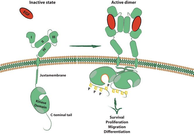
Figure 3. A model of EGFR ligand-induced homo-dimerization as an archetype of
ErbB receptors activation.
Homo- or hetero-dimerization of the ErbB receptors is necessary for their
activation via a characteristic asymmetric interaction of their kinase domains71
(Fig. 3). In the dimeric conformation, the C-lobe of the kinase domain of one
of the receptors involved (the activator) binds a specific region in the N-lobe
of the other (the receiver) via a conserved hydrophobic patch. This interaction
induces an active conformation of the receiver, culminating in trans-phosphor-
ylation of various tyrosine residues in the C-terminal tail of the activator.
Binding of the juxtamembrane segment of the receiver receptor to the homol-
ogous segment and the kinase C-lobe of the activator further stabilizes the
dimer72,73.
25Notably, ErbB3, which has a reduced kinase activity due to substitutions of key residues in the catalytic pocket, can still act as an activator in the asym- metric dimer71. However, due to the inability of ErbB3 to act as a fully active kinase, the N-lobe sequence that stabilizes the interaction with the activator has diverged compared to the other members of the family. Thus, an arrange- ment in which ErbB3 would cover the receiver position is prevented. As a consequence of the activation by dimerization of the ErbB receptor, the multiple phosphorylations of the C-terminal tails generate docking sites for adaptor proteins containing SH2 or Phosphotyrosine Binding (PTB) do- mains, whose binding initiates downstream signalling cascades. 3.2 The ErbB receptors activate signalling cascades implicated in cell survival, differentiation and migration Eleven known ligands bind, with different selectivity, the ErbB receptors74. EGF, transforming growth factor α (TGFα), amphiregulin and epigen bind ex- clusively to EGFR; while heparin-binding EGF, betacellulin and epiregulin can bind both EGFR and ErbB4. ErbB4 is activated, as well, by neuregulin (Nrg) -1 to -4, while ErbB3 binds Nrg-1 and -2. The ErbB ligands can be syn- thetized either as transmembrane proteins (prone, however, to proteolytic cleavage into soluble forms), or as shorter secreted isoforms75–77, both of which can bind to the receptors. It has been reported that, despite binding to the same receptor, different ligands have the potential to induce different ac- tivation kinetics and divergent endocytic sorting of the bound receptor78. Ad- ditionally, the signalling outcome can also be affected by cleavage of the lig- and79. To further complicate the picture, although the pathways activated by the four ErbBs largely overlap, the particular identity of the dimerization part- ners can also modulate the triggered signalling80. As previously mentioned, the phosphorylation of the C-terminal tail of ac- tivated ErbB receptors promotes binding of adaptor proteins. Among them, Shc (SH2-containing Collagen-related protein) and Grb2 are of critical im- portance in recruiting the GEF Sos (Son Of Sevenless), which in turn activates Ras81,82. Activated Ras binds to Raf and relieves its autoinhibition, initiating the MAPK/ERK (Mitogen-Activated Protein Kinase/Extracellular signal– Regulated Kinase) pathway74. The MAPK/ERK pathway culminates in phos- phorylation of transcription factors such as c-Fos (cellular-FBJ murine Oste- osarcoma viral oncogene homolog), which promotes entry into the cell cycle and induction of mitogenic genes83,84. Additionally, MAPK/ERK signalling stimulates cell migration-inducing cytoskeletal rearrangements85. Ras activa- tion can also orchestrate cytoskeletal dynamics directly, by supporting the GEF-dependent activation of Cdc4286. 26
Another important branch of the ErbB-activated pathways is initiated by
the binding and direct phosphorylation of Phospholipase C-γ (PLCγ)87. Phos-
phorylated PLCγ then catalyses the hydrolyzation of PIP2 (phosphatidylino-
sitol 4,5-bisphosphate) into two secondary messengers: Diacylglycerol
(DAG) and Inositol 1,4,5-triphosphate (IP3)88. DAG subsequently stimulates
PKC (Protein Kinase C) activity at the membrane. The activation of PKC is
further potentiated by the release of intracellular calcium stores promoted by
the binding of IP3 to dedicated receptors at the endoplasmic reticulum89. PKC,
in turn, phosphorylates a number of substrates in the cell, and the best studied
functional consequences of its activation are cytoskeletal remodelling and mi-
gration90.
    Another branch of ErbB signalling that promotes cell migration is initiated
by the direct binding of JAK to the receptors, and the consequent activation
of the STAT-dependent transcriptional program91.
    ErbB receptors, and in particular heterodimers containing ErbB3, also pro-
mote the activation of the PI3K/Akt pathway. PI3K is recruited at the cellular
membrane by direct binding of its regulatory subunit p85 to specific phos-
phorylated residues on the C-terminal tail of ErbBs92. The enzymatically ac-
tive subunit of PI3K, p110, can then phosphorylate PIP2 to PIP3, generating
a docking site for pleckstrin-homology domain-containing proteins93,94.
Among them, Akt is the most important downstream effector of PI3K. The
activation of Akt (whose mechanism has been described in section 2.3.2) re-
sults in the phosphorylation of a series of proteins involved in inhibiting apop-
tosis and activating proliferation. Akt inhibits apoptosis by phosphorylation
of the pro-apoptotic protein BAD (Bcl-2-associated death promoter) and by
promoting the nuclear translocation of Mdm2, which in turns ubiquitinates
p53 and primes it for degradation 95,96. Additionally, Akt reprograms gene ex-
pression by inhibiting the Forkhead-box transcription factor FOXO and pro-
moting degradation of the NF-κB inhibitor IκB, thus enhancing transcription
of anti-apoptotic genes97,98. Moreover, Akt induces proliferation by mediating
exclusion of the cell cycle inhibitor p27 from the nucleus, and by inhibiting
Glycogen Synthase Kinase 3 (GSK3), which controls the cell cycle by down-
regulating Cyclin D and E99,100. Altogether, the PI3K/Akt signalling pathway
promotes survival and proliferation and suppresses apoptosis, accounting for
its strong implications in cancer101.
    Collectively, the numerous signalling cascades triggered by ErbBs activa-
tion result in increased motility, decreased apoptosis and cell cycle progres-
sion, determining the pivotal roles of this family of receptors in development,
physiology and disease.

                                                                              27
3.3 ErbB-dependent signalling drives development and neoplastic transformation As previously mentioned, EGF – the founder ErbB ligand – was first discov- ered for its role in promoting eyelid opening and teeth eruption in mice65. Ever since, a large number of studies, fuelled also by the advent of genetic engi- neering, highlighted the importance of ErbB receptors in development. The deletion of either of the genes encoding ErbB2, ErbB3 and ErbB4 in mice is lethal in utero102–104. In particular, lack of ErbB2 causes abnormalities in cardiac development and defects in formation of sensory nerves derived from the neural crest102. ErbB2-/- embryos also suffer impairment in the devel- opment of motor nerves, and lack Schwann cells105. Analogously, the deletion of the ErbB4 gene results in aborted ventricular formation and generates al- tered nervous system phenotypes, in particular deficient innervation of the hindbrain104. Mice embryos lacking ErbB3 also display degeneration of sen- sory and motor neurons and lack of Schwann cells103. The outcome of EGFR knockout phenotypes, on the other hand, is depend- ent on the genetic background and results in death between mid-gestation and post-natal day 20106–109. When born alive, mice display severe phenotypes in- cluding defects in brain, skin, lungs, kidneys, liver and gastrointestinal tract. Moreover, EGFR knock-out mice also display placental defects, which cause death in utero in some strains107,108. The expression of dominant negative EGFR has additionally unveiled its role in post-natal development of the mammary gland110. In adult life, ErbB receptors have a critical pathophysiological role in a wide range of human cancers. In particular, EGFR has been found to be over- expressed or overactivated in a high percentage of lung cancers111. In non- small-cell lung cancers (NSCLC), EGFR overactivation is associated with ac- tive STAT3, ERK and PI3K/Akt pathways112,113. The most common mutation of EGFR spontaneously occurring in tumours is a deletion of exons 2 to 7, which generates a constitutively active truncated form named EGFRvIII114. Expression of EGFRvIII has been found to correlate with poor prognosis in glioblastoma, breast and lung cancers114–116. Additionally, activating muta- tions in the kinase domain have frequently been reported in NSCLCs and are important in the clinic, as they identify a subset of tumours responsive to ther- apy with the EGFR inhibitors gefitinib and erlotinib117–119. The constitutive activation of EGFR often occurs in tumours as a result of autocrine loops in- duced by overexpression of its ligands, which has been reported to occur in breast, lung, prostate and gastrointestinal tumors120–123. As the activity of ErbB2 does not depend on direct ligand binding, its over- expression alone is potentially oncogenic124,125. The tumorigenic role of the overexpression of ErbB2 has been extensively characterized in breast cancer, with an incidence of over 20% in malignant tumours, and it constitutes a widely recognized marker of poor prognosis and resistance to ER-directed 28
therapies126–128. However, its overexpression has also been detected in bladder,
NSCLC and stomach cancers129–131. Interestingly, ErbB2 and ErbB3 are often
co-overexpressed in cancer, and form a very potent mitogenic com-
plex124,132,133.
    It has been reported that the overexpression of ErbB3 alone cannot drive
tumorigenesis134. However, it becomes of critical importance when accompa-
nied with dysregulation of other members of the family or transactivation by
the receptor Met135. In particular, ErbB3 overexpression has been found in
melanoma and colorectal cancers136,137. Increased ErbB3 expression is also a
common feature in breast cancers, where it correlates with tumour size, me-
tastasis and relapse138–140. Additionally enhanced activity of ErbB3 generates
therapeutic resistance to ErbBs kinase inhibitors, chemotherapy and castration
in prostate cancers141, suggesting the importance of targeting ErbB3 to in-
crease the efficacy of anti-cancer therapy.
    The role of ErbB4 in human cancers has been less studied in comparison
to the other members of the family. However, ErbB4 is overexpressed in colon
cancer and its expression correlates with poor prognosis of breast cancer pa-
tients, regardless of ErbB2 expression status142,143. Interestingly, ErbB4-acti-
vating mutations have also been registered in NSCLC144.
    The overactivation of EGFR and other family members in various malig-
nancies has prompted the development of drugs directed to block ErbB sig-
nalling, and a number of small kinase inhibitors and monoclonal antibodies
have successfully been introduced in the clinic in the past years114,145,146. The
effectiveness of these therapeutic approaches, however somewhat hindered by
the emergence of resistance, highlights the importance of the study of ErbB
receptors and their regulation in the fight against cancer.

3.4 A wide range of mechanisms modulate ErbB
receptors-dependent signalling
Given the physiological importance of ErbB receptors, tight control of the
strength, duration and localisation of their activities is required to prevent
high-risk aberrant behaviours of this system. Due to its primary role, most of
what we know about the regulation of ErbB receptors comes from studies on
EGFR, which will therefore be the focus of this section. The specific features
of ErbB3 regulation will be instead addressed in section 3.5.

                                                                              29
3.4.1 ErbB receptors signalling is spatiotemporally controlled via
regulated trafficking
An important mechanism that prevents uncontrolled activation of ErbBs sig-
nalling is the spatial segregation of receptors and ligands at the cell mem-
brane147. In polarized epithelial cells, ErbBs are prevalently trafficked from
the trans-Golgi network or recycling endosomes to the basolateral membrane,
due to the presence of a sorting signal in the juxtamembrane domain148–150.
Their ligands, however, are primarily exposed on the apical surface147,151.
These physical constraints prevent autocrine stimulation under physiological
settings. ErbBs activation to promote restoration of integrity is however al-
lowed when the tight junctions (which preserve polarisation) are disrupted by
an injury147. Notably, perturbations of the trafficking route of newly synthe-
tized receptors to the appropriate membrane compartment causes transfor-
mation in epithelial cells151.
Many factors contribute to limiting the signalling kinetics of activated ErbB
receptors. The spatiotemporal control of ErbBs signalling is particularly tight
at the level of endocytosis and intracellular trafficking of the receptors152. In
absence of ligand stimulation, ErbBs are subjected to slow constitutive inter-
nalisation followed by rapid recycling, resulting in a prevalent membrane lo-
calisation of the receptors153,154.
The rate of ErbB receptors internalisation is increased upon ligand binding
and dimerization, which prevents high concentrations of activated receptors
to stay in the plasma membrane and cause sustained signalling. Certain clath-
rin-independent mechanisms of receptor endocytosis have been proposed, and
they seem to be particularly important in presence of high concentrations of
ligand155–157. However, the regulatory process of ErbB receptors internalisa-
tion is thought to be predominantly mediated by CME152,158,159.
Phosphorylation of EGFR has been considered for a long time to be re-
quired for localisation of EGFR in clathrin-coated pits, possibly due to the
reported dependency on the interaction with Grb2159–164. However, it has been
more recently suggested that it is the ligand-induced dimerization, rather than
kinase activity, which is required for CME165,166
Additionally, the identity of the partner of dimerization influences the per-
manence at the membrane. Overexpression of ErbB2, for example, favours
the formation of ErbB2-EGFR over homo-dimers, thereby promoting sus-
tained signalling167–169. This has been ascribed to both resistance of ErbB2 to
endocytosis and its tendency to undergo rapid recycling170.
As previously mentioned, Ack1 has been extensively implicated in the reg-
ulation of CME of EGFR. However, no clear conclusion regarding its mecha-
nism of action in this context has been drawn yet18,19,171.
Upon CME internalization, EGFR-containing clathrin-coated pits fuse with
early endosomes. Interestingly, EGFR and its ligand do not dissociate at the
mildly acidic pH of endosomes, preserving dimerization, association with
30You can also read