The Ig-like domain of Punctin/MADD-4 is the primary determinant for interaction with the ectodomain of neuroligin NLG-1
←
→
Page content transcription
If your browser does not render page correctly, please read the page content below
JBC Papers in Press. Published on September 14, 2020 as Manuscript RA120.014591
         The latest version is at https://www.jbc.org/cgi/doi/10.1074/jbc.RA120.014591
Platsaki et al., revised                                                              Main ms

The Ig-like domain of Punctin/MADD-4 is the primary determinant
for interaction with the ectodomain of neuroligin NLG-1
Semeli Platsaki1, Xin Zhou2, Bérangère Pinan-Lucarré2, Vincent Delauzun1, Haijun Tu2,5,
Pascal Mansuelle3, Patrick Fourquet4, Yves Bourne1, Jean-Louis Bessereau2, and Pascale
Marchot1*
1
  CNRS / Aix-Marseille Univ, Laboratory ‘Architecture et Fonction des Macromolécules Biologiques’
(AFMB), Marseille - France.
2
  Univ Lyon / Univ Claude Bernard Lyon 1 / CNRS / INSERM, Institut NeuroMyoGène (INMG), Lyon -
France.
3
  CNRS / Aix-Marseille Univ, Institut de Microbiologie de la Méditerranée (IMM), Plateforme Marseille
Protéomique MaP-IBiSA, Marseille – France
4
  Aix-Marseille Univ / INSERM / CNRS, Institut Paoli-Calmettes, Centre de Recherche en Cancérologie
de Marseille (CRCM), Marseille Proteomics, Marseille - France
5
    Present address: Institute of Neuroscience, Hunan University, China

*Corresponding author: Lab ‘Architecture et Fonction des Macromolécules Biologiques’ (AFMB,

                                                                                                                    Downloaded from http://www.jbc.org/ by guest on November 5, 2020
CNRS/AMU UMR-7257), Faculté des Sciences Campus Luminy - case 932, 163 av. de Luminy, 13288
Marseille CEDEX 09, France. Tel: +33 (0) 491 825 579. Email: pascale.marchot@univ-amu.fr.

Running title: Molecular bases of Punctin/MADD-4 interaction with NLG-1

Keywords: ADAMTS-like, GABA receptor, Punctin/MADD-4, muscle, neuroligin, neuromuscular junction,
nicotinic acetylcholine receptor, postsynaptic membrane, protein-protein interaction, synapse.

Abbreviations: ADAMTS(L), A disintegrin and metalloproteinase with thrombospondin type 1 repeats (-
like); ECM, extracellular matrix; GABAAR, GABA type A receptor; HS, heparan sulphate; Ig,
immunoglobulin; MDGA, MAM domain-containing GPI-anchored; NLG, neuroligin; NMJ, neuromuscular
junction; NRX, neurexin; SEC-MALS, size-exclusion chromatography coupled to multi-angle light
scattering; TSP, thrombospondin.

This article contains Supp. Figures S1–S6 and Supp. Experimental Procedures
______________________________________________________________________________________

Punctin/MADD-4, a member of the ADAMTSL                        bind NLG-1. These data identify the Ig-like
extracellular matrix protein family, was                       domain as the primary determinant for N-
identified as an anterograde synaptic organizer in             MADD-4B interaction with NLG-1 in vitro. We
the nematode Caenorhabditis elegans. At                        further demonstrate in vivo that this Ig-like
GABAergic neuromuscular junctions, the short                   domain is essential, albeit not sufficient per se,
isoform MADD-4B binds the ectodomain of                        for efficient recruitment of GABA A receptors at
neuroligin NLG-1, itself a postsynaptic organizer              GABAergic synapses in C. elegans. The
of inhibitory synapses. To identify the molecular              interaction of N-MADD-4B with NLG-1 is also
bases of their partnership, we generated                       disrupted by heparin, used as a surrogate for the
recombinant forms of the two proteins and                      extracellular matrix      component,      heparan
carried out a comprehensive biochemical and                    sulphate, and whose high-affinity binding to the
biophysical     study    of   their    interaction,            Ig-like domain may proceed from surface charge
complemented by an in vivo localisation study.                 complementarity, as suggested by homology 3D
We show that spontaneous proteolysis of MADD-                  modelling. These data point to N-MADD-4B
4B first generates a shorter N-MADD-4B form,                   processing and cell-surface proteoglycan binding
which comprises four thrombospondin (TSP) and                  as two possible mechanisms that can regulate the
one Ig-like domains and binds NLG-1. A second                  interaction between MADD-4B and NLG-1 at
processing event eliminates the C-terminal Ig-like             GABAergic synapses.
domain along with the ability of N-MADD-4B to                  _________________________________________

                                                       1
Platsaki et al., revised                                                                                     Main ms

    Synapse formation is fundamental for                          MADD-4L, is present at both cholinergic and
communication between neurons and their target                    GABAergic synapses. (19). At GABAergic
cells. The life cycle of a synapse involves attraction,           synapses, MADD-4B interacts with the ectodomain
recognition and interaction between cells,                        of NLG-1, the sole NLG in C. elegans (20-21), to
differentiation of the cell membrane into pre- and                cluster GABA type A receptors (GABAARs) (19,
postsynaptic terminals, and eventually maturation                 22). MADD-4B might also interact with NRX at
and elimination (1). Proteins involved in synaptic                GABAergic NMJs (23). Surprisingly, in the MADD-
organization fall into two categories: cell adhesion              4B knockout mutant, NLG-1 and GABAAR are
molecules acting upon contact, and secreted factors               distributed in both the cholinergic and GABAergic
acting either locally in the extracellular matrix                 synapses, indicating that MADD-4B is essential for
(ECM) as scaffolding proteins or at longer range                  retaining NLG-1 at GABAergic synapses, while
distances by diffusion (2). A cardinal pair of cell-              preventing its incorrect recruitment at cholinergic
adhesion molecules essential for excitatory                       synapses (19). Altogether, these data imply that
(glutamatergic and cholinergic) and inhibitory                    specific domains of MADD-4L and MADD-4B
(GABAergic and glycinergic) synapse function                      instruct the proper recruitment of receptors at
involves the membrane-tethered neuroligin (NLG)                   cholinergic versus GABAergic synapses. In this
and alpha- or beta-neurexin (NRXα/β) proteins (3-                 light, it was proposed that MADD-4L and MADD-
5), whose ectodomains interact trans-synaptically to              4B undergo intermolecular association, and that this
bridge the pre- and post-synaptic terminals (3, 6-8).             event inactivates the ability of MADD-4L to recruit
Besides the NRXs, the extracellular interactome of                NLG-1 and prevents incorrect GABAAR clustering

                                                                                                                          Downloaded from http://www.jbc.org/ by guest on November 5, 2020
the NLGs also includes secreted partners, such as                 at cholinergic NMJs (19, 22).
thrombospondin, whose interaction with NLG
accelerates synapse formation (9); hevin, which is                   Punctin/MADD-4 belongs to the poorly
secreted by astrocytes and bridges isoforms NLG1B                 understood ADAMTSL (A Disintegrin and
and NRXα1 at excitatory synapses (10); the MAM                    Metalloproteinase with ThromboSpondin type 1
domain-containing GPI-anchored (MDGA) proteins,                   repeats - Like) family of secreted multi-domain
which act as negative regulators of synaptic activity             glycoproteins. Members in the family comprise,
by challenging NRX binding to the NLGs (11). The                  from the N- to the C-terminal, a Cys-rich domain, a
importance of correct synapse formation for                       ‘spacer’    domain,      a   varying    number     of
cognition is highlighted by the link between                      thrombospondin type I repeats (TSPs, elsewhere
mutations in genes encoding synaptic proteins and                 also referred to as ‘TSRs’), and unique combinations
the occurrence of neurodegenerative conditions,                   of several immunoglobulin (Ig)-like domains with
justifying the emerging concept of 'synaptopathies'               most often one exemplary of protease and lacunin
(12-13). For instance, mutations in the NLG and/or                (PLAC) or netrin- or Kunitz-type domains (24, and
NRX genes have been consistently detected in                      references therein) (Fig. 1A). As such, ADAMTSL
individuals with autism spectrum disorder (ASD),                  proteins resemble, in their domain composition, the
and mutations in the NRX genes have been                          C-terminal ancillary part of ADAMTS proteins, yet
associated with schizophrenia (14). Knockdown of                  they are devoid of the N-terminal pro-peptide and
the MDGA2 gene in mice resulted in increased                      catalytic and disintegrin domains and of the protease
excitatory neurotransmission and ASD-associated                   activity that characterise ADAMTS proteins (24).
behaviour (15), while polymorphism in the MDGA1                   The importance of ADAMTS(L) proteins is
gene has been linked to schizophrenia (16-17).                    highlighted by their role in binding and remodelling
                                                                  the ECM, an active component in synapse
   Genetic screens for C. elegans mutants with                    formation. The ECM, which is rich in heparan
abnormal synaptic organization identified gene                    sulphate     (HS)     and     chondroitin    sulphate
Punctin/madd-4, whose protein products specify                    proteoglycans (25-26), promotes the structural
cholinergic versus GABAergic identity of                          consolidation of formed synapses. Conversely, ECM
neuromuscular junctions (NMJs) (reviewed in 18).                  degradation and remodelling allows synapse
In C. elegans, body-wall muscles are innervated by                plasticity and promotes neurite growth (27, 28-30).
both cholinergic and GABAergic motoneurons that                   ADAMTS(L) proteins actively remodel the ECM by
express and secrete distinct Punctin/MADD-4                       degrading      HS     and     chondroitin    sulphate
isoforms (19). The long MADD-4L (isoforms a and                   proteoglycans (26, 31), binding fibrillins and
c, which differ by the presence/absence of two                    participating in microfibril assembly/turnover (24,
neighbouring residues) is exclusively present at                  32-33) and procollagen processing (34). In
cholinergic synapses where it recruits ionotropic                 schizophrenic individuals, overexpression of
nicotinic acetylcholine receptors. The short MADD-                chondroitin sulphate proteoglycans leads to
4B (isoform b), which lacks the N-terminal third of               abnormal architecture of ECM peri-neuronal nets

                                                          2
Platsaki et al., revised Main ms
(27, 30). ADAMTS(L) dysregulation is linked to the TSP1-4 and Ig-like domains, herein referred to
ECM-related pathologies including arthritis (31), as ‘N-MADD-4B’ (Fig. 1A). N-MADD-4B contains
melanoma growth (35), bronchial epithelial a CXX(S/T)CXXG consensus motif for O-
dysplasia (36) among others. Interestingly, gene fucosylation (38, 39) at the beginning of each TSP
ADAMTSL3, one of the two human madd-4 domain, along with a NX(T/S) consensus motif for
homologs, is expressed in the central nervous system N-glycosylation at Asn362 in the Ig-like domain
and was identified as a susceptibility gene for (Fig. 1B). Structural models of the TSP1-4 and Ig-
schizophrenia (27). like domains point to an elongated, flexible TSP1-4
domain arrangement, connected to the globular Ig-
MADD-4 is the first member of the ADAMTSL like domain by an equally flexible hinge (Fig. 1B,
family to be identified as a synaptic regulator (19). C). The net positive charge of N-MADD-4B
To better understand the function of MADD-4B at (theoretical pI: 8.8) is reflected in the even
GABAergic synapses, we generated recombinant distribution of electropositive potentials at the
forms of MADD-4B and of the ectodomain of NLG- surface of the molecule, apart from the presence of
1 and carried out a comprehensive biochemical and two pockets, respectively more electropositive than
biophysical analysis of their interaction, buttressed the overall surface and highly electronegative, on
by a complementary in vivo analysis. We show that opposite faces of the Ig domain (Fig. 1C).
MADD-4B inevitable proteolysis at a well defined
site first generates a shorter entity encompassing the N-MADD-4B processing in vitro eliminates the Ig-
N-terminal four TSP and sole Ig-like C2-type like domain
Downloaded from http://www.jbc.org/ by guest on November 5, 2020
domains (herein referred to as ‘N-MADD-4B’), N-MADD-4B was produced, purified and
while further processing eliminates the Ig-like analysed using standard procedures (see
domain to generate an even shorter entity Experimental procedures) (Fig. 2). Electrophoretic
comprising the TSP domains only. Quantitative analysis of the affinity-purified protein revealed a
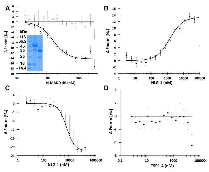
characterisation of the interaction of N-MADD-4B major entity migrating slightly above the 45 kDa
and of its maturation products with NLG-1 points to marker, along with two to three smaller entities
the Ig-like domain as the primary determinant migrating at ca. 35 kDa, 25 kDa and 15 kDa,
responsible for NLG-1 binding in vitro and for respectively (Figs. 2A and 3A). Peptide mass
efficient GABAAR recruitment at GABAergic fingerprinting analysis confirmed that all three
synapses in vivo. Distinct relative affinities of these entities belonged to N-MADD-4B, albeit with
proteins for heparin, used as a surrogate for heparan distinct levels of C-terminal coverage (Supp. Fig.
sulphate, suggest a role of cell-surface proteoglycans S1). Intact N-MADD-4B and the 35 kDa entity
as regulators of the N-MADD-4B interaction with (45.781 kDa and 31.345 kDa, respectively, by
NLG-1. We propose that N-MADD-4B maturation, MALDI-TOF-TOF mass spectrometry (MS), Supp.
resulting in elimination of the Ig-like domain, is a Fig. S2) could be separated from each other by
plausible regulation mechanism at C. elegans cation exchange (Fig. 2B) but not size-exclusion
GABAergic synapses. chromatography (Fig. 2C). Consistently, analytical
size-exclusion chromatography coupled to multi-
Results and discussion angle light scattering (SEC-MALS) pointed to two
overlapping peaks of 45.7 kDa and 33.7 kDa,
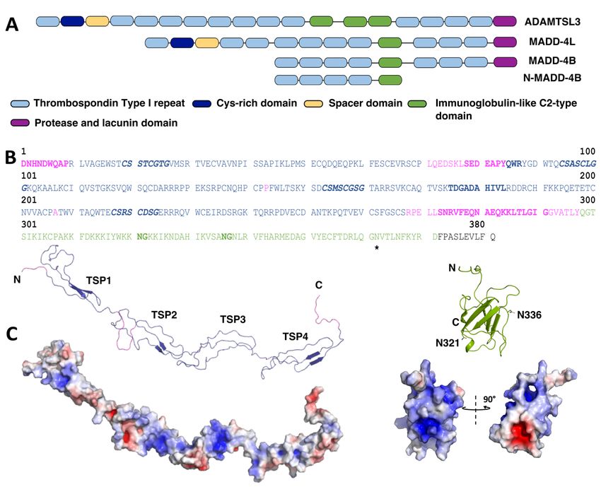
The ADAMTSL protein MADD-4B comprises, respectively (Supp. Fig. S3). The ca. 2.9 kDa
from the N- to the C-terminal, four TSP domains, difference between the theoretical mass of the naked
one Ig-like C2-type module, three additional TSP N-MADD-4B polypeptide (42.9 kDa) and the
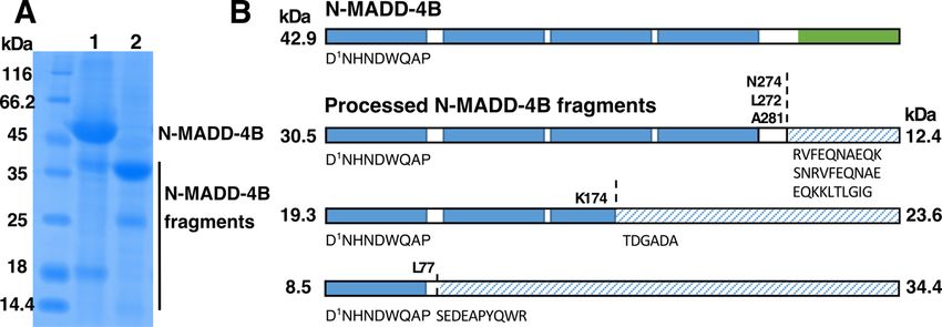
domains and a PLAC domain (Fig. 1A). Initial experimental values obtained by electrophoresis, MS
analysis of a recombinant form of MADD-4B and SEC-MALS, may reflect, e.g., presence of a N-
secreted from mammalian (HEK) cells revealed glycan linked to Asn362 and/or O-glycans at
critical cleavage of most of the protein population in consensus motifs (see Fig. 1). Over time, the
two pieces during the earliest steps of production. purified 35 kDa entity further led to a ca. 25 kDa
Western blot tracking of the C-terminal Fc-tag piece (Fig. 3A). Thorough analysis of the
coupled with N-terminal sequencing of the pieces fragmentation process pointed to a time- and
lead to locate the cleavage site upstream to sequence temperature-dependent process not prevented by
VQVSKED in the linker region that follows the Ig- protease inhibitors (Supp. Fig. S4).
like domain (data not shown). Since previous
analysis of MADD-4B as a guidance cue in vivo To identify the cleavage sites in N-MADD-4B,
demonstrated that the C-terminal TSP5-7 and PLAC samples of intact and processed N-MADD-4B were
domains were dispensable for protein functionality subjected to N-terminal sequencing (Fig. 3A, Table
(37), we generated a new construct comprising only 1). The most abundant sequence detected (ca. 90%)
3
Platsaki et al., revised                                                                                       Main ms

corresponded to the N-MADD-4B N-terminus (Fig.                   the concept of deamidation regulating protein
3B). Minor abundance sequences (≤10%) indicated                  functionality. Whether this is the case for MADD-
cleavage between the TSP1 and TSP2 domains (S78-                 4B in vivo would be worth investigating.
R87), within the TSP3 domain (sequence T175-L184)
and between the TSP4 and Ig-like domains (S273-                  The Ig-like domain of N-MADD-4B is essential for
E282, R275-K284 and E282-G291). The entity cleaved               the interaction with NLG-1 in vitro
between TSP1 and TSP2 corresponds to the most                        The ability of N-MADD-4B and N-MADD-4BΔIg
abundant fragment of ca. 25 kDa (Fig. 3A, lane 2).               to interact with the ectodomain of NLG-1 was
A similar fragment was generated upon spontaneous                assayed using microscale thermophoresis (MST),
processing of a recombinant ‘TSP1-4’ protein (see                along with data fitting to binding equations
below, and Supp Fig. S4). In the absence of further              considering either the law of mass action
processing, cleavage before the T175-L184 sequence               (calculation of the equilibrium dissociation constant,
would generate N- and C-terminal moieties of ca. 19              Kd) or the Hill model (calculation of the half
kDa and 23 kDa, respectively, while cleavage before              maximal effective concentration, EC50) (Fig. 4,
or within the S273-E282 sequence would generate N-               Table 2). Direct binding of N-MADD-4B to labelled
and C-terminal moieties of ca. 31 kDa and 10 kDa,                NLG-1 occurred with comparable Kd or EC50
respectively. The two N-terminal moieties were                   values in the 0.5 µM range, while N-MADD-4BΔIg
occasionally detected by electrophoresis (Figs. 2A,              generated no binding signal (Fig. 4A), suggesting
3A, 4A), while the C-terminal moieties were                      that the Ig-like domain is the primary determinant
detected neither by N-terminal sequencing nor by                 for N-MADD-4B interaction with NLG-1. Full

                                                                                                                            Downloaded from http://www.jbc.org/ by guest on November 5, 2020
electrophoresis. These data suggest that N-MADD-                 competition between labelled and unlabelled NLG-1
4B processing readily eliminates the C-terminal Ig-              for binding to N-MADD-4B led to Kd and EC50
like domain to generate a ‘N-MADD-4BΔIg’ moiety                  values again comparable to each other, and less than
comprising only the TSP1-4 domains.                              3-fold higher than those obtained for direct binding
                                                                 (i.e., corresponding to only minimal differences in
    Inability to prevent N-MADD-4B processing                    free energy requirements for binding), thereby
using protease inhibitors led us to suspect a                    confirming the binding specificity (Fig. 4B). If the
maturation mechanism distinct from accidental                    Hill coefficient is fixed at the value of 1 (for a 1:1
proteolysis. Spontaneous cleavage at physiological               binding stoichiometry), the Hill model reverts to the
pH was first reported for cytochrome c (40) and                  Kd model. However, except for binding reactions of
rabbit muscle aldolase (41) and proposed to act as a             higher order stoichiometry, the Hill model may also
'molecular clock', i.e., a natural mechanism                     apply to binding events involving multiple steps
regulating protein activity (42). Spontaneous                    (46). Here, the multi-domain organization of N-
cleavage occurs often, but not exclusively, at                   MADD-4B may trigger primary and secondary
Asn/Asp-Gly dipeptide sites, through non-enzymatic               binding events accounting for the slightly better
deamidation of Asn or dehydration of Asp, leading                statistics obtained upon data fitting to the Hill
to intermediates prone to further hydrolysis (43).               model.
The ability of a protein to undergo a deamidation
reaction depends on the conformation and/or solvent                  Finally, to preclude possible bias associated with
exposure of the Asn-Gly doublet (42-43). The Ig-                 the use of processed proteins, we generated
like domain of N-MADD-4B contains two such                       recombinant forms of the TSP1-4 and Ig-like
dipeptides (N 321G, N336G), likely located in surface            domains and explored their respective interaction
loop regions (Fig. 1B). However, Ala substitution of             with NLG-1. NLG-1 was found to bind the labelled
these two Asn residues did not prevent the loss of               Ig-like domain with Kd/EC50 values in the 6 µM
the Ig-like domain (Supp. Fig. S5). Asn/Asp-X                    range (Fig. 4C, Table 2), i.e., an affinity lower, by
dipeptides comprising a second residue other than                one order of magnitude, than that for the intact N-
Gly (Ala>Ser>Leu>Val>Ile in order of reactivity)                 MADD-4B, while no binding of TSP1-4 to labelled
can also undergo degradation (43). Such doublets                 NLG-1 was observed (Fig. 4D). These data confirm
are present throughout the N-MADD-4B sequence                    the primary requirement of the Ig-like domain for N-
(D74S, D124A, D176G, N201V, D222S, D250A, N280A), of             MADD-4B interaction with NLG-1, and support
which D74S and D176G are near or within low                      secondary involvement of at least one of the TSP1-4
abundance N-terminal sequences (S78-R87 and T175-                modules, recruited upon binding proximity when
L184, respectively) (Fig. 3, Table 1). The implication           tethered to the Ig-like domain through the flexible
of deamidation in physiological responses in vivo,               intervening linker (see Fig. 1), albeit not able to bind
including signal transduction during DNA-damage                  NLG-1 by its/their own.
induced cell apoptosis (44) and in the enhanced cell-
adhesion properties of ECM proteins (45), supports

                                                         4
Platsaki et al., revised                                                                                      Main ms

    MADD-4 was shown to be essential in muscle                    absence/presence of heparin, confirmed heparin-
arm guidance during the process of NMJ formation                  driven destabilisation of the N-MADD-4B
in C. elegans, where it acts as a molecular cue, while            interaction with NLG-1 (Fig. 5B). To preclude
initial analysis of MADD-4B suggested that its                    possible bias from molecular heterogeneity of
TSP1 and Ig-like domains are both crucial for                     commercial heparin, we repeated this experiment
protein functionality (37). It was also suggested that            using highly purified samples of decameric heparin
the Ig-like domain alone is sufficient for MADD-4B                and HS: the same results were obtained (data not
interaction with NRX, while domains TSP3-4 may                    shown).
contribute some degree of synergy (23). Consistent
with these reports is the radical effect of the loss of               The relative affinities of N-MADD-4B
the Ig-like domain on N-MADD-4B interaction with                  (processed sample containing both the intact protein
NLG-1 in vitro. In addition, the reduced affinity of              and N-MADD-4BΔIg moiety), TSP14 and NLG-1 for
the isolated Ig-like domain for NLG-1, relative to                heparin where assessed by affinity chromatography
that of N-MADD-4B, suggests that one or more of                   with conductivity recording and by SDS-PAGE
the TSP1-4 domains may reinforce the primary                      (Fig. 5C). All proteins were retained on immobilised
binding of the Ig-like domain. Intramolecular                     heparin, yet elution of N-MADD-4B required a ca.
synergy has been reported previously within the                   3-fold higher ionic strength (900 mM NaCl, 81.9
ADAMTS proteins, where the catalytic domain                       mS/cm) than elution of N-MADD-4BΔIg, TSP1-4 or
alone is often not fully active and needs the                     NLG-1 (elution at ca. 22 mS/cm, i.e., 340 mM
disintegrin domain for substrate binding, while the               NaCl). A similar analysis of the interaction of two

                                                                                                                           Downloaded from http://www.jbc.org/ by guest on November 5, 2020
ancillary domains define substrate specificity (47-               variants of the Hedgehog morphogen (a key
48). In turn, NLG complexes with demonstrated or                  mediator of embryonic development) with
proposed synergistic ligand binding are exemplified               immobilised heparin correlated elution at 37 mS/cm
by those formed by mammalian NLGs with MDGA                       and 52 mS/cm conductivity with Kd values of 6.8
or NRXα, respectively. Indeed, distinct Ig-like                   µM and 0.8 µM, respectively, as measured by
domains of MDGA were shown to bind distinct sites                 surface     plasmon     resonance      (54).    Linear
at the surface of the NLG dimer, thereby clamping                 extrapolation from these two sets of data led to
the two subunits together (11, 49-50). Differently,               estimated Kd values of ca. 11 nM and 58 µM for
overlaying the LNS6 domain in the L-shaped, long                  heparin binding by N-MADD-4B and by the other
NRXα molecule with that forming the short NRXβ                    three proteins, respectively. To confirm the
molecule as bound to NLG pointed to the LNS3                      significant (104-fold) difference in estimated
and/or LNS4 domains in NRXα as possible                           affinities, we also loaded the recombinant Ig-like
secondary binders reinforcing, on the same NLG                    domain onto the same heparin column, albeit using a
subunit, the primary interaction of domain LNS6                   high salt starting buffer to compensate its lower
common to the two NRX forms (51-52).                              solubility (Fig. 5D). Not only did the Ig-like domain
                                                                  fully bind to heparin, despite its loading at an ionic
Heparin interacts with NLG-1, N-MADD-4B and                       strength higher than those leading to N-MADD-
TSP1-4 with distinct relative affinities                          4BΔIg , TSP1-4 and NLG-1 elution from the column,
    Proteoglycans are active components of the                    but also its elution required as high a ionic strength
ECM. The negatively charged HS chains of HS                       as did elution of intact N-MADD-4B. Hence,
proteoglycans regulate the interactions between                   heparin-driven destabilisation of the N-MADD-4B
synaptic components, such as the mammalian NRX                    interaction with NLG-1 proceeds from specific, high
proteins and their NLG or Leucine-Rich Repeat                     affinity binding of heparin to the Ig-like domain of
TransMembrane (LRRTM) partners (53). To explore                   N-MADD-4B.
the potential effect of HS on the N-MADD-4B
interaction with NLG-1, we used heparin as a                          ADAMTS proteins were shown to degrade the
surrogate for HS, along with complementary MST                    protein core of sulphated proteoglycans such as
and affinity chromatography approaches (Fig. 5,                   aggrecan and versican (25-26). The critical
Table 3). Upon direct binding in the presence of 250              contribution of the negatively charged sulphated
µM heparin, the affinity of intact N-MADD-4B for                  glycan chains to this phenomenon suggested a
labelled NLG-1 was found to be reduced by ca. 4-                  primarily electrostatic interaction (48). While the
fold, indicating heparin binding to either N-MADD-                ADAMTSL proteins are devoid of the catalytic
4B or NLG-1, or both (Fig. 5A). Full competition                  domain and activity found in their ADAMTS
between heparin binding and formation of the N-                   relatives, they share some of the ADAMTS ancillary
MADD-4B/labelled NLG-1 complex, with an EC50                      domains (TSP, Ig, PLAC) (Fig. 1A). Our structural
value intermediate to those determined for formation              model of the Ig-like domain evidences a highly
of the N-MADD-4B/labelled NLG-1 complex in                        electropositive surface pocket (Fig. 1C) of a suitable

                                                          5
Platsaki et al., revised Main ms
size for lodging heparin with high affinity. In vivo, recruitment at GABAergic synapses. Autocatalytic
HS or chondroitin sulphate proteoglycans are processing of the ADAMTS2 or ADAMTS4 C-
thought to generate signalling gradients guiding terminus has been reported to generate cleavage
protein interactions and/or secreted protein release, products thought to regulate catalytic activity (34,
as is the case for the HS-dependent interaction 56). Similarity of ADAMTSL proteins to the
between the secreted glycoprotein, Wnt, and its ancillary domains of ADAMTS suggested that these
partner, Frizzled (55). Binding of ADAMTS4/5 to domains may act as regulators of ADAMTS activity
syndecan has been proposed to trigger signalling (57). Papillin, an ADAMTSL relative, was shown to
events through activation of the MAP kinase act in vitro as a non-competitive inhibitor of
pathway (28). Whether, and how, HS proteoglycans ADAMTS2 (58). At the C. elegans NMJ, MADD-
regulate the N-MADD-4B interaction with NLG-1 4B processing could either be controlled by a matrix
in C. elegans remains to be investigated. metalloprotease (potentially an ADAMTS family
member) or be an auto-processing event. Taking a
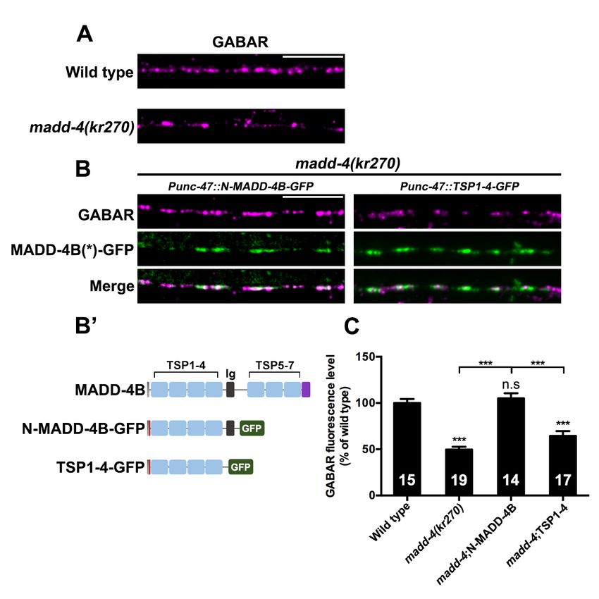
The Ig-like domain of N-MADD-4B is necessary step back, one may wonder whether the MADD-4B
but not sufficient for rescuing GABAAR clustering primary cleavage event identified from the
in C. elegans madd-4 mutants conditioned culture medium reflects a regulatory
To investigate in vivo the functional relevance of mechanism in vivo, aimed at generating not only N-
the interaction between the Ig-like domain and MADD-4B as described herein, but also a
NLG-1 observed in vitro, we expressed GFP-fused complementary ‘C-MADD-4B’ moiety, comprising
N-MADD-4B or TSP1-4 in madd-4 null mutants the three TSP and one PLAC domains C-terminal to
Downloaded from http://www.jbc.org/ by guest on November 5, 2020
expressing RFP-tagged GABAAR from the MADD-4 (Fig. 1A). While N-MADD-4B acts at the
CRISPR/Cas9-engineered locus unc-49::rfp(kr296) inhibitory, GABAergic synapse, C-MADD-4B could
(Fig. 6A, B’). Compared to wild-type animals, in migrate to excitatory, cholinergic NMJs, where its
madd-4 null mutants, synaptic expression of interaction with MADD-4L would prevent improper
GABAAR was found to be reduced by 50% (Fig. 6 GABAAR recruitment, as previously proposed for
A, C). Expression of N-MADD-4B-GFP in intact MADD-4B (19). Whether the C-MADD-4B
GABAergic neurons rescued GABAAR expression moiety could prevent the Ig-like domain from
to wild-type levels (Fig. 6B, C). Moreover, N- interacting with NLG-1 in the MADD-4L context
MADD-4B-GFP was found to colocalize with UNC- remains to be assayed. Finally, from an evolutionary
49, a feature further supporting the hypothesis that perspective it must be pointed out that the Ig-like
N-MADD-4B alone can promote GABAAR domain in MADD-4 resembles more, in length and
clustering. In contrast, expression of TSP1-4-GFP sequence, the first of the three Ig-like domains in
did not rescue GABAAR expression nor promoted human ADAMTSL3 (36% identity, versus 23 and
GABAAR clustering (Fig. 6B, C). These data point 26% for the other two domains - BLASTn search,
to a critical role of the Ig-like domain for MADD-4 data not shown) (Fig. 1A). It would be worth
function at GABAergic synapses in vivo. investigating whether this domain plays a central
role for ADAMTSL3 function, as does the Ig-like
To further explore the role of this domain in vivo, domain for MADD-4B function.
we expressed a GFP-fused Ig-like domain in the
madd-4 null mutants and assessed its ability to Conclusions
rescue the madd-4 null phenotype (Supp. Fig. S6). Previous studies identified the short MADD-4B
The Ig-GFP fusion protein did not sharply localize isoform as a key molecule for the organization of
to GABAergic synapses, but instead, made enlarged GABAergic synapses (18). How MADD-4B activity
punctae in the nerve cord region. Moreover, the Ig- is regulated remains, however, elusive. The current
GFP fusion protein did not rescue GABAAR study evidences a critical role of the Ig-like domain
expression defects, indicating that the Ig-like domain contained in the central part of MADD-4B, both for
alone is not sufficient for GABAAR recruitment. in vitro binding to NLG-1 and for in vivo clustering
Hence, in vivo, synergistic interaction of the Ig-like of GABAAR, and points to two novel regulatory
domain and one or more TSP domains with NLG-1 mechanisms of MADD-4B activity. First, we show
is likely required for ensuring N-MADD-4B that the Ig-like domain is the primary determinant
synaptogenic function. for N-MADD-4B binding to heparin, whose
competition with NLG-1 binding suggests a
Our results, in demonstrating the essential role of regulatory role by cell-surface proteoglycans at the
the Ig-like domain of MADD-4B for NLG-1 binding synapse. Second, we identified an unexpected self-
in vitro and in vivo, raise the question of whether the maturation process of N-MADD-4B in vitro, which
shedding of the Ig-like domain could be a yet sheds the Ig-like domain. An increasing number of
undescribed mechanism for regulation of GABAAR studies report the shedding of synaptic protein
6
Platsaki et al., revised                                                                                  Main ms

domains by various domains in vivo. Whether self-                 All liquid chromatography was performed at
processing is also used in vivo to regulate the               room temperature using an ÄKTA Purifier apparatus
activity of synaptic determinants is an appealing             (GE Healthcare). Maintaining protease inhibitors
hypothesis.                                                   throughout all purification steps was not
                                                              manageable, while their presence was reported not
Experimental procedures                                       to prevent proteolysis of ADAMTSL proteins (32).
                                                              Instead, we tested their impact on the kinetics of
Production of recombinant proteins                            proteolysis of purified intact N-MADD-4B at three
    The C. elegans genes and construct boundaries             temperatures (see below). Conditioned culture
used for recombinant protein production are as                medium (500 ml) containing the secreted, Fc-fused
follows: (i) the ectodomain of NLG-1 (residues                NLG-1, N-MADD-4B or TSP1-4 protein was
Tyr18-Glu607; Uniprot C40C9.5); (ii) a truncated              harvested, dialysed against sodium phosphate 100
version of MADD-4B encompassing the TSP1-4                    mM, pH 7.4, NaCl 200 mM (8L, 4°C, overnight),
repeats and subsequent Ig-like C2-type domain (’N-            then loaded on a Protein-A affinity column (5 ml;
MADD-4B’) (residues Asp369-Ser743 of MADD-4                   GE Healthcare). The Fc fragment was removed by
isoform a, Uniprot F53B6.2); (iii) the TSP1-4                 in-column digestion by GST-tagged 3C-Protease
repeats of N-MADD-4B (‘TSP1-4’) (residues                     (800 µl at 0.5 mg/ml, overnight, 4°C), and the
Asp369-Phe645 of MADD-4 isoform a); (iv) the Ig-              protein of interest was eluted from the Protein-A
like C2-type domain of N-MADD-4B (‘Ig-like’)                  column through a GSTrap column (1 ml, GE
(residues Glu646-Ser743 of MADD-4 isoform a).                 Healthcare) in the same buffer. For N-MADD-4B

                                                                                                                       Downloaded from http://www.jbc.org/ by guest on November 5, 2020
                                                              only, the fractions eluted from the Protein-A/GST-
   The NLG-1, N-MADD-4B and TSP1-4 encoding                   Trap columns were diluted 10-fold in Hepes 20 mM,
sequences were N-terminally fused to sequences for            pH 7.5, loaded on a MonoS column (1 ml, GE
a signal peptide ensuring protein secretion from              Healthcare) and eluted using a linear gradient of
mammalian cells (secreted human placental alkaline            NaCl (0 to 1 M over 20 ml). For all three proteins,
phosphatase peptide for NLG-1; mouse IgK peptide              the fractions of interest were concentrated by
for N-MADD-4B and TSP1-4), C-terminally                       ultrafiltration (10 kDa cut-off membrane, Amicon,
prolonged by sequences encoding a 3C-Protease                 Merck-Millipore) and further purified by size-
cleavage site and a human Fc domain, and inserted             exclusion chromatography on Superdex-200 (16/60
into a pYD7 vector (National Research Council                 column) or Superdex-75 (10/300 column) in Hepes
Canada, Biotechnology Research Institute) using In-           20 mM, pH 7.5, NaCl 200 mM.
Fusion cloning (Takara-Bio). The constructs were
transfected into adherent HEK293-EBNA cells (59)                 Bacterial pellets containing the Ig-like domain
using polyethylenimine (Polysciences) and stable              were resuspended in Hepes 20 mM, pH 7.5, NaCl
cell lines were generated using blasticidine (20-50           500 mM (buffer A), supplemented with DTT 1 mM,
µg/ml) for selection pressure. The proteins were              DNase 10 µg/ml, MgCl2 2 mM and a cocktail of
routinely produced from 500 ml of stable cell lines           protease inhibitors (Roche). The soluble fraction of
cultured at 37°C in Dulbecco's Modified Eagle                 the lysate was loaded on Ni Sepharose 6 Fast Flow
Medium (DMEM) supplemented with foetal bovine                 resin (5 ml in batch, GE Healthcare), which was then
serum (FBS) 10% (v/v) and blasticidine 5 µg/ml.               washed with buffer A supplemented with NaCl up to
Proteins were then expressed over 5-8 days in                 1 M and imidazole 30 mM. The protein was eluted
DMEM supplemented with FBS 2% (v/v) and                       in buffer A supplemented with DTT 1 mM and
valproic acid 1.25 mM, at 37°C for NLG-1 and at               imidazole 300 mM, and dialysed against buffer A
30°C for N-MADD-4B and TSP1-4.                                (4°C, overnight). Finally, the protein was
                                                              concentrated (3 kDa cut-off membrane, Amicon,
   The Ig-like encoding sequence was N-terminally             Merck-Millipore), and further purified by size-
fused to sequences for a 6xHis tag followed by a              exclusion chromatography on Superdex-75 (10/300
thrombin cleavage site, and cloned into a pET28a              column) in the dialysis buffer.
vector (Twist Bioscience) suitable for bacterial
expression. The construct was transformed into                   Proteins were analysed by SDS-PAGE under
Escherichia coli (BL21_DE3) and cells were grown              denaturing and reducing conditions (loading buffer
at 37°C. Protein expression was induced at OD600              containing SDS 4% (w/v) and DTT 500 mM) using
0.2-0.6 using IPTG 0.5 mM and allowed to proceed              15% (NLG-1, N-MADD-4B, TSP1-4) or 18% gels
overnight at 17°C.                                            (Ig-like) stained with Instant Blue (Expedeon). In all
                                                              cases, protein identity and intact mass were verified
Protein purification                                          by MS procedures (see below). Purified proteins
                                                              were quantified by absorbance on a NanoDrop

                                                      7
Platsaki et al., revised                                                                                         Main ms

(Thermo Scientific) or UV-vis spectrophotometer                        Samples of intact N-MADD-4B or its processing
(Carry Eclipse), using the theoretical molar                       products, either in the liquid state or transferred from
extinction coefficients, ε at 280 nm, calculated using             SDS-PAGE gels to PVDF membranes, were
the ProtParam tool in Expasy (NLG-1: 82.65 mM-1                    subjected to N-terminal sequencing by automated
cm-1; N-MADD-4B: 67.06 mM-1 cm-1; TSP1-4:                          Edman degradation. For liquid samples, 500 pmol of
55.47 mM-1 cm-1; Ig-like: 12.55 mM-1 cm-1). They                   protein were diluted to 100 µl in TFA 0.1% (v/v),
were stored at 4°C or -20°C. For the time-course                   pH 2.0, and loaded on a ProSorb membrane pre-
analysis of N-MADD-4B processing, the protein (ca.                 treated with 10 µl methanol. The membrane was
0.4 mg/ml) was maintained at -20°C, 4°C or room                    washed with TFA 0.1% (3 x 100 µl) to fix and desalt
temperature in the absence or presence of an EDTA-                 the protein sample, and then loaded on a Shimadzu
free    protease     inhibitor     cocktail   (Roche)              PPSQ 31B sequencer. Samples on PVDF
supplemented with EDTA 2 mM, according to the                      membranes were stained with Ponceau red, then the
manufacturer's instructions. Aliquots were removed                 membrane was washed three times with ethanol
at selected time points, denatured and reduced as                  90%, dried and loaded on the sequencer as above
above described, and stored at 4°C until SDS-PAGE                  described. Seven to ten cycles were performed. N-
analysis.                                                          MADD-4B was found to be preceded by an extra
                                                                   Asp residue arising from the cloning procedure (see
Heparin-affinity chromatography                                    Supp. Fig. S1).
    Chromatography was performed using an ÄKTA
Purifier apparatus equipped with an on-line                        Mass spectrometry (MS)

                                                                                                                              Downloaded from http://www.jbc.org/ by guest on November 5, 2020
conductimeter. Stock NLG-1, N-MADD-4B and                             Protein identity in the SDS-PAGE bands was
TSP1-4 proteins, diluted to 0.8 mg/ml in Hepes 20                  confirmed by peptide mass fingerprinting using
mM, pH 7.5, to lower the NaCl concentration to less                Orbitrap LC-MS/MS while the monoisotopic masses
than 50 mM, were loaded on a heparin column (5ml,                  of intact N-MADD-4 and its processing fragments
GE Healthcare) pre-equilibrated in and then washed                 were determined by MALDI-TOF-TOF MS, as
with the same buffer, and eluted using a linear                    described in the Supporting Information.
gradient of NaCl (0 to 1.0 M over 40 ml). Stock Ig-
like protein (0.8 mg/ml) in buffer A (see above) was               Microscale thermophoresis (MST)
loaded on the column, pre-equilibrated in and then                    Recombinant NLG-1, N-MADD-4B and TSP1-4
washed with the same buffer, and eluted with a                     proteins were fluorescently labelled on Lys residues
linear gradient of NaCl (500 mM to 1.0 M over 45                   using Alexa647 fluor (Thermo-Fischer) and NHS
ml). Fractions of interest were analysed by SDS-                   ester conjugation according to the manufacturer's
PAGE (see above).                                                  instructions. Briefly, the protein at 20 µM was
                                                                   incubated with a 2-3-fold molar excess of dye (1 h,
Size-exclusion chromatography coupled to multi-                    room temperature) and then loaded on a desalting
angle light scattering (SEC-MALS)                                  column (NAP-5, GE Healthcare) to remove excess
    The SEC-MALS-RI-UV setup consisted of an                       dye. Labelling efficiency was assessed by a protein-
Ultimate3000       HPLC        apparatus      (including           to-dye molar ratio of ca. 2:1, as measured at 280 nm
quaternary pump, autosampler, and UV-VIS variable                  and 650 nm, respectively. Differently, the Ig-like
wavelength (diode array) detector) (Thermo                         domain at 10 µM was labelled on the N-terminal
Scientific) in line with a DAWN8 multi-angle laser                 6xHis tag using the Monolith His-Tag labelling kit
light scattering detector calibrated with titrated BSA,            RED-tris-NTA 2nd generation (NanoTemper
and an Optilab relative refractive interferometer                  Technologies) and the manufacturer's instructions (5
(Wyatt Technology, Santa              Barbara, CA).                µM, 1 h incubation at room temperature), given for
Chromatography was performed at 20°C on                            labelling half of the protein population.
Superdex-200 (10/300 column, GE Healthcare)
using Hepes 20 mM, pH 7.5, NaCl 200 mM, a flow                         Labelled NLG-1 at 35 nM was incubated with
rate of 0.6 ml/min, and 30 µl of protein at 2 mg/ml.               unlabelled N-MADD-4B or N-MADD-4BΔIg in the
UV absorbance of the eluents was recorded at 280                   73 nM to 12.5 µM concentration range or with
nm. The Agilent software was used to control the                   unlabelled TSP1-4 in the broader 0.8 nM to 25 µM
HPLC, and the Wyatt Astra V software was used for                  range, in the absence or presence of 250 µM heparin
data collection and analysis. Peak alignment and                   sodium (Euromedex) or HPLC-purified decameric
band broadening correction between the UV,                         heparin or HS (a gift from Hugues Lortat-Jacob,
MALS, and RI detectors were performed using                        IBS, Grenoble) (1.5 h, room temperature). Samples
Astra software algorithms.                                         were briefly centrifuged and loaded into standard-
                                                                   treated capillaries (NanoTemper Technologies).
N-terminal (Edman) sequencing                                      Measurements were performed at 24°C in Hepes 20

                                                           8
Platsaki et al., revised                                                                                   Main ms

mM, pH 7.5, NaCl 200 mM, supplemented with
CaCl2 2 mM, Tween-20 0.1% (v/v) and BSA 0.2%                    Plasmids and germline transformation
(w/v), using the Monolith NT.115 apparatus                         The control strain used in this study was EN296
(NanoTemper Technologies) with 40% led power                    unc-49(kr296::tagRFP) (62). Plasmids were
and 80% IR-laser power. At least three independent              pXZ066           [Punc-47::madd-4(TSP1-4)-GFP],
experiments were performed, while each series of                pXZ067 [Punc-47::madd-4(TSP1-4+Ig)-GFP] and
capillaries was read twice at 20 min interval. To               pXZ111 [pmyo-3::madd-4(Ig)-GFP]. Transgenes
record the Ig-like interaction with NLG-1 with                  were created by conventional microinjection of
optimal thermophoretic signal amplitude, we                     plasmids at 15 ng/µl in the gonad of EN3218 madd-
labelled the smaller Ig-like protein and incubated it           4(kr270); unc-49(kr296::tagRFP) animals, and
at 150 nM with unlabelled NLG-1 in the 4.5 nM to                named krEx1319 and krEx1320 for [Punc-47::madd-
149 µM concentration range. Here, premium                       4(TSP1-4)-GFP] and krEx1345 for [pmyo-3::madd-
capillaries, 95% led power and 40% IR-laser power               4(Ig)-GFP]. A single copy insertion allele was
were used for data recording. Competition                       created using the miniMos technique for pXZ067
experiments were performed using a mix of labelled              krSi93[miniMos_Punc-47::madd-4(TSP1-4+Ig)-
NLG-1 (35 nM) and unlabelled N-MADD-4B (1.25                    GFP] (63).
µM) along with either unlabelled NLG-1 (7.6 nM to
125 µM) or heparin (4.6 nM to 150 µM) as a                      Spinning disk microscopy imaging and
competitor, and the same experimental conditions as             quantification
for the N-MADD-4B interaction with NLG-1.                           Young adult hermaphrodites (24h post L4 larva

                                                                                                                        Downloaded from http://www.jbc.org/ by guest on November 5, 2020
    MST data were analysed with the NanoTemper                  stage) were used for imaging. Live worms were
MO.Affinity Analysis software v2.2.4. Binding and               mounted on 2% agarose dry pads with 1%
competition curves were fitted to the data points               polylysine beads in M9 buffer. Fluorescence images
using either the Law of mass action (Kd model, with             were captured using an Andor spinning disk system
calculation of the equilibrium dissociation constant,           installed on a Nikon-IX86 microscope (Olympus,
Kd) or the Hill model (calculation of the half-                 Japan) equipped with a 60/1.2 oil immersion
maximal effective concentration, EC50) and the                  objective and an Evolve EMCCD camera. Each
equations provided by the analysis software (see                animal was imaged as a stack of optical sections (0.2
Supporting information).                                        µm apart) containing 30-39 slices and projected
                                                                along the Z-axis. Images were quantified using
Homology 3D modelling of the TSP1-4 and Ig-like                 ImageJ (v1.48 by NIH) with Fiji plugin add-ons.
domains                                                         Synaptic GABAARs were quantified as described
    Homologues of N-MADD-4B with available 3D                   previously (22, 64). Acquisition settings were the
structures were identified from sequence processing             same for each experimental group. For fluorescence
by the HHpred server (60). Structures 1W0R and                  intensity measurement, 30 µm (wide) x 5 µm (high)
1XIW were identified as suitable homologues (rank               regions along ventral (between VD6 and VD7
1) for the TSP1-4 and Ig-like parts of N-MADD-4B,               neuron) or dorsal cord near the anterior mid-body
respectively, and used as molecular templates for               were cropped and analysed. For the MADD-
homology modelling with MODELLER (61) using                     4B(TSP1-4)-GFP rescue group, two independent
default parameters. Fig. 1C was generated with                  extra-chromosome arrays were quantified, and the
PyMol (The PyMol Molecular Graphics System,                     values were pooled together.
version 2.2.3, Schrödinger, LLC).

Data availability – The raw mass spectrometry data associated to Supp. Fig. S1 have been deposited to the
ProteomeXchange Consortium via the PRIDE partner repository (https://www.ebi.ac.uk/pride/) with dataset
identifier PXD020639.

Acknowledgements - This project was supported by the French ANR-15-CE11-0016 grant (to JLB and
PM); the Campus France PRESTIGE-2017-1-0027 co-financing grant (to SP); the European Research
Council ERC_Adg C.NAPSE #695295 grant (to JLB); and the French Infrastructure for Integrated Structural
Biology (FRISBI) through ANR-10-INBS-05 grant. The Marseille Proteomics facility (marseille-
proteomique.univ-amu.fr) is supported by ‘Infrastructures Biologie Santé et Agronomie’ (IBiSA);
‘Plateformes Technologiques Aix-Marseille’; ‘Cancéropôle PACA’; ‘Région Sud Provence-Alpes-Côte
d'Azur’; ‘Institut Paoli-Calmettes’; ‘Centre de Recherche en Cancérologie de Marseille’ (CRCM); ‘Fonds
Européen de Développement Régional’ (FEDER); and ‘Plan Cancer’. We thank Hugues Lortat-Jacob
(Institut de Biologie Structurale, Grenoble, France) for the kind gift of purified heparin and HS samples, and

                                                        9
Platsaki et al., revised                                                                              Main ms

Maria Mate (Biophysical techniques facility, AFMB Lab, Marseille, France) for SEC-MALS. SP was a
laureate of the Marie Sklodowska-Curie Actions Seal of Excellence Award on this project in 2017.

Author contributions - SP, BPL, JLB and PMarchot designed the research; SP produced, purified,
characterised the recombinant proteins, performed the MST experiments, generated the structural models;
XZ performed the in vivo experiments; VD created stable cell lines for production of recombinant NLG-1
and N-MADD-4B; BPL and HT generated the plasmidic constructs; PMansuelle performed N-terminal
sequencing; PF performed mass spectrometry analyses; YB generated initial 3D models and defined
construct boundaries; SP, BPL, PMansuelle, PF, JLB and PMarchot analysed the data; SP and PMarchot
wrote the manuscript with contribution from all authors. All authors agreed on this version of the manuscript.

Conflict of interest: The authors declare that they have no conflicts of interest with the contents of this
article.

References
1. Biederer, T., and Massimiliano, S. (2008) Signalling by synaptogenic molecules. Curr. Opin. Neurobiol.
    18, 261-269.
2. Yuzaki, M. (2018) Two classes of secreted synaptic organizers in the central nervous system. Annu. Rev.
    Phys. 80, 243-262.
3. Graf, E. R., Zhang, X., Jin, S-X., Linhoff, M. W., and Craig, A. M. (2004) Neurexins induce

                                                                                                                   Downloaded from http://www.jbc.org/ by guest on November 5, 2020
    differentiation of GABA and glutamate postsynaptic specializations via neuroligins. Cell 119, 1013-1026.
4. Levinson, J. N., Chéry, N., Huang, K., Wong, T. P., Gerrow, K., Kang, R., Prange, O., Wang, Y. T., and
    El-Husseini E. (2005) Neuroligins mediate excitatory and inhibitory synapse formation: involvement of
    PSD-95 and neurexin-1 in neuroligin-induced synaptic specificity. J. Biol. Chem. 280, 17312-17319.
5. Chubykin, A. A., Atasoy, D., Etherton, M. R., Brose, N., Kavalali, E. T., Gibson, J. R., and Su dhof, T.
    C. (2007) Activity-dependent validation of excitatory vs. inhibitory synapses by Neuroligin-1 vs.
    Neuroligin-2. Neuron 54, 919-931.
6. Scheiffele, P., Fan, J., Choih,, J., Fetter, R., and Serafini, T. (2000) Neuroligin expressed in non-neuronal
    cells triggers presynaptic development in contacting axons. Cell 101, 657-669.
7. Fu, Z., Washbourne, P., Ortinski, P., and Vicini, S. (2003) Functional excitatory synapses in HEK293 cells
    expressing neuroligin and glutamate receptors. J. Neurophysiol. 90, 3950-3957.
8. Ko, J., Zhang, C., Arac, D., Boucard, A. A., Brunger, A. T., and Südhof, T. C. (2009) Neuroligin-1
    performs neurexin-dependent and neurexin-independent functions in synapse validation. EMBO J. 28,
    3244-3255.
9. Xu, J., Xiao, N., and Xia, J. (2010) Thrombospondin 1 accelerates synaptogenesis in hippocampal neurons
    through neuroligin 1. Nat. Neurosci. 13, 22-24.
10. Singh, S. K., Stogsdill, J. A., Pulimood, N. S., Dingsdale, H., Kim, Y. H., Pilaz, L. J., Kim, I. H.,
    Manhaes, A. C., Rodrigues, W. S. Jr., Pamukcu, A., Enustun, E., Ertuz, Z., Scheiffele, P., Soderling, S.
    H., Silver, D. L., Ji, R. R., Medina, A. E., and Eroglu, C. (2016) Astrocytes assemble thalamocortical
    synapses by bridging NRX1a and NL1 via hevin. Cell 164, 183-196.
11. Elegheert, J., Cvetkovska, V., Clayton, A. J., Heroven, C., Vennekens, K. M., Smukowski, S. N., Regan,
    M. C., Jia, W., Smith, A. C., Furukawa, H., Savas, J. N., de Wit, J., Begbie, J., Craig, A-M., and
    Aricescu, A. R. (2017) Structural mechanism for modulation of synaptic neuroligin-neurexin signalling
    by MDGA Proteins. Neuron 95, 896-913.
12. Ting, J. T., Peça, J., and Feng, G. (2012) Functional consequences of mutations in postsynaptic
    scaffolding proteins and relevance to psychiatric disorders. Annu. Rev. Neurosci. 35, 49-71.
13. Selkoe, D. J. (2002) Alzheimer’s disease is a synaptic failure. Science 298, 789-791.
14. Südhof, T. C. (2008) Neuroligins and neurexins link synaptic function to cognitive disease. Nature 455,
    903-911.
15. Connor, S. A., Ammendrup-Johnsen, I., Chan, A. W., Kishimoto, Y., Murayama, C., Kurihara, N., Tada,
    A., Ge, Y., Lu, H., Yan, R., LeDue, J. M., Matsumoto, H., Kiyonari, H., Kirino, Y., Matsuzaki, F.,
    Suzuki, T., Murphy, T. H., Wang, Y. T., Yamamoto, T., and Craig, A-M. (2016) Altered cortical
    dynamics and cognitive function upon haploinsufficiency of the autism-linked excitatory synaptic
    suppressor MDGA2. Neuron 91, 1052-1068.
16. Kähler, A. K., Djurovic, S., Kulle, B., Jönsson, E. G., Agartz, I., Hall, H., Opjordsmoen, S., Jakobsen, K.

                                                      10
Platsaki et al., revised                                                                              Main ms

   D., Hansen, T., Melle, I., Werge, T., Steen, V. M., and Andreassen, O. A. (2008) Association analysis of
   schizophrenia on 18 genes involved in neuronal migration: MDGA1 as a new susceptibility gene. Am. J.
   Med. Genet. B Neuropsychiatr. Genet. 147B, 1089-1100.
17. Li, J., Liu, J., Feng, G., Li, T., Zhao, Q., Li, Y., Hu, Z., Zheng, L., Zeng, Z., He, L., Wang, T., and Shi,
   Y. (2011) The MDGA1 gene confers risk to schizophrenia and bipolar disorder. Schizophr. Res. 125, 194-
   200.
18. Zhou, X., and Bessereau, J-L. (2019) Molecular architecture of genetically-tractable GABA synapses in
   C. elegans. Front. Mol. Neurosci. 12, 304 (Review).
19. Pinan-Lucarré, B., Tu, H., Pierron, M., Ibáñez Cruceyra, P., Zhan, H., Stigloher, C., Richmond, J. E., and
   Bessereau, J-L. (2014) C. elegans Punctin specifies cholinergic versus GABAergic identity of
   postsynaptic domains. Nature 511, 466-470.
20. Hunter, J. W., Mullen, G. P., McManus, J. R., Heatherly, J. M., Duke, A., and Rand, J. B. (2010)
   Neuroligin-deficient mutants of C. elegans have sensory processing deficits and are hypersensitive to
   oxidative stress and mercury toxicity. Dis. Model. Mech. 3, 366-376.
21. Hu, Z., Hom, S., Kudze, T., Tong, X-J., Choi, S., Aramuni, G., Zhang, W., and Kaplan, J. M. (2012)
   Neurexin and neuroligin mediate retrograde synaptic inhibition in C. elegans. Science 337, 980-984.
22. Tu, H., Pinan-Lucarré, B., Ji, T., Jospin, M., and Bessereau, J-L. (2015) C. elegans punctin clusters
   GABAA receptors via neuroligin binding and UNC-40/DCC recruitment. Neuron 86, 1407-1419.
23. Maro, G. S., Gao, S., Olechwier, A. M., Hung, W. L., Liu, M., Özkan, E., Zhen, M., and Shen, K. (2015)

                                                                                                                   Downloaded from http://www.jbc.org/ by guest on November 5, 2020
   MADD-4/Punctin and neurexin organize C. elegans GABAergic postsynapses through neuroligin.
   Neuron 86, 1420-1432.
24. Hubmacher, D., and Apte, S. S. (2015) ADAMTS proteins as modulators of microfibril formation and
   function. Matrix Biol. 47, 34-43.
25. Ferrer-Ferrer, M., and Dityatev, A. (2018) Shaping synapses by the neural extracellular matrix. Front.
   Neuroanat. 12, 40-56.
26. Krishnaswamy, V. R., Benbenishty, A., Blinder, P., and Sagi, I. (2019) Demystifying the extracellular
   matrix and its proteolytic remodeling in the brain: structural and functional insights. Cell. Mol. Life Sci.
   76, 3229-3248.
27. Dow, D. J., Huxley-Jones, J., Hall, J. M., Francks, C., Maycox, P. R., Kew, J. N. C., Gloger, I. S., Mehta,
   N. A. L., Kelly, F. M., Muglia, P., Breen, G., Jugurnauth, S., Pederoso, I., St.Clair, D., Rujescu, D., and
   Barnes, M. R. (2011) ADAMTSL3 as a candidate gene for schizophrenia: Gene sequencing and ultra-
   high density association analysis by imputation. Schizophr. Res. 127, 28-34.
28. Hamel, M. G., Ajmo, J. M., Leonardo, C. C., Zuo, F., Sandy, J. D., and Gottschall, P. E. (2008)
   Multimodal signaling by the ADAMTSs (a disintegrin and metalloproteinase with thrombospondin
   motifs) promotes neurite extension. Exp. Neurol. 210, 428-440.
29. Chelini, G., Pantazopoulos, H., Durning, P., and Berretta, S. (2018) The tetrapartite synapse: a key
   concept in the pathophysiology of schizophrenia. Eur. Psychiatry 50, 60-69.
30. Pantazopoulos, H., Woo, T-U. W., Lim, M. P., Lange, N., and Berretta, S. (2010) Extracellular matrix-
   glial abnormalities in the amygdala and entorhinal cortex of subjects diagnosed with schizophrenia. Arch.
   Gen. Psychiatry. 67, 155-166.
31. Gendron, C., Kashiwagi, M., Lim, N. H., Enghild, J. J., Thøgersen, I. B., Hughes, C., Caterso, B., and
   Nagase, H. (2007) Proteolytic activities of human ADAMTS-5 comparative studies with ADAMTS-4. J.
   Biol. Chem. 282, 18294-18306.
32. Tsutsui, K., Manabe, R., Yamada, T., Nakano, I., Oguri, Y., Keene, D. R., Sengle, G., Sakai, L. Y., and
   Sekiguchi, K. (2010) ADAMTSL-6 is a novel extracellular matrix protein that binds to fibrillin-1 and
   promotes fibrillin-1 fibril formation. J. Biol. Chem. 285, 4870-4882.
33. Bader, H. L., Wang, L. W., Ho, J. C., Tran, T., Holden, P., Fitzgerald, J., Atit, R. P., Reinhardt, D. P.,
   and Apte, S. S. (2012) A disintegrin-like and metalloprotease domain containing thrombospondin type 1
   motif-like 5 (ADAMTSL5) is a novel fibrillin-1-, fibrillin-2-, and heparin-binding member of the
   ADAMTS superfamily containing a netrin-like module. Matrix Biol. 31, 398-411.
34. Colige, A., Ruggiero, F., Vandenberghe, I., Dubail, J., Kesteloot, F., Van Beeumen, J., Beschin, A., Brys,
   L., Lapière, C. M., and Nusgens, B. (2005) Domains and maturation processes that regulate the activity of
   ADAMTS-2, a metalloproteinase cleaving the aminopropeptide of fibrillar procollagens types I–III and
   V. J. Biol. Chem. 280, 34397-34408.
35. Rao, N., Ke, Z., Liu, H., Ho, C-J., Kumar, S., Xiang, W., Zhu, Y., and Ge, R. (2013) ADAMTS4 and its

                                                      11
Platsaki et al., revised                                                                             Main ms

   proteolytic fragments differentially affect melanoma growth and angiogenesis in mice. Int. J. Cancer 133,
   294-307.
36. Hubmacher, D., Wang, L. W., Mecham, R. P., Reinhardt, D. P., and Apte S. S. (2015) Adamtsl2 deletion
   results in bronchial fibrillin microfibril accumulation and bronchial epithelial dysplasia – a novel mouse
   model providing insights into geleophysic dysplasia. Disease Model Mech. 8, 487-499.
37. Seetharaman, A., Selman, G., Puckrin, R., Barbier, L., Wong, E., D’Souza, S. A., and Roy, P. J. (2011)
   MADD-4 is a secreted cue required for midline-oriented guidance in Caenorhabditis elegans. Dev. Cell
   21, 669-680.
38. Vasudevan, D., and Haltiwanger, R. S. (2014) Novel roles for O-linked glycans in protein folding.
   Glycoconj. J. 31, 417-426.
39. Kozma, K., Keusch, J. J., Hegemann, B., Luther, K. B., Klein, D., Hess, D., Haltiwanger, R. S., and
   Hofsteenge, J. (2006) Identification and characterization of a β1,3-Glucosyltransferase that synthesizes
   the Glc-β1,3-Fuc disaccharide on thrombospondin type 1 repeats. J. Biol. Chem. 281, 36742-36751.
40. Flatmark, T., and Sletten, K. (1968) Multiple forms of cytochrome c oxidase in the rat. J. Biol. Chem.
   243, 1623-1629.
41. Midelfort, C. F., and Mehler, A. H. (1972) Deamidation in vivo of an asparagine residue of rabbit muscle
   aldolase. Proc. Natl. Acad. Sci. U. S. A. 69, 1816-1819.
42. Robinson, N. E., and Robinson, A. B. (2001) Molecular clocks. Proc. Natl. Acad. Sci. U. S. A. 98, 944-
   949.

                                                                                                                  Downloaded from http://www.jbc.org/ by guest on November 5, 2020
43. Stephenson, R. C., and Clarke, S. (1989) Succinimide formation from aspartyl and asparaginyl peptides
   as a model for the spontaneous degradation of proteins. J. Biol. Chem. 264, 6164-6170.
44. Deverman, B. E., Cook, B. L., Manson, S. R., Niederhoff, R. A., Langer, E. M., Rosova, I., Kulans, L.
   A., Fu, X., Weinberg, J. S., Heinecke, J. W., Roth, K. A., and Weintraub, S. J. (2002) Bcl-xL deamidation
   is a critical switch in the regulation of the response to DNA damage. Cell 111, 51-62.
45. Curnis, F., Longhi, R., Crippa, L., Cattaneo, A., Dondossola, E., Bachi, A., and Corti, A. (2006)
   Spontaneous formation of L-isoaspartate and gain of function in fibronectin. J. Biol. Chem. 281, 36466-
   36476.
46. Wilkinson, K. D. (2004) Quantitative analysis of protein-protein interactions. Methods Mol. Biol. 261,
   15-32.
47. Gao, G., Westling, J., Thompson, V. P., Howell, T. D., Gottschall, P. E., and Sandy, J. D. (2002)
   Activation of the proteolytic activity of ADAMTS4 (Aggrecanase-1) by C-terminal truncation. J. Biol.
   Chem. 277, 11034-11041.
48. Kashiwagi, M., Enghild, J. J., Gendron, C., Hughes, C., Caterson, B., Itoh, Y., and Nagase, H. (2004)
   Altered proteolytic activities of ADAMTS-4 expressed by C-terminal processing. J. Biol. Chem. 279,
   10109-10119.
49. Kim, J. A., Kim, D., Won, S. Y., Han, K. A., Park, D., Cho, E., Yun, N., An, H. J., Um, J. W., Kim, E.,
   Lee, J. O., Ko, J., and Kim, H. M. (2017) Structural insights into modulation of neurexin-neuroligin trans-
   synaptic adhesion by MDGA1/neuroligin-2 complex. Neuron 94, 1121-1131.
50. Gangwar, S. P., Zhong, X., Seshadrinathan, S., Chen, H., Machius, M., and Rudenko, G. (2017)
   Molecular mechanism of MDGA1: regulation of neuroligin 2: neurexin trans-synaptic bridges. Neuron
   94, 1132-1141.
51. Miller, M. T., Mileni, M., Comoletti, D., Stevens, R. C., Harel, M., and Taylor, P. (2011) The crystal
   structure of the α-neurexin-1 extracellular region reveals a hinge point for mediating synaptic adhesion
   and function. Structure 19,767-78.
52. Chen, F., Venugopal, V., Murray, B., and Rudenko, G. (2011) The structure of neurexin 1α reveals
   features promoting a role as synaptic organizer. Structure 19, 779-789.
53. Zhang, P., Lu, H., Peixoto, R. T., Pines, M. K., Ge, Y., Oku, S., Siddiqui, T. J., Xie, Y., Wu, W., Archer-
   Hartmann, S., Yoshida, K., Tanaka, K. F., Aricescu, A. R., Azadi, P., Gordon, M. D., Sabatini, B. L.,
   Wong, R. O. L., and Craig A-M. (2018) Heparan sulfate organizes neuronal synapses through neurexin
   partnerships. Cell 174, 1-15.
54. Whalen, D. M., Malinauskas, T., Gilbert, R. J. C., and Siebold C. (2013) Structural insights into
   proteoglycan-shaped Hedgehog signalling. Proc. Natl. Acad. Sci. U. S. A. 110, 16420-16425.
55. Ai, X., Do, A-T., Lozynska, O., Kusche-Gullberg, M., Lindahl U., and Emerson C. P. Jr. (2003) QSulf1
   remodels the 6-O sulfation states of cell surface heparan sulfate proteoglycans to promote Wnt signaling.
   J. Cell Biol. 162, 341-351.

                                                      12
You can also read