American Association of Orthodontists - White Paper: Obstructive Sleep Apnea and Orthodontics - AAO Member
←
→
Page content transcription
If your browser does not render page correctly, please read the page content below
1

                                                                             (Amended 3-15-19)

                  American Association of Orthodontists

         White Paper: Obstructive Sleep Apnea and Orthodontics

I. Introduction

The specialty of orthodontics involves much more than just moving teeth, and the management
of sleep apnea bears witness to this. As such, there is increasing interest in the role of the
orthodontist both in screening for obstructive sleep apnea (OSA) and as a practitioner who may
be valuable in the multidisciplinary management of OSA in both children and adults. As experts
in the science of facial growth and development, combined with our knowledge of oral devices,
orthodontists are well suited to collaborate with physicians and other allied health providers in
the treatment of OSA.
While OSA can only be definitively diagnosed by a physician, the orthodontist may be called on
to screen for OSA, contribute to the identification of underlying dentofacial components, and
assist the physician in managing the disease. As such, the orthodontist is not able to manage
this care alone, and a cooperative, shared effort between the orthodontist and other medical
professionals is preferred to optimize care of patients with OSA.
A patient with suspected OSA may come to the orthodontist several different ways. A patient
who has been medically diagnosed with OSA may be referred to the orthodontist by a physician
who prescribes an oral appliance and/or suggests orthodontic/orthopedic therapy to assist in
the management of the OSA. At other times, a patient or caregiver may present to the
orthodontist with concerns about breathing during sleep. Additionally, patients may present to
the orthodontist unaware of their obstructive sleep apnea, and orthodontic screening may
unveil the need for further evaluation by a physician.
In November 2017, the Board of Trustees of the American Association of Orthodontists tasked a
panel of medical and dental experts in sleep medicine and dental sleep medicine to create a
document designed to offer guidance to practicing orthodontists on the suggested role of the
specialty of orthodontics in the management of OSA. The panel completed an exhaustive
review of the available literature as well as contributed their own personal expertise gleaned
from managing these patients in both academic centers and within private practice settings. In
considering the literature, it was obvious that there is broad interest in OSA, as evidenced by
the development of guidelines for the consideration and treatment of OSA that spans the world
and involves many different communities. The topic has been covered by physicians, dentists,
and scientists from a variety of organizations including the American Dental Association,
American Academy of Dental Sleep Medicine, American Academy of Sleep Medicine, European
2

Respiratory Society, Australian Dental Association, American Association of Oral and
Maxillofacial Surgeons, American College of Prosthodontists, American Academy of Pediatric
Dentistry, Canadian Dental Sleep Medicine, Canadian Thoracic Society, American Academy of
Pediatrics, and the U.S. Preventative Respiratory Society, among others.
However, the task force could not identify any formal OSA guidance for orthodontists. This was
surprising in that orthodontists have specialized knowledge, skill, and experience that would be
beneficial in the management and care of patients with OSA. In addition, orthodontists
typically have a broad patient population (children, adolescents, and adults), with contact
maintained over a long period of time. Moreover, orthodontists have a long and productive
history of working with others in medicine and dentistry to provide collaborative care for
patients with special needs (e.g., cleft lip and palate, craniofacial syndromes, complex
restorative cases, and orthognathic surgery).
Given that OSA can be a serious, even life-threatening disorder and given the quality of patient
management and care that can be provided by orthodontists, the task force determined that it
was very important to develop specific recommendations that would be useful to an
orthodontist in practice. The following represents a summary of their findings and
recommendations.

II. Adult OSA

Sleep-related breathing disorders (SRBD) is a diagnostic category of disease that encompasses
obstructive phenomena including primary snoring, upper airway resistance syndrome and
obstructive sleep apnea (OSA), along with the related entities of central sleep apnea and sleep-
related hypoventilation. This document focuses on OSA, beginning with this section on the
adult patient (i.e., greater than 18 years of age). Clinical concerns for other forms of SRBD and
additional types of sleep disorders (e.g., insomnia, central disorders of hypersomnolence,
circadian rhythm sleep-wake disorders, sleep-related movement disorders, and parasomnias), if
identified, should be referred to a physician for evaluation and treatment; a sleep medicine
physician is preferred.
Etiology
Obstructive sleep apnea occurs as a function of increased collapsibility of the upper airway.
The pharyngeal critical closing pressure (Pcrit) is the pressure at which the upper airway
collapses. This collapsibility is influenced further by impaired neuromuscular tone. Respiratory
effort increases to maintain airflow through a constricted airway, accompanied by relative
increase in serum carbon dioxide (hypercarbia) and decrease in serum oxygen (hypoxemia).
The increased work of breathing causes a cortical arousal from sleep, which in turn raises
sympathetic neural activity leading to increased heart rate and blood pressure and a tendency
for cardiac arrhythmia. With the cortical arousal from sleep comes an increase in airway
patency and resumption of normal airflow, with subsequent return to sleep and recurrence of
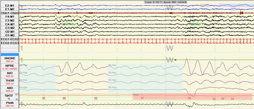
3

sleep-related upper airway collapsibility. This disruption in breathing may occur multiple times
per hour for the entire duration of the patient’s sleep.
The complexity of obstructive sleep apnea is exemplified by its multifactorial etiology. Such
etiologies involve the craniofacial structures, neuromuscular tone, and other related factors.
Collapsibility of the upper airway is influenced further by hormonal fluctuation (e.g., pregnancy
or menopause), obesity, rostral fluid shifts, and genetic predisposition that influences
craniofacial anatomy. OSA severity is heterogeneous among patients with the disorder. This
wide range of presentation leads to variations in management approach and differences in
treatment response.
Prevalence
Estimates of the prevalence of obstructive sleep apnea in adults vary in the literature; OSA
commonly is thought to involve 14% of men and 5% of women. Prevalence rates are higher in
certain populations, such as obese patients considered for bariatric surgery and post-stroke
patients. Under-recognition of OSA likely leads to under-diagnosis and a false reduction of the
true prevalence of disease.
Risk Factors
Individuals with certain characteristics appear to be predisposed to OSA. Conditions that may
be risk factors for the development of OSA in adults include obesity, menopause, gender
(male), and increasing age. [Obesity is considered to exist if the BMI is equal to or greater than
30] Genetic influences on craniofacial structure leads to higher OSA prevalence in certain
ethnic groups that have been studied. Some genetic syndromes, particularly those with
associated craniofacial anomalies, also are associated with an increased risk of OSA.
Craniofacial morphologies that may predispose to OSA include retrognathia, long and narrow
faces, dolichocephalic facial type, narrow and deep palate, steep mandibular plane angle,
anterior open bite, midface deficiency, and lower hyoid position. It should be noted, however,
that the strength of the relationship between these craniofacial morphologies and the
development of OSA is not well established.
Symptoms
Patients with obstructive sleep apnea often have a history of snoring, gasping respiration or
choking, and witnessed pauses in breathing (apneas) during sleep. Common clinical symptoms
of untreated obstructive sleep apnea include frequent nocturnal awakenings, non-restorative
sleep, morning headaches, and excessive daytime sleepiness. Patients with OSA often describe
difficulty with attention and concentration, mood disturbance, and difficulty controlling other
medical comorbidities such as diabetes mellitus, hypertension, and obesity.
Diagnosis
Diagnostic confirmation of obstructive sleep apnea is performed by a sleep medicine specialist
using the gold standard of an in-center overnight sleep study (polysomnography or PSG) or out-
of-center sleep testing (OCST) for appropriately selected patients. Home sleep apnea testing
(HSAT) is a type of OCST. Attended PSG includes at least 7 channels of recording, including
electroencephalography (EEG), monitoring of sleep, airflow through the nose and mouth, pulse
4

oximetry, respiratory effort, electrocardiography, and leg movement. HSAT includes 4-7
channels. It is important to note that HSAT typically does not include EEG monitoring of sleep.
According to the International Classification of Sleep Disorders1 obstructive sleep apnea can be
diagnosed by either of two sets of criteria. The first set of diagnostic criteria for OSA includes
the presence of at least one of the following: (1) the patient has sleepiness, nonrestorative
sleep, fatigue or insomnia symptoms, (2) the patient wakes with breath holding, gasping or
choking, (3) a bed partner or other observer reports habitual snoring, breathing interruptions,
or both, during the patient’s sleep, (4) the patient has been diagnosed with hypertension, a
mood disorder, cognitive dysfunction, coronary artery disease, stroke, congestive heart failure,
atrial fibrillation, or type 2 diabetes mellitus AND polysomnography or OCST shows 5 or more
predominantly obstructive events (obstructive or mixed apneas, hypopneas, or respiratory
effort related arousals (RERAs) per hour of sleep during a PSG or per hour of monitoring on
OCST.
Secondly, obstructive sleep apnea can be diagnosed if PSG or OCST shows 15 or more
predominantly obstructive events (obstructive or mixed apneas, hypopneas, or respiratory
effort related arousals (RERAs) per hour of sleep during a PSG or per hour of monitoring on
OCST). Examples of apnea and hypopnea are shown in Appendix 1.
A few different terms are used in the classification of obstructive sleep apnea. The respiratory
disturbance index (RDI) includes the number of apneas, hypopneas, and RERAs per hour of
sleep. The apnea-hypopnea index (AHI) includes the number of apneas and hypopneas per
hour of sleep. Thus, a patient’s RDI may be higher than the AHI. Some publications reference
AHI while others reference RDI, so it is important for clinicians and researchers to understand
the difference between these two measurements. Compared with PSG, OCST often
underestimates the frequency of obstructive events per hour because OCST typically does not
measure total sleep time as determined by EEG. The respiratory event index (REI) can be used
to indicate the frequency of respiratory events based on total recording time (rather than total
sleep time).
Severity
Severity of obstructive sleep apnea is classified based on the RDI; categories are mild (RDI ≥5
and 30). The minimum oxygen saturation
also should be considered when making clinical assessment of the magnitude of obstructive
sleep apnea, although there are no consensus classifications for the severity of oxygen
desaturation.
Significance
Untreated OSA can lead to many serious consequences. Excessive daytime sleepiness increases
the risk of motor vehicle accidents and diminishes quality-of-life. Neurocognitive impairment
leads to decreased scholastic and occupational performance. Chronic intermittent hypoxemia
and heightened sympathetic neural activity, endothelial damage and heightened inflammation
are related to metabolic dysfunction and end-organ sequelae. Untreated obstructive sleep
apnea increases risk of insulin resistance, coronary artery disease, congestive heart failure,
myocardial infarction, hypertension, stroke, cardiac arrhythmia, and sudden cardiac death.
5

III. The Role of Orthodontics in Adult OSA

The orthodontist is well positioned to perform an OSA screening assessment and refer at-risk
patients for diagnostic evaluation. Once the diagnosis of OSA is confirmed, physicians (and
advanced practice providers supervised by physicians) may prescribe orthodontic appliances or
procedures in appropriately-selected adult patients as part of OSA management.
Medical and Dental History
Orthodontists should be familiar with the signs and symptoms of OSA in adult patients.
Thorough history-taking is critically important in this regard for this establishes the presence of
pre-existing conditions, a basis for a diagnosis, the need for referral, and a baseline for
evaluating the effects of treatment. Orthodontists also should include assessment of a
patient’s height, weight, and neck size to screen adult patients for OSA.
The following items should be considered when constructing a health history that is sensitive to
OSA:

                A prior diagnosis of Obstructive          Excessive daytime sleepiness*
                Sleep Apnea (OSA)

                A prior diagnosis of other forms of       Fatigue during the day
                Sleep Related Breathing Disorders
                (SRBD)

                Height*                                   Choking or gasping respirations during
                                                          sleep

                Weight*                                   Habitual or loud snoring*

                Gender*                                   Observed episodes of pauses in
                                                          breathing*

                Age*                                      Abrupt awakening and shortness of
                                                          breath

                High blood pressure*                      Awakening with dry mouth or sore throat

                Mouth breathing                           Morning headaches

                Menopause                                 Difficulty staying asleep

                Alterations in performance                Enuresis or unexplained Nocturia

                Disordered mood                           Attention or memory problems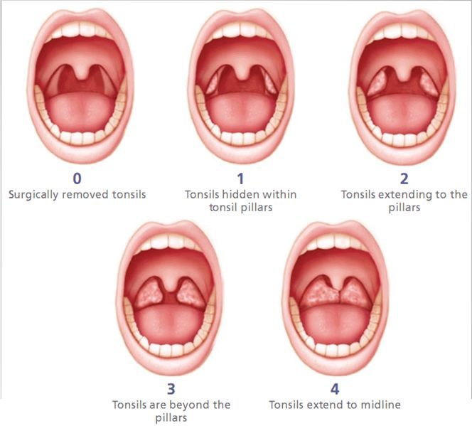
6
Restlessness during sleep Sweating
Nasal obstruction Bruxism
Type 2 Diabetes Neck Circumference
*Component of the STOP-Bang questionnaire
Screening Tools
In adults, a validated tool for OSA risk assessment is the STOP-Bang questionnaire (Appendix
2),2-3 which asks yes or no questions based on its acronym: snoring (S), tiredness (T), observed
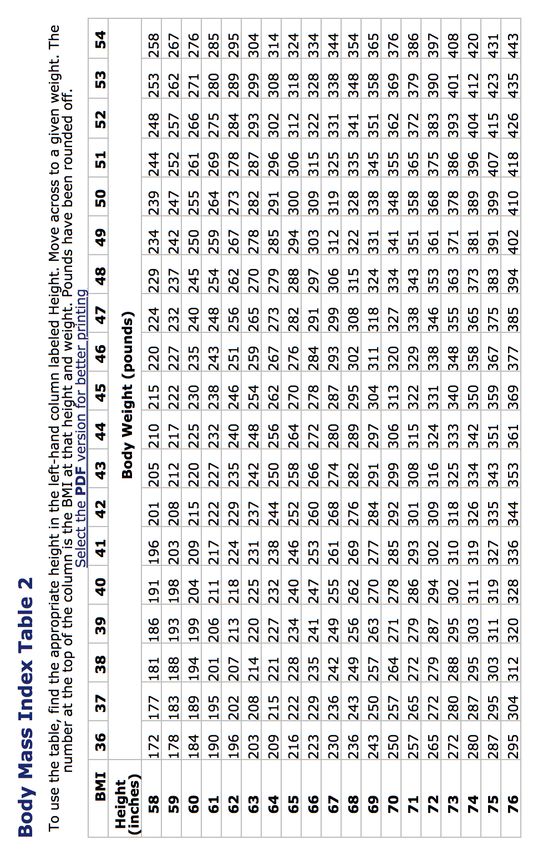
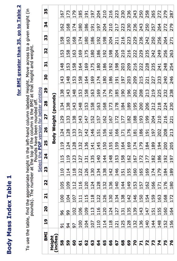
pauses in breathing (O), high blood pressure (P), BMI higher than 35 kg/m2 (B), age older than
50 years (A), neck circumference of 17 inches or larger in males, or 16 inches or larger in
females (N), and if patient’s gender is male (G). A patient is considered to be at low risk for OSA
if the questionnaire has 2 or less “yes” answers, at intermediate risk if here are 3 to 4 “yes”
answers, and at high risk if there are more than 5 “yes” answers.
The patient also is considered at high risk if there are 2 “yes” answers from the STOP section,
combined with either male gender, high BMI, or large neck size. For AHI ≤5, AHI between 5 and
15, and AHI ≥30, the sensitivities were 84%, 93%, and 100% and the specificities were 56%,
43%, and 37%, respectively. The STOP-Bang Questionnaire has a high sensitivity for identifying
patients with moderate-to-severe OSA. This sensitivity gives the practitioner an excellent tool
for identifying patients who have the condition. This questionnaire can be completed in a few
minutes when incorporated into an orthodontist’s workflow.
Clinical Examination
The clinical examination is an important part of the screening process. In addition to regular
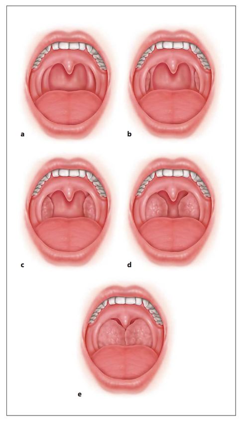
orthodontic screening, the orthodontist can use the Modified Mallampati Classification to
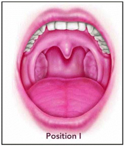
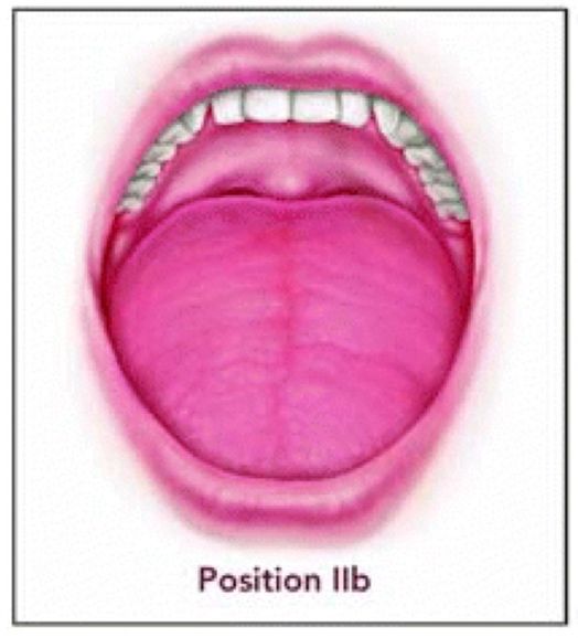
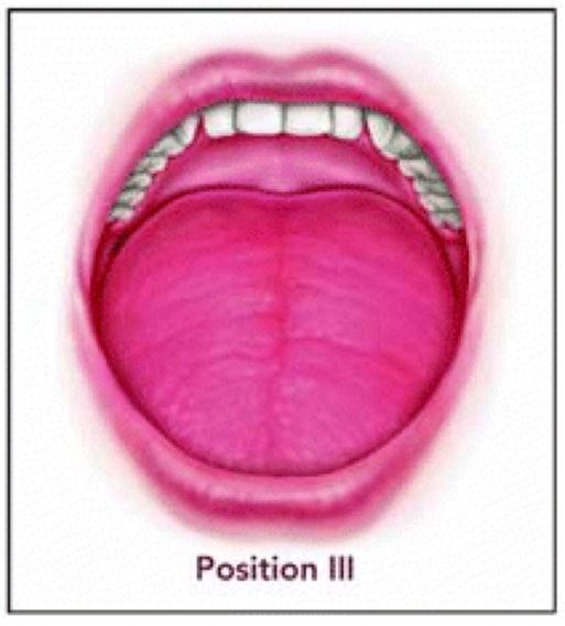
describe the patency of the oral airway (Appendix 3).4-10 Three steps are followed to determine
the MM Class: Step 1. Patients are asked to take a seated or supine position. Step 2. Patients
are asked to protrude their tongue as far forward as they can without emitting a sound. Step 3.
The examiner observes the relationship between the palate, tongue base and other soft tissue
structures to determine the MM Classification defined as Class Ι: Soft palate, fauces (the arched
opening at the back of the mouth leading to the pharynx), uvula, and tonsillar pillars are visible;
Class ΙΙ: Soft palate, fauces, and uvula are visible; Class ΙΙΙ: Soft palate and base of uvula are
visible; Class ΙV: Soft palate is not visible.
This clinical assessment framework can help orthodontists identify patients who may be at risk
for upper airway obstruction during sleep. It should be noted that the MM Class may vary over
the course of a pregnancy, so the MM Class may need to be reassessed at various times during
pregnancy. The Modified Mallampati Classification is a helpful part of the OSA screening
process; it should not, however, be used in isolation to predict OSA presence or severity.
Many other OSA screening questionnaires have been developed and studied in various
populations, with wide ranging specificities and sensitivities. The Epworth Sleepiness Scale
(Appendix 4)11 asks patients to self-rate their level of sleepiness in eight different sedentary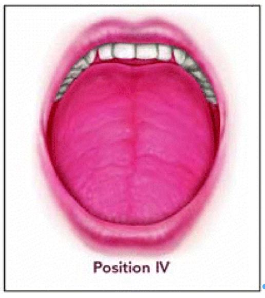
7

situations. The Epworth Sleepiness Scale may be used to gauge or track symptomatic
impairment (or response to treatment). However, it is not a screening tool for OSA, as it
detects abnormalities in level of daytime sleepiness regardless of the cause of sleepiness.
Practitioners also may find the Friedman Tongue Classification System (Appendix 5),12 the
Kushida Index,13 and the Berlin Questionnaire for Sleep Apnea14 useful.
Orthodontic Radiographs
The use of imaging in the assessment of OSA is often limited in a typical orthodontic setting.
Conventional cephalometric images are dimensionally limited. Thus, airway imaging using a
lateral cephalogram does not portray mediolateral information in the oropharyngeal airway
and may give misleading information as to the volume and minimal cross-sectional area.
Cone Beam Computed Tomographic (CBCT) images have been shown to be useful in diagnostic
and morphometric analysis of the hard and soft tissues in routine orthodontic treatment, but
they have certain limitations regarding the diagnosis of OSA. CBCT provides no information on
neuromuscular tone, susceptibility to collapse, or actual function of the airway. There are
significant positional and functional differences when the patient is asleep versus awake. It is a
snapshot of a specific moment of the breathing cycle. Additionally, there currently is no
minimal cross-sectional area or volume of the airway that has been validated as a minimal
threshold level at which an individual is at higher risk of having OSA. Thus, orthodontic records
may be taken by the orthodontist, but currently no radiographic methods have been reported
to have high enough sensitivity or specificity to serve as a risk assessment tool for OSA.
Three-dimensional imaging of the airway should not be used to diagnose sleep apnea or any
other sleep-related breathing disorders because such imaging currently does not represent a
proper risk assessment technique or screening method. On the other hand, three-dimensional
imaging of the airway, when available, may be used for monitoring or treatment
considerations. If radiographic records are taken as part of orthodontic diagnosis and
treatment planning, the airway and surrounding structure should be analyzed comprehensively.

IV. Diagnosis and Treatment Planning in Adult OSA

Obstructive sleep apnea and other sleep-related breathing disorders can only be definitively
diagnosed by a physician. It is not in the scope of the orthodontist or any other dentist to
definitively diagnose obstructive sleep apnea or any other sleep-related breathing disorder. If
the patient is found to have OSA, the physician will prescribe the appropriate course of action;
the orthodontist should consider working in a collaborative way with the physician, providing
related orthodontic treatment when necessary, as long as it does not interfere with medical
treatment.
The OSA treatment plan should be based on careful consideration of the patient’s individual
needs and treatment goals. If the treatment plan involves orthodontics, a plan for treatment,
monitoring, and long-term follow up care should be developed by all practitioners involved.
8

Care should be coordinated via communication between the orthodontist and any other
practitioners participating in the treatment of the patient. It is recommended that treatment
and/or management of obstructive sleep apnea not take place without a referral from a
physician (or provider supervised by a physician).

V. Treatment of OSA in Adults by Physicians and Surgeons

Positive airway pressure (PAP) therapy is the gold standard treatment method for obstructive
sleep apnea in adults. PAP acts as a pneumatic splint that maintains patency of the upper
airway. PAP is delivered through a mask interface as either continuous positive airway pressure
(CPAP), bi-level positive airway pressure (BPAP) or auto-titrating positive airway pressure
(APAP). Of note, CPAP and BPAP devices are available in conventional and auto-titrating
modes. CPAP use can decrease OSA-related cognitive impairment along with improving
objective and subjective measures of sleepiness, particularly in patients with severe OSA (AHI ≥
30/hour).15 BPAP may be used for patients with OSA who are intolerant to CPAP or those who
have other forms of sleep-related breathing disorders (e.g., sleep-related hypoventilation).
APAP may be considered for patients with OSA patients who do not have contraindications to
APAP use (e.g., congestive heart failure, lung disease such as chronic obstructive pulmonary
disease, obesity hypoventilation syndrome, or central sleep apnea).
Studies on PAP non-adherence report wide ranging results. While definitions of non-adherence
vary across studies, a common definition of PAP non-adherence is mean use ≤ 4 hours per
night. Estimates of PAP non-adherence range from 29% to 83%.16-17 Early adherence to PAP use
predicts longer-term PAP use; a study of 100 patients started on CPAP showed that CPAP use
for at least 4 hours per night 3 days after starting therapy was predictive of CPAP adherence 30
days after treatment initiation.18 Factors that affect PAP adherence include OSA severity, ability
to tolerate the prescribed pressure setting, mask fit, spousal support, and other psychological
and social influences.19
Other treatment options include positional therapy (avoidance of back-sleeping) and long-term
weight reduction as indicated. Nasal congestion and allergic rhinitis may be managed with
nasal steroids and other oral medications as indicated. For some patients, nasal surgery may be
performed as adjunctive therapy to decrease intranasal resistance and facilitate better
adherence to PAP therapy. For selected patients, multilevel surgery including nasal and/or
palatal surgery with or without mandibular surgery, genioglossus advancement, and hyoid
suspension may be considered. Other soft tissue surgeries might be indicated that involve the
tonsils, adenoids, frena, and tongue. Hypoglossal nerve stimulation addresses the impaired
neuromuscular tone in obstructive sleep apnea and may be considered in certain patients with
OSA.
9

VI. Orthodontic Management in Adult OSA

Following diagnosis of OSA by a physician, a patient may be referred to (or back to) an
orthodontist for one or more types of care.

Informed Consent
Prior to initiating care, informed consent appropriate to OSA must be obtained before any
treatment is provided. The proposed treatment plan should be described in detail, and
treatment alternatives also should be discussed. The orthodontist should describe the benefits,
risks, short and long-term side effects, and complications that might arise. The need for
compliance, long-term monitoring, and follow-up care should be discussed. An estimate of the
nightly duration of oral appliance therapy use should be provided, and a realistic estimate of
the probability of success with the treatment protocol should be presented. Given the serious
nature of untreated OSA, it is recommended that the orthodontist carefully document the
informed consent process.
Oral Appliance Therapy
Oral appliances (OA), which include both mandibular advancing oral appliances (OAm) and
tongue retaining devices, are usually effective options for OSA management in appropriately
selected patients. OAm are intended to hold the mandible and/or the associated soft tissues
forward, resulting in an increased caliber of the upper airway at the oropharyngeal level. A
substantial body of research supports the use of oral appliances for patients with OSA.
Specifically, OAs may be used for treatment of mild to moderate OSA and for treatment of
patients with severe OSA who are unwilling or unable to use PAP therapy. Published guidelines
(American Academy of Sleep Medicine/American Academy of Dental Sleep Medicine) describe
how oral appliances fit into the OSA management paradigm.20-21
Functional appliances and OAm are considered the first line of treatment for patients with OSA
that prefer OA over PAP and for those patients that do not respond to PAP therapy. While
typically well tolerated, it should also be noted that not all patients with OSA will respond to
OAm treatment, with this form of therapy reported to be completely effective in 36% to 70% of
OSA cases.
Many types of oral appliances are used in the treatment of OSA in adults. The appliances vary
based on the coupling design, mode of fabrication and activation, titration capability, degree of
vertical opening, lateral jaw movement, and whether they are custom made or prefabricated.
Proper indications for each design should be considered.
Oral Appliance Titration
Oral appliances initially are delivered with the mandible advanced to a position approximating
2/3 of maximum protrusion. After a period of accommodation, based on subjective feedback
from the patient regarding their OSA symptoms and sleep quality, the amount of protrusion can
be titrated or increased until optimum symptom relief is obtained. Unattended (type 3 or 4)
10

portable monitors may be employed by the orthodontist to help define the optimal target
position of the mandible. Then typically the physician involved will request a sleep study with
the OAm in place. Should the physician deem the calibrated position to be sub-therapeutic, the
physician and orthodontist should discuss the possibility of further titration or alternative
treatment.

Monitoring
During treatment for OSA, the patient should be monitored, which may involve subjective
reports as well as objective observations. Reports on usage of the OA may be obtained from
the patient and bedpartner or caregiver. Compliance should be evaluated, and the appliance
should be checked for fit and comfort, the need for titration, and the development of
undesirable side effects. At present, most data on adherence to OA therapy rely on subjective
reports. Use of a thermal sensor23 has been studied in an effort to have objective
measurement of OA adherence, although such measures currently are not part of routine
clinical care.
It has been suggested that monitoring be conducted at least once every 6 months during the
first year and then annually. Routine monitoring should result in regular communications
between the physician and orthodontist. If the patient has worsening of OSA-related
symptoms, and/or changes to overall health, a consultation with the physician is strongly
recommended.
The Goals of Treatment
The endpoints of treatment include:
                  §   Reduced or eliminated snoring
                  §   Resolution of the patient’s initial symptoms of OSA
                  §   Normalization of the AHI
                  §   Normalization of oxyhemoglobin saturation
No pretreatment risk factors have been consistently shown to predict success for oral
appliances in reaching treatment goals.
Change in Occlusion
Oral appliances used in sleep apnea treatment move teeth. In the global field of dentistry,
orthodontists generally are considered the experts in the management of malocclusion because
of their education and clinical experience. Improved awareness of both OSA and the
effectiveness of oral appliances has resulted in increased numbers of OSA patients being
treated with oral appliances by non-orthodontists. While successful OSA treatment may be
evident over the short term in many of these patients, non-orthodontic providers may be
unaware of the unwanted effects of OAs can have on their patient’s occlusion over the long
term. Orthodontists can be helpful in providing our medical and dental colleagues valued
oversight, and sometimes treatment, of unexpected and unwanted occlusal changes occurring
with long-term oral appliance wear.
11 Typical changes include a reduction in overjet and overbite, changes in facial height, development of anterior crossbites, and posterior openbite. Changes are progressive with ongoing oral appliance use. In that many patients ultimately will be treated for a protracted period of their lifetime, appliance-generated malocclusions often become significant over the long term and may require treatment to reverse the dentoskeletal adaptations that may occur. Orthodontists may be asked to assess and treat oral appliance-related malocclusions, a condition that has become a more frequent occurrence in recent years. When considering treatment of these malocclusions, orthodontists need to be aware that the patient will not be able to wear their oral appliance during treatment; therefore, the patient may need to use PAP therapy during the period of orthodontic care. Communication with the physician helps ensure the patient’s OSA is still being managed appropriately. Should the patient return to using an oral appliance for OSA following orthodontic treatment, then the malocclusion may also return. Consequently, such patients often switch to PAP therapy or can be evaluated for surgical treatment options. MMA and SARME Patients who are unable to tolerate or adhere to PAP and/or oral appliance therapy with an underlying sagittal skeletal discrepancy may be candidates for maxillomandibular advancement (MMA) or telegnathic (>10mm) jaw advancement surgery. MMA generally is reserved for patients with severe OSA who are unable to tolerate PAP therapy, and for those patients who also have an orthodontic indication for the procedure. The severity of OSA is not the only determinant of candidacy for MMA; these patients often require detailed evaluation and counseling before MMA is selected as a treatment option. Such patients typically should proceed with routine orthodontic diagnosis and treatment planning, including comprehensive soft tissue facial evaluation to assure optimal pre-surgical preparation and that the surgery performed will not affect facial esthetics adversely. Orthodontic care is usually a beneficial adjunct for patients to facilitate obtaining optimal occlusion while simultaneously reducing the risk of post-operative malocclusion. Patients with ideal or minimal Class I malocclusion may not require extensive pre-surgical orthodontics in that the two jaws may have a similar interdigitation following symmetric maxillary and mandibular advancement. Telegnathic surgery is not recommended for patients who are already bimaxillary protrusive; such patients should usually be reevaluated by the team to explore alternative treatment options. One of the concerns of telegnathic surgery in this situation involves esthetics. As such, each practitioner and patient should decide for themselves if the benefits of the surgery outweigh the risks involved. Significantly less data exists for surgically assisted rapid maxillary expansion (SARME), which aims to correct a maxillary transverse deficiency. In OSA patients with maxillary transverse deficiency, normalizing the width of the maxilla with SARME and developing a functional and esthetic occlusion with comprehensive orthodontic treatment afterward has been suggested to improve PSG parameters.22
12

Possible Treatments on the Horizon
New treatment modalities such as mini-implant (aka, miniscrew or temporary anchorage
device) supported rapid maxillary expansion (MARME) are appearing as possible alternatives
for surgical assisted rapid maxillary expansion. However, to date there is very limited PSG
evidence for its use in the management of OSA patients. Future studies are needed, and with
time mini-implant supported expansion may become a viable adjunctive form of treatment for
OSA management in adult patients.

VII. Pediatric OSA (Under 18 Years of Age)

Etiology
As with adult OSA, impaired neuromuscular tone underlies upper airway collapsibility in
children. In addition to etiologic factors similar to those in adults, exacerbating factors for
pediatric OSA often include lymphoid hyperplasia and growth-related changes in the size of the
upper airway.
As the upper airway is narrowed or completely occluded, the patient’s effort during breathing
progressively increases. Due to the airflow restriction, there is a relative increase in serum
carbon dioxide (hypercarbia) and decrease in serum oxygen (hypoxemia). The escalating
respiratory effort causes a cortical arousal from sleep, which results in the upper airway
opening so that normal airflow is reestablished. Once the patient falls back asleep, the upper
airway may collapse again with recurrence of the above-noted process. This breathing
sequence may have significant consequences for the child.
Risk Factors
As the obesity epidemic also affects children, obesity is becoming a greater factor for childhood
OSA. However, as untreated OSA may contribute to growth restriction, some children with OSA
paradoxically may be underweight. Thus, it is recommended that a clinical risk assessment for
OSA be performed even in normal weight or underweight children.
In addition, it is thought that certain craniofacial morphologies can increase a child’s risk for
having OSA. For instance, mandibular retrognathia, long and narrow faces, narrow and deep
palate, steep mandibular plane angle, anterior open bite, and midface deficiency may
predispose a child to develop OSA. However, the presence of OSA cannot be determined by
craniofacial morphology alone; these physical findings should be interpreted in the context of
the clinical history.
Genetic syndromes that are associated with craniofacial anomalies can confer an increased risk
of OSA. For example, patients with Pierre Robin sequence24 and syndromic craniosynostosis25
have a high prevalence of OSA. Children with Down syndrome26 also have an increased OSA
prevalence. Orthodontists who care for children with these and other genetic syndromes that
affect craniofacial morphology should pay attention to clinical features that may suggest the
presence of untreated OSA.
13

Symptoms
Children with OSA may present with snoring, witnessed apneas, and choking or gasping during
sleep. Parents or caregivers may describe that the child sleeps in unusual positions, such as
having the neck hyperextended or with the head hanging off the side of the bed, as well as
appearing very restless with frequent position changes during sleep.
Some children with OSA may present with sleepiness; those who previously had discontinued
daytime napping may resume daily or near-daily naps. In other children, untreated OSA may
manifest as hyperactivity rather than excessive sleepiness. While obesity may be a contributor
to the pathogenesis of OSA in some children, others may present with failure to thrive. As such,
it is recommended that the evaluation for OSA in every child should be part of an orthodontist’s
comprehensive clinical assessment.
Diagnosis
Diagnosis of OSA in children is confirmed only by the gold standard PSG. Diagnostic evaluation
of childhood OSA has evolved in recent years. In addition to standard recording channels, all
pediatric PSG is now conducted with carbon dioxide (CO2) monitoring. Measurement with
either end-tidal CO2 (the partial pressure of CO2 present at the end of exhalation) or
transcutaneous CO2 monitoring also is acceptable.
According to the International Classification of Sleep Disorders,1 obstructive sleep apnea can be
diagnosed by either of two sets of diagnostic criteria. The first set of criteria for obstructive
sleep apnea includes the presence of at least one of the following: (1) snoring, (2) labored,
paradoxical, or obstructed breathing during the child’s sleep, or (3) sleepiness, hyperactivity,
behavioral problems, or learning problems AND polysomnography shows one or more
obstructive apneas, mixed apneas, or hypopneas per hour of sleep.
Alternatively, obstructive sleep apnea can be diagnosed if the PSG shows a pattern of
obstructive hypoventilation, which is defined as at least 25% of total sleep time with
hypercapnia (PaCO2 > 50 mm Hg) associated with at least one of the following: (1) snoring, (2)
flattening of the inspiratory nasal pressure waveform, or (3) paradoxical thoracoabdominal
motion. These OSA diagnostic criteria are for children below age 18 years, though adult OSA
diagnostic criteria may be used for children ages 13-18 years per the American Academy of
Sleep Medicine Manual for the Scoring of Sleep and Associated Events.27

HSAT is not indicated in patients less than 18 years of age.28-29

Severity
Published studies on childhood OSA have included various diagnostic criteria; some studies use
the adult criteria of AHI ≥5/hour. Other studies define childhood OSA as mild (AHI 1-5/hour),
moderate (AHI 5-10/hour) and severe (AHI ≥10/hour). Of note, scoring of obstructive apneas
and hypopneas on PSG differs slightly for children compared with adults. For adults, event
14

duration is at least 10 seconds whereas for children obstructive event duration is defined as at
least 2 breaths.
Prevalence
Prevalence of childhood OSA is obscured by different diagnostic criteria used in published
studies. Epidemiologic data from 2008 indicate prevalence of parent-reported ‘‘always’’
snoring to be 1.5% to 6%, prevalence of parent-reported apneic events during sleep to be 0.2%
to 4%, and OSA diagnosed by varying criteria to be 1% to 4%. Multiple studies have shown that
during certain phases of growth, childhood OSA remits without any intervention. These data
indicate that prevalence of childhood OSA changes across periods of growth and development.
Specific populations, such as children with certain craniofacial or other genetic syndromes and
those who are obese, have a higher prevalence of OSA compared with the general population.

Significance
Consequences of OSA in children include impaired growth and cardiovascular dysfunction. The
impaired neurocognitive function seen in children with untreated OSA can have an effect on
academic performance. Behavioral problems also can result. Persistent snoring and nocturnal
enuresis (bedwetting), which can result from untreated OSA, can be embarrassing for children
in social settings and thus affect interpersonal interactions.

VIII. Pediatric OSA: Skeletal and Soft Tissue Growth

Orthodontists are aware of the impact facial growth has on orthodontic treatment outcome.
Facial growth also influences the size and shape of the upper airway in the pediatric population.
One approach to understanding the interaction of hard and soft tissue growth on upper airway
morphology can be described as follows. The hard tissue boundaries of the upper airway
include the upper and lower incisors and the piriform rim in the anterior, the cranial base
superiorly, the cervical vertebrae posteriorly and the hyoid bone inferiorly. Laterally, the size of
the airway is related to the width of the palate, the middle cranial fossa, and the distance
between the ascending rami. Together these structures define the bony skeletal boundaries of
the airway. Soft tissues then line this hard tissue framework. These tissues include the
pharyngeal muscles, the tongue, the soft palate, the turbinates, as well as the pharyngeal
tonsils, adenoids, and nares.
Importantly, growth of the bony components effectively increases the size of the skeletal
boundaries in the following ways. The anterior cranial base increases in length via growth at
the spheno-ethmoidal synchondrosis up to age 7 years. Increases in posterior cranial base
length are similarly related to growth at the spheno-occipital synchondrosis up to age 13 years.
The anterior cranial base carries the nasomaxillary complex forward at the same time as the
individual bones of the midface are displaced in an anterior and inferior direction.
Simultaneously, the mandible elongates and is displaced downward and forward with
deposition of bone on the posterior and superior borders of the ramus, increasing the height of
15

the rami (bony pharyngeal height) and increasing the distance between the ascending rami
(bony pharyngeal width). Concurrently, resorption on the anterior border of the ramus
increases corpus length (oropharyngeal length). While all these bony changes are occurring,
the hyoid bone also is displaced anteriorly and inferiorly. Thus, the normal facial growth
process results in dramatic increases in all three dimensions of the skeletal framework.30
While the skeletal boundaries of the airway are increasing, the major lymphatic tissues of the
upper airway (tonsils and adenoids) are shrinking. This combination of increases in skeletal
dimensions along with decreases in soft tissue mass results in enormous increases in the size of
the upper airway over infancy, childhood, adolescence, and teenage years. These changes in
airway due to growth far exceed any orthodontic or orthopedic effects on airway shape and/or
size. Knowledge of these changes is important in the understanding of the dynamics of OSA in
children.31

IX. The Role of Orthodontics in Pediatric OSA

It is strongly recommended that the orthodontist perform a clinical risk assessment for OSA and
refer at-risk patients to the appropriate physician for definitive diagnosis of OSA. Subsequently,
orthodontists may be involved in treatment of pediatric OSA if the treating physician refers the
patient back to the orthodontist to address an underlying skeletal discrepancy thought to
contribute to the child’s OSA.
Medical and Dental History
Orthodontists should be familiar with the signs and symptoms of OSA in pediatric patients.
Questions concerning the health history of a pediatric patient should solicit information on
snoring, sleep-related behaviors, daytime sleepiness, difficulty concentrating, and/or formal
diagnosis of attention deficit hyperactivity disorder. The American Academy of Pediatric Sleep
Physicians recommends that if a patient reports snoring, more thorough questioning is
warranted; the guidelines state, “If they snore, you must do more.”32
A thorough history and examination are critically important in that they establish the presence
of pre-existing conditions, a basis for a diagnosis, the need for referral, and a baseline for
evaluating the effects of treatment. Orthodontists also should include assessment of a
patient’s height, weight, and neck size to screen pediatric patients for OSA.
The following items should be considered when performing a pediatric evaluation that is
sensitive to OSA:

             A prior diagnosis of Obstructive     Loud snoring
             Sleep Apnea (OSA)
16
A prior diagnosis of other forms Mouth breathing during sleep
of Sleep Related Breathing
Disorders (SRBD)
Height Poor school performance
Weight Aggressive behavior
Medications Developmental delays
Age Bed wetting that is not age-
appropriate
Attention problems Hard to wake up in the morning
Trouble breathing during sleep Morning headaches
Pauses in breathing during sleep Fall asleep quickly
Nasal obstruction ADHD
Screening Tools
One potential screening tool that has been validated and used in orthodontic offices is the
Pediatric Sleep Questionnaire (PSQ)(Appendix 6).33-35 This questionnaire has a positive
predictive value of 0.4 (i.e., patients with a positive PSQ score will be diagnosed with OSA 40%
of the time) and a negative predictive value of 0.99, (i.e., patients with a negative PSQ score will
only have a 1% chance of being diagnosed with OSA). The PSQ often is a valuable first step in
screening patients presenting to the orthodontic office without a history of OSA. The Epworth
Sleepiness Scale for Children and Adolescents (Appendix 7)36 may be helpful to assess for
problematic sleepiness, but this tool cannot identify a specific cause of daytime sleepiness. The
Epworth scale has been validated only for children ages 12-18 years.11
Clinical Examination
In addition to the usual orthodontic clinical examination that evaluates the dental occlusion,
range of mandibular motion, soft tissue frenum attachments, gingival health, and TMD, the
orthodontist should also note the degree to which the tonsils impinge on the pharyngeal
airway. A commonly accepted tonsil classification system called the Brodsky scale grades the
clinical manifestation of tonsil hypertrophy from 1 to 5 based on the percentage of the
oropharyngeal airway taken up by the two tonsils (Appendix 8).37 The Friedman Tonsil Grading
System (Appendix 9)38 also may be a useful tool to evaluate the size of the tonsils. As tonsil size
does not correlate to OSA severity, there is no set cut-point about which level of tonsillar
hypertrophy necessitates a referral to an otolaryngologist (ENT physician) for further
evaluation;39 thus, this decision is best made in the patient-specific context of symptoms and
physical exam findings. The clinical evaluation of OSA in children should include evaluation of17 tongue size and position, the presence of obesity, and the patient’s overall growth and development. Orthodontic Records The typical orthodontic record set captures some important information that can be useful for further evaluation of the upper airway. For example, the adenoid mass and the hyoid bone can be seen on both the lateral cephalogram and the cone beam computed tomographic (CBCT) image. A low position of the hyoid bone when measured from the inferior border of the mandible has been shown to be an indicator of low muscle tonicity and has been linked with OSA. Three-dimensional imaging is more accurate than two-dimensional imaging for assessment of airway volume and area of maximum constriction. Airway imaging using a cephalogram does not portray mediolateral changes in the oropharyngeal airway and may give misleading information as to the volume and minimal cross-sectional area. As in adult patients, while CBCT images have been shown to be useful in diagnostic and morphometric analysis of the hard and soft tissues in routine orthodontic treatment, there are limitations regarding the screening of OSA. CBCT provides no information on neuromuscular tone, susceptibility to collapse, or actual function of the airway. Although both two dimensional and three-dimensional imaging of the airway are helpful, they cannot be used to diagnose sleep apnea or any other sleep-related breathing disorders alone, and they do not provide a proper risk assessment technique or screening method. Importantly, there is no direct link between any radiographic measures of airway size or shape and PSG results. Therefore, imaging values should be interpreted cautiously and in conjunction with other clinical signs and symptoms. Three-dimensional imaging of the airway, when available, also may be used for monitoring or treatment planning. If radiographic records are taken for orthodontic purposes, the airway and surrounding structures, specifically the adenoids in children, should be evaluated. X. Diagnosis and Treatment Planning in Pediatric OSA As mentioned previously, orthodontists should not assume the responsibility for the definitive diagnosis of OSA. The definitive diagnosis is appropriately made by a physician. If the patient is found to have OSA, the physician should decide on an appropriate course of action for the treatment of OSA. The orthodontist may choose to work in a collaborative way with the physician, providing orthodontic treatment when necessary and when it does not interfere with ongoing medical treatment. The plan for treating pediatric OSA should be based on consideration of the patient’s individual needs and treatment goals. If the OSA treatment regimen involves orthodontics, a plan for treatment, monitoring, and long-term follow up care should be considered by all medical and dental practitioners involved. Care should be coordinated via communication between the
18

orthodontist and all other practitioners who are working to treat the patient’s obstructive sleep
apnea.
The orthodontic treatment plan for patients with OSA should follow the same orthodontic
principles for correction of dental and skeletal deformities. Two orthodontic procedures that
may change upper airway physiology are rapid maxillary expansion (RME) and mandibular
advancement appliances for Class II correction. With both types of interventions, the primary
objective of the orthodontic appliance should be to improve the occlusion and address the
underlying skeletal discrepancy.
It would be appropriate, for example, to recommend rapid maxillary expansion (RME) for
patients diagnosed with maxillary transverse deficiency. In this situation, the primary
treatment goals would be to normalize the transverse width of the maxilla and establish a
normal occlusion. Secondary effects of this treatment may result in reduction of nasal airway
resistance and increase in the volume of the nasopharynx and nasal cavity. Both secondary
effects of RME have the potential to improve OSA.
In the case of mandibular advancement devices for mandibular retrognathia, the primary goals
should be to correct the skeletal discrepancy and the Class II molar relationship. A secondary
effect of mandibular advancement devices may be the increase in the caliber of the
oropharyngeal airway. The same applies to maxillary advancement appliances used in the
treatment of Class III malocclusions.
It is possible that an OSA patient might be referred for expansion but does not have a
transverse discrepancy. Likewise, it is possible a patient with OSA might be referred for
mandibular advancement (or maxillary advancement) where no sagittal discrepancy exists. In
such situations, the treatment alternatives should be considered on a case-by-case basis by the
medical and dental practitioners involved. In such situations, it is appropriate to prioritize the
treatments to serve the best interests of the patient.

XI. Treatment of Pediatric OSA

In the growing child, OSA management is dramatically different than for the adult. It is
recommended that orthodontists become aware of the vast array of potential treatment
modalities that are available and that they work in unison with medical and dental practitioners
when managing pediatric OSA. Hypertrophic tonsils and/or adenoids are the most common risk
factor for OSA in the pediatric population, with tonsillectomy and/or adenoidectomy typically
considered the first-line of treatment.
Various forms of pharmacologic agents may be prescribed by the attending physician to reduce
the size of the nasal soft tissues if there is suspicion of these tissues being a potential cause of
OSA. Nasal surgery, including turbinate reduction and deviated septum correction, also may be
considered in selected cases. For the obese child, weight reduction management should be
considered as part of the treatment plan. Positive airway pressure (PAP) also may be utilized in
19

severe cases. Possible negative craniofacial consequences of longitudinal usage of PAP on the
developing facial structures should be considered.
Dentofacial orthopedic management, which is within the scope of the orthodontic specialist,
also may be considered. For instance, rapid maxillary expansion is a well-known orthodontic
treatment option for patients with a narrow maxilla. There is growing evidence, although of
low-level, that in mixed-dentition patients who are properly diagnosed with OSA, RME can
decrease AHI in the short and long term.40 Unfortunately untreated control groups generally
were not used in the studies considered. Regardless of the presence of OSA, it is recommended
the orthodontist use these devices only when there is an appropriate underlying skeletal
condition. There is no indication in the literature that prophylactic application of maxillary
expansion prevents the future development of OSA.
Based on a few studies that were performed on mixed dentition samples, mandibular anterior
repositioning appliances also can produce a decrease in AHI. Long-term stability of these
changes has not been studied. Untreated control groups generally were not used in these
studies as well. Regardless of the presence of OSA, it is recommended the orthodontist use
these devices only when there is an indication that a related retrognathic condition exists.
There is no clear indication in the literature, however, that prophylactic use of mandibular
anterior repositioning appliances prevents later development of obstructive sleep apnea.
In addition, the orthodontist should be aware that some children who remain PAP intolerant
may require airway support while sleeping. The use of mandibular advancing devices may be
prescribed by the physician, and this prescription is not predicated solely on Angle’s
classification of occlusion. In this instance, treatment with an oral device is directed primarily
toward airway maintenance and less so with dentofacial orthopedic management. Careful
monitoring of facial growth and development is important during this time.
For Class III patients, there are no studies that have assessed the impact of maxillary
protraction on AHI. Only assessment of pharyngeal dimensions has been published so far. It
appears inappropriate for the clinician to make the jump from enlarged airway dimensions to
improvement in airway function or sleep-related breathing parameters. Regardless of the
presence of OSA, it is recommended the orthodontist use these devices when there is an
underlying skeletal issue.
Orthognathic surgery usually is not indicated until craniofacial growth is completed. As a result,
the pediatric patient that presents with clear skeletal issues should typically be managed to
adulthood in the normal fashion with corrective jaw surgery planned later when the timing of
the surgery is appropriate. An exception might be considered in a case where the patient has
OSA and a severe skeletal discrepancy. After considering the potential benefits and risks
involved (including the need for later surgical revision), orthognathic or telegnathic surgery
could be considered.
In summary, much is known regarding treatment for OSA in adults. In contrast, information on
the treatment of OSA in pediatric patients is much more limited. Thus, care should be taken
regarding the indications for orthodontic and orthopedic treatment intended to treat OSA in
the young patient. Clearly defined treatment goals, focusing on the orthodontic and orthopedic
20

components, should be articulated to the responsible parties involved. Improvement of the
OSA should be highlighted as a “possible,” or some studies say “anticipated,” outcome of
treatment. But, no guarantees of OSA resolution can be implied or stated emphatically by the
treating orthodontist.

XII. Fallacies About Orthodontics in Relation to OSA

Conventional orthodontic treatment never has been proven to be an etiologic factor in the
development of obstructive sleep apnea. When one considers the complex multifactorial
nature of the disease, assigning cause to any one minor change in dentofacial morphology is
not possible. However, misinformation exists regarding the potential airway related sequelae
of orthodontic treatment performed with dental extractions or orthopedic headgear.
The specific effects on the dental arches and the muscles and soft tissues of the oral cavity
following orthodontic extractions can differ significantly, depending on the severity of dental
crowding, the amount of protrusion of the anterior teeth and the specific mechanics used to
close the extraction spaces. The indication for extractions varies from patient to patient, as
does the resulting change to the width, length and arch perimeter of the dentition—all may
increase, decrease, or stay the same following treatment. The impact that orthodontic
treatment with or without dental extractions may have on the dimensions of the upper airway
also has been examined directly, first with two-dimensional cephalograms and more recently
with three-dimensional CBCT imaging.41
In certain instances, namely in patients with significant protrusion of both upper and lower
anterior teeth where skeletal anchorage or extractions are used to retract the anterior teeth as
much as possible to reduce lip protrusion in profile, reductions in the cross-sectional area of the
oropharynx have been reported. More frequently, as in patients where extractions are
performed to help address dental crowding or improve the occlusion, there is no discernible
change in airway dimensions when extractions are employed.42-43 The studies examining these
effects in children and adolescents have reported increases in airway volumes and cross-
sectional areas in patients both with and without extractions performed as part of their
orthodontic treatment.44-46 These effects may likely be related to normal growth changes.
In discussing orthodontic treatment to changes in the dimensions of the upper airway, it also is
helpful to understand that an initial small or subsequently reduced or increased size does not
necessarily result in a change in airway function. Reflecting the higher significance of
neuromuscular control on airway function during sleep, it has been demonstrated that a
narrow airway does not result in OSA, but rather it is an inability for a patient’s airway muscles
to compensate adequately that leads to obstruction and sleep-disordered breathing.47
As such, future investigations should aim to place greater emphasis on the effects of airway
function following orthodontic treatment, as opposed to focusing solely on quantifying airway
dimensions. One such study assessed dental extractions as a cause of OSA later in life with a
large retrospective examination of dental and medical records.48 Researchers reviewed the
health records of more than 2700 adults with four missing premolars and evaluated whether
You can also read