Skin Barrier Abnormalities and Immune Dysfunction in Atopic Dermatitis - MDPI
←
→
Page content transcription
If your browser does not render page correctly, please read the page content below
International Journal of
            Molecular Sciences

Review
Skin Barrier Abnormalities and Immune Dysfunction
in Atopic Dermatitis
Gabsik Yang 1 , Jin Kyung Seok 2 , Han Chang Kang 2 , Yong-Yeon Cho 2 , Hye Suk Lee 2
and Joo Young Lee 2, *
 1    Department of Pharmacology, College of Korean Medicine, Woosuk University, Jeonbuk 55338, Korea;
      yangboncho@gmail.com
 2    BK21plus Team, College of Pharmacy, The Catholic University of Korea, Bucheon 14662, Korea;
      jinkyung.seok@gmail.com (J.K.S.); hckang@catholic.ac.kr (H.C.K.); yongyeon@catholic.ac.kr (Y.-Y.C.);
      sianalee@catholic.ac.kr (H.S.L.)
 *    Correspondence: joolee@catholic.ac.kr; Tel.: +82-2-2164-4095

 Received: 30 March 2020; Accepted: 17 April 2020; Published: 20 April 2020

 Abstract: Atopic dermatitis (AD) is a common and relapsing skin disease that is characterized by
 skin barrier dysfunction, inflammation, and chronic pruritus. While AD was previously thought
 to occur primarily in children, increasing evidence suggests that AD is more common in adults
 than previously assumed. Accumulating evidence from experimental, genetic, and clinical studies
 indicates that AD expression is a precondition for the later development of other atopic diseases,
 such as asthma, food allergies, and allergic rhinitis. Although the exact mechanisms of the disease
 pathogenesis remain unclear, it is evident that both cutaneous barrier dysfunction and immune
 dysregulation are critical etiologies of AD pathology. This review explores recent findings on AD
 and the possible underlying mechanisms involved in its pathogenesis, which is characterized by
 dysregulation of immunological and skin barrier integrity and function, supporting the idea that AD
 is a systemic disease. These findings provide further insights for therapeutic developments aiming to
 repair the skin barrier and decrease inflammation.

 Keywords: atopic dermatitis; immunity; skin; homeostasis

1. Introduction
      Atopic dermatitis (AD) is a common chronic relapsing inflammatory skin disease that repeatedly
passes through the following stages: exacerbation and improvement. AD manifests as severe pruritus,
distinct shapes and distributions of skin lesions, and genetic factors that show a personal or familial
history of atopic diseases. AD is the first stage in the Allergic March, which is the sequential occurrence
of allergic diseases, such as asthma and allergic rhinitis with increasing age [1]. AD typically starts
in infancy or early childhood and improves or disappears with age; however, approximately 10% of
patients experience symptoms up to adulthood. If continued until adolescence or adulthood, the illness
is usually categorized as severe AD; such patients often have severe problems in terms of aesthetics
and social adaptation, in addition to distress from the dermatitis itself. Symptoms of AD include
edema, xerosis, excoriations, erythema, oozing, erosions, crusting, and lichenification, but these vary
from person to person [2].
      AD has a complex etiology including genetic, immunological, and environmental factors that
cause skin barrier abnormalities and immune dysfunctions (Figure 1), which are considered crucial to
the pathogenesis of AD [3]. Research is underway on the close connection between genetic mutations
and AD occurrence. Major contributors to the pathogenesis of skin barrier abnormalities in AD include
decreased filaggrin, ceramides, and antimicrobial peptides; increased serine protease (SP); decreased

Int. J. Mol. Sci. 2020, 21, 2867; doi:10.3390/ijms21082867                           www.mdpi.com/journal/ijms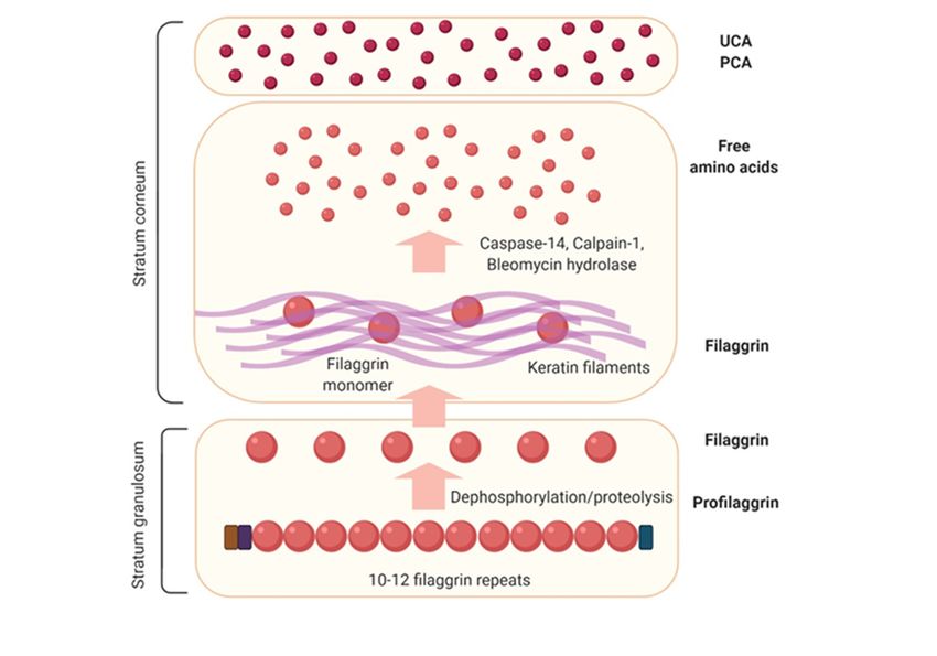
Int. J. Mol. Sci. 2020, 21, 2867 2 of 14
Int. J. Mol. Sci. 2020, 21, x FOR PEER REVIEW 2 of 14
dysfunction
SP inhibitors; inand
ADdisordered
involves an increase
tight in serum
junctions immunoglobulin
[4]. Pathogenesis relatedEto(IgE)
immune levels, sensitization
dysfunction in ADto
allergens,
involves an predominance
increase in serum of Th2 cytokines, an increase
immunoglobulin in T cells
E (IgE) levels, expressing
sensitization to cutaneous lymphocyte-
allergens, predominance
associated antigen,
of Th2 cytokines, anan increase
increase in Tin FcεRI
cells expression
expressing in inflammatory
cutaneous dendritic epidermal
lymphocyte-associated antigen, ancells and
increase
Langerhans cells, and
in FcεRI expression in increased
inflammatory expression
dendritic of epidermal
thymic stromal
cells andlymphopoietin
Langerhans cells,(TSLP) and(Figure 1).
increased
Recently,
expression in of
both the skin
thymic barrier
stromal and immunological
lymphopoietin aspects,1).genetic
(TSLP) (Figure mutations
Recently, in both related
the skintobarrier
AD haveand
been discovered.aspects,
immunological With respectgenetictomutations
the skin barrier,
relatedthere
to AD is have
typically
beena discovered.
mutation in Withthe filaggrin gene
respect to the
(FLG). There are
skin barrier, also
there single nucleotide
is typically a mutation polymorphisms
in the filaggriningenethe SP inhibitor
(FLG). ThereSPINK5
are alsoand thenucleotide
single SP KLK7,
and mutations in the
polymorphisms in thetightSPjunction
inhibitor protein
SPINK5claudin 1. InSPterms
and the KLK7,of and
immunological
mutations in responses,
the tightthere are
junction
mutations in the1.IgE
protein claudin In receptor FcεRb, the innate
terms of immunological immunity-related
responses, genes NOD1
there are mutations in theand
IgE-2receptor
and TLR2, -4
FcεRb,
and
the -9 and mutations
innate in the acquired
immunity-related genes NOD1immunity-related
and -2 and TLR2,genes -4
IL-4,
and-5,-9-9,
and-10,mutations
-12, -13, -18, andacquired
in the -31 and
TSLP [4].
immunity-related genes IL-4, -5, -9, -10, -12, -13, -18, and -31 and TSLP [4].
1. Skin
Figure 1.
Figure Skinbarrier
barrierabnormalities andand
abnormalities immune dysfunction
immune are theare
dysfunction main
thefeatures
main of atopic dermatitis.
features of atopic
dermatitis.
2. Skin Barrier Formation and Function
2. SkinThe most important
Barrier Formationfunction of the skin is to provide an effective barrier between the internal and
and Function
external environments of an organism. Thus, the skin acts as an interface between the organism and
The most important function of the skin is to provide an effective barrier between the internal
its external environment, providing both protection and support to the organism it encloses [5].
and external environments of an organism. Thus, the skin acts as an interface between the organism
Our understanding of the skin barrier is continuously evolving, in parallel with advances in
and its external environment, providing both protection and support to the organism it encloses [5].
research methods [6]. The epidermal barrier serves three primary functions: limiting passive
Our understanding of the skin barrier is continuously evolving, in parallel with advances in research
water loss, restricting environmental chemical absorption, and preventing microbial infection [7].
methods [6]. The epidermal barrier serves three primary functions: limiting passive water loss,
The epidermal barrier provides an outside-inside barrier that protects against mechanical, chemical,
restricting environmental chemical absorption, and preventing microbial infection [7]. The epidermal
and microbial injury through the formation of terminally differentiated keratinocytes, a process termed
barrier provides an outside-inside barrier that protects against mechanical, chemical, and microbial
keratinization [8]. During keratinization, epidermal cells progressively mature from the basal epidermal
injury through the formation of terminally differentiated keratinocytes, a process termed
layers to form flattened cells of the stratum corneum (SC) [8]. Within the epidermis, keratinocyte
keratinization [8]. During keratinization, epidermal cells progressively mature from the basal
proliferation is restricted to the basal cell layers. After mitosis in the basal layer, keratinocytes
epidermal layers to form flattened cells of the stratum corneum (SC) [8]. Within the epidermis,
keratinocyte proliferation is restricted to the basal cell layers. After mitosis in the basal layer,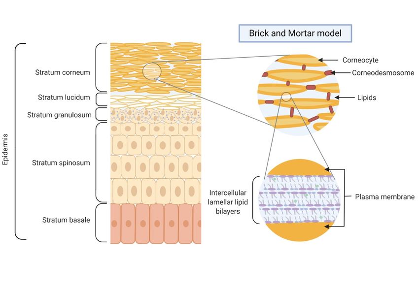
Int. J. Mol. Sci. 2020, 21, 2867 3 of 14
Int. J. Mol. Sci. 2020, 21, x FOR PEER REVIEW 3 of 14
keratinocytes
differentiate anddifferentiate
migrate throughand migrate through the
the epidermis epidermis
towards towards
the SC. the SC. The differentiation
The differentiation process yields
process yields several keratinocyte layers within the
several keratinocyte layers within the epidermis: the stratum basale, stratum epidermis: the stratum spinosum,
basale, stratumstratum
spinosum, stratum granulosum, and SC. Distinct marker genes are expressed
granulosum, and SC. Distinct marker genes are expressed by keratinocytes at each of the differentiation by keratinocytes at each
of the differentiation stages [9]. As the outermost layer of the skin, with a thickness of 10–20 μm, the
stages [9]. As the outermost layer of the skin, with a thickness of 10–20 µm, the SC is the primary mediator
SC is the primary mediator of the epidermal permeability barrier, accounting for over 90% of the
of the epidermal permeability barrier, accounting for over 90% of the functionality of the skin [6].
functionality of the skin [6]. Therefore, proper development and maintenance of the SC are essential
Therefore, proper development and maintenance of the SC are essential for maintaining its remarkable
for maintaining its remarkable ability to defend the body against both chemical and microbial attacks
ability to defend the body against both chemical and microbial attacks and dehydration [10]. A major
and dehydration [10]. A major defensive function of the skin is to maintain homeostasis by
defensive function of the skin is to maintain homeostasis by preventing the uncontrolled loss of water,
preventing the uncontrolled loss of water, ions, and serum proteins. A diverse set of strategies is used
ions,
by and
the SCserum proteins. epidermal
to maintain A diverse set of strategies
integrity, is used
including by the SC
enzymatic to maintain
reactions, epidermal
commensal integrity,
bacterial
including enzymatic reactions, commensal bacterial colonization, immune
colonization, immune signaling, antimicrobial lipids and peptides, low pH, and natural moisturizing signaling, antimicrobial
lipids and
factors [8].peptides,
The complex low tissue
pH, andof thenatural moisturizing
SC supports execution factors [8].strategies
of these The complexand istissue of the
composed of SC
supports execution of these strategies and is composed of corneocytes and
corneocytes and a matrix of intercellular lipids (ceramide, cholesterol, and free fatty acids), with botha matrix of intercellular
lipids (ceramide,
components cholesterol,
derived from theand free fatty
terminal acids), with
differentiation both components
process of keratinocytes derived
[11]. from the terminal
differentiation
Considerable process of keratinocytes
efforts have been made [11].to elucidate the full structure, function, and biochemistry
Considerable
of the efforts have
SC. Approximately threebeen made
decades ago,toElias
elucidate
proposedthe full
the structure,
“brick andfunction, and biochemistry
mortar model”, in which
ofcorneocytes (bricks) are embedded
the SC. Approximately three decadesin a continuous matrix of specialized
ago, Elias proposed the “brick andintercellular lipids (mortar)
mortar model”, in which
(Figure 2) [8].
corneocytes The corneocytes
(bricks) are embedded are responsible for protection
in a continuous matrix of against chemical
specialized and mechanical
intercellular lipidsinjury,
(mortar)
with the
(Figure lipid
2) [8]. Thematrix providing
corneocytes arethe essential for
responsible component
protection of against
the water barrier and
chemical [8]. The bulk of the
mechanical injury,
mechanical resistance offered by the epidermal barrier is due to corneocytes.
with the lipid matrix providing the essential component of the water barrier [8]. The bulk of the A protein shell termed
the corneocyte
mechanical envelope
resistance surrounds
offered by theeach corneocyte;
epidermal its components
barrier include loricrin,
is due to corneocytes. involucrin,
A protein and
shell termed
filaggrin.
the Beyond
corneocyte the corneocyte
envelope surrounds and each
in immediate
corneocyte; contact its with it sits the include
components corneocyte lipid envelope,
loricrin, involucrin,
which is a structure of specialized lipids. These lipids and protein-rich
and filaggrin. Beyond the corneocyte and in immediate contact with it sits the corneocyte corneocytes are critical for the
lipid
formation of the functional skin barrier. Thus, the barrier function of the normal epidermis is a
envelope, which is a structure of specialized lipids. These lipids and protein-rich corneocytes are critical
product of the quality of its brick and mortar components [12].
for the formation of the functional skin barrier. Thus, the barrier function of the normal epidermis is
a product of the quality of its brick and mortar components [12].
Figure 2. Schematic structure of the skin barrier and “brick and mortar” model.
3. Skin Barrier Abnormalities in Atopic Dermatitis
Accumulating evidence supports a permeability barrier dysfunction in AD. Decreased levels of
Figure 2. Schematic structure of the skin barrier and “brick and mortar” model.
total ceramides and bound ceramides in the SC have been reported [13]. In addition, an abnormal
expression of epidermal
3. Skin Barrier differentiation-related
Abnormalities in Atopic Dermatitismolecules, such as filaggrin, loricrin, and involucrin,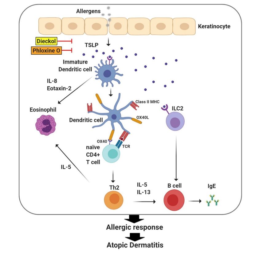
Int. J. Mol. Sci. 2020, 21, 2867 4 of 14
has been demonstrated in AD patients [14,15], and these molecules are expected to affect permeability
barrier homeostasis.
3.1. Lipids
Decreases in ceramide in both lesional and non-lesional skin of patients with atopic dermatitis are
uniquely observed, especially in those with filaggrin abnormalities. Moreover, it has been reported
that the ratio of ceramide and cholesterol is reduced in these patients [16]. In the stratum corneum
of atopic dermatitis patients, an increased pH and an elevated serine proteinase activity promote
inactivation and degradation of acid sphingomyelinase and β-glucocerebrosidase, which are the
necessary enzymes for ceramide synthesis [17]. An elevated serine proteinase activity reduces lamellar
body secretion through plasminogen activator type 2 (PAR2) signaling and results in the abnormal
transfer of various substances that are secreted from the lamellar body. This is ultimately related to
the reduced stratum corneum reported in patients with atopic dermatitis [18]. In addition, cytokine
cascades that are observed in various skin diseases associated with atopic dermatitis and skin barrier
abnormalities reduce the synthesis of ceramide by increasing interferon alpha (IFN-α) levels [17].
In the lesions of patients with atopic dermatitis, the chain lengths of ceramide, free fatty acids, and
esterified fatty acids are also shortened, which causes abnormalities in epidermal lipid organization
and results in epidermal barrier permeability abnormalities [19]. In patients with chronic atopic
dermatitis, increased IFN-α decreases two fatty acid elongases (i.e., elongation of very long chain fatty
acids protein (ELOVL) 1 and ELOVL4), resulting in shorter N-acyl chain lengths of free fatty acids and
ceramides [17]. The excessive increase in kallikrein activity may also lead to these changes in lipid
structure by inducing the degradation of elongation of very long chain fatty acids protein (ELOV) [17].
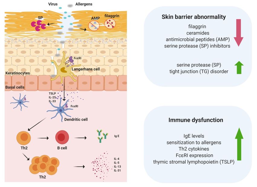
3.2. Filaggrin
Filaggrin is an important structural protein that is responsible for the keratinization, moisturization,
and antimicrobial peptide functions of the skin [20]. Filaggrin is a major component of keratohyalin
granules (KGs), which are membraneless protein deposits that eventually contribute to the formation
of a flattened, dead cell barrier at the skin surface [21]. The phase-separation property of filaggrin
family proteins is important for KG assembly and disassembly [21]. Genetic abnormalities related
to filaggrin are known to be strongly associated with atopic dermatitis [22]. In particular, filaggrin
deficiency has been reported to cause atopic dermatitis at an early age, increase the sensitivity and
severity of allergies, and increase infection vulnerability [23]. Filaggrin can be broken down into free
amino acids and converted into urocanic acid (UCA), which maintains the acidity level in the skin,
and pyrrolidine carboxylic acid (PCA), which acts as a natural moisturizer. Filaggrin abnormalities are
closely related to transepidermal water loss (TEWL) and dry skin in patients with atopic dermatitis.
A truncated mutation in filaggrin results in alteration in phase-separation dynamics and is linked to
skin barrier disorders [21]. Thus, characterizing the genetic abnormalities in filaggrin is important
for understanding the outside-inside mechanism in atopic dermatitis (Figure 3). However, 40% of
filaggrin mutation carriers do not develop atopic dermatitis, and filaggrin mutations are only found in
15%–50% of patients with atopic dermatitis [22]. Since genetic abnormalities in filaggrin alone do not
explain all the skin barrier dysfunctions of atopic dermatitis, further research is needed to clarify the
contribution of environmental and other possible factors to this condition. Recent studies have shown
that in addition to filaggrin abnormalities, abnormalities in the filaggrin-like proteins, hornerin and
Filaggrin family member 2 (FLG2), are also involved in either the lesional or non-lesional skin barrier
symptoms of atopic dermatitis [24]. Even patients with atopic dermatitis who do not harbor genetic
defects related to filaggrin can have decreased filaggrin levels later, suggesting that filaggrin is closely
related to the mechanism underlying the development of atopic dermatitis [25].Int. J. Mol. Sci. 2020, 21, 2867 5 of 14
Int. J. Mol. Sci. 2020, 21, x FOR PEER REVIEW 5 of 14
Figure 3. Life cycle of filaggrin. Filaggrin exists as profilaggrin within keratohyaline granules in
the granular
Figure 3.layer of theofepidermis.
Life cycle Profilaggrin
filaggrin. Filaggrin is degraded
exists to form
as profilaggrin filaggrin
within during the
keratohyaline terminal
granules in the
differentiation process. Then, in the upper part of the stratum corneum (SC), filaggrin
granular layer of the epidermis. Profilaggrin is degraded to form filaggrin during the terminalis degraded
into differentiation
amino acids and plays Then,
process. a crucial roleupper
in the in maintaining
part of theSC hydration
stratum and pH
corneum (SC),byfilaggrin
forming is
natural
degraded
moisturizing factors, including pyrrolidine carboxylic acid (PCA) and urocanic acid (UCA).
into amino acids and plays a crucial role in maintaining SC hydration and pH by forming natural
3.3. Tightmoisturizing factors, including pyrrolidine carboxylic acid (PCA) and urocanic acid (UCA).
Junctions (TJs)
TJsTight
3.3. are extremely
Junctions complex
(TJs) intracellular barriers that selectively control the cellular permeability
of soluble materials. As a skin barrier, TJs exist in the cell membranes of keratinocytes of the epidermal
TJs are extremely complex intracellular barriers that selectively control the cellular permeability
granule layer, and thus, these structures act as a second physical barrier in the epidermis [18]. Impaired
of soluble materials. As a skin barrier, TJs exist in the cell membranes of keratinocytes of the
TJs are attributed to abnormal skin barrier function in AD [26]. Knockout of claudin-1 (CLDN1),
epidermal granule layer, and thus, these structures act as a second physical barrier in the epidermis
which is the most important adhesion protein in TJs, in mice, results in a critical lethal epidermal barrier
[18]. Impaired TJs are attributed to abnormal skin barrier function in AD [26]. Knockout of claudin-1
defect, which highlights the importance of epidermal TJs and CLDN1 [27]. Reduced CLDN1 expression
(CLDN1), which is the most important adhesion protein in TJs, in mice, results in a critical lethal
in the nonlesional areas of patients with atopic dermatitis and the consequent TJ abnormalities have
epidermal barrier defect, which highlights the importance of epidermal TJs and CLDN1 [27].
been reported [28,29]. Yuki and colleagues stated that abnormalities in TJs adversely affect epidermal
Reduced CLDN1 expression in the nonlesional areas of patients with atopic dermatitis and the
lipids and metabolic processes associated with filaggrin [30].
consequent TJ abnormalities have been reported [28,29]. Yuki and colleagues stated that
abnormalities
4. Immune in TJs in
Dysfunction adversely affect epidermal lipids and metabolic processes associated with
Atopic Dermatitis
filaggrin [30].
The pathology of AD is accompanied by an imbalance in immunity involving Th1, Th2, and
Treg4.cells,
Immuneculminating in alterations
Dysfunction in Th1-
in Atopic and Th2-mediated immune responses and IgE-mediated
Dermatitis
hypersensitivity [31]. The expression levels of the Th2 cytokines IL-4 and IL-13 are elevated in AD
The pathology
lesions. Dupilumab, of AD is accompanied
a monoclonal antibody by an imbalance
against in immunity
the IL-4/IL-13 involving
receptor, Th1, Th2,
was effective forand
ADTreg
cells, culminating
treatment in alterations
in clinical studies, in Th1-
suggesting androle
a key Th2-mediated immune
of Th2 cytokines responses
in the pathologyandofIgE-mediated
AD [32].
hypersensitivity [31]. The expression levels of the Th2 cytokines IL-4 and IL-13
In an AD mouse model, the Treg cell population with a Th2 cytokine profile is increased, showing are elevated in AD
that pathogenic Treg cells are increased to exacerbate AD symptoms [33]. The immune system in AD AD
lesions. Dupilumab, a monoclonal antibody against the IL-4/IL-13 receptor, was effective for
treatment
becomes moreinheterogeneous
clinical studies,
andsuggesting a keythe
complex with role of Th2 cytokines
activation in the pathology
of other immune of as
cells, such ADTh22
[32]. In
an AD mouse
and Th17 cells [34]. model, the Treg cell population with a Th2 cytokine profile is increased, showing that
pathogenic Treg cells are increased to exacerbate AD symptoms [33]. The immune system in AD
becomes
5. TSLP more heterogeneous
Regulates Immune Responses and complex
in Atopicwith the activation of other immune cells, such as Th22
Dermatitis
and Th17 cells [34].
TSLP belongs to the cytokine family, is closely related to IL-7, and is expressed primarily by
epithelial cells in the skin, gastrointestinal tract, and lung [35]. Keratinocytes in AD skin express
5. TSLP Regulates Immune Responses in Atopic Dermatitis
high levels of TSLP, contributing to the initiation and exacerbation of immune responses in the skin
TSLP belongs to the cytokine family, is closely related to IL-7, and is expressed primarily by
epithelial cells in the skin, gastrointestinal tract, and lung [35]. Keratinocytes in AD skin express highInt. J. Mol. Sci. 2020, 21, 2867 6 of 14
Int. J. Mol. Sci. 2020, 21, x FOR PEER REVIEW 6 of 14
levels4)
(Figure of [35].
TSLP,TSLPcontributing
inducestothetheactivation
initiation and exacerbation
of dendritic cellsoftoimmune
interactresponses
with TSLP in the skin (Figure
receptor (TSLPR)
4) [35]. TSLP induces the activation of dendritic cells to interact with TSLP receptor
and interleukin-7-receptor subunit alpha (IL-7Rα), leading to the activation of intracellular signaling (TSLPR) and
interleukin-7-receptor subunit alpha (IL-7Rα), leading to the activation of intracellular signaling
pathways, such as signal transducer and activator of transcription (STAT)-5 [36]. TSLP induces the
pathways, such as signal transducer and activator of transcription (STAT)-5 [36]. TSLP induces the
expression of OX40 ligand (OX40L) in dendritic cells, which in turn induces the differentiation of naive
expression of OX40 ligand (OX40L) in dendritic cells, which in turn induces the differentiation of
CD4(+) T cells into Th2 cells to generate the Th2 cytokines IL-4, -5, and -13, with downregulation of
naive CD4(+) T cells into Th2 cells to generate the Th2 cytokines IL-4, -5, and -13, with downregulation
IL-10 and IFNγ [37–39]. TSLP/OX40L-induced Th2 responses are critical for the development of atopic
of IL-10 and IFNγ [37–39]. TSLP/OX40L-induced Th2 responses are critical for the development of
dermatitis. In addition, TSLP has been shown to activate other innate immune cells, such as natural
atopic dermatitis. In addition, TSLP has been shown to activate other innate immune cells, such as
killer T cells
natural andT basophils
killer and to modulate
cells and basophils B cell maturation
and to modulate [40]. Therefore,
B cell maturation TSLP is considered
[40]. Therefore, TSLP is
anconsidered
efficient therapeutic target to treat AD (Figure 4). Downregulation of TSLP expression
an efficient therapeutic target to treat AD (Figure 4). Downregulation of TSLP expression by dieckol,
a phlorotannin compound, is
by dieckol, a phlorotannin well corroborated
compound, by its efficacy
is well corroborated by itsin efficacy
improving AD-like skin
in improving symptoms
AD-like skin
in symptoms
a house dust in amite-induced AD model with
house dust mite-induced NC/Nga
AD model with mice
NC/Nga(Figuremice4) [41]. Similarly,
(Figure phloxine O,
4) [41]. Similarly,
a cosmetic
phloxinedye,O, a reduced
cosmeticTSLP expression
dye, reduced in keratinocytes
TSLP expression in and mouse skin,
keratinocytes andcorrelating
mouse skin,with alleviation
correlating
of with
AD-like symptoms and a decrease in serum IgE and histamine levels
alleviation of AD-like symptoms and a decrease in serum IgE and histamine levels in mice in mice (Figure 4) [42].
These results
(Figure show
4) [42]. the positive
These relationship
results show between
the positive the downregulation
relationship between the of TSLP expression
downregulation and the
of TSLP
expression
treatment and symptoms.
of AD the treatment of AD symptoms.
Figure 4. Thymic stromal lymphopoietin (TSLP) initiates the innate and adaptive phases of allergic
Figure 4.
immune Thymic stromal
responses lymphopoietin
in the skin. (TSLP)the
TSLP induces initiates the innate
maturation and adaptive
of dendritic cellsphases of allergic
to express OX40L,
immune
which responses
in turn in the skin.
differentiates naiveTSLP
CD4+ induces
T cells the
intomaturation
Th2 cells to ofproduce
dendriticTh2
cells to express
cytokines OX40L,
such as IL-4,
IL-5, and IL-13, leading to the secretion of IgE from B cells. Together with the activation of IL-4,
which in turn differentiates naive CD4+ T cells into Th2 cells to produce Th2 cytokines such as Group
IL-5, and
2 innate IL-13, leading
lymphoid to the secretion
cells (ILC2s), of IgE from
TSLP initiates B cells.and
the innate Together withimmune
adaptive the activation of Group
responses 2
of atopic
innate lymphoid cells (ILC2s), TSLP initiates the innate and adaptive immune responses
dermatitis. Dieckol and phloxine O reduce atopic dermatitis-like inflammatory symptoms by inhibiting of atopic
dermatitis.
TSLP Dieckol and phloxine O reduce atopic dermatitis-like inflammatory symptoms by
production.
inhibiting TSLP production.
6. The Involvement of ILC2s in the Pathogenesis of Atopic Dermatitis
6. The Involvement of ILC2s in the Pathogenesis of Atopic Dermatitis
Innate lymphoid cells (ILCs) are derived from common lymphoid progenitors and belong to the
lymphoid lineage. However, ILCs do not express antigen-specific receptors or myeloid or dendriticInt. J. Mol. Sci. 2020, 21, 2867 7 of 14
cell markers. ILCs play an important role in innate immune responses to infection and the regulation
of inflammation and metabolism. ILCs include natural killer (NK) cells, noncytotoxic groups of ILCs,
and lymphoid tissue-inducer (LTi) cells. ILCs are classified based on transcriptional profiles, effector
cytokines, and potential effector functions (Table 1). Among ILCs, Group 2 ILCs (ILC2s) have recently
drawn much attention because they participate in type 2 immunity. ILC2s respond to local Th2 antigens
derived from helminth and viral infection and produce type 2 cytokines, such as interleukin (IL)-4,
IL-5, IL-9, and IL-13, leading to the allergic inflammation process [43,44]. ILC2s are distributed in
various tissues, including skin, lung, liver, small intestine, bone marrow, spleen, and adipose tissue,
contributing to the regulation of Th2 immune responses, tissue homeostasis and repair, and metabolism.
Table 1. Classification of ILC subsets.
Cytokines
Transcription Stimulating Cytokine
Cell Type Required for Biological Function
Factors Cytokines Production
Development
T-bet IL-12 IFN-γ Immunity to virus and cancer
NK cell IL-15
EOMES IL-18 TNF Chronic inflammation
Group 1
ILCs Immunity to intracellular bacteria
IL-7 IL-12 IFN-γ
ILC1 T-bet and protozoa
IL-15 IL-18 TNF
Chronic inflammation
BCL11B IL-4 (in humans)
IL-25 Immunity to helminths
Group 2 GFI1 IL-5
ILC2 IL-7 IL-33 Asthma and allergic disease
ILCs EST1 IL-13
TSLP Metabolic homeostasis
GATA3 AREG
IL-23 IL-17A/IL-17F
LTi cell IL-7 RORγt
IL-1β IL-22
AHR IL-23 IL-17A/IL-17F Lymphoid tissue developments
Group 3 NCR- ILC3 IL-7
RORγt IL-1β IL-22 Intestinal homeostasis
ILCs
Immunity to extracellular bacteria
AHR Chronic inflammation
IL-23
NCR+ ILC3 IL-7 RORγt IL-22
IL-1β
T-bet
Dysregulation or chronic activation of ILCs leads to the exacerbation of allergic inflammatory
diseases, including atopic dermatitis. Although ILC2s are present in healthy human skin, the ILC2
population is increased in the AD lesional skin of patients [45]. In transgenic mice expressing interleukin
33 with atopic dermatitis symptoms, ILC2s were greatly increased in the lesional skin, regional lymph
nodes, and peripheral blood [46]. ILCs in the skin are activated by TSLP, IL-33, and IL-25, which are
highly expressed in atopic dermatitis. These findings suggest a critical role of ILC2s in allergic skin
diseases, such as atopic dermatitis.
7. Toll-Like Receptors and Atopic Dermatitis
It has been suggested that Toll-like receptors are associated with the pathology and severity of AD
immune responses. Impaired TLR2 function in the lesional skin of AD patients disrupts the normal
immune response to S. aureus, a commensal bacterium in the skin, thereby increasing the colonization of
S. aureus in the lesional skin area of AD patients. In addition, TLR2 levels were reduced in macrophages
or peripheral blood mononuclear cells (PBMCs) that were isolated from AD patients. TLR2 ligand
stimulation induces less production of Th1/Th17 cytokines such as IFNγ, IL-12, and IL-17F, and more
production of the Th2 cytokine IL-5 in macrophages or PBMCs of AD patients than in macrophages
and PBMCs from non-AD patients. Although TLR2 signaling in response to S. aureus is important for
protective immune responses during the acute and initial phases of AD, continuous activation of TLR2
promotes Th1 immune responses, leading to the exacerbation of inflammation at the later stage of AD.
Certain strains of Candida species reside in atopic skin, and viruses such as herpes simplex virus
may exacerbate other infections and thereby worsen the inflammatory symptoms of AD. TLR 1, 2,
6, and 9 are responsible for recognizing pathogen invasion and activating host immune responses,
suggesting a possible role of TLRs in the immune responses in AD lesional skin.Int. J. Mol. Sci. 2020, 21, 2867 8 of 14
Genetic polymorphisms in TLRs alter innate immune responses to infections, making AD skin
vulnerable to bacterial or viral infections. Monocytes from AD patients with heterozygous R753Q
polymorphism in TLR2 showed higher production of IL-6 and IL-12 in response to TLR2 than monocytes
with no mutation [47]. The frequency of the R753Q in TLR2 was significantly higher in Italian AD
children (16%) than in controls (6%), with a more severe phenotype [48]. However, there is a contrary
report showing no correlation between the R753Q mutation in TLR2 and the severity of AD in children
from Turkey [49]. The promoter polymorphism C-1237T in the TLR9 gene was associated with impaired
immunity in some cases of AD [50].
8. The Emerging Role of Inflammasomes in Atopic Dermatitis Symptoms
House dust mite allergens and S. aureus activate the NOD-, LRR- and pyrin domain-containing
protein 3 (NLRP3) inflammasome, suggesting a possible role of the NLRP3 inflammasome in
the pathogenesis of AD. Dermatophagoides pteronyssinus induces caspase-1 activation and IL-1β
secretion in keratinocytes. The assembly of the inflammasome complex, which consists of NLRP3,
apoptosis-associated speck-like protein containing a CARD (ASC), and caspase-1, was induced by
D. pteronyssinus. Knockdown of NLRP3, ASC, or caspase-1 results in suppression of the secretion of
IL-1β and IL-18 from keratinocytes stimulated with D. pteronyssinus [51].
The expression of NLRP3 and caspase-1 is downregulated in lesional AD skin compared with
healthy skin. The expression of caspase-1 is differentially regulated by Th1 and Th2 cytokines.
Th1 cytokines enhance, while Th2 cytokines reduce, the expression of caspase-1. Therefore, monocytes
from AD patients have impaired IL-1β secretion upon bacterial infection [52].
Since IL-1β induces TSLP expression in an NFκB-dependent manner [53], it is interesting
that inflammasome activation negatively regulates dibutyl phthalate-induced TSLP expression in
keratinocytes [54]. In addition, IL-33, another cytokine produced in AD skin to promote Th2 responses,
is inactivated by caspase-1 [55]. These results suggest that inflammasome activation may downregulate
Th2 immune responses due to the inhibition of TSLP and IL-33 expression.
Absent in melanoma 2 (AIM2), a double-stranded DNA receptor, forms inflammasomes that
produce IL-1β. AIM2 protein expression is elevated in AD skin, suggesting its role in the regulation of
AD immunity [56].
The relationship between single-nucleotide polymorphisms in NLRP3 and atopic dermatitis
susceptibility was studied in the Swedish AD population [57]. The polymorphism rs10733113 in
NLRP3 is associated with increased total IgE antibodies in male AD patients, but not in female
patients [57]. The significance of polymorphism in NLRP3 and its inflammasome components needs to
be further elucidated.
9. Crosstalk between the Skin Barrier and Immune System in Atopic Dermatitis
The normal intact skin barrier is considered the first host defense mechanism against microbiomes
and allergens that are linked with AD and often represents a part of the innate immune system.
Conversely, the host immune system and cytokines are interconnected with skin barrier protein
expression and function. Therefore, there exists a crosstalk between the skin barrier and immunity in
AD pathology.
Kuo et al. reported that increased TLR2 activity strengthens TJs, while the activity of TLR2 is
decreased in atopic dermatitis [58]. Enhanced TLR2 activity may help restore skin barrier function
through the restoration of TJ function in normal skin. TLR2 activation induces the expression of the TJ
protein CLDN1 and the antimicrobial peptides, β-defensins and cathelicidin, in normal keratinocytes.
However, TLR2 expression and function are altered in the lesional skin of AD patients, resulting in
decreased expression of TJ proteins and antimicrobial peptides.
These results support the hypothesis that decreased TJ function is strongly related to the abnormal
skin barrier in atopic dermatitis. A decrease in CLDN1 expression in atopic dermatitis is related to
increased susceptibility to herpes simplex virus-1 infection, the expression of Th2 immune markers,Int. J. Mol. Sci. 2020, 21, 2867 9 of 14
and the number of serum IgEs and eosinophils [59]. This indicates that CLDN1 is also associated with
the immune abnormalities of patients with atopic dermatitis and their susceptibility to infection.
In response to barrier disruption, keratinocytes produce type 2 mediators, such as TSLP, IL-25,
and IL-33, that activate basophils, ILC2s, and DCs [60]. In turn, ILC2s directly target TJs and reduce
the barrier integrity of human keratinocytes [61]. E-cadherin, a ligand of the lectin inhibitory receptor
KLRG1, has a regulatory role in ILC2 activation in atopic dermatitis. E-cadherin suppresses IL-5
and IL-13 production by ILC2s [45]. Since downregulation of E-cadherin is correlated with filaggrin
insufficiency, ILC2 activity is associated with dysregulation of skin barrier integrity [45].
Defects in the filaggrin gene disrupt the integrity of the skin barrier and facilitate bacterial
colonization and exposure to environmental factors, including allergens, leading to a skewed
polarization towards the Th2 phenotype [62]. Filaggrin mutation leads to loss of skin barrier function
and an increase in the ILC2 population, promoting acute skin inflammation and the development
of AD. Filaggrin mutation in mice results in spontaneous skin inflammation and increases in the
ILC2 population, IL-1β production, and other cytokines related to AD in skin. IL-1β and IL-1R1
signaling plays a critical role in chronic dermatitis inflammation in Filaggrin-mutant mice because
anti-IL-1β antibody treatment alleviated dermatitis symptoms [63]. Among 137 patients, IL-1β levels
are elevated in corneocytes from AD patients with filaggrin mutations compared with those of AD
patients without these mutations. Filaggrin mutations are correlated with a reduction in natural
moisturizing factors. Filaggrin-deficient mice show enhanced expression of IL-1β and IL-1RA mRNA
in skin and keratinocytes. These results suggest that there is a regulatory mechanism between filaggrin
and inflammasome activity in AD [64].
Protease-activated receptor 2 (PAR2) promotes Th2 inflammation and pruritus, and reduces the
integrity of the TJ barrier by disrupting claudin-1 and occluding proteins, suggesting a role for PAR2
in TJ expression and AD pathogenesis [65,66].
10. Development of Treatment Restoring Skin Barrier Abnormalities and Immune Dysfunction
Current mainstay treatments, including emollients, steroids, calcineurin inhibitors,
and immunosuppressants, have limited efficacy with potentially serious side effects. Recent studies
on the pathogenesis of AD have resulted in new therapies that target specific pathways with
increased efficacy and the potential for fewer systemic side effects. Impaired TJs are attributed
to abnormal skin barrier function in AD and may be an important mechanism contributing to
skin inflammation and unbalanced immune responses. Crisaborole and dupilumab are two new
FDA-approved therapies for AD [67,68]. Treatment of crisaborole, a phosphodiesterase-4B inhibitor
blocking cytokine expression, to AD patients downregulated genes involved in inflammation (MMP12),
Th2 (CCL22), Th1 (CXCL9, CXCL10), and Th17 (CXCL1, CXCL2), while it upregulated tight junction
claudin 8 (CLDN8) [69]. Dupilumab is a monoclonal antibody for IL-4 receptor α, blocking IL-4 and
IL-13 pathways, while increasing the expression of filaggrin, loricrin, claudins, and ELOVL3 to reverse
skin abnormalities [70]. Of the emerging therapies, the JAK-STAT inhibitors including baricitinib,
upadacitinib, PF-04965842, ASN002, tofacitinib, ruxolitinib, and delgocitinib show the most promising
results by restoring skin barrier function with increased filaggrin expression, as well as reducing
inflammatory signaling [71]. The aryl hydrocarbon receptor modulating agent tapinarof ameliorates
skin inflammation and induces barrier proteins such as filaggrin, hornerin, and involucrin [72,73].
IL-4/IL-13 antagonists, lebrikizumab and tralokinumab, showed clinical efficacy in AD patients,
demonstrating the critical role of IL-13 in AD pathology [74,75]. Since IL-4 and IL-13 downregulate
the expression of skin barrier proteins, such as filaggrin, loricrin, and involucrin, it is expected that
the treatment of IL-13 antagonists, such as lebrikizumab and tralokinumab, would influence the
recovery of skin barrier integrity [14]. Novel targeted therapeutic strategies are likely to lead to new
possibilities in generating tailored treatments and enhanced clinical efficacy to combat pediatric and
adult AD. Nanoparticles (NPs) have been proposed for the topical delivery of drugs used to treat skin
diseases [76]. NPs may help to reduce the adverse effects of classical drugs (e.g., topical corticosteroids),Int. J. Mol. Sci. 2020, 21, 2867 10 of 14
as NPs show an improved safety profile since lower doses are required, as a result of site-specific
delivery. NPs have also been put forward to address the problems of poor drug solubility and limited
skin permeability, thereby increasing skin bioavailability [77]. Several types of NPs have been proposed
for the topical delivery of sets of different drugs useful in AD (e.g., antibiotics and corticosteroids) [78].
Our study shows that the liposomal encapsulation of X-shaped oligonucleotides that act as TLR9
agonists magnifies the in vivo anti-AD efficacy of topical application, enabling the penetration of the
skin surface [79].
11. Conclusions
Although AD is not a life-threatening disease, it severely disrupts patients’ quality of life. The future
for AD patients is optimistic as many new therapies are being developed, and the pathogenesis of AD
is becoming clearer. Skin barrier abnormalities and immune dysfunction are important aspects for AD
pathology. They are interconnected and influence each other to initiate and aggravate AD symptoms.
The underlying mechanisms and key molecules regulating skin barrier function and immune responses
in AD are uncovered. The unveiling knowledge sheds a light to develop more efficient therapies
with less adverse effects for AD patients. In the near future, personalized care for patients with AD is
anticipated as these treatment methods include targeted therapeutics utilizing differential strategies
based on clinical phenotypes and endotypes of AD, biomarkers and molecular analyses, and patient
co-morbidities and complications.
Author Contributions: G.Y. and J.Y.L. contributed for the literature research, writing, and editing of the manuscript.
G.Y., J.K.S., and J.Y.L. conceptualized and finalized Figures and a Table. H.C.K. and J.Y.L. contributed to the project
administration and funding acquisition. Y.-Y.C. and H.S.L. supervised and contributed to the manuscript revision
and editing. All authors have read and agreed to the published version of the manuscript.
Funding: This study was supported by grants from the National Research Foundation of Korea
(NRF-2019R1A2C2085739 and NRF-2020R1A4A2002894) funded by the Korean government (Ministry of Science,
ICT and Future Planning).
Acknowledgments: Figures 1–4 were created with BioRender.com.
Conflicts of Interest: The authors declare no conflict of interest.
References
1. Spergel, J.M.; Paller, A.S. Atopic dermatitis and the atopic march. J. Allergy Clin. Immunol. 2003, 112, S118–S127.
[CrossRef] [PubMed]
2. Ellis, C.N.; Mancini, A.J.; Paller, A.S.; Simpson, E.L.; Eichenfield, L.F. Understanding and managing atopic
dermatitis in adult patients. Semin. Cutan. Med. Surg. 2012, 31, S18–S22. [CrossRef] [PubMed]
3. Novak, N.; Leung, D.Y. Advances in atopic dermatitis. Curr. Opin. Immunol. 2011, 23, 778–783. [CrossRef]
[PubMed]
4. Zaniboni, M.C.; Samorano, L.P.; Orfali, R.L.; Aoki, V. Skin barrier in atopic dermatitis: Beyond filaggrin.
An. Bras. Dermatol. 2016, 91, 472–478. [CrossRef]
5. Lee, S.H.; Jeong, S.K.; Ahn, S.K. An update of the defensive barrier function of skin. Yonsei Med. J. 2006, 47,
293–306. [CrossRef]
6. Darlenski, R.; Kazandjieva, J.; Tsankov, N. Skin barrier function: Morphological basis and regulatory
mechanisms. J. Clin. Med. 2011, 4, 36–45.
7. Wickett, R.R.; Visscher, M.O. Structure and function of the epidermal barrier. Am. J. Infect. Control. 2006, 34,
S98–S110. [CrossRef]
8. Pouillot, A.; Dayan, N.; Polla, A.S.; Polla, L.L.; Polla, B.S. The stratum corneum: A double paradox. J. Cosmet.
Dermatol. 2008, 7, 143–148. [CrossRef]
9. Elsholz, F.; Harteneck, C.; Muller, W.; Friedland, K. Calcium—A central regulator of keratinocyte
differentiation in health and disease. Eur. J. Dermatol. EJD 2014, 24, 650–661. [CrossRef]
10. Menon, G.K.; Cleary, G.W.; Lane, M.E. The structure and function of the stratum corneum. Int. J. Pharm.
2012, 435, 3–9. [CrossRef]
11. Elias, P.M. Skin barrier function. Curr. Allergy Asthma Rep. 2008, 8, 299–305. [CrossRef] [PubMed]Int. J. Mol. Sci. 2020, 21, 2867 11 of 14
12. Nemes, Z.; Steinert, P.M. Bricks and mortar of the epidermal barrier. Exp. Mol. Med. 1999, 31, 5–19. [CrossRef]
[PubMed]
13. Imokawa, G.; Abe, A.; Jin, K.; Higaki, Y.; Kawashima, M.; Hidano, A. Decreased level of ceramides in stratum
corneum of atopic dermatitis: An etiologic factor in atopic dry skin? J. Investig. Dermatol. 1991, 96, 523–526.
[CrossRef] [PubMed]
14. Howell, M.D.; Kim, B.E.; Gao, P.; Grant, A.V.; Boguniewicz, M.; Debenedetto, A.; Schneider, L.; Beck, L.A.;
Barnes, K.C.; Leung, D.Y. Cytokine modulation of atopic dermatitis filaggrin skin expression. J. Allergy Clin.
Immunol. 2007, 120, 150–155. [CrossRef]
15. Kim, B.E.; Leung, D.Y.; Boguniewicz, M.; Howell, M.D. Loricrin and involucrin expression is down-regulated
by Th2 cytokines through STAT-6. Clin. Immunol. 2008, 126, 332–337. [CrossRef]
16. Jungersted, J.M.; Scheer, H.; Mempel, M.; Baurecht, H.; Cifuentes, L.; Hogh, J.K.; Hellgren, L.I.; Jemec, G.B.;
Agner, T.; Weidinger, S. Stratum corneum lipids, skin barrier function and filaggrin mutations in patients
with atopic eczema. Allergy 2010, 65, 911–918. [CrossRef]
17. Elias, P.M.; Wakefield, J.S. Mechanisms of abnormal lamellar body secretion and the dysfunctional skin
barrier in patients with atopic dermatitis. J. Allergy Clin. Immunol. 2014, 134, 781–791. [CrossRef]
18. Wolf, R.; Wolf, D. Abnormal epidermal barrier in the pathogenesis of atopic dermatitis. Clin. Dermatol. 2012,
30, 329–334. [CrossRef]
19. Janssens, M.; van Smeden, J.; Gooris, G.S.; Bras, W.; Portale, G.; Caspers, P.J.; Vreeken, R.J.; Hankemeier, T.;
Kezic, S.; Wolterbeek, R.; et al. Increase in short-chain ceramides correlates with an altered lipid organization
and decreased barrier function in atopic eczema patients. J. Lipid Res. 2012, 53, 2755–2766. [CrossRef]
20. Malik, K.; Heitmiller, K.D.; Czarnowicki, T. An Update on the Pathophysiology of Atopic Dermatitis.
Dermatol. Clin. 2017, 35, 317–326. [CrossRef]
21. Quiroz, F.G.; Fiore, V.F.; Levorse, J.; Polak, L.; Wong, E.; Pasolli, H.A.; Fuchs, E. Liquid-liquid phase separation
drives skin barrier formation. Science 2020, 367, 6483. [CrossRef] [PubMed]
22. Palmer, C.N.; Irvine, A.D.; Terron-Kwiatkowski, A.; Zhao, Y.; Liao, H.; Lee, S.P.; Goudie, D.R.; Sandilands, A.;
Campbell, L.E.; Smith, F.J.; et al. Common loss-of-function variants of the epidermal barrier protein filaggrin
are a major predisposing factor for atopic dermatitis. Nat. Genet. 2006, 38, 441–446. [CrossRef] [PubMed]
23. Szegedi, A. Filaggrin mutations in early- and late-onset atopic dermatitis. Br. J. Dermatol. 2015, 172, 320–321.
[CrossRef] [PubMed]
24. Pellerin, L.; Henry, J.; Hsu, C.Y.; Balica, S.; Jean-Decoster, C.; Mechin, M.C.; Hansmann, B.; Rodriguez, E.;
Weindinger, S.; Schmitt, A.M.; et al. Defects of filaggrin-like proteins in both lesional and nonlesional
atopic skin. J. Allergy Clin. Immunol. 2013, 131, 1094–1102. [CrossRef]
25. Thyssen, J.P.; Kezic, S. Causes of epidermal filaggrin reduction and their role in the pathogenesis of atopic
dermatitis. J. Allergy Clin. Immunol. 2014, 134, 792–799. [CrossRef]
26. De Benedetto, A.; Rafaels, N.M.; McGirt, L.Y.; Ivanov, A.I.; Georas, S.N.; Cheadle, C.; Berger, A.E.; Zhang, K.;
Vidyasagar, S.; Yoshida, T. Tight junction defects in patients with atopic dermatitis. J. Allergy Clin. Immunol.
2011, 127, 773–786. [CrossRef]
27. Furuse, M.; Hata, M.; Furuse, K.; Yoshida, Y.; Haratake, A.; Sugitani, Y.; Noda, T.; Kubo, A.;
Tsukita, S. Claudin-based tight junctions are crucial for the mammalian epidermal barrier: A lesson
from claudin-1-deficient mice. J. Cell Biol. 2002, 156, 1099–1111. [CrossRef]
28. Bergmann, S.; von Buenau, B.; Vidal, Y.S.S.; Haftek, M.; Wladykowski, E.; Houdek, P.; Lezius, S.; Duplan, H.;
Basler, K.; Dahnhardt-Pfeiffer, S.; et al. Claudin-1 decrease impacts epidermal barrier function in atopic
dermatitis lesions dose-dependently. Sci. Rep. 2020, 10, 2024. [CrossRef]
29. Tokumasu, R.; Yamaga, K.; Yamazaki, Y.; Murota, H.; Suzuki, K.; Tamura, A.; Bando, K.; Furuta, Y.;
Katayama, I.; Tsukita, S. Dose-dependent role of claudin-1 in vivo in orchestrating features of atopic
dermatitis. Proc. Natl. Acad. Sci. USA 2016, 113, E4061–E4068. [CrossRef]
30. Yuki, T.; Komiya, A.; Kusaka, A.; Kuze, T.; Sugiyama, Y.; Inoue, S. Impaired tight junctions obstruct stratum
corneum formation by altering polar lipid and profilaggrin processing. J. Dermatol. Sci. 2013, 69, 148–158.
[CrossRef]
31. Sheikhi, A.; Giti, H.; Heibor, M.R.; Jafarzadeh, A.; Shakerian, M.; Baharifar, N.; Niruzad, F.; Moghaddam, A.S.;
Kokhaei, P.; Baghaeifar, M. Lactobacilus delbrueckii subsp. bulgaricus modulates the secretion of Th1/Th2
and Treg cell-related cytokines by PBMCs from patients with atopic dermatitis. Drug Res. 2017, 67, 724–729.
[CrossRef] [PubMed]Int. J. Mol. Sci. 2020, 21, 2867 12 of 14
32. Roesner, L.M.; Werfel, T.; Heratizadeh, A. The adaptive immune system in atopic dermatitis and implications
on therapy. Expert Rev. Clin. Immunol. 2016, 12, 787–796. [CrossRef] [PubMed]
33. Moosbrugger-Martinz, V.; Tripp, C.H.; Clausen, B.E.; Schmuth, M.; Dubrac, S. Atopic dermatitis induces the
expansion of thymus-derived regulatory T cells exhibiting a Th2-like phenotype in mice. J. Cell. Mol. Med.
2016, 20, 930–938. [CrossRef] [PubMed]
34. Brunner, P.M.; Guttman-Yassky, E.; Leung, D.Y. The immunology of atopic dermatitis and its reversibility
with broad-spectrum and targeted therapies. J. Allergy Clin. Immunol. 2017, 139, S65–S76. [CrossRef]
35. Cianferoni, A.; Spergel, J. The importance of TSLP in allergic disease and its role as a potential therapeutic
target. Expert Rev. Clin. Immunol. 2014, 10, 1463–1474. [CrossRef]
36. Quentmeier, H.; Drexler, H.; Fleckenstein, D.; Zaborski, M.; Armstrong, A.; Sims, J.; Lyman, S. Cloning of
human thymic stromal lymphopoietin (TSLP) and signaling mechanisms leading to proliferation. Leukemia
2001, 15, 1286–1292. [CrossRef]
37. Ito, T.; Wang, Y.-H.; Duramad, O.; Hori, T.; Delespesse, G.J.; Watanabe, N.; Qin, F.X.-F.; Yao, Z.; Cao, W.;
Liu, Y.-J. TSLP-activated dendritic cells induce an inflammatory T helper type 2 cell response through
OX40 ligand. J. Exp. Med. 2005, 202, 1213–1223. [CrossRef]
38. Soumelis, V.; Reche, P.A.; Kanzler, H.; Yuan, W.; Edward, G.; Homey, B.; Gilliet, M.; Ho, S.; Antonenko, S.;
Lauerma, A. Human epithelial cells trigger dendritic cell–mediated allergic inflammation by producing TSLP.
Nat. Immunol. 2002, 3, 673–680. [CrossRef]
39. Liu, Y.-J. Thymic stromal lymphopoietin and OX40 ligand pathway in the initiation of dendritic cell–mediated
allergic inflammation. J. Allergy Clin. Immunol. 2007, 120, 238–244. [CrossRef]
40. Zhang, Y.; Zhou, X.; Zhou, B. DC-derived TSLP promotes T h2 polarization in LPS-primed allergic airway
inflammation. Eur. J. Immunol. 2012, 42, 1735–1743. [CrossRef]
41. Yang, G.; Oh, J.W.; Lee, H.E.; Lee, B.H.; Lim, K.M.; Lee, J.Y. Topical Application of Dieckol Ameliorates
Atopic Dermatitis in NC/Nga Mice by Suppressing Thymic Stromal Lymphopoietin Production. J. Investig.
Dermatol. 2016, 136, 1062–1066. [CrossRef] [PubMed]
42. Lee, H.E.; Yang, G.; Kim, K.B.; Lee, B.M.; Lee, J.Y. Phloxine O, a Cosmetic Colorant, Suppresses the Expression
of Thymic Stromal Lymphopoietin and Acute Dermatitis Symptoms in Mice. Biomol. Ther. 2018, 26, 481–486.
[CrossRef] [PubMed]
43. Moro, K.; Yamada, T.; Tanabe, M.; Takeuchi, T.; Ikawa, T.; Kawamoto, H.; Furusawa, J.-I.; Ohtani, M.; Fujii, H.;
Koyasu, S. Innate production of TH 2 cytokines by adipose tissue-associated c-Kit+ Sca-1+ lymphoid cells.
Nature 2010, 463, 540–544. [CrossRef] [PubMed]
44. Neill, D.R.; Wong, S.H.; Bellosi, A.; Flynn, R.J.; Daly, M.; Langford, T.K.; Bucks, C.; Kane, C.M.; Fallon, P.G.;
Pannell, R. Nuocytes represent a new innate effector leukocyte that mediates type-2 immunity. Nature 2010,
464, 1367–1370. [CrossRef]
45. Salimi, M.; Barlow, J.L.; Saunders, S.P.; Xue, L.; Gutowska-Owsiak, D.; Wang, X.; Huang, L.C.; Johnson, D.;
Scanlon, S.T.; McKenzie, A.N.; et al. A role for IL-25 and IL-33-driven type-2 innate lymphoid cells in atopic
dermatitis. J. Exp. Med. 2013, 210, 2939–2950. [CrossRef]
46. Imai, Y.; Yasuda, K.; Sakaguchi, Y.; Haneda, T.; Mizutani, H.; Yoshimoto, T.; Nakanishi, K.; Yamanishi, K.
Skin-specific expression of IL-33 activates group 2 innate lymphoid cells and elicits atopic dermatitis-like
inflammation in mice. Proc. Natl. Acad. Sci. USA 2013, 110, 13921–13926. [CrossRef]
47. Niebuhr, M.; Langnickel, J.; Draing, C.; Renz, H.; Kapp, A.; Werfel, T. Dysregulation of toll-like receptor-2
(TLR-2)-induced effects in monocytes from patients with atopic dermatitis: Impact of the TLR-2 R753Q
polymorphism. Allergy 2008, 63, 728–734. [CrossRef]
48. Salpietro, C.; Rigoli, L.; Miraglia Del Giudice, M.; Cuppari, C.; Di Bella, C.; Salpietro, A.; Maiello, N.;
La Rosa, M.; Marseglia, G.L.; Leonardi, S.; et al. TLR2 and TLR4 gene polymorphisms and atopic dermatitis
in Italian children: A multicenter study. Int. J. Immunopathol. Pharm. 2011, 24, 33–40. [CrossRef]
49. Can, C.; Yazicioglu, M.; Gurkan, H.; Tozkir, H.; Gorgulu, A.; Sut, N.H. Lack of Association between Toll-like
Receptor 2 Polymorphisms (R753Q and A-16934T) and Atopic Dermatitis in Children from Thrace Region of
Turkey. Balkan Med. J. 2017, 34, 232–238. [CrossRef]
50. Novak, N.; Yu, C.F.; Bussmann, C.; Maintz, L.; Peng, W.M.; Hart, J.; Hagemann, T.; Diaz-Lacava, A.;
Baurecht, H.J.; Klopp, N.; et al. Putative association of a TLR9 promoter polymorphism with atopic eczema.
Allergy 2007, 62, 766–772. [CrossRef]Int. J. Mol. Sci. 2020, 21, 2867 13 of 14
51. Dai, X.; Sayama, K.; Tohyama, M.; Shirakata, Y.; Hanakawa, Y.; Tokumaru, S.; Yang, L.; Hirakawa, S.;
Hashimoto, K. Mite allergen is a danger signal for the skin via activation of inflammasome in keratinocytes.
J. Allergy Clin. Immunol. 2011, 127, 806–814. [CrossRef] [PubMed]
52. Niebuhr, M.; Baumert, K.; Heratizadeh, A.; Satzger, I.; Werfel, T. Impaired NLRP3 inflammasome expression
and function in atopic dermatitis due to Th2 milieu. Allergy 2014, 69, 1058–1067. [CrossRef] [PubMed]
53. Yamanaka, K.; Tanaka, M.; Tsutsui, H.; Kupper, T.S.; Asahi, K.; Okamura, H.; Nakanishi, K.; Suzuki, M.;
Kayagaki, N.; Black, R.A.; et al. Skin-specific caspase-1-transgenic mice show cutaneous apoptosis and
pre-endotoxin shock condition with a high serum level of IL-18. J. Immunol. 2000, 165, 997–1003. [CrossRef]
[PubMed]
54. Schuepbach-Mallepell, S.; Philippe, V.; Bruggen, M.C.; Watanabe, H.; Roques, S.; Baldeschi, C.; Gaide, O.
Antagonistic effect of the inflammasome on thymic stromal lymphopoietin expression in the skin. J. Allergy
Clin. Immunol. 2013, 132, 1348–1357. [CrossRef] [PubMed]
55. Cayrol, C.; Girard, J.P. The IL-1-like cytokine IL-33 is inactivated after maturation by caspase-1. Proc. Natl.
Acad. Sci. USA 2009, 106, 9021–9026. [CrossRef] [PubMed]
56. De Koning, H.D.; Bergboer, J.G.; van den Bogaard, E.H.; van Vlijmen-Willems, I.M.; Rodijk-Olthuis, D.;
Simon, A.; Zeeuwen, P.L.; Schalkwijk, J. Strong induction of AIM2 expression in human epidermis in acute
and chronic inflammatory skin conditions. Exp. Dermatol. 2012, 21, 961–964. [CrossRef]
57. Bivik, C.; Verma, D.; Winge, M.C.; Lieden, A.; Bradley, M.; Rosdahl, I.; Soderkvist, P. Genetic variation in the
inflammasome and atopic dermatitis susceptibility. J. Investig. Dermatol. 2013, 133, 2486–2489. [CrossRef]
58. Kuo, I.H.; Carpenter-Mendini, A.; Yoshida, T.; McGirt, L.Y.; Ivanov, A.I.; Barnes, K.C.; Gallo, R.L.;
Borkowski, A.W.; Yamasaki, K.; Leung, D.Y.; et al. Activation of epidermal toll-like receptor 2 enhances tight
junction function: Implications for atopic dermatitis and skin barrier repair. J. Investig. Dermatol. 2013, 133,
988–998. [CrossRef]
59. Leung, D.Y. New insights into atopic dermatitis: Role of skin barrier and immune dysregulation. Allergol. Int.
Off. J. Jpn. Soc. Allergol. 2013, 62, 151–161. [CrossRef]
60. Dainichi, T.; Kitoh, A.; Otsuka, A.; Nakajima, S.; Nomura, T.; Kaplan, D.H.; Kabashima, K. The epithelial
immune microenvironment (EIME) in atopic dermatitis and psoriasis. Nat. Immunol. 2018, 19, 1286–1298.
[CrossRef]
61. Halim, T.Y.; Steer, C.A.; Mathä, L.; Gold, M.J.; Martinez-Gonzalez, I.; McNagny, K.M.; McKenzie, A.N.;
Takei, F. Group 2 innate lymphoid cells are critical for the initiation of adaptive T helper 2 cell-mediated
allergic lung inflammation. Immunity 2014, 40, 425–435. [CrossRef] [PubMed]
62. Mu, Z.; Zhao, Y.; Liu, X.; Chang, C.; Zhang, J. Molecular biology of atopic dermatitis. Clin. Rev. Allergy
Immunol. 2014, 47, 193–218. [CrossRef] [PubMed]
63. Schwartz, C.; Moran, T.; Saunders, S.P.; Kaszlikowska, A.; Floudas, A.; Bom, J.; Nunez, G.; Iwakura, Y.;
O’Neill, L.; Irvine, A.D.; et al. Spontaneous atopic dermatitis in mice with a defective skin barrier is
independent of ILC2 and mediated by IL-1beta. Allergy 2019, 74, 1920–1933. [CrossRef] [PubMed]
64. Kezic, S.; O’Regan, G.M.; Lutter, R.; Jakasa, I.; Koster, E.S.; Saunders, S.; Caspers, P.; Kemperman, P.M.;
Puppels, G.J.; Sandilands, A.; et al. Filaggrin loss-of-function mutations are associated with enhanced
expression of IL-1 cytokines in the stratum corneum of patients with atopic dermatitis and in a murine model
of filaggrin deficiency. J. Allergy Clin. Immunol. 2012, 129, 1031–1039. [CrossRef] [PubMed]
65. Henehan, M.; De Benedetto, A. Update on protease-activated receptor 2 in cutaneous barrier, differentiation,
tumorigenesis and pigmentation, and its role in related dermatologic diseases. Exp. Dermatol. 2019, 28,
877–885. [CrossRef] [PubMed]
66. Nadeau, P.; Henehan, M.; De Benedetto, A. Activation of protease-activated receptor 2 leads to impairment
of keratinocyte tight junction integrity. J. Allergy Clin. Immunol. 2018, 142, 281–284. [CrossRef]
67. Blauvelt, A.; de Bruin-Weller, M.; Gooderham, M.; Cather, J.C.; Weisman, J.; Pariser, D.; Simpson, E.L.;
Papp, K.A.; Hong, H.C.-H.; Rubel, D. Long-term management of moderate-to-severe atopic dermatitis
with dupilumab and concomitant topical corticosteroids (LIBERTY AD CHRONOS): A 1-year, randomised,
double-blinded, placebo-controlled, phase 3 trial. Lancet 2017, 389, 2287–2303. [CrossRef]
68. Dina Coronado, B.; Zane, L.T. Crisaborole topical ointment, 2%: A nonsteroidal, topical, anti-inflammatory
phosphodiesterase 4 inhibitor in clinical development for the treatment of atopic dermatitis. J. Drugs Dermatol.
2016, 15, 390–396.You can also read