Apple of Sodom (Calatropis procera) Callus Extract, a Novel Skincare Active and Its Biological Activity in Skin Models When Combined with Dead Sea ...
←
→
Page content transcription
If your browser does not render page correctly, please read the page content below
Journal of Cosmetics, Dermatological Sciences and Applications, 2018, 8, 73-91
                                                                                                http://www.scirp.org/journal/jcdsa
                                                                                                           ISSN Online: 2161-4512
                                                                                                             ISSN Print: 2161-4105

Apple of Sodom (Calatropis procera) Callus
Extract, a Novel Skincare Active and Its
Biological Activity in Skin Models When
Combined with Dead Sea Water

Meital Portugal-Cohen1,2*, Eliran Ish-Shalom1,2, Ruben Mallon3, Patricia Corral3,
Franck Michoux3, Ze’evi Ma’or1,2
1
 Ahava Dead Sea Laboratories, Lod, Israel
2
 The Dead Sea Laboratory for Skin Biochemistry and Biotechnology, Dead Sea and
Arava Science Center, Masada, Israel
3
 Evonik Advanced Botanicals, Parçay Meslay, France

How to cite this paper: Portugal-Cohen,       Abstract
M., Ish-Shalom, E., Mallon, R., Corral, P.,
Michoux, F. and Ma’or, Z. (2018) Apple of     Background: Calotropis procera (C. procera), is an authentic plant naturally
Sodom (Calatropis procera) Callus Extract,    grown in the flora of Dead Sea region. Despite its toxicity, C. procera presents
a Novel Skincare Active and Its Biological
Activity in Skin Models When Combined
                                              healing properties. However, it has not been implemented yet in cosmetics as
with Dead Sea Water. Journal of Cosmetics,    an active ingredient. Objective: The biological effects of C. procera callus ex-
Dermatological Sciences and Applications,     tract on skin were elucidated solely and in combination with Dead Sea water
8, 73-91.
                                              (DSW). Methods: The capability of C. procera extract to protect against skin
https://doi.org/10.4236/jcdsa.2018.82010
                                              inflammation and irritation was tested on ex vivo human skin organ culture
Received: March 22, 2018                      by LPS and SDS addition respectively. Viability and cytokine secretion were
Accepted: June 26, 2018                       evaluated. The combination of C. procera extract with Dead Sea water was
Published: June 29, 2018
                                              tested on full thickness skin equivalents. Gene expression and relevant bio-
Copyright © 2018 by authors and               chemical markers for glycolysis, hypoxia and extracellular matrix balance
Scientific Research Publishing Inc.           were tested. Results: C. procera extract exhibits a protective biological activity
This work is licensed under the Creative      against skin irritation and inflammation at the biochemical level. Further-
Commons Attribution International
License (CC BY 4.0).
                                              more, a combination of C. procera extract and DSW demonstrates a potential
http://creativecommons.org/licenses/by/4.0/   contribution for skin wellbeing via enhance energy production, resistance to
                Open Access                   hypoxia and extracellular matrix balance. Conclusions: Topical application of
                                              C. procera callus extract might support skin balance and wellbeing at the mo-
                                              lecular level. Hence, it is recommended for new cosmetic formulae as standa-
                                              lone or in combination with Dead Sea water, in the effort to achieve an-
                                              ti-aging bio-activity that is working beyond skin aging symptoms, especially
                                              via skin calming effects and skin energy enhancement.

DOI: 10.4236/jcdsa.2018.82010        Jun. 29, 2018               73          J. Cosmetics, Dermatological Sciences and Applications
M. Portugal-Cohen et al.

                                  Keywords
                                  C. procera Callus Extract, Alternative Skin Models, Inflammation, Irritation,
                                  Aging, Dead Sea Water

                                1. Introduction
                                Authenticity is an increasing trend in last years’ beauty business, changing pop-
                                ular list of ingredients to include more endemic ingredients in new cosmetic
                                launches, especially selective minerals collected from specific lands, local springs,
                                spas and seas, and extracts of endemic plants [1]. The growing awareness to side
                                effects of synthetic drugs invigorates scientists exploring ethnic medicinal reme-
                                dies as potential new actives for dermatological and cosmetic formulae.
                                  The plant Calotropis procera, also known as “Apple of Sodom” is grown as a
                                woody shrub in desert oases in the hot regions and is a native plant in the flora
                                of Dead Sea region [2] [3]. The extracts derived from C. procera’s different plant
                                parts are believed to possess various powerful deeds including anti-bacterial, an-
                                ti-inflammatory, anti-diabetic and anti-cancer [2] [3] [4]. In spite of its poison-
                                ing threats, various skin problems, such as wounds, sores, external infections,
                                swelling and eczema, are treated by C. procera’s preparations “prescribed” by
                                traditional healers [2] [3]. Yet, C. procera has not been exploited yet as an active
                                to be formulated in modern skincare products.
                                  The benefits of Dead Sea minerals for skin health and beauty are well estab-
                                lished and formulating Dead Sea different extracts, i.e. DS Mud, DS solid Salts
                                and DS brines, are widespread in modern cosmetics [5]-[10]. Blending Dead Sea
                                minerals and plant extracts were proven to have skin beneficial effects and dif-
                                ferent mineral-plant extract combinations are formulated and patented as inno-
                                vative complexes for new cosmetic products [9] [11] [12]. Due to regulatory
                                banning of animal tests [13], skin models are effectively used today, looking for
                                the bio mechanism beyond natural and pathogenic skin phenomena. The wish to
                                successfully attenuate skin aging is the main engine of huge beauty industry [14].
                                Skin aging reflects the accumulation of many damaged molecules during skin
                                exposure to a broad spectrum of stressors [15]. This inevitable accumulation
                                leads to the activation of “pro-aging” biochemical pathways and could eventually
                                result with the appearance of skin aging symptoms and skin pathologies, derma-
                                titis, rosacea, seborrheic and other pathological skin conditions including skin
                                cancers [16] [17] [18]. Inflammatory processes are key mediators of the different
                                pathways leading to skin aging and therefore, many research activities are tar-
                                geting protective effects against inflammation using modern skin laboratory in
                                vitro and ex vivo models [19].
                                  This work describes the results of a research, focused on the biological an-
                                ti-aging and anti-inflammatory skin effects of a callus extract of the plant C.
                                procera when topically applied and combined with Dead Sea water.

DOI: 10.4236/jcdsa.2018.82010                         74         J. Cosmetics, Dermatological Sciences and Applications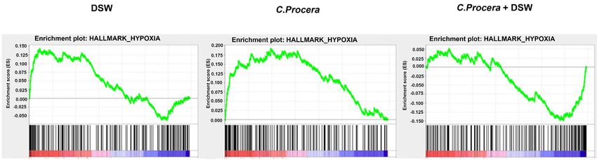
M. Portugal-Cohen et al.

                                2. Methods
                                2.1. Preparation of Test Materials
                                Test materials consist of either C. procera callus extract, either Dead Sea water
                                (DSW) extract (Osmoter), or their combination.

                                2.2. Calotropis procera Extract
                                Calotropis procera callus was developed from root explants grown under aseptic
                                conditions on Murashige and Skoog medium [20] supplemented with 3% su-
                                crose, 27 µM naphthalene acetic acid, 0.5 µM Thidiazuron and solidified with
                                0.25% gelrite.
                                  Calotropis procera dried callus was grounded and callus powder was extracted
                                with hot water by using a Soxhlet extractor during 3 h. After extraction, the sol-
                                vent was partially evaporated, using a rotary evaporator (RC600, KNF). The
                                concentrated solvent with the crude extract was frozen at −80˚C for 24 h and
                                freeze-dried until complete dryness. Crude extract was ground and solubilized in
                                vegetable glycerol and water at a final ratio of 4% crude extract, 16% water and
                                80% glycerol.
                                   Chemical analysis by HPLC and mass spectroscopy of Calotropis procera cal-
                                lus extract: The dry extract was re-suspended in 250 µL of 80% MeOH. Prior to
                                injection (20 µl) into the HPLC system, the sample was filtered through a 0.45
                                µm filter (Minisart RC4, Sartorius). Analysis of the presence of cardenolides was
                                performed by an HPLC Jasco System instrument consisting of a pump (Pu-2089
                                Plus) and a diode array detector (MD 2018 Plus). The HPLC column was Luna 2
                                C18 (250 × 4.6 mm, 5 µm) fitted with a guard cartridge (Securityguard system,
                                Phenomenex, France). A linear gradient at a flow of 0.7 ml/min was used for 25
                                min. The mobile phase consisted of a solvent A (ultra pure water) and a solvent
                                B (pure Acetonitrile). Gradient was as follow: 0 - 4 min 25% solvent B; 24 min
                                solvent B increased to 50%; 29 min solvent B increased to 70% and 31 min 100%
                                solvent B. All solvents used were of HPLC grade quality. Detection wavelength
                                was 220 nm. Mass identification of major peaks was performed with an
                                UPLC-ESI/MS consisted of a LC system Acquity I-Class UPLC with a PDA de-
                                tector linked to a mass spectrometry Instrument Waters LCT Premier (Milford,
                                MA, USA). The chromatographic separation was realized with a C18 UPLC
                                Column (50 × 2.1 mm, 1.7 μm). The mobile phase consisted of A: water + 0.9%
                                formic acid and B: 100% MeOH. The gradient (from 30% to 80% of B in 3 min)
                                was eluted at a flow rate of 0.31 mL/min. Source voltage: 2000 V; sample cone:
                                30 V; desolvation: 350˚C; source: 120˚C; Gas Flow Cone: 10 L/h; desolvation gas:
                                400 L/h; using the full scan mode and a m/z range of 100 - 1000.

                                2.3. Dead Sea Water (DSW) Extract
                                The major constituents of DSW (OsmoterTM by AHAVA) are the following ions:
                                Mg2+ (92,500 mg/L), Ca2+ (38,000 mg/L), K+ (1400 mg/L), Na+ (2000 mg/L), Sr2+
                                (800 mg/L), Cl- (345,000 mg/L) and Br- (11,500 mg/L).

DOI: 10.4236/jcdsa.2018.82010                        75         J. Cosmetics, Dermatological Sciences and Applications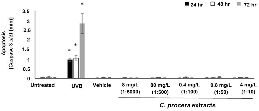
M. Portugal-Cohen et al.

                                2.4. Human Skin Organ Culture for Biological Tests
                                Ex vivo human skin organ culture (HSOC) [19] was used as a representative skin
                                laboratory model for testing C. procera extract safety threshold and its protec-
                                tion against inflammation and irritation.
                                  Human skin cultures were obtained from healthy females (age 23 - 45) un-
                                dergoing abdominal plastic surgery. The study was initiated at the day of sur-
                                gery. Fixed size of skin pieces (0.64 cm2) were cut from the skin tissue, using a
                                designated press apparatus. The skin pieces were placed in culture medium
                                (DMEM supplemented with 100 U/ml penicillin and 100 μg/ml streptomycin),
                                dermal side down in the medium and epidermis up. The pieces were incubated
                                overnight at 37˚C with 5% CO2 for recovery for 24 hr.

                                2.5. Treatment with C. procera Callus Extracts for Dose Response
                                     Analysis
                                After recovery, C. procera callus extract was applied on the skin pieces topically
                                at five different concentrations by a serial dilution of 1:10, 1:50, 1:100, 1:500,
                                1:5000. Topical application of 3 μl was mounted on the skin. The dilution of the
                                C. procera callus extract was carried out in its original formulation i.e., Glyce-
                                rol:DDW (80%:20%) mixture containing C. procera at a concentration of 40 g/L,
                                as in detailed in the formulation section.
                                  The Control group represents Naïve skin. Vehicle treated group (glyce-
                                rol:water) served as negative control. 10% SDS and UVB (400 mJ/cm2) served as
                                positive control for cell viability and apoptois respectively.
                                  The skin pieces were incubated for 24, 48 and 72 hr at 37˚C with 5% CO2.
                                  After 24- and 48-hr, the second and third time point incubation groups were
                                applied similarly. At the end of all incubations, the epidermis was peeled and its
                                viability and apoptosis were evaluated.

                                2.6. Inflammation Induction by Lipopolysaccharides (LPS) and
                                     Treatment with C. procera Extracts
                                Fixed size of skin pieces (0.64 cm2) was cut from the skin tissue, using a desig-
                                nated press apparatus. The skin pieces were laid in 6-well culture plates con-
                                taining skin culture medium (DMEM supplemented with 100 U/ml penicillin
                                and 100 µg/ml streptomycin), dermal side down in the medium and epidermis
                                up. The pieces were incubated overnight at 37˚C with 5% CO2 for recovery for
                                24 hr. To induce inflammation characteristics, fresh culture medium was sup-
                                plemented with LPS (10 μg/ml), which was added to the skin pieces after recov-
                                ery. Culture medium without supplements was used as negative, unstimulated
                                control. In addition, glycerol:DDW (80:20) mixture was used as vehicle control
                                group.
                                  The dilutions of the C. procera extract were carried out in their original for-
                                mulation, i.e., Glycerol: DDW (80%:20%) mixture. Naïve and LPS-stimulated
                                cultures were treated without or with three non-toxic concentrations of the ex-

DOI: 10.4236/jcdsa.2018.82010                         76         J. Cosmetics, Dermatological Sciences and Applications
M. Portugal-Cohen et al.
tracts by applying them on the epidermis topically (3 µL) 15 min prior to LPS
stimulation. These concentrations were made by a serial dilution of 1:50, 1:100,
1:200. The positive control contained LPS, without addition of other agents.
The pieces were incubated for 48 hr at 37˚C with 5% CO2. At the end of incu-
bation the epidermis was peeled and its viability was evaluated by MTT assay.
Concomitantly, spent medium from treated skin cultures was collected under
standardized conditions (~1000 µl) and centrifuged at 1500 g for 5 min to re-
move particulates and cells. Clear supernatants were frozen at −70˚C for cyto-
kines (IL-1β, TNFα) quantification.
2.7. Irritation Induction by Sodium Dodecyl Sulfate (SDS) and
Treatment with C. procera Extracts
Fixed size of skin pieces (0.64 cm2) was cut from the skin tissue, using a desig-
nated press apparatus. The skin pieces were laid in 6-well culture plates con-
taining skin culture medium (DMEM supplemented with 100 U/ml penicillin
and 100 µg/ml streptomycin), dermal side down in the medium and epidermis
up. The pieces were incubated overnight at 37˚C with 5% CO2 for recovery for
24 hr. To induce irritation, 10% w/w of Sodium Dodecyl Sulfate (SDS) was ap-
plied topically (3 µl). 15 min later, the cultures were treated without or with
three concentrations of non-toxic concentrations of the extracts by applying
them on the epidermis topically (3 µL) 15 min prior to LPS stimulation. These
concentrations were made by a serial dilution of 1:200, 1:100, 1:50. The pieces
were incubated for 48 hr at 37˚C with 5% CO2. At the end of incubation the epi-
dermis was peeled and its viability was evaluated by MTT assay. Concomitantly,
spent medium from treated skin cultures was collected under standardized con-
ditions (~1000 l) and centrifuged at 1500 g for 5 min to remove particulates and
cells. Clear supernatants were frozen at −70˚C for cytokines (IL-1β, TNFα)
quantification and Prostaglandin E2 (PGE2) determination.
2.8. MTT Cell Viability Assay
Viability was evaluated by mitochondrial reduction using,
5-dimethylthiazol-2-yl]-2,5-diphenyl tetrazolium bromide (MTT) assay. Epi-
dermis units were treated with 1 mg/mL MTT solution for 3 hours at 37˚C. The
solution was then removed and replaced with isopropanol, with further 2 hours
incubation at room temperature. Two aliquots of every sample were transferred
to a 96-well plate for the reading. The absorbance was read at the wavelength of
540/570 nm with a colorimeter equipped with a microplate reader Fluoroskan
Ascent spectrofluorimeter (Thermo Scientific).
2.9. Cell Apoptosis by Caspase 3 Activity
Apoptosis determination by caspase 3 assay was carried by caspase-3 activity
measurement as previously described [10]. The collected epidermis pieces were
DOI: 10.4236/jcdsa.2018.82010 77 J. Cosmetics, Dermatological Sciences and ApplicationsM. Portugal-Cohen et al.
moved into a 96-well plate, each well filled by 125 µL of aspase-3 specific sub-
strate solution (10 µM) Ac-DEVD-AMC; Merck, Darmstadt, Germany) with
0.02% Triton X-100 (J. T. Baker, Phillipsburg, NJ, USA) and 10 mm DTT (TCI,
Tokyo, Japan), at 37˚C in a 96-well plate. The enzyme’s fluorescent product
emission level (355/460 nm) was measured at 2-min intervals using fluorescence
plate reader Fluostar-BMG spectrofluorimeter at 37˚C for 40 min. Apoptosis
level was later deduced from the slope value of enzymatic activity in the linear
range.
2.10. Cytokines and Prostaglandin Quantification
Cytokines quantification of skin culture supernatants were analyzed with specif-
ic ELISA kits for TNFα, IL-1α and IL-1β (Biolegend) and for Prostaglandin E2
(PGE2, Enzo Life Sciences) [8]. Briefly, wells in 96-well plates were coated with
capture antibody. Following blocking, spent media as well as standard cytokine
solutions were incubated in the coated wells. Wells were washed and biotiny-
lated detection antibody solution was incubated in the wells. Following addi-
tional washes, extravidin-peroxidase conjugate solution was incubated in the
wells. The HRP fluorogenic substrate QuantaBlue (Thermo Scientific, Rockford,
IL, USA) was added to the wells and emission was measured using a Fluoroskan
Ascent spectrofluorimeter (Thermo Scientific) fluorescence plate reader
(320/405 nm) every 2 min for 40 min. The level of peroxidase activity was de-
duced from the slope value of enzymatic activity in the linear range. The level of
the cytokines in the spent media was deduced from a standard curve based on
the activity levels of the standard cytokine solutions.
2.11. Reconstructed Skin Model
MatTek Full Thickness Tissues were placed into a 6-well plate containing 2.5 ml
of assay medium and incubated overnight at 37˚C ± 2˚C and 5% ± 1% CO2 [8].
Then, the assay medium was replaced with 5ml of fresh medium (37˚C ± 2˚C)
and the tissues were treated topically with 0.4 g/L C. procera extract alone, 5 g/L
DSW alone or with the combination of 0.4 g/L C. procera extract and 5 g/L DSW
for 48 hours. At the end of the incubation period the surface of the tissues were
rinsed with PBS to remove the test materials after which the tissues were homo-
genized (for RNA/proteins isolations) and the tissue culture media was collected
(for ELISA methods).
2.12. RNA Isolation
Total RNA was extracted from 48 h treated MatTek EFT-400 full thickness skin
tissues by RNAqueous kit (Ambion) followed by mRNA Amplification protocol
(Ambion MessageAmp aRNA kit). Total RNA was reverse-transcribed to cDNA
using a T7 oligo (dT) primer. Second-strand cDNA was synthesized and in vitro
transcribed to aRNA. The RNA concentration was determined by Ribogreen as-
say reagent (Molecular Probes) using Thermo Labsystems Fluorskan Ascent FL
DOI: 10.4236/jcdsa.2018.82010 78 J. Cosmetics, Dermatological Sciences and ApplicationsM. Portugal-Cohen et al.
fluorometer and the RNA quality was evaluated via gel electrophoresis.
2.13. Microarray Experiment
aRNA probes from untreated, C. procera extract alone, DSW alone or with the
combination of C. procera extract and DSW samples were labeled with Cy3 or
Cy5, respectively, using PerkinElmer ASAP RNA Labeling Kit and Purified by
Millipore Microcon YM-30 filter column and TE buffer. After purification, the
fluorescent aRNA probes prepared above were hybridized to DNA Microarray
SureHyb Chip (Agilent Technologies) for 17 h at 65˚C. The microarrays signal
was scanned by Axon GenePix 4100A Scanner with the scanning resolution set
to 5 μm and analyzed with GenePix Pro software. During the initial scan the
PMT gains for the scanner were adjusted such that the cy5/cy3 image count ra-
tios were between 0.95 and 1.05. Fluorescence intensities for the microarrays
were subjected to global normalization by quantile method and were express as
adjusted logFCs [8].
2.14. Gene Set Enrichment Analysis (GSEA)
The GSEA software was downloaded from the Broad Institute
(http://software.broadinstitute.org/gsea/index.jsp). The adjusted logFCs were
used for ranking the whole transcriptome and GSEA (cut-off independent) was
carried out as described previously (Subramanian et al., PNAS 2005) using the
MSigDB v6.1 database category H. Enrichment of gene sets was considered sta-
tistically significant if the false discovery rate (FDR) wasM. Portugal-Cohen et al.
by the addition of 100 μl of stop solution (1 N sulfuric acid). The plate was read
using a microplate reader at 450 nm.
2.17. Statistical Analysis
Averaged values are presented as the mean ± SEM. When comparing two
groups, statistical significance was determined using a one way ANOVA or a
two-tailed Student t test. p value < 0.05 was considered statistically significant.
3. Results
3.1. Phytochemical Analysis of Calotropis procera Callus Extract
Callus extract of C. procera was generated by hot water extraction and detailed
chemical analysis as performed with a combination of HPLC-DAD and
UPLC-MS. Previously identified toxic compounds from Calotropis plant species,
along with their molecular mass are Calotropin (523.63 Da), Calactin (523.63
Da), Calotoxin (548.63 Da), Uscharidin (530.61 Da), Calotropagenin (404.50
Da), Uscharin (587.67 Da), Gigantin (154.12 Da), 3’O acetyl calotropin (syn-
onyme of asclepin, 574,67 Da), Calotropin 3’ glucoside (694.77 Da), Calotropone
(468.59 Da), Calotroposide A and B (1189.44 Da and 1205.44 Da), Frugoside
(536.66 Da), Proceroside (548.63 Da), Uzarigenin (374.52 Da), Voruscharin
(589.74 Da) and 2’Oxovoruscharin (603.73 Da) [2].
The sustainable generation of C. procera biomass using plant biotechnology
had a strong impact on the phytochemical profile of the water-based extract, as
none of the known toxic compounds was present in detectable amount in the
extract. We could only detect one compound (data not shown) with a molecular
mass of 520 Da which could correspond to an unknown cardiac glycoside pre-
viously described in a close relative plant species, Calotropis gigantea [21], sug-
gesting a potential lower toxicity of such callus extract than the plant itself.
3.2. Dose Response for Safety Threshold
The aim of this experiment was to determine the concentration and time range,
where each C. procera extract tolerated by the skin without causing a decrease in
epidermis vitality.
Figure 1 shows the dose-response and time course analyses in terms of viabil-
ity and apoptosis. Figure 1(a) shows the impact of the different test items on cell
viability by MTT. Except of SDS (10% vol/vol), which served as a positive con-
trol and significantly reduced viability in all time points by 41% - 58%, no sig-
nificant effect was detected by all C. procera extracts and their vehicle.
Figure 1(b) shows the impact of the different test items on Caspase-3 activa-
tion, which is indicative to apoptosis. Other than the positive control group
(UVB), which significantly enhanced caspase 3 activity in all time points by 20 -
100 fold, all C. procera extracts and their vehicle detected no significant effect.
These results indicate that C. procera extracts at concentration between 4 g/L -
8 mg/L have no effect on cell viability and apoptosis.
DOI: 10.4236/jcdsa.2018.82010 80 J. Cosmetics, Dermatological Sciences and ApplicationsM. Portugal-Cohen et al.
(a)
(b)
Figure 1. Dose response analysis of C. procera extracts for skin safety threshold. The
HSOC pieces were incubated without or with C. procera different extract concentrations
for 24-, 48- and 72-hr. Then, epidermis viability was measured by MTT assay (a). Epi-
dermal apoptosis was evaluated by caspase-3 activity kinetically (b). Values are expressed
as means ± SEM; *p < 0.05 for differences from Control. SDS = positive control for via-
bility. UVB = positive control for apoptosis.
3.3. Biological Activity of C. procera Extract against Skin
Inflammation and Irritation
Based on the dose response tests results, three different C. procera extracts con-
centrations, which did not affect cells viability or apoptosis were selected: 0.2
g/L, 0.4 g/l, 0.8 g/L. These concentrations were tested for a protective effect
against inflammation and irritation.
Inflammation was performed by LPS (10 μg/ml) addition to medium. Al-
though LPS did no affect cell viability (Figure 2(a)), the results show a signifi-
cant 3 fold and 4 fold induction of the inflammatory cytokines IL-1β and TNFα
respectively than the control (Figure 2(b), Figure 2(c)). Treatment with C. pro-
cera extracts at concentrations of 0.4 g/L, 0.8 g/L significantly attenuated IL-β
induction by 42% and 57% respectively (Figure 2(b)). These two concentrations
also significantly attenuated TNFα secretion by 24% and 35% (Figure 2(c)). Un-
like C. procera extracts attenuation effect, treatment with their vehicle did not
attenuate cytokines secretion induced by LPS.
DOI: 10.4236/jcdsa.2018.82010 81 J. Cosmetics, Dermatological Sciences and ApplicationsM. Portugal-Cohen et al.
(a)
(b) (c)
Figure 2. The impact of C. procera extract on LPS-induced inflammation. The HSOC pieces were incubated with LPS (10 μg/ml)
to induce inflammation. Concomitantly, the explants were treated without or with the indicated concentrations for 48 hr. Then,
TNFα (a) and IL-1β (b) levels in the spent medium were evaluated by ELISA. In addition, epidermis viability was measured by
MTT assay (c). Values are expressed as means ± SEM. *p < 0.05 for differences from the control.; #p < 0.05 for differences from
the LPS-treated control.
Irritation was induced by SDS (10% w/w) topical application. As shown in
Figure 3(a), SDS application led to a significant decrease in epidermal viability
by 47% in untreated skin. Treatment with all C. procera extracts significantly
abolished this viability reduction. SDS application also enhanced significantly
the irritation-induced cytokines, IL-α and TNFα, by 4 fold and 2.5 fold than the
untreated (control) respectively (Figure 3(b), Figure 3(c)). Treatment with C.
procera extracts at concentrations of 0.4 g/L, 0.8 g/L significantly attenuated the
induction of IL-1α by 2 fold (Figure 3(b)). Additionally to inflammatory cyto-
kine secretion, the lipid, prostaglandin 2 (PGE2), secretion was significantly in-
duced by 2.8 fold following topical application of SDS than control (Figure
3(d)). Treatment with C. procera extracts at concentrations of 0.4 g/L and 0.8
g/L significantly attenuated PGE2 induction by 40%.
Unlike the effect of C. procera extract, treatment with their vehicle neither at-
tenuated the induction of cytokines and PGE2 nor abolished the reduction in
epidermis viability.
3.4. Biological Activity of C. procera Extract in Combination with
DSW Extract on Skin
In the second part of the study C. procera extract was combined with another
Dead Sea regional active ingredient, Dead Sea water (DSW) to test their effect on
DOI: 10.4236/jcdsa.2018.82010 82 J. Cosmetics, Dermatological Sciences and ApplicationsM. Portugal-Cohen et al.
(a) (b)
(c) (d)
Figure 3. The impact of C. procera extract on SDS-induced irritation. The HSOC pieces were incubated with 10% SDS (vol/vol) to
induce irritation. Concomitantly, the explants were treated without or with the indicated concentrations for 48 hr. Then, epider-
mis viability was measured by MTT assay (a). In addition, TNFα (b), IL-1α (c) and Prostagland in E2 (PGE2) (d) levels in the spent
medium were evaluated by ELISA. Values are expressed as means ± SEM and normalized to cell viability. *p < 0.05 for differences
from the Vehicle control; #p < 0.05 for differences from the SDS-treated control.
skin gene expression. The effects of these extracts on various biological func-
tional gene sets were analyzed by the GSEA approach. To this end, 0.4 g/L C.
procera extract and 5 g/L DSW extract alone or in combination were topically
applied on reconstructed full thickness skin tissues. Comparison of the gene
profiles of the treatment groups to whole transcriptome GSEA revealed that bi-
ological processes pertaining to hypoxia (Figure 4(a)), glycolysis (Figure 4(b))
and epithelial mesenchymal transition (EMT, Figure 4(c)) were enriched. The
GSEA analyses further confirmed that those three biological pathways were
found to have an opposite direction in the combined treatment group versus
treatment with each ingredient as stand-alone. In the treatment group with a
combination of C. procera and DSW all three biological pathways were signifi-
cantly down-regulated (hypoxia FDR = 0, glycolysis FDR = 0.036 and EMT FDR
= 0) while in the treatment groups with C. procera or DSW separately, the three
biological pathways were up-regulated (hypoxia: FDR = 0.014 for DSW and FDR
= 0 for C. procera extract, glycolysis: FDR = 0.021 for DSW and FDR = 0.11 for
C. procera extract and EMT FDR = 0.022 for DSW and FDR = 0.077 for C. pro-
cera extract).
The above-mentioned enriched biological pathways flowed by C. procera ex-
tract + DSW were further tested in the protein level by evaluating representative
DOI: 10.4236/jcdsa.2018.82010 83 J. Cosmetics, Dermatological Sciences and ApplicationsM. Portugal-Cohen et al.
(a)
(b)
(c)
Figure 4. Gene Set Enrichment Analysis (GSEA) in reconstructed skin upon treatment with C. procera extract and DSW. MatTek
full thickness reconstructed skin were treated with C. procera extract alone (left panels), DSW alone (middle panels) or C. procera
extract plus DSW (right panels) and subjected to GSEA for (a) Hypoxia; (b) Glycolysis and (c) Epithelial Mesenchymal Transition
cellular pathways (FDR < 0.05).
protein/enzymatic biomarkers for each pathway. To investigate the role of hy-
poxia pathway in human skin under the combined treatment of C. procera ex-
tract + DSW, the expression of hypoxia-inducible factor 1 (HIF1) was measured.
HIF1 is a ubiquitously expressed heterodimeric transcription factor, which is
known to be associated with mammalian cells ability to adapt hypoxia condi-
tions [22] and involved also in skin biological processes, including wound heal-
ing and infections. Using immunoblotting approach, a significant increase ex-
pression of HIF1 by 15% was observed only after treatment with a combination
of C. procera extract + DSW, unlike the untreated skin or skin treated with each
DOI: 10.4236/jcdsa.2018.82010 84 J. Cosmetics, Dermatological Sciences and ApplicationsM. Portugal-Cohen et al.
of the tested materials alone (Figure 5(a)). Glycolysis pathway was evaluated by
the same experimental methodology for the marker Phosphfructokinase-1
(PFK1), a key enzyme involves the hydrolysis of ATP, a critical step in deter-
mining glycolytic flux and has been correlated with changes in cellular metabol-
ism and physiology [23]. The results show a significant enhancement in PFK1
expression by 20% when skin was topically applied with combination of C. pro-
cera extract + DSW extract relatively to untreated skin or when treated sepa-
rately with each of the tested ingredients alone (Figure 5(b)). The ability of C.
procera extract and DS water extract to affect EMT physiologic processes was
assessed by evaluating the amount of fibronectin secreted to culture medium.
Fibronectin is a key master of the extracellular matrix (ECM), forming essential
connections between cell surface integrin receptors and structural components
of the ECM, enhancing skin cells differentiation, assisting in regeneration and
healing [24]. Figure 5(c) indicates that both topical treatment with C. procera
extract alone and the mixture of C. procera extract and DSW extract were ob-
served to significantly increase fibronectin production by 22% and 20%, respec-
tively.
4. Discussion
The fear from skin appearance of an old person is the engine of cosmetic indus-
try and therefore, anti-aging products are the gold core of the huge worldwide
cosmetic business. The anti-aging cosmetology main perception focus for many
years had been on diminishing the typical skin aging symptoms or hiding them
using a long list of skincare preparations, including products targeting skin deep
wrinkles, fine-lines, age spots, discoloration, loss of skin young appearance re-
lated to reduction of moisture, elasticity and glow. Recently, a new anti-aging
strategy has been evolved, proposing to fight against the biological causes
beyond the appearance of symptoms, i.e. trying to reduce the damage of natural
processes involved in its cellular level.
Skin aging reflects the accumulation of damaged molecules following skin
exposure to a wide range of stressors [25] [26]. This unavoidable accumulation
leads to an activation of “pro-aging” biochemical and metabolic pathways that
could eventually result with the appearance of skin aging symptoms or activate
skin pathologies [16] [17] [18]. Inflammatory processes are recognized as key
mediators of the different pathways leading to skin aging. These process are
characterized by enhancement inflammatory cytokine secretion (e.g. IL-1α, IL-6,
IL-8, TNFα), matrix metalloproteinase (MMPs) activation and resulting in ac-
cumulation of reactive oxygen species (ROS) [10] [26]. Therefore, lots of re-
search efforts target attenuation of skin inflammation while skin models, such as
reconstructed in-vitro skin and human ex-vivo donated skin are used as impor-
tant research tools [8] [9] [10] [19].
The new anti-aging strategy, coping not only with skin symptoms, but rather,
aiming to affect the biological mechanisms beyond symptoms appearance, has
DOI: 10.4236/jcdsa.2018.82010 85 J. Cosmetics, Dermatological Sciences and ApplicationsM. Portugal-Cohen et al.
(a)
(b) (c)
Figure 5. DSW and C. procera extract combined treatment induced skin related biological activities. MatTek full thickness recon-
structed skin was treated without or with C. procera extract, DSW extract and their combination for 48 h. Then, tissues were ho-
mogenized and subjected to immunoblotting for testing (a) Hypoxia Inducible Factor 1 and (b) Phosphofructokinase expression
levels. Simultaneously, the tissue culture media was collected and tested for Fibronectin accumulation (c) by ELISA. Values are
expressed as mean RFU ± SEM normalized to GAPDH expression ((a) + (b)) or as ng/ml(c). *p < 0.05 for differences from the
untreated control.
been evolved due to a better understanding of biological pathways in different
levels as well as selection of more potent actives to affect the relevant biological
mechanism. Many skin active ingredients are derived from botanical sources in-
cluding plant extracts and oils. Some of the plants that are extracted known to be
toxic, yet when formulated in moderate dose, i.e. small concentrations, are not
poisoning and exhibit positive therapeutic skin effects [27]. This phenomenon is
in line with the theory of “hormesis”, which takes “toxic known substance” to be
utilized as beneficial, when used in smaller concentrations [28]. Hence, more
sophisticated treatment strategy can be designed by exploiting a diversity of sub-
stances considered as toxic. Furthermore, a combination of several substances
might lead to additional positive outcomes, which were not observed on each
ingredient as stand-alone.
The use of C. procera plant extract is an excellent example for taking a toxic
plant and making it beneficial by treatment with small doses. Calotropis procera,
grows in Dead Sea natural environment as a protected plant. Choosing Callus
technology for plant extraction, in addition to attenuate the expression of toxic
DOI: 10.4236/jcdsa.2018.82010 86 J. Cosmetics, Dermatological Sciences and ApplicationsM. Portugal-Cohen et al.
compounds, enable a controlled preparation of big amounts of extract in
well-ordered quality without threatening the prevalence of a natural specie of its
local flora. Plant extraction via callus technology is in line with modern trends
and sustainable approach.
The new C. procera callus extract was then tested for biological activity on ex
vivo human skin models, which might serve as a predictive tool. a. Different ex-
tract dilutions at concentrations from 4 g/L to 8 mg/ml were tested for safety. All
of them did not show decrease in vitality parameters (viability and apopto-
sis).Three extract concentrations, at the safe range (Figure 1) were chosen for
further activity testing in terms of protection against inflammation and irrita-
tion, common skin manifestations also relevant to cosmetic treatment. Accepta-
ble models for inflammation and irritation induction in laboratory are by the
addition of LPS and SDS respectively [29] [30]. Still, clinical tests should be per-
formed on human volunteers in order to support safety and activity claims.
The LPS-induced skin inflammation is an experimental system to observe an
inflammatory response, imitating bacteria invade, known to induce the secretion
of the cytokines TNFα and IL-1β [31] [32]. Indeed, the obtained results follow-
ing adding LPS using human skin cultures model demonstrated a significant in-
crease in TNFα and IL-1secretion (Figure 2). Treatment with C. procera extract
significantly attenuated these inflammatory markers using concentrations of 0.8
and 4 g/L, while vehicle control did not demonstrate any protective effect. Thus,
we conclude C. procera extract possess anti-inflammatory properties.
The SDS-induced skin irritation model is an experimental system allows
monitoring the ability of the tested item to attenuate the deleterious impact of
the SDS strong detergent of the irritant on skin surface. The hallmark of this
system is the reduction of epidermis viability and the concomitant induction of
specific inflammatory markers, particularly IL-1α, TNFα and PGE2 [33] [34]. As
expected, the viability decreased and the levels of three measured inflammation
markers were significantly increased, following SDS treatment (Figure 3). In
addition, the vehicle control was not differing from the untreated control.
Treatment with C. procera extract significantly abolished the decrease in epi-
dermal viability and attenuated the elevation in all three tested biomarkers.
These results indicate that C. procera extract capable to alleviate irritation
symptoms.
Due to the capability of the new C. procera extract to have a protective effect
against inflammation and irritation, it had been further elucidated by it combi-
nation with Dead Sea water, another regional skin active. Dead Sea minerals are
reported in literature for their therapeutic capabilities to treat a variety of skin
diseases as well as for their beautifying cosmetic effects [5] [35] [36]. Dead Sea
mineral-rich water (DSW; Osmoter™, a natural commercial composition of Dead
Sea water) was combined with C. procera extract. In this part of the study the
purpose was to test on skin models the effect of a mixture of C. procera + DSW
extracts versus each active ingredient as stand-alone to search for the involve-
ment of biological pathways that can contribute to skin well-being. The tests
DOI: 10.4236/jcdsa.2018.82010 87 J. Cosmetics, Dermatological Sciences and ApplicationsM. Portugal-Cohen et al.
were carried out on skin equivalents. As a first step, a full micro-array gene ex-
pression screening was performed. A deep analysis on transcription level of gene
expression could give a preliminary prediction of biological process, which
might be involved. Notably, analysis only in gene level is a good starting point,
but is not sufficient and additional tests are needed in the protein level. Com-
parison of the gene profiles of the treatment groups to whole transcriptome
GSEA revealed interesting findings, in which biological processes that were sig-
nificantly enriched, have an opposite direction in the combined treatment group
compared to the DSW extract and the C. procera extract groups that were
treated separately (Figure 4). These biological processes are linked to skin res-
ponses to hypoxia, glycolysis and to EMT. The observed processes can contri-
bute to skin maintenance via better coping with stress, optimizing metabolic
balance, and regeneration respectively. Therefore, they were further examined by
evaluating representative protein/enzymatic biomarkers of relevant detected
pathways to strengthen the assumption of their involvement. For each biological
process, a representative biomarker was tested. HIF1 enzyme plays a key role in
the cell resistance to hypoxia conditions [37]. PFK1 enzyme is crucial for ATP
formation as part of glycolysis process [23]. Only treatment with the combina-
tion of C. procera extract + DSW demonstrated a significant induction of these
two enzymes expression compared to each extract solely. Thus, combination of
C. procera extract + DSW has surprising effect by their contribution to the
processes of energy production and resistance to hypoxia. Together with gene
expression results, a patent application had been submitted for the unexpected
biological activity of this extracts combination (patent no. 2489269 RIGO/j). C.
procera extract + DSW combination demonstrated also a significant induction
in fibronectin enzyme expression. Of note, fibronectin participates in regenera-
tion and healing process and might be related to EMT process [24]. Yet, treat-
ment with C. procera extract as stand-alone also enhanced fibronectin expres-
sion.
In summary, the combination of C. procera extract + DSW is suggested to
have superiority by affecting skin in both gene expression level and protein level
following topical application. These effects might be in terms of glycolysis and
energy production, coping with hypoxia and ECM regeneration.
5. Conclusion
The biological effects of C. procera callus extract on skin were elucidated in this
study, solely and in combination with Dead Sea minerals. C. procera callus ex-
tract is a new active ingredient in cosmetic formulae, lately introduced to INCI
list of ingredients and is the only cosmetic ingredient extracted of the plant C.
procera. Study results on laboratory skin models reveal that C. procera extract
exhibits a protective biological activity against irritation and inflammation at the
biochemical level. Furthermore, a combination of C. procera extract and DSW
extract demonstrates a potential contribution for skin wellbeing via increase
energy production, resistance to hypoxia and ECM balance. Taken together, all
DOI: 10.4236/jcdsa.2018.82010 88 J. Cosmetics, Dermatological Sciences and ApplicationsM. Portugal-Cohen et al.
presented results suggest that C. procera callus extract might support skin bal-
ance and wellbeing at the molecular level. Hence, it is recommended for new
cosmetic formulae as standalone or in combination with Dead Sea water, in the
effort to achieve anti-aging bio-activity that works on the molecular level beyond
skin aging symptoms, especially via skin calming effects and skin energy en-
hancement.
Acknowledgements
This study was supported by the Israeli Chief Scientist under project number
55243. The authors thank Mr. Robert Holtz of BioInnovation Laboratories Inc.,
Lakewood, USA. The authors thank Mr. Ron Michael, AHAVA CEO for his vi-
sion to adopt C. procera plant as a new active ingredient for cosmetic applica-
tion.
References
[1] Spencer, N. (2017) Top Glubal Consumer Trends 2017 Part 2: Authenticity and
Personalization.
https://www.cosmeticsdesign-asia.com/Article/2017/02/01/Top-global-consumer-tr
ends-2017-authenticity-and-personalisation
[2] Imosemi, I.O. (2016) Evaluation of the Toxicity, Medicinal Use and Pharmacologi-
cal Actions of Calotropis Procera. EJPMR, 3, 28-39.
[3] Yaniv, Z. and Koltai, H. (2018) Calotrtopis procera, Apple of Sodom. Ethnobotani-
cal Review and Medicinal Activities. Israel Journal of Plant Sciences, Accepted for
Publication.
[4] Parrotta, J.A. (2001) Healing Plants of Peninsular India. CAB International, Wal-
lingford, UK and New York, 944. https://doi.org/10.1079/9780851995014.0000
[5] Moses, S.W., et al. (2006) The Dead Sea, a Unique Natural Health Resort. The Israel
Medical Association Journal, 8, 483-488.
[6] Oron, M., Portugal-Cohen, M., Horev, L., Tauber, G., Cohen, D., David, M., Ma’or,
Z., Ben-Yehuda Greenwald, M., Cohen, D., Kohen, R., Hodak, E. and Pavlovsky, L.
(2017) Complementary Non-Invasive Methods to Assess Disease State in Psoriasis
Vulgaris Patients. Clinical & Experimental Dermatology and Therapies, 2017,
1-10.
[7] Portugal-Cohen, M., Miriam, O., Merrik, E., Ma’or, Z., Ben-Amitai, D., Yogev, H.
and Zvulunov, A. (2011) A Dead Sea Water-Enriched Body Cream Improves Skin
Severity Scores in Children with Atopic Dermatitis. Journal of Cosmetics, Derma-
tological Sciences and Applications, 1, 71-78.
https://doi.org/10.4236/jcdsa.2011.13012
[8] Portugal-Cohen, M., Dominguez, F.M., Oron, M., Holtz, R. and Ma’or, Z. (2015)
Dead Sea Minerals-Induced Positive Stress as an Innovative Resource for Skincare
Actives. Journal of Cosmetics, Dermatological Sciences and Applications, 5, 22-35.
https://doi.org/10.4236/jcdsa.2015.51004
[9] Portugal-Cohen, M., et al. (2017) Antipollution Skin Protection—A New Paradigm
and Its Demonstration on Two Active Compounds. Clinical, Cosmetic and Investi-
gational Dermatology, 10, 185-193. https://doi.org/10.2147/CCID.S129437
[10] Portugal-Cohen, M., et al. (2009) Protective Effects of a Cream Containing Dead
DOI: 10.4236/jcdsa.2018.82010 89 J. Cosmetics, Dermatological Sciences and ApplicationsM. Portugal-Cohen et al.
Sea Minerals against UVB-Induced Stress in Human Skin. Experimental Dermatol-
ogy, 18, 781-788. https://doi.org/10.1111/j.1600-0625.2009.00865.x
[11] Ma’or, Z., Meshulam-Simon, G., Yehuda, S. and Gavrieli, J.A. (1999) Antiwrinkle
and Skin Moisturizing Effects of a Mineral-Algal-Botanical Complex. Journal of
Cosmetic Science, 51, 27-36.
[12] Wineman, E., et al. (2012) Photo-Damage Protective Effect of Two Facial Products,
Containing a Unique Complex of Dead Sea Minerals and Himalayan Actives. Jour-
nal of Cosmetic Dermatology, 11, 183-192.
[13] Parliament, E.N.o.t.E., Regulation (EC) No 1223/2009 of the European Parliament
and of the Council of 30 November 2009 on Cosmetic Products 2009.
[14] Datta, H.S. and Paramesh, R. (2010) Trends in Aging and Skin Care: Ayurvedic
Concepts. Journal of Ayurveda and Integrative Medicine, 1, 110-113.
https://doi.org/10.4103/0975-9476.65081
[15] Uitto, J. (1997) Understanding Premature Skin Aging. The New England Journal of
Medicine, 337, 1463-1465. https://doi.org/10.1056/NEJM199711133372011
[16] Hashizume, H. (2004) Skin Aging and Dry Skin. The Journal of Dermatology, 31,
603-609. https://doi.org/10.1111/j.1346-8138.2004.tb00565.x
[17] Helfrich, Y.R., Sachs, D.L. and Voorhees, J.J. (2008) Overview of Skin Aging and
Photoaging. Dermatology Nursing, 20, 177-183.
[18] Hoeijmakers, J.H. (2009) DNA Damage, Aging, and Cancer. The New England
Journal of Medicine, 361, 1475-1485. https://doi.org/10.1056/NEJMra0804615
[19] Portugal-Cohen, M., et al. (2011) Skin Organ Culture as a Model to Study Oxidative
Stress, Inflammation and Structural Alterations Associated with UVB-Induced
Photodamage. Experimental Dermatology, 20, 749-755.
https://doi.org/10.1111/j.1600-0625.2011.01317.x
[20] Murashige, T. and Skoog, F. (1962) Revised Medium for Rapid Growth and Bioas-
says with Tobacco Tissue Cultures. Physiologia Plantarum, 15, 473-497.
https://doi.org/10.1111/j.1399-3054.1962.tb08052.x
[21] Tripathi, P.K., Awasthi, S., Kanojiya, S., Tripathi, V. and Mishra, D.K. (2013) Callus
Culture and in Vitro Biosynthesis of Cardiac Glycosides from Calotropis gigantea in
Vitro Cellular & Developmental Biology. Plant, 49, 455-460.
[22] Manresa, M.C. and Taylor, C.T. (2017) Hypoxia Inducible Factor (HIF) Hydrox-
ylases as Regulators of Intestinal Epithelial Barrier Function. Cellular and Molecular
Gastroenterology and Hepatology, 3, 303-315.
https://doi.org/10.1016/j.jcmgh.2017.02.004
[23] Mor, I., Cheung, E.C. and Vousden, K.H. (2011) Control of Glycolysis through
Regulation of PFK1: Old Friends and Recent Additions. Cold Spring Harbor Sym-
posia on Quantitative Biology, 76, 211-216.
https://doi.org/10.1101/sqb.2011.76.010868
[24] Stone, R.C., et al. (2016) Epithelial-Mesenchymal Transition in Tissue Repair and
Fibrosis. Cell and Tissue Research, 365, 495-506.
https://doi.org/10.1007/s00441-016-2464-0
[25] Fisher, G.J., et al. (2002) Mechanisms of Photoaging and Chronological Skin Aging.
Archives of Dermatology, 138, 1462-1470.
https://doi.org/10.1001/archderm.138.11.1462
[26] Pillai, S., Oresajo, C. and Hayward, J. (2005) Ultraviolet Radiation and Skin Aging:
Roles of Reactive Oxygen Species, Inflammation and Protease Activation, and
Strategies for Prevention of Inflammation-Induced Matrix Degradation—A Review.
DOI: 10.4236/jcdsa.2018.82010 90 J. Cosmetics, Dermatological Sciences and ApplicationsM. Portugal-Cohen et al.
International Journal of Cosmetic Science, 27, 17-34.
https://doi.org/10.1111/j.1467-2494.2004.00241.x
[27] Belz, R.G. and Duke, S.O. (2014) Herbicides and Plant Hormesis. Pest Management
Science, 70, 698-707. https://doi.org/10.1002/ps.3726
[28] Mattson, M.P. (2008) Hormesis Defined. Ageing Research Reviews, 7, 1-7.
https://doi.org/10.1016/j.arr.2007.08.007
[29] Desai, A., et al. (2009) META060 Inhibits Multiple Kinases in the NF-kappaB
Pathway and Suppresses LPS—Mediated Inflammation in Vitro and ex Vivo. In-
flammation Research, 58, 229-234. https://doi.org/10.1007/s00011-008-8162-y
[30] Le, T.K., et al. (1997) Effect of a Topical Corticosteroid, a Retinoid and a Vitamin
D3 Derivative on Sodium Dodecyl Sulphate Induced Skin Irritation. Contact Der-
matitis, 37, 19-26. https://doi.org/10.1111/j.1600-0536.1997.tb00369.x
[31] Matsukawa, A., et al. (1997) Analysis of the Inflammatory Cytokine Network
among TNF Alpha, IL-1 Beta, IL-1 Receptor Antagonist, and IL-8 in LPS-Induced
Rabbit Arthritis. Laboratory Investigation, 76, 629-638.
[32] Reimann, T., et al. (1994) Lipopolysaccharide Induces Activation of the Raf-1/MAP
Kinase Pathway. A Putative Role for Raf-1 in the Induction of the IL-1 Beta and the
TNF-Alpha Genes. The Journal of Immunology, 153, 5740-5749.
[33] Corsini, E. and Galli, C.L. (1998) Cytokines and Irritant Contact Dermatitis. Toxi-
cology Letters, 102-103, 277-282.
[34] Gibbs, S. (2009) In Vitro Irritation Models and Immune Reactions. Skin Pharma-
cology and Physiology, 22, 103-113. https://doi.org/10.1159/000178869
[35] Halevy, S. and Sukenik, S. (1998) Different Modalities of Spa Therapy for Skin Dis-
eases at the Dead Sea Area. Archives of Dermatology, 134, 1416-1420.
https://doi.org/10.1001/archderm.134.11.1416
[36] Ma’or, Z., Yehuda, S. and Voss, W. (1997) Skin Smoothing Effects of Dead Sea
Minerals: Comparative Profilometric Evaluation of Skin Surface. International
Journal of Cosmetic Science, 19, 105-110.
https://doi.org/10.1111/j.1467-2494.1997.tb00173.x
[37] Wynn, T.A. (2008) Cellular and Molecular Mechanisms of Fibrosis. The Journal of
Pathology, 214, 199-210. https://doi.org/10.1002/path.2277
DOI: 10.4236/jcdsa.2018.82010 91 J. Cosmetics, Dermatological Sciences and ApplicationsYou can also read