The Microbiome of Neotropical Water Striders and Its Potential Role in Codiversification
←
→
Page content transcription
If your browser does not render page correctly, please read the page content below
insects
Article
The Microbiome of Neotropical Water Striders and Its
Potential Role in Codiversification
Anakena M. Castillo 1,2 , Kristin Saltonstall 3 , Carlos F. Arias 3,4 , Karina A. Chavarria 3,5 ,
Luis A. Ramírez-Camejo 1,6 , Luis C. Mejía 1,3 and Luis F. De León 1,3,4,6, *
 1    Centro de Biodiversidad y Descubrimiento de Drogas, Instituto de Investigaciones Científicas y Servicios de
      Alta Tecnología (INDICASAT-AIP), P.O. Box 0843-01103 Panamá 5, Panama;
      anakenamar@gmail.com (A.M.C.); ramirezcamejo@gmail.com (L.A.R.-C.); lmejia@indicasat.org.pa (L.C.M.)
 2    Department of Biotechnology, Acharya Nagarjuna University, Guntur 522 510, Andhra Pradesh, India
 3    Smithsonian Tropical Research Institute, P.O. Box 0843-03092 Amador, Naos, Panama;
      SaltonstallK@si.edu (K.S.); AriasC@si.edu (C.F.A.); kari07cha@berkeley.edu (K.A.C.)
 4    Department of Biology, University of Massachusetts Boston, Boston, MA 02125, USA
 5    Department of Civil and Environmental Engineering, University of California, Berkeley, CA 94720, USA
 6    Coiba Scientific Station (COIBA-AIP), City of Knowledge, P.O. Box 0843-01853 Balboa, Panama
 *    Correspondence: luis.deleonreyna@umb.edu

 Received: 3 August 2020; Accepted: 26 August 2020; Published: 31 August 2020

 Simple Summary: Insects host a highly diverse bacterial community. Although we have a good
 understanding of the role that this microbiome plays in insects, the composition and diversity of
 microbiomes associated with Neotropical freshwater insects is virtually unknown. Here, we describe,
 for the first time, the microbiome associated with six species of Neotropical water striders in Panama.
 We also performed phylogenetic analyses to explore potential codiversification or coevolution between
 water strider species and their associated microbiome. We found a diverse microbiome associated with
 the six species of water striders, with the dominant bacterial taxa belonging to the phyla Proteobacteria
 and Tenericutes. Although some bacterial lineages were shared across species, some lineages were also
 uniquely associated with different water strider species. Our results suggest that both environmental
 variation and host phylogenetic identity are important drivers of the microbiome associated with
 water striders. Understanding the evolution of the host-microbiome interaction is crucial to our
 understanding of Neotropical freshwater ecosystems.

 Abstract: Insects host a highly diverse microbiome, which plays a crucial role in insect life. However,
 the composition and diversity of microbiomes associated with Neotropical freshwater insects is
 virtually unknown. In addition, the extent to which diversification of this microbiome is associated
 with host phylogenetic divergence remains to be determined. Here, we present the first comprehensive
 analysis of bacterial communities associated with six closely related species of Neotropical water
 striders in Panama. We used comparative phylogenetic analyses to assess associations between
 dominant bacterial linages and phylogenetic divergence among species of water striders. We found a
 total of 806 16S rRNA amplicon sequence variants (ASVs), with dominant bacterial taxa belonging to
 the phyla Proteobacteria (76.87%) and Tenericutes (19.51%). Members of the α- (e.g., Wolbachia) and
 γ- (e.g., Acinetobacter, Serratia) Proteobacteria, and Mollicutes (e.g., Spiroplasma) were predominantly
 shared across species, suggesting the presence of a core microbiome in water striders. However,
 some bacterial lineages (e.g., Fructobacillus, Fluviicola and Chryseobacterium) were uniquely associated
 with different water strider species, likely representing a distinctive feature of each species’ microbiome.
 These findings indicate that both host identity and environmental context are important drivers of
 microbiome diversity in water striders. In addition, they suggest that diversification of the microbiome
 is associated with diversification in water striders. Although more research is needed to establish the
 evolutionary consequences of host-microbiome interaction in water striders, our findings support
 recent work highlighting the role of bacterial community host-microbiome codiversification.

Insects 2020, 11, 578; doi:10.3390/insects11090578                                  www.mdpi.com/journal/insects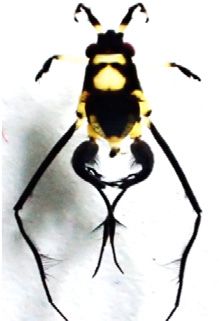
Insects 2020, 11, 578 2 of 15
Keywords: amplicon sequence variants (ASVs); bacterial community; microbiome; Neotropical;
codiversification; water striders; Wolbachia
1. Introduction
Insects host a highly diverse microbiome, which plays a crucial role in insect life. This bacterial
community is involved in a variety of functions, ranging from food processing [1,2], to protection
against pathogens [2–6] and regulation of developmental and life cycles [2,7]. In addition, recent studies
have highlighted the contribution of the microbiome to diversification [8–10], including the evolution
of reproductive isolation between species [10–12]. For instance, the presence of bacterial taxa such
as Wolbachia, Rickettsia and Cardinium have been associated with mating incompatibility [13–17] and
speciation in many insects (e.g., the plant-sap feeding Hemiptera and gall wasp (Cynipidae) [12,18–20].
Thus, understanding the nature and consequences of host-microbiome interactions in insects is crucial
to our understanding of diversification in nature. However, despite the functional and evolutionary
consequences of host-microbiome interactions [9,16,21], the composition and diversity of the bacterial
community associated with Neotropical freshwater insects remains unexplored. In addition, questions
regarding the evolution of host-microbiome interaction and its potential association with diversification
in Neotropical freshwater insects have received little attention to date. Here, we advance these
issues by assessing bacterial community composition and diversity associated with six closely related
species of Neotropical water striders in Panama. We also use a comparative phylogenetic approach
to test for associations between dominant bacterial linages and genetic divergence among species of
water striders.
Water striders (family Gerridae) are a conspicuous group of semi-aquatic insects that are typical
of freshwater and estuarine ecosystems. They are found in a variety of environments including rivers,
streams, lakes and even the open ocean [22,23]. A prominent feature of water striders is their ability to
walk on water via specialized hydrophobic legs that distribute their weight over a large surface area
and take advantage of the high surface tension of water [22,23]. Water striders are dominant predators,
providing a crucial functional role to aquatic ecosystems [23–27]. Taxonomically, they are highly
diverse, with nearly 450 known species [23], and many more remaining to be described, particularly
in the Neotropics. We have taken advantage of this diversity and specialized life strategy to explore
host-microbiome associations in Neotropical water striders.
2. Materials and Methods
2.1. Sampling Sites
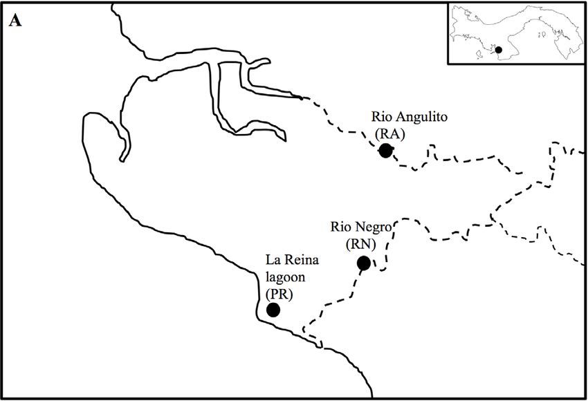
We collected water striders from three sites located in Llano de Catival on the Western Azuero
Peninsula on the Pacific coast of Panama (Figure 1A). The three sites are relatively close to each
other (4 to 5 km), but vary in salinity levels due to sea water intrusion. Playa Reina lagoon (PR;
7◦ 370 31.1” N, 81◦ 000 16.7” O) has salinity levels ranging from 0.4 to 11 ppt, sandy substrate, and is
surrounded by a mix of mangrove (Rizhophora mangle) and cativo (Prioria copaifera) forest. Río Angulito
(RA; 7◦ 380 22.0” N, 80◦ 580 17.0” O) represents a typical estuarine site with salinity levels ranging from
0.1 to values >10 ppt. This site has a combination of rocky and sandy substrate, and is surrounded by
mangrove and cativo trees and secondary forest. Río Negro (RN; 7◦ 380 22.0” N, 80◦ 580 36.6” O) is a
typical freshwater river, with salinity levels ranging from 0 to 0.4 ppt. This site has gravel substrate
and is surrounded by secondary forest.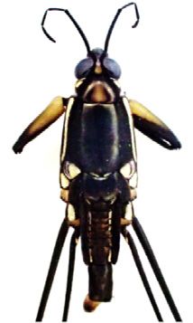
individuals of six species of water striders: Platygerris assimetricus (RN), Potamobates horvathi (RA,
RN), Potamobates tridentatus (RA), Rheumatobates bergrothi (PR), Rheumatobates ornatus (PR) and
Telmatometra withei (PR, RA, RN) (Figure 1B–G; Table 1). Of the six water strider species sampled
across sites, only three species were present at any given site, and only one species (T. withei) was
present
Insects 2020,at11,all
                   578three sites. This pattern of species assemblage is likely due to habitat preference
                                                                                                    3 of 15
[22,23,28], given that our sampling sites included both fresh and brackish water sites (Figure 1A).

                        A

                                                                   Rio Angulito
                                                                       (RA)

                                                               Rio Negro
                                                                  (RN)

                                                     Playa Reina
                                                     lagoon (PR)

                         Pacific Ocean
                        B                     C                             D

                        E                       F                          G

     Figure 1. Sampling sites (A) and water striders species Platygerris assimetricus (Hungerford, 1932; B),
     Figure 1. Sampling sites (A) and water striders species Platygerris assimetricus (Hungerford, 1932; B),
     Potamobates horvathi (Esaki, 1926; C), Potamobates tridentatus (Esaki, 1926, D), Rheumatobates bergrothi
     Potamobates horvathi (Esaki, 1926; C), Potamobates tridentatus (Esaki, 1926, D), Rheumatobates bergrothi
     (Meinert, 1895; E), Rheumatobates ornatus (Polhemus and Cheng, 1976; F), Telmatometra withei (Bergroth,
     (Meinert, 1895; E), Rheumatobates ornatus (Polhemus and Cheng, 1976; F), Telmatometra withei
     1908; G). Photo credits: Pamela Polanco and Anakena Castillo.
     (Bergroth, 1908; G). Photo credits: Pamela Polanco and Anakena Castillo.
2.2. Water Strider Species
     Table 1. Number of individuals of six water strider species sampled at three sites on the Western
     We   visited
     Azuero       each site
              Peninsula,    in December 2018 or January 2019 and collected a minimum of three adult
                         Panama.
individuals of six species of water striders: Platygerris assimetricus (RN), Potamobates horvathi (RA, RN),
Potamobates tridentatus (RA),Site                        Species
                               Rheumatobates bergrothi (PR),                     Number
                                                              Rheumatobates ornatus  (PR) and Telmatometra
withei (PR, RA, RN) (Figure 1B–G; Table 1). Rheumatobates        bergrothi
                                                  Of the six water   strider species5sampled across sites,
                     Playapresent
only three species were     Reina lagoon          Rheumatobates
                                    at any given site,            ornatus
                                                       and only one                  3 was present at all
                                                                      species (T. withei)
three sites. This pattern of species assemblage isTelmatometra
                                                    likely due towithei
                                                                   habitat preference5 [22,23,28], given that
our sampling sites included both fresh and brackish water sites (Figure 1A). 4
                                                   Potamobates  horvathi
                         Río Angulito            Potamobates tridentatus             3
                                                   Telmatometra withei               5
                          Río Negro              Platygerris assimetricus            5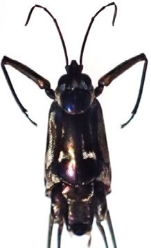
Insects 2020, 11, 578                                                                                 4 of 15

      Table 1. Number of individuals of six water strider species sampled at three sites on the Western
      Azuero Peninsula, Panama.

                               Site                 Species                  Number
                                             Rheumatobates bergrothi             5
                        Playa Reina lagoon   Rheumatobates ornatus               3
                                              Telmatometra withei                5
                                              Potamobates horvathi               4
                          Río Angulito       Potamobates tridentatus             3
                                              Telmatometra withei                5
                                             Platygerris assimetricus            5
                            Río Negro         Potamobates horvathi               4
                                               Telmatometra withei               5

2.3. DNA Extraction and Amplification
      Before DNA extraction, we surface-sterilized each insect by submerging it in 70% ethanol for
1 min, then rinsing three times in sterile water [29]. Whole individuals were then immersed in 0.01 M
solution of sterile phosphate buffered saline (PBS) at 1× for 5 min [29] and macerated with a pestle in a
1.5 mL tube. DNA was extracted using a DNeasy Blood & Tissue Kit (Qiagen, Valencia, CA, USA),
following the manufacturer’s protocol, with a final elution volume of 100 µL in buffer AE (buffer AE
(elution buffer for genomic DNA).
      To characterize the bacterial community associated with water striders, we used 16S rDNA
primers (515 F and 806 R) [30] to amplify a 251 bp portion of the V4 region, which is one of the most
effective regions for assessing bacterial diversity [31]. Triplicate PCR amplifications were prepared
in 11 µL reaction volumes, containing 4.0 µL of molecular water, 5 µL of Platinum 2× Mastermix
(ThermoFisher, Foster City, CA, USA), 0.5 µL primers 515F and 806R which included a partial Illumina
adapter on their 50 ends, and 1 µL DNA extract. Reaction conditions included a denaturation step of
94 ◦ C for 3 min, followed by 35 cycles of denaturation at 94 ◦ C for 45 s, annealing at 50 ◦ C for 1 min,
and elongation at 72 ◦ C for 1.5 min, followed by a 10 min final elongation at 72 ◦ C. We ran 2 µL of the
PCR products on an agarose gel to verify amplification.

2.4. Library Preparation
     We pooled our PCR triplicates and performed a second PCR to add barcode indexes and Illumina
adapters in 12 µL reactions using 4 µL of molecular grade water, 5 µL of PlatinumTM master mix
(Thermo Fisher 2×), 0.5 µL of each index (Forward and Reverse) and 2 µL of pooled PCR product.
PCR started with a denaturation step of 94 ◦ C for 3 min, followed by 6 cycles of denaturation at 94 ◦ C,
for 45 s, annealing at 50 ◦ C for 1 min and elongation at 72 ◦ C for 1.5 min, and ending with a 10 min final
elongation at 72 ◦ C. All resulting PCR reactions were cleaned and normalized with PCR purification
and normalization plates (Charm Biotech, San Diego, CA, USA). All samples were combined and the
library was concentrated and clean using KAPA pure beads (Kapa BioSystems, Wilmington, MA, USA).
Final library concentration was determined using a Qubit fluorometer (Turner BioSystems, Foster city,
CA, USA) and quality was checked on a BioAnalyzer (Agilent). Finally, the library was sequenced
on an Illumina MiSeq sequencing platform (Illumina Inc., San Diego, CA, USA), on a 2 × 250 bp pair
end run.

2.5. Data Analysis
     We used the Quantitative Insights Into Microbial Ecology (QIIME 2.0) pipeline to process all raw
bacterial 16S rRNA sequences associated with water striders [32]. In brief, we used Divisive Amplicon
Denoising Algorithm (DADA2) [33], as implemented in R package version 4.0.2 (Kongens Nytorv,
Denmark) to dereplicate and quality filter sequences. Then, we imported the sequence table into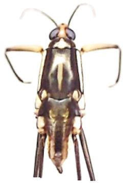
Insects 2020, 11, 578                                                                              5 of 15

QIIME2 for following analysis. Representative amplicon sequence variants (ASVs) were assigned
taxonomic classification with the SILVA database [34,35]. All ASVs assigned to mitochondrial and
chloroplast sequences as well as those with less than 10 counts were removed from the dataset. Finally,
the all data files generated with Qiime 2 were uploaded to the R software [36], for further statistical
analyses and plotting.

2.5.1. Bacterial Diversity and Community Composition
      Sequences were rarefied to a depth of 9000 sequence per sample before performing diversity
estimates. To estimate alpha diversity based on ASVs, we calculated Faith’s phylogenetic diversity
(Faith’s PD), followed by nonparametric Kruskal-Wallis to examine statistical differences among site
and species. We then quantified beta diversity among sites and species within sites based on weighted
UniFrac distance and visualized it using principle coordinates analyses (PCoA) as implemented in the
ggfortify and ggplot2 package [37,38].
      To quantify variation in bacteria community composition across sites and species, as well as
among species within site, we performed ANOSIM analyses in the vegan package [39]. We ran
1000 permutations for each analysis. Given our low sample size (3–5 individual per species/site;
Table 1), and the fact that not all water strider species were present at each sampling site, we were
not able to include the species vs. site effect into a single variance analysis. To further explore the
effect of site, we tested (using ANOSIM) for variation in the bacterial community associated with
T. withei, the only species present at all three sampling sites. Finally, the number of shared and unique
bacterial taxa across water strider species and sites were visualized with a Venn diagram using the
VennDiagram package [40].

2.5.2. Exploring Phylogenetic Associations between Water Striders and Associated Microbiome
     To determine phylogenetic relationships among water strider species, we amplified the Cytochrome
oxidase I (COI) region from each of the six species of water striders, using the primers set
LCO1490/HCO2198 [41] and dgLCO1490/dgHCO2198 [42]. We followed similar PCR protocols
as in De León et al., 2020 [43]. We aligned the sequences using MAFFT, and then built a phylogenetic
tree following RA × ML bootstrapping with 1000 iterations in Geneious 10.0.6 [44], and following
the model GTR GAMMA. Then, we mapped the relative abundance of ASVs of the 29 most common
bacterial taxa onto the phylogeny of the six water strider species using the vegan [39] and gplots
packages [45].

3. Results
     After trimming and filtering we obtained a total of 738,729 bacterial sequences, with an average of
18,941 ± 937 per sample. A rarefaction curve at a sequencing depth of 9000 showed the majority of
bacterial diversity associated with water striders was captured at a relatively low number of reads
(Figure S1).

3.1. Community Composition and Diversity of Water Strider Microbiomes
     After quality filtering, we found 806 ASVs associated with the six species of water striders.
These ASVs were classified into 31 phyla, 59 classes, 138 orders, 222 families and 373 genera.
Overall, the most abundant bacterial taxa were represented by the Phyla Proteobacteria (80.89%)
and Tenericutes (13.81%), including the classes α- and γ-Proteobacteria and Mollicutes (Figure 2A,B).
Other less abundant phyla such as Actinobacteria (Actinobacteria), Bacteroidetes (Bacteroidia) and
Firmicutes (Bacilli) were also associated with some sites and water strider species (Figure 2A,B).
Nine genera (Actinobacteria Sp. 1, Geobacillus, Candidatus cardinium, Weeksellaceae Sp. 1, Wolbachia,
Acinetobacter, Serratia, Enterobacteriaceae (unknown) and Spiroplasma) were abundant across sites and
species (Figure 2C,D).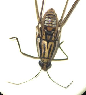
Insects 2020, 11, 578 6 of 15
Insects 2020, 11, x 6 of 15
100 100
A B
75 75
Relative abundance
Relative abundance
Class
Actinobacteria (Actinobacteria)
Bacteroidia (Bacteroidetes)
50 50 Bacilli (Firmicutes)
α-Proteobacteria (Proteobacteria)
γ-Proteobacteria (Proteobacteria)
Mollicutes (Tenericutes)
Other bacteria
25 25
0 0
C 100 D 100
75
75 Actinobacteria Sp.1
Relative abundance
Relative abundance
Geobacillus (Bacilli)
Candidatus Cardinium (Bacteroidia)
50 Weeksellaceae Sp.1 (Bacteroidia)
Wolbachia (α-Proteobacteria)
Acinetobacter (γ-Proteobacteria)
50 Serratia (γ-Proteobacteria)
25 unknown (Enterobacteriaceae/ γ-Proteobacteria)
Spiroplasma (Mollicutes)
Other bacteria
25 0
P. horvathi (RN)
T. withei (PR)
P. assimetricus (RN)
P. tridentatus (RA)
R. bergrothi (PR)
R. ornatus (PR)
P. horvathi (RA)
T. withei (RA)
0 T. withei (RN)
Playa Reina Rio Angulito Rio Negro
lagoon
Site Species
Figure 2. Relative
Figure abundance
2. Relative abundanceof dominant
of dominantbacteria taxataxa
bacteria associated withwith
associated water striders.
water Abundance
striders. Abundance
waswas
estimated at the
estimated at level of bacterial
the level classclass
of bacterial across sitessites
across (A) and species
(A) and within
species sitessites
within (B), (B),
as well as atas at
as well
the level of genus across sites (C) and species within sites (D). Only bacterial taxa with >5%
the level of genus across sites (C) and species within sites (D). Only bacterial taxa with >5% sequencesequence
abundance
abundanceare shown
are shownfor both taxonomic
for both levels.
taxonomic levels.
OurOur
estimates of alpha
estimates diversity
of alpha diddid
diversity not not
show significant
show differences
significant among
differences amongsitessites
or species
or species
(Kruskal-Wallis
(Kruskal-Wallis H, pH,> p0.05; Figure
> 0.05; 3A,B).
Figure By contrast,
3A,B). betabeta
By contrast, diversity analyses
diversity based
analyses on weighted
based on weighted
Unifrac distance
Unifrac showed
distance significant
showed differences
significant amongamong
differences sites across
sites species (ANOSIM
across species R = 0.09,R =
statistic: statistic:
(ANOSIM
p < 0.09,
0.05;pFigure 3C), and among water strider species across sites (R = 0.36, p < 0.001;
< 0.05; Figure 3C), and among water strider species across sites (R = 0.36, p < 0.001; Figure Figure 3D).3D).
However,
However,in both cases
in both the the
cases ANOSIM
ANOSIM R statistic suggested
R statistic thatthat
suggested the the
microbial community
microbial community share many
share many
taxa. Similarly, we found significant differences among species in Río Angulito
taxa. Similarly, we found significant differences among species in Río Angulito and Negro (RA: and Negro (RA:R =
R =0.43,
0.43,pp 0.05), 3D).
(Figure 3D).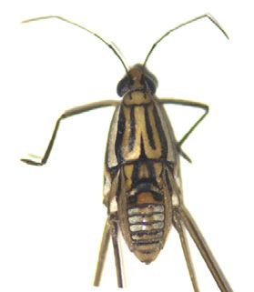
Insects 2020, 11, 578                                                                                                   7 of 15
      Insects 2020, 11, x                                                                                         7 of 15

      FigureFigure  3. Bacterial
               3. Bacterial      diversityassociated
                              diversity     associated with
                                                       with water
                                                            water striders.
                                                                   striders.Graphs
                                                                               Graphs represent estimates
                                                                                         represent         of alpha
                                                                                                    estimates    of alpha
            diversity based on Faith’s phylogenetic diversity (Faith’s PD) for each site (A) and species (B), as well
      diversity based on Faith’s phylogenetic diversity (Faith’s PD) for each site (A) and species (B), as well
            as beta diversity principle coordinates analyses (PCoA) based on weighted UniFrac distance among
      as beta diversity principle coordinates analyses (PCoA) based on weighted UniFrac distance among
            sites (C) and species within sites (D).
      sites (C) and species within sites (D).
            The majority of ASVs (77.7%) were unique to a site and only 7.4% were shared among all sites,
     The majority of ASVs (77.7%) were unique to a site and only 7.4% were shared among all sites,
     with both brackish water sites (Playa Reina lagoon and Río Angulito) showing the largest number of
with unique
     both brackish   water
            ASVs (Figure     sites
                           4A).    (Playa Reina
                                In addition,       lagoon
                                             we found        and
                                                        a large   Río Angulito)
                                                                proportion  of ASVsshowing
                                                                                     that werethe largest
                                                                                               unique      number
                                                                                                       to each
of unique  ASVs   (Figure  4A).  In addition,   we  found    a large  proportion   of ASVs   that were
     water strider species (Figure 4): 15.1–44.9% in Playa Reina lagoon (Figure 4B), 7.01–46.8% in Río   unique  to
each Angulito
     water strider  species
               (Figure       (Figure
                       4C), and       4): 15.1–44.9%
                                 18.4–41.9%             in Playa
                                              in Río Negro         Reina
                                                              (Figure 4D).lagoon  (Figure
                                                                            Overall,       4B),few
                                                                                     however,   7.01–46.8%
                                                                                                   (2.4–5.8%)in Río
     ASVs(Figure
Angulito   were shared   among
                   4C), and     species within
                            18.4–41.9%    in Ríosites
                                                 Negro(Figure  4B,D).
                                                          (Figure  4D). Overall, however, few (2.4–5.8%) ASVs
were shared among species within sites (Figure 4B,D).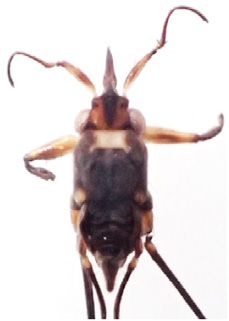
Insects 2020, 11, 578 8 of 15
Insects 2020, 11, x 8 of 15
A Playa Reina
lagoon 49
Rio Angulito B R. bergrothi
3
R. ornatus
(6.1%) 250 172 (0.8%) 58
230
(28.5%) (31.0%) (44.9%) (15.1%)
60 9
(7.4%) (2.4%)
44
(5.5%) 26 21 6
(3.2%) (5.5%) (1.6%)
147 T. withei
(18.2%) 114
(29.8%)
Rio Negro
C D
P. horvathi 25 P. tridentatus
P. assimetricus 18 P. horvathi
180 (6.5%) 27 (6.5%)
(46.8%) (7.0%) 51 116
(18.4%) (41.9%)
17 16
(4.4%) (5.8%)
14 6 8 5
(3.6%) (1.6%) (2.9%) (1.8%)
T. withei T. withei
116 63
(30.1%) (22.7%)
Figure 4. Distribution of bacteria
Figure 4. Distribution amplicon
of bacteria ampliconsequence variants
sequence variants (ASVs)
(ASVs) associated
associated with
with water water striders.
striders.
Venn diagrams
Venn show
diagramstheshow
number (whole(whole
the number values) and and
values) proportion (in(inparenthesis)
proportion ofunique
parenthesis) of unique and shared
and
ASVs amongshared ASVs
sites (A)among sites (A) and
and species withspecies
sites,with sites, including
including Playa Playa
Reina Reina lagoon
lagoon (B),Río
(B), Río Angulito
Angulito (C) and
Río Negro (C) and Río Negro (D).
(D).
3.2. Phylogenetic Associations
3.2. Phylogenetic Associations
Our results showed that some bacterial taxa were uniquely associated with different water
Our results showed
strider species that some
(sequence found inbacterial taxa
at least 40% were
of the uniquely
samples associated
for given with
species). For different
instance, P. water
strider species (sequence
assimetricus showed found
bacterialin atsuch
taxa least 40% of the(Figure
as Fructobacillus samples5). P.for givenshowed
tridentatus species). For instance,
unknown
(Diplorickettsiaceae/Gammaproteobacteria)
P. assimetricus (Figure 5). R. ornatus
showed bacterial taxa such as Fructobacillus (Figurehosted
5). Fluviicola, Chryseobacterium
P. tridentatus showed unknown
and Bacteroidia Sp. 1 (Figure 5). Finally, T. withei showed Vibrio and Rickettsiella (Figure 5). Overall,
(Diplorickettsiaceae/Gammaproteobacteria) (Figure 5). R. ornatus hosted Fluviicola, Chryseobacterium
less than 24% of ASVs were shared among water strider species, and each water strider was often
and Bacteroidia Sp.with
associated 1 (Figure 5).bacterial
a different Finally, T. withei
cluster showed
(Figure 5). Vibrio and Rickettsiella (Figure 5). Overall,
less than 24% of ASVs were shared among water strider species, and each water strider was often
associated with a different bacterial cluster (Figure 5).Insects2020,
Insects 11,x578
2020,11, 99ofof15
15
Figure 5. Amplicon sequence variants (ASVs) of dominant bacterial taxa associated with the phylogeny
Figure 5. Amplicon
water striders. sequence
Only bacteria variants
with (ASVs)
a relative abundance of >0.5%bacterial
of dominant taxa associated
were included with the
in the analysis.
phylogeny water striders. Only bacteria with a relative abundance of >0.5% were included in the
4. Discussion
analysis.
The unique ecological niche occupied by water striders represents a fascinating opportunity to
4.explore
Discussion
the evolution of host-microbiome interactions in freshwater and estuarine environments. Here,
we assess, for the
The unique first time,
ecological the occupied
niche bacterial community associated
by water striders with asix
represents closely related
fascinating speciesto
opportunity of
Neotropical water striders in Panama. We also explore potential phylogenetic associations
explore the evolution of host-microbiome interactions in freshwater and estuarine environments. between
thesewe
Here, bacterial
assess,communities and water
for the first time, strider community
the bacterial species. associated with six closely related species
of Neotropical water striders in Panama. We also explore potential phylogenetic associations between
4.1. Bacterial Diversity and Core Microbiome of Water Striders
these bacterial communities and water strider species.
Overall, we found 806 ASVs of bacterial lineages associated with the six species of water striders.
4.1. Bacterial Diversity
The most common andand Core Microbiome
abundant bacterialoftaxa
Water Stridersphyla such as Proteobacteria and Tenericutes.
included
These phyla we
Overall, were dominated
found 806 ASVsbyoffacultative endosymbionts,
bacterial lineages associatednonsymbiont, and pathogenic
with the six species bacteria
of water striders.
such as α- (e.g., Wolbachia) and γ- (e.g., Acinetobacter, Serratia and Enterobacteriaceae)
The most common and abundant bacterial taxa included phyla such as Proteobacteria and Proteobacteria
and Mollicutes
Tenericutes. These Spiroplasma).
(e.g.,phyla Given their high
were dominated frequency across
by facultative sites and species,
endosymbionts, these bacterial
nonsymbiont, and
taxa represent the core microbiome of Neotropical water striders. These taxa
pathogenic bacteria such as α- (e.g., Wolbachia) and γ- (e.g., Acinetobacter, Serratia have also been previously
and
associated with both
Enterobacteriaceae) aquatic and and
Proteobacteria terrestrial insects
Mollicutes [46–51],
(e.g., suggesting
Spiroplasma). thattheir
Given theyhigh
are afrequency
common
component
across of thespecies,
sites and microbiome
these of insects in
bacterial general,
taxa and play
represent important
the core functional
microbiome roles in theirwater
of Neotropical insect
striders. These taxa have also been previously associated with both aquatic and terrestrial insects [46–Insects 2020, 11, 578 10 of 15
hosts [3,6,46,52]. For example, a recent study found that the genus Wolbachia is widespread in aquatic
Hemipteran, including Gerridae, from Southwest Cameroon [53]. In addition, the genera Wolbachia
and Spiroplasma are known to influence host ecology and evolution [1,3,6,53], and could be involved in
diversification in water striders (see below).
We also found bacterial taxa that have not been previously associated with water striders.
These include Fructobacillus, which was associated with P. assimetricus. This bacterial genus has also
been reported in terrestrial insects [54–57], with some species offering protection against pathogens
in bees such as the American foulbrood [58]. Rickettsiella, which was found in T. withei, is known
to reduce mortality and decreases fungal sporulation in insects [59], however the genus is also an
important pathogen in arthropods [60]. Vibrio, which was also associated with T. withei, is considered a
pathogen of aquatic organisms [61,62], and can cause high mortality and severe economic losses in
marine fisheries [61]. Chryseobacterium, which was found in R. ornatus, has been reported in other
insects [63], and some of species can be pathogenic to humans and other animals [64]. However,
the role of these genera in water striders is currently unknown.
Our most striking result was the high proportion of bacterial ASVs that were uniquely associated
with different water strider species, with only 2.4–5.8% of ASVs being shared among species at each site.
Although the functional consequences of this microbiome disparity is currently unknown, this finding
suggests that species identity is likely a major factor driving microbiome diversity in water striders.
Unfortunately, our small sample size prevented us from determining if this pattern is consistent across
species or if some bacterial taxa show a stronger contribution to microbiome diversity than others.
Microbiome composition could also be influenced by habitat type [65–70], particularly because we
sampled one fresh and two brackish-water sites. Indeed, we found significant differences in the
number of ASVs across sites, with brackish-water sites showing the highest number of unique bacterial
taxa. This is consistent with previous work showing that some bacteria taxa belonging to α, γ and
β-Proteobacteria present affinities for different levels of salinity, and that saline environments often
host a higher bacterial diversity than freshwater habitats [67]. However, we believe that common
environmentally derived bacterial taxa were infrequent in our samples, in part because we sterilized
the external body of our water strider specimens before DNA extraction (i.e., our sampling was focused
on the internal body). Thus, differences in microbiome composition across sites are likely confounded
by the strong species effect (i.e., different water strider species were present at different sites), but more
data are needed to disentangle these effects statistically.
Another factor influencing microbiome diversity is host diet [71], but we currently know little
about the diet of our water strider species. However, given that water striders are opportunistic
predators that feed mostly on insects that fall on the water surface [23], we may expect low variation in
diet across species. Moreover, the fact that T. withei, the species that was present at all sites, showed
low variation in microbiome composition across sites suggests that both diet and habitat type are less
important in determining water strider microbiomes. However, further research is needed to confirm
this possibility, particularly because some closely related species of aquatic Hemiptera (Veliidae and
Gerridae) show differences in prey capture and feeding behavior [72].
4.2. Codiversification between Water Striders and Their Microbiome
Our phylogenetic analyses showed that some of the most abundant bacterial taxa were uniquely
associated with different water strider species (Figure 5). In addition, it appears that some closely
related species of water striders also host closely related bacterial microbiome (Figure 5). This was
particularly evident for some bacterial taxa such as Actinobacteria (sp.1 and sp.2), Geobacillus and
Wolbachia, which were hosted by P. horvathi and P. tridentatus (Figure 5). The results are consistent with
recent studies showing strong associations between host phylogenetic divergence and phylogenetic
divergence of the associated microbiome in several taxa, including humans [72,73], mice [74], birds [75],
lizards [76] and insects [77].Insects 2020, 11, 578 11 of 15
Thus, codiversification between hosts and associated microbiomes appears to be a common
evolutionary consequence of host-microbiome interaction. This is a tantalizing possibility in water
striders, given that they host a high diversity of bacterial taxa that are known to affect several aspects
of the reproductive biology in insects—including reproductive isolation. For example, we found a
high abundance of Wolbachia, which has been associated with cytoplasmic incompatibility in other
insect taxa [13,14,19,20].
Of particular interest are the genera Wolbachia and Spiroplasma which are common in
insects [14,78,79], and are involved in a variety of functions [3,6]. This includes fecundity in some
species of beetles (Family Curculionidae) [80,81], parthenogenesis [82], feminization [14,82,83], as well
as cytoplasmic incompatibility [13,14]. Wolbachia has also been implicated in reproductive isolation and
speciation in insects [78,84,85]. On the other hand, Spiroplasma is associated with male-killing [47,53,86].
Although more work is need to assess the evolutionary consequences of host-microbiome interaction
in water striders, our results suggest the possibility that some bacterial taxa, such as Wolbachia and
Spiroplasma, are involved in codiversification in water striders. Thus, future work should assess the
diversity of these bacterial taxa in a larger number of water strider species. Experimental analyses are
also necessary to confirm the potential role of these taxa in driving reproductive isolation.
5. Conclusions
In summary, our findings show that Neotropical water striders host a diverse bacterial
community. Some of these bacterial taxa are uniquely associated with different water strider species,
and these associations are likely influenced by both environmental context and host phylogenetic
history. This suggests that diversification in water strider microbiomes is likely associated with
host phylogenetic divergence. Assessing these associations is crucial to our understanding of the
evolution host-microbiome interaction and its role in diversification and codiversification Neotropical
freshwater organisms.
Supplementary Materials: The following are available online at http://www.mdpi.com/2075-4450/11/9/578/s1,
Figure S1: Rarefaction curves of bacterial phylogenetic diversity (Faith’s PD, ±SE) associated with (A) our three
sampling sites, and (B) six species of Neotropical water striders across the three sites. Raw sequence data and
metadata are available at: https://doi.org/10.6084/m9.figshare.12855158.
Author Contributions: Conceptualization A.M.C. and L.F.D.L.; formal analysis A.M.C., K.A.C., and K.S.;
methodology A.M.C.; investigation A.M.C., L.F.D.L., K.A.C., K.S., C.F.A., L.A.R.-C., and L.C.M.; writing—original
draft preparation, A.M.C., and L.F.D.L.; writing—review and editing A.M.C., L.F.D.L., K.A.C., K.S., C.F.A.,
L.A.R.-C., and L.C.M.; funding acquisition L.F.D.L. All authors have read and agreed to the published version of
the manuscript.
Funding: This work was supported by the Secretaría Nacional de Ciencia, Tecnología e Innovación (SENACYT,
Panamá) in the form of a doctoral fellowship to A.M.C. (No. 270-2013-284) and a research grant (No. FID16-116)
to L.F.D.L. Additional support was provided by Instituto para la Formación y Aprovechamiento de los Recursos
Humanos in the form of a doctoral fellowship to A.M.C, L.A.R.-C., and L.C.M. are supported by the Sistema
Nacional de Investigación (SNI, Panamá). L.F.D.L. is supported by the University of Massachusetts Boston.
Acknowledgments: We thank Eyda Gómez for logistical support in the laboratory.
Conflicts of Interest: The authors declare no conflict of interest.
References
1. Fukatsu, T.; Hosokawa, T. Capsule-transmitted gut symbiotic bacterium of the Japanese common plataspid
stinkbug, Megacopta punctatissima. Appl. Environ. Microbiol. 2002, 68, 389–396. [CrossRef] [PubMed]
2. Engel, P.; Moran, N.A. The gut microbiota of insects—Diversity in structure and function. FEMS Microbiol.
Rev. 2013, 37, 699–735. [CrossRef] [PubMed]
3. Hedges, L.M.; Brownlie, J.C.; O’Neill, S.L.; Johnson, K.N. Wolbachia and Virus Protection in Insects.
Science 2008, 322, 702. [CrossRef] [PubMed]
4. Koch, H.; Schmid-Hempel, P. Socially transmitted gut microbiota protect bumble bees against an intestinal
parasite. Proc. Natl. Acad. Sci. USA 2011, 108, 19288–19292. [CrossRef]Insects 2020, 11, 578 12 of 15
5. Motta, E.V.S.; Raymann, K.; Moran, N.A. Glyphosate perturbs the gut microbiota of honey bees. Proc. Natl.
Acad. Sci. USA 2018, 115, 10305–10310. [CrossRef]
6. Teixeira, L.; Ferreira, Á.; Ashburner, M. The bacterial symbiont Wolbachia induces resistance to RNA viral
infections in Drosophila melanogaster. PLoS Biol. 2008, 6, 2753–2763. [CrossRef]
7. Coon, K.L.; Valzania, L.; McKinney, D.A.; Vogel, K.J.; Brown, M.R.; Strand, M.R. Bacteria-mediated hypoxia
functions as a signal for mosquito development. Proc. Natl. Acad. Sci. USA 2017, 114, E5362–E5369.
[CrossRef]
8. Rennison, D.J.; Rudman, S.M.; Schluter, D. Parallel changes in gut microbiome composition and function in
parallel local adaptation and speciation. bioRxiv 2019, 1–29. [CrossRef]
9. Dillon, R.J.; Webster, G.; Weightman, A.J.; Dillon, V.M.; Blanford, S.; Charnley, A.K. Composition of Acridid
gut bacterial communities as revealed by 16S rRNA gene analysis. J. Invertebr. Pathol. 2008, 97, 265–272.
[CrossRef]
10. Janson, E.M.; Stireman, J.O.; Singer, M.S.; Abbot, P. Phytophagous insect-microbe mutualisms and adaptive
evolutionary diversification. Evolution 2008, 62, 997–1012. [CrossRef]
11. Vavre, F.; Kremer, N. Microbial impacts on insect evolutionary diversification: From patterns to mechanisms.
Curr. Opin. Insect Sci. 2014, 4, 29–34. [CrossRef] [PubMed]
12. Schuler, H.; Egan, S.P.; Hood, G.R.; Busbee, R.W.; Driscoe, A.L.; Ott, J.R. Diversity and distribution of Wolbachia
in relation to geography, host plant affiliation and life cycle of a heterogonic gall wasp. BMC Evol. Biol. 2018,
18, 37. [CrossRef] [PubMed]
13. Poinsot, D.; Charlat, S.; Merçot, H. On the mechanism of Wolbachia-induced cytoplasmic incompatibility:
Confronting the models with the facts. BioEssays 2003, 25, 259–265. [CrossRef]
14. Kikuchi, Y. Endosymbiotic Bacteria in Insects: Their Diversity and Culturability. Microbes Environ. 2009, 24,
195–204. [CrossRef] [PubMed]
15. Perlman, S.J.; Hunter, M.S.; Zchori-Fein, E. The emerging diversity of Rickettsia. Proc. R. Soc. B Biol. Sci. 2006,
273, 2097–2106. [CrossRef]
16. Moran, N.A.; McCutcheon, J.P.; Nakabachi, A. Genomics and Evolution of Heritable Bacterial Symbionts.
Annu. Rev. Genet. 2008, 42, 165–190. [CrossRef]
17. Zchori-Fein, E.; Perlman, S.J. Distribution of the bacterial symbiont. Mol. Ecol. 2004, 2009–2016. [CrossRef]
18. Poff, K.E.; Stever, H.; Reil, J.B.; Seabourn, P.; Ching, A.J.; Aoki, S.; Logan, M.; Michalski, J.R.; Santamaria, J.;
Adams, J.W.; et al. The native Hawaiian insect microbiome initiative: A critical perspective for Hawaiian
insect evolution. Insects 2017, 8, 130. [CrossRef]
19. Shropshire, J.D.; Bordenstein, S.R. Speciation by symbiosis: The microbiome and behavior. MBio 2016, 7.
[CrossRef]
20. Brucker, R.M.; Bordenstein, S.R. Speciation by symbiosis. Trends Ecol. Evol. 2012, 27, 443–451. [CrossRef]
21. Lundgren, J.G.; Lehman, R.M.; Chee-sanford, J. Bacterial Communities within Digestive Tracts of Ground
Beetles (Coleoptera: Carabidae). Ann. Entomol. Soc. Am. 2007, 100, 275–282. [CrossRef]
22. Cheng, L. Marine Insects; Scripps Institution of Oceanography, University of California: La Jolla, CA,
USA, 2005.
23. Stonedahl, G.M.; Lattin, J.D. The Gerridae or Water Striders of Oregon and Washington (Hemiptera:
Heteroptera). Tech. Bull. Agric. Exp. Station. Oregon State Univ. 1982, 144, 1–36.
24. Izabella, O.; Pawel, B.; Piotr, J.; Lee, S.D. Diet of Water Striders (Gerris lacustris L. 1758) in a Rice Field Near
Seoul, Korea. J. Asia. Pac. Entomol. 2007, 10, 85–88. [CrossRef]
25. Merritt, R.W.; Cummins, K.W.; Berg, M.B. An Introduction to Aquatic Insects of North America, 4th ed.;
Kendall/Hunt Publishing Company: Dubuque, IA, USA, 2008.
26. Merritt, R.W.; Cummins, K.W.; Berg, M.B.; Novak, J.A.; Higgins, M.J.; Wessell, K.J.; Lessard, J.L. Development
and application of a macroinvertebrate functional-group approach in the bioassessment of remmant river
oxbows in southwest Florida. J. N. Am. Benthol. Soc. 2002, 21, 290–310. [CrossRef]
27. Nummelin, M. Waterstriders (Het: Gerridae) as predators of hatching mosquitoes. Bicovas 1988, 1, 121–125.
28. Pacheco, B. Diversidad Taxonómica y Distribución de los Chinches Patinadores (Hemiptera: Gerridae) en
Costa Rica. Licentiate Thesis, Universidad de Costa Rica, San Pedro, Costa Rica, 2012.
29. Chen, B.; Teh, B.S.; Sun, C.; Hu, S.; Lu, X.; Boland, W.; Shao, Y. Biodiversity and Activity of the Gut Microbiota
across the Life History of the Insect Herbivore Spodoptera littoralis. Sci. Rep. 2016, 6, 1–14. [CrossRef]
[PubMed]Insects 2020, 11, 578 13 of 15
30. Caporaso, J.G.; Lauber, C.L.; Walters, W.A.; Berg-lyons, D.; Lozupone, C.A.; Turnbaugh, P.J.; Fierer, N.;
Knight, R. Global patterns of 16S rRNA diversity at a depth of millions of sequences per sample. Proc. Natl.
Acad. Sci. USA 2011, 108, 4516–4522. [CrossRef]
31. Mizrahi-Man, O.; Davenport, E.R.; Gilad, Y. Taxonomic Classification of Bacterial 16S rRNA Genes Using
Short Sequencing Reads: Evaluation of Effective Study Designs. PLoS ONE 2013, 8, e53608. [CrossRef]
32. Bokulich, N.A.; Kaehler, B.D.; Rideout, J.R.; Dillon, M.; Bolyen, E.; Knight, R.; Huttley, G.A.; Gregory
Caporaso, J. Optimizing taxonomic classification of marker-gene amplicon sequences with QIIME 2’s
q2-feature-classifier plugin. Microbiome 2018, 6, 1–17. [CrossRef]
33. Callahan, B.J.; McMurdie, P.J.; Rosen, M.J.; Han, A.W.; Johnson, A.A.; Holmes, S.P. DADA2: High resolution
sample inference from Illumina amplicon data. Nat. Methods 2016, 13, 4–5. [CrossRef]
34. Suenami, S.; Konishi Nobu, M.; Miyazaki, R. Community analysis of gut microbiota in hornets, the largest
eusocial wasps, Vespa mandarinia and V. simillima. Sci. Rep. 2019, 9, 1–13. [CrossRef] [PubMed]
35. Nowinski, B.; Smith, C.B.; Thomas, C.M.; Esson, K.; Marin, R.; Preston, C.M.; Birch, J.M.; Scholin, C.A.;
Huntemann, M.; Clum, A.; et al. Microbial metagenomes and metatranscriptomes during a coastal
phytoplankton bloom. Sci. Data 2019, 6, 1–7. [CrossRef]
36. R Development Core. R: A Language and Environment for Statistical Computing; R Foundation for Statistical
Computing: Vienna, Austria, 2008.
37. Callahan, B.J.; Sankaran, K.; Fukuyama, J.A.; McMurdie, P.J.; Holmes, S.P. Bioconductor workflow for
microbiome data analysis: From raw reads to community analyses. F1000Research 2016, 5, 1492. [CrossRef]
[PubMed]
38. Tang, Y.; Horikoshi, M.; Li, W. Ggfortify: Unified interface to visualize statistical results of popular r packages.
R J. 2016, 8, 478–489. [CrossRef]
39. Oksanen, J.; Blanchet, F.G.; Friendly, M.; Kindt, R.; Legendre, P.; Mcglinn, D.; Minchin, P.R.; O’hara, R.B.;
Simpson, G.L.; Solymos, P.; et al. Vegan: Community Ecology Package. R Package Version 2.0-10. 2017.
Available online: https://cran.r-project.org/web/packages/vegan/index.html (accessed on 18 December 2017).
40. Wilkinson, L. Exact and approximate area-proportional circular Venn and Euler diagrams. IEEE Trans. Vis.
Comput. Graph. 2012, 18, 321–331. [CrossRef] [PubMed]
41. Folmer, O.; Black, M.B.; Black, M.; Hoeh, W.; Lutz, R. DNA primers for amplification of mitochondrial
Cytochrome c Oxidase subunit I from diverse metazoan invertebrates. Mol. Mar. Biol. Biotechnol. 1994, 3,
294–299. [PubMed]
42. Meyer, C.P. Molecular systematics of cowries (Gastropoda: Cypraeidae) and diversification patterns in the
tropics. Biol. J. Linn. Soc. 2003, 79, 401–459. [CrossRef]
43. De León, L.F.; Cornejo, A.; Gavilán, R.G.; Aguilar, C. Hidden biodiversity in Neotropical streams: DNA
barcoding uncovers high endemicity of freshwater macroinvertebrates at small spatial scales. PLoS ONE
2020, 15, e0231683. [CrossRef]
44. Kearse, M.; Moir, R.; Wilson, A.; Stones-Havas, S.; Cheung, M.; Sturrock, S.; Buxton, S.; Cooper, A.;
Markowitz, S.; Duran, C.; et al. Geneious Basic: An integrated and extendable desktop software platform for
the organization and analysis of sequence data. Bioinformatics 2012, 28, 1647–1649. [CrossRef]
45. Warnes, G.R.; Bolker, B.; Bonebakker, L.; Gentleman, R.; Liaw, W.H.A.; Lumley, T.; Maechler, M.;
Magnusson, A.; Moeller, S.; Schwartz, M.; et al. Package “gplots”: Various R programming tools for plotting
data. R Packag. Version 2.17.0. 2016, pp. 1–68. Available online: https://cran.r-project.org/package=gplots
(accessed on 28 August 2020).
46. Azambuja, P.; Feder, D.; Garcia, E.S. Isolation of Serratia marcescens in the midgut of Rhodnius prolixus:
Impact on the establishment of the parasite Trypanosoma cruzi in the vector. Exp. Parasitol. 2004, 107, 89–96.
[CrossRef]
47. Montenegro, H.; Solferini, V.N.; Klaczko, L.B.; Hurst, G.D.D. Male-killing Spiroplasma naturally infecting
Drosophila melanogaster. Insect Mol. Biol. 2005, 14, 281–287. [CrossRef] [PubMed]
48. Minard, G.; Tran, F.H.; Raharimalala, F.N.; Hellard, E.; Ravelonandro, P.; Mavingui, P.; Valiente Moro, C.
Prevalence, genomic and metabolic profiles of Acinetobacter and Asaia associated with field-caught Aedes
albopictus from Madagascar. FEMS Microbiol. Ecol. 2013, 83, 63–73. [CrossRef] [PubMed]
49. Sazama, E.J.; Bosch, M.J.; Shouldis, C.S.; Ouellette, S.P.; Wesner, J.S. Incidence of Wolbachia in aquatic insects.
Ecol. Evol. 2017, 7, 1165–1169. [CrossRef] [PubMed]Insects 2020, 11, 578 14 of 15
50. Kolasa, M.; Kubisz, D.; Mazur, M.A.; Ścibior, R.; Kajtoch, Ł. Wolbachia prevalence and diversity in selected
riverine predatory beetles (Bembidiini and Paederini). Bull. Insectol. 2018, 71, 193–200.
51. González, C.T.; Saltonstall, K.; Fernández-marín, H. Garden microbiomes of Apterostigma dentigerum and
Apterostigma pilosum fungus-growing ants (Hymenoptera: Formicidae ). J. Microbiol. 2019, 57. [CrossRef]
52. Abbott, D.W.; Boraston, A.B. Structural Biology of Pectin Degradation by Enterobacteriaceae. Microbiol. Mol.
Biol. Rev. 2008, 72, 301–316. [CrossRef] [PubMed]
53. Esemu, S.N.; Dong, X.; Kfusi, A.J.; Hartley, C.S.; Ndip, R.N.; Ndip, L.M.; Darby, A.C.; Post, R.J.; Makepeace,
B.L. Aquatic Hemiptera in Southwest Cameroon: Biodiversity of Potential Reservoirs of Mycobacterium
ulcerans and multiple Wolbachia sequence types revealed by metagenomics. Diversity 2019, 11, 225.
[CrossRef]
54. Endo, A.; Tanizawa, Y.; Tanaka, N.; Maeno, S.; Kumar, H.; Shiwa, Y.; Okada, S.; Yoshikawa, H.; Dicks, L.;
Nakagawa, J.; et al. Comparative genomics of Fructobacillus spp. and Leuconostoc spp. reveals niche-specific
evolution of Fructobacillus spp. BMC Genom. 2015, 16. [CrossRef]
55. Maddaloni, M.; Hoffman, C.; Pascual, D.W. Paratransgenesis feasibility in the honeybee (Apis mellifera) using
Fructobacillus fructosus commensal. J. Appl. Microbiol. 2014, 117, 1572–1584. [CrossRef]
56. Endo, A.; Maeno, S.; Tanizawa, Y.; Kneifel, W.; Arita, M.; Dicks, L.; Salminen, S. Fructophilic lactic acid
bacteria, a unique group of fructose-fermenting microbes. Appl. Environ. Microbiol. 2018, 84, 1–14. [CrossRef]
[PubMed]
57. Endo, A. Fructophilic lactic acid bacteria inhabit fructose-rich niches in nature. Microb. Ecol. Health Dis. 2012,
23. [CrossRef] [PubMed]
58. Janashia, I.; Choiset, Y.; Rabesona, H.; Hwanhlem, N.; Bakuradze, N.; Chanishvili, N.; Haertlé, T. Protection
of honeybee Apis mellifera by its endogenous and exogenous lactic flora against bacterial infections.
Ann. Agrar. Sci. 2016, 14, 177–181. [CrossRef]
59. Su, Q.; Zhou, X.; Zhang, Y. Symbiont-mediated functions in insect hosts. Commun. Integr. Biol. 2013, 6.
[CrossRef] [PubMed]
60. Bouchon, D.; Cordaux, R.; Grève, P. Rickettsiella, Intracellular Pathogens of Arthropods. In Manipulative
Tenants; Zchori-Fein, E., Bourtzis, K., Eds.; CRC Press: Boca Raton, FL, USA, 2012; pp. 127–148.
61. Abdelaziz, M.; Ibrahem, M.D.; Ibrahim, M.A.; Abu-Elala, N.M.; Abdel-moneam, D.A. Monitoring of
different vibrio species affecting marine fishes in Lake Qarun and Gulf of Suez: Phenotypic and molecular
characterization. Egypt. J. Aquat. Res. 2017, 43, 141–146. [CrossRef]
62. Hughes, J. Population Genetic Structure in Stream Insects: What Have We Learned. In Aquatic Insects:
Challenges to Populations; Lancaster, J., Briers, R.A., Eds.; CAB International: Wallingford, UK, 2008;
pp. 268–288.
63. Dugas, J.E.; Zurek, L.; Paster, B.J.; Keddie, B.A.; Leadbetter, E.R. Isolation and characterization of a
Chryseobacterium strain from the gut of the American cockroach, Periplaneta americana. Arch. Microbiol. 2001,
175, 259–262. [CrossRef]
64. Bloch, K.C.; Nadarajah, R.; Jacobs, R. Chryseobacterium meningosepticum: An emerging pathogen among
immunocompromised adults. Report of 6 cases and literature review. Medicine 1997, 76, 30–41. [CrossRef]
65. Pruzzo, C.; Huq, A.; Colwell, R.R.; Donelli, G. Pathogenic vibrio species in the marine and estuarine
environment. Ocean. Health Pathog. Mar. Environ. 2005, 217–252. [CrossRef]
66. Thompson, F.; Iida, T.; Swings, J. Biodiversity of Vibrios. Microbiol. Mol. Biol. Rev. 2004, 68, 403–431.
[CrossRef]
67. Morrissey, E.M.; Franklin, R.B.; Kent, A. Evolutionary history influences the salinity preference of bacterial
taxa in wetland soils. Front. Microbiol. 2015, 6, 1–12. [CrossRef]
68. Zhang, M.; Sun, Y.; Liu, Y.; Qiao, F.; Chen, L.; Liu, W.T.; Du, Z.; Li, E. Response of gut microbiota to salinity
change in two euryhaline aquatic animals with reverse salinity preference. Aquaculture 2016, 454, 72–80.
[CrossRef]
69. Gallardo, K.; Candia, J.E.; Remonsellez, F.; Escudero, L.V.; Demergasso, C.S. The ecological coherence of
temperature and salinity tolerance interaction and pigmentation in a non-marine Vibrio isolated from salar
de atacama. Front. Microbiol. 2016, 7, 1–10. [CrossRef] [PubMed]
70. Truong, A.; Sondossi, M.; Clark, J.B. Genetic characterization of Wolbachia from Great Salt Lake brine flies.
Symbiosis 2017, 72, 95–102. [CrossRef]Insects 2020, 11, 578 15 of 15
71. Krams, I.A.; Kecko, S.; Jõers, P.; Trakimas, G.; Elferts, D.; Krams, R.; Luoto, S.; Rantala, M.J.; Inashkina, I.;
Gudrā, D.; et al. Microbiome symbionts and diet diversity incur costs on the immune system of insect larvae.
J. Exp. Biol. 2017, 220, 4204–4212. [CrossRef] [PubMed]
72. Ditrich, T.; Papáček, M. Differences in prey capture in semiaquatic bugs (Heteroptera: Gerromorpha).
Entomol. Sci. 2016, 19, 34–41. [CrossRef]
73. Falush, D.; Wirth, T.; Linz, B.; Pritchard, J.K.; Stephens, M.; Kidd, M.; Blaser, M.J.; Graham, D.Y.; Vacher, S.;
Perez-perez, G.I.; et al. Traces of Human Migrations in Helicobacter pylori Populations. Science 2003, 299,
1582–1585. [CrossRef]
74. Moeller, A.H.; Gomes-Neto, J.C.; Mantz, S.; Kittana, H.; Segura Munoz, R.R.; Schmaltz, R.J.; Ramer-Tait, A.E.;
Nachman, M.W. Experimental Evidence for Adaptation to Species-Specific Gut Microbiota in House Mice.
mSphere 2019, 4. [CrossRef]
75. Kropáčková, L.; Těšický, M.; Albrecht, T.; Kubovčiak, J.; Čížková, D.; Tomášek, O.; Martin, J.F.; Bobek, L.;
Králová, T.; Procházka, P.; et al. Codiversification of gastrointestinal microbiota and phylogeny in passerines
is not explained by ecological divergence. Mol. Ecol. 2017, 26, 5292–5304. [CrossRef]
76. Ren, T.; Kahrl, A.F.; Wu, M.; Cox, R.M. Does adaptive radiation of a host lineage promote ecological diversity
of its bacterial communities? A test using gut microbiota of Anolis lizards. Mol. Ecol. 2016, 25, 4793–4804.
[CrossRef]
77. Moran, N.A.; Sloan, D.B. The Hologenome Concept: Helpful or Hollow? PLoS Biol. 2015, 13, 1–10. [CrossRef]
78. Hilgenboecker, K.; Hammerstein, P.; Schlattmann, P.; Telschow, A.; Werren, J.H. How many species are
infected with Wolbachia?—A statistical analysis of current data. FEMS Microbiol. Lett. 2008, 281, 215–220.
[CrossRef] [PubMed]
79. Gauthier, J.P.; Outreman, Y.; Mieuzet, L.; Simon, J.C. Bacterial communities associated with host-adapted
populations of pea aphids revealed by deep sequencing of 16S ribosomal DNA. PLoS ONE 2015, 10, e0120664.
[CrossRef] [PubMed]
80. Chen, S.-J.; Lu, F.; Cheng, J.-A.; Jiang, M.-X.; Way, M.O. Identification and Biological Role of the Endosymbionts
Wolbachia in Rice Water Weevil (Coleoptera: Curculionidae). Environ. Entomol. 2012, 41, 469–477. [CrossRef]
[PubMed]
81. Zchori-Fein, E.; Borad, C.; Harari, A.R. Oogenesis in the date stone beetle, Coccotrypes dactyliperda, depends
on symbiotic bacteria. Physiol. Entomol. 2006, 31, 164–169. [CrossRef]
82. Stouthamer, R.; Breeuwer, J.A.J.; Hurst, G.D.D. Wolbachia Pipientis: Microbial Manipulator of Arthropod
Reproduction. Annu. Rev. Microbiol. 1999, 53, 71–102. [CrossRef]
83. Hiroki, M.; Kato, Y.; Kamito, T.; Miura, K. Feminization of genetic males by a symbiotic bacterium in a
butterfly, Eurema hecabe (Lepidoptera: Pieridae). Naturwissenschaften 2002, 89, 167–170. [CrossRef]
84. Hurst, G.D.D.; Jigging, F.M.; Von Der Schulenburg, J.H.G.; Bertrand, D.; West, S.A.; Goriacheva, I.I.;
Zakharov, I.A.; Werren, J.H.; Stouthamer, R.; Majerus, M.E.N. Male-killing Wolbachia in two species of insect.
Proc. R. Soc. B Biol. Sci. 1999, 266, 735–740. [CrossRef]
85. Telschow, A.; Hammerstein, P.; Werren, J.H. The effect of Wolbachia versus genetic incompatibilities on
reinforcement and speciation. Evolution 2005, 59, 1607–1619. [CrossRef]
86. Harumoto, T.; Fukatsu, T.; Lemaitre, B. Common and unique strategies of male killing evolved in two distinct
Drosophila symbionts. Proc. R. Soc. B Biol. Sci. 2018, 285. [CrossRef]
© 2020 by the authors. Licensee MDPI, Basel, Switzerland. This article is an open access
article distributed under the terms and conditions of the Creative Commons Attribution
(CC BY) license (http://creativecommons.org/licenses/by/4.0/).You can also read