Long-Term Effects of Snake Envenoming - Review - MDPI
←
→
Page content transcription
If your browser does not render page correctly, please read the page content below
toxins
Review
Long-Term Effects of Snake Envenoming
Subodha Waiddyanatha 1,2 , Anjana Silva 1,2 , Sisira Siribaddana 1              and
Geoffrey K. Isbister 2,3, *
 1    Faculty of Medicine and Allied Sciences, Rajarata University of Sri Lanka, Saliyapura 50008, Sri Lanka;
      subodhawaid@gmail.com (S.W.); nkanjanasilva@gmail.com (A.S.); sisira.siribaddana@gmail.com (S.S.)
 2    South Asian Clinical Toxicology Research Collaboration, Faculty of Medicine, University of Peradeniya,
      Peradeniya 20400, Sri Lanka
 3    Clinical Toxicology Research Group, University of Newcastle, Callaghan, NSW 2308, Australia
 *    Correspondence: geoff.isbister@gmail.com or Geoff.Isbister@newcastle.edu.au; Tel.: +612-4921-1211

 Received: 14 March 2019; Accepted: 29 March 2019; Published: 31 March 2019

 Abstract: Long-term effects of envenoming compromise the quality of life of the survivors of
 snakebite. We searched MEDLINE (from 1946) and EMBASE (from 1947) until October 2018 for clinical
 literature on the long-term effects of snake envenoming using different combinations of search terms.
 We classified conditions that last or appear more than six weeks following envenoming as long term
 or delayed effects of envenoming. Of 257 records identified, 51 articles describe the long-term effects
 of snake envenoming and were reviewed. Disability due to amputations, deformities, contracture
 formation, and chronic ulceration, rarely with malignant change, have resulted from local necrosis
 due to bites mainly from African and Asian cobras, and Central and South American Pit-vipers.
 Progression of acute kidney injury into chronic renal failure in Russell’s viper bites has been reported
 in several studies from India and Sri Lanka. Neuromuscular toxicity does not appear to result in
 long-term effects. Endocrine anomalies such as delayed manifestation of hypopituitarism following
 Russell’s viper bites have been reported. Delayed psychological effects such as depressive symptoms,
 post-traumatic stress disorder and somatisation have been reported. Blindness due to primary and
 secondary effects of venom is a serious, debilitating effect. In general, the available studies have
 linked a clinical effect to a snakebite in retrospect, hence lacked accurate snake authentication, details
 of acute management and baseline data and are unable to provide a detailed picture of clinical
 epidemiology of the long-term effects of envenoming. In the future, it will be important to follow
 cohorts of snakebite patients for a longer period of time to understand the true prevalence, severity,
 clinical progression and risk factors of long-term effects of snake envenoming.

 Keywords: long-term; chronic; delayed; envenoming; snakebite

 Key Contribution: The present review highlights the lack of authenticated data on the long-term
 effects of snake envenoming globally. We highlight the need for long-term follow-up of snakebite
 cohorts to understand the true burden and the clinical epidemiology of long-term effects of
 snake envenoming.

1. Introduction
     Snakebite is a neglected tropical disease that mainly affects poor farming communities in the
rural tropics [1]. Snake envenoming is often under-reported, and there is limited accurate data on the
global burden of snakebites. The high estimates suggest that there are 5.5 million bites, 1.8 million
envenomings and 94,000 deaths annually due to snakebite [2]. The largest burden of snakebite occurs
in South Asia, Southeast Asia, Sub-Saharan Africa and Latin America.

Toxins 2019, 11, 193; doi:10.3390/toxins11040193                                   www.mdpi.com/journal/toxins
Toxins 2019, 11, 193 2 of 13
Snake envenoming can cause acute local and systemic effects due to the actions of toxic
components in the venom. Some elapid and viperid snakes cause tissue injury at the bite site,
manifesting initially with oedema, pain, redness and blistering. In more severe cases, there may
be subsequent dermonecrosis and myonecrosis, occasionally requiring debridement and rarely
amputation. The commonest important systemic manifestations of snake envenoming are venom
induced consumption coagulopathy, neuromuscular paralysis, acute kidney injury, myotoxicity and
cardiovascular collapse [3–6]. Initial treatment is with antivenom and supportive care, depending on
the specific clinical effects. The vast majority of patients are then discharged once these effects have
resolved. Occasionally patients with complications require a more prolonged hospital stay.
Most snakebite patients are not followed up once they are discharged from hospital and the
acute effects have resolved. They rarely have further contact with the healthcare system in relation
to the snakebite. Although some acute pathological effects of envenoming might completely resolve
within a few days of the bite, other pathological effects or their consequences may last for months or
years [7–10]. However, due to the lack of follow up clinically and in research studies, the long-term
effects of snake envenoming are poorly defined. In addition, some effects, such as the psychological
effects resulting from the snakebite, are likely to have a delayed onset [11].
In this review, we aim to summarise our current knowledge of the long-term effects of snake
envenoming and identify the knowledge gaps.
2. Long-term Sequalae of Local Effects
2.1. Local Necrosis Resulting in Amputation
Most viperid and some elapid envenomings cause local tissue injury. Occasionally, this is
more severe with varying degrees of necrosis of the skin, subcutaneous tissues and muscles [12,13].
In addition to the toxin mediated tissue necrosis, rapidly developing oedema can lead to a compartment
syndrome, which can result in limb ischaemia [14]. Secondary infections at the bite site can further
aggravate the tissue injury and prolong the recovery [15]. The above conditions usually require surgical
management. Uncommonly, partial amputation of the limb at different levels and/or digits is required
in order to stop further spread of tissue injury [16]. Even if amputation is not required, tissue loss
resulting from necrosis, subsequent fibrosis and formation of contractures of various tissues can lead
to impaired or loss of function in limbs.
Detailed descriptions of the long-term sequalae of the local effects of authenticated snake
envenoming are rare. In particular, literature on the long-term socioeconomic burden following
amputations due to snake envenoming is scarce. Several studies report the acute stages of snakebites
that result in amputation. This provides some insight into their long-term impact. In a study from
Nigeria, of 16 snakebite patients who presented late and lost an upper or lower limb due to
amputation, the median age was 12 years (2–55). This demonstrates the duration of the disability
in the survivors, being so young at the time of the bite [16]. Cobras [13,16–19], true vipers and
pit-vipers [20–25] are reported most commonly to cause extensive local tissue injuries. In a recent
population-based cross-sectional study from Sri Lanka, of 816 snakebite victims, 26 (3.2%) had a range
of musculoskeletal disabilities that persisted for an average period of 13.4 years [9]. Despite the
limitations of a population-based study, in which case-authentication is lacking, the study reported
a range of long-term disabilities due to local envenoming following snakebite. These included
contractures and deformities, muscle wasting, joint stiffness, reduced range of movement and impaired
balance. Some of these effects, such as reduced range of movements are mostly reported in those who
did not undergo an exercise program to improve the range of motions, hence likely preventable [9].
2.2. Chronic Ulcers
Another important local injury is the development of a chronic ulcer at the bite site. This can cause
extensive scarring followed by transformation into squamous cell carcinoma, similar to a MarjolinToxins 2019, 11, 193 3 of 13
ulcer. Chronic ulcers have been reported in a possible pit-viper bite in Brazil [26] and a black-necked
spitting cobra (Naja nigricollis) bite [27]. These consequences are extremely rare, but can result in
severe morbidity.
Toxins 2019, 11, x FOR PEER REVIEW 3 of 13
2.3. Chronic
2.3. Local Pain
Chronic Local Pain and
and Swelling
Swelling
Less severe
Less severe effects
effects of
of local
local envenoming
envenoming have have also
also been
been reported
reported for for some
some snakes.
snakes. In
In aa follow-up
follow-up
telephone survey
telephone surveyconducted
conductedin in California, 6 of6the
California, of13thepatients with rattlesnake
13 patients bites reported
with rattlesnake localised
bites reported
pain, numbness or paraesthesia, abnormal skin peeling and discolouration at the at
localised pain, numbness or paraesthesia, abnormal skin peeling and discolouration bite
thesite,
bitewith
site,
persistent
with weakness
persistent weaknessof the of bitten extremity
the bitten extremityfor for
7 months
7 months to to1212years
years[28].
[28].Similar
Similar effects were
effects were
experienced for weeks to months after the bite by snakebite survivors in Sri Lanka [10]. Complex
experienced for weeks to months after the bite by snakebite survivors in Sri Lanka [10]. Complex
regional pain
regional pain syndrome
syndrome has has been
been reported
reported following
following viperviper bites
bites from South Korea,
from South Korea, Turkey
Turkey and and
Norway [29–31].
Norway [29–31].This This symptom
symptom complexcomplex lastssixover
lasts over weekssixand weeks andallodynia
includes includesandallodynia
hyperalgesia,and
hyperalgesia, in addition to pain at the bite site. The burden of persistent local pain and its impact on
in addition to pain at the bite site. The burden of persistent local pain and its impact on the post-bite
the post-bite
quality of lifequality
requiresof further
life requires further investigation,
investigation, as it is likelyas it ismany
that likelysnakebite
that manysurvivors
snakebiteexperience
survivors
experience such, without seeking medical care. Persistent swelling in 27 of 145 patients who
such, without seeking medical care. Persistent swelling in 27 of 145 patients who had Malayan had
pit-viper
Malayan pit-viper (Callocellasma rhodostoma) envenoming has been reported from Thailand [32].
(Callocellasma rhodostoma) envenoming has been reported from Thailand [32].
Therefore, not
Therefore, not only
only the
the consequences
consequences of severe local
of severe local effects,
effects, but
but also the burden
also the burden due
due to to mild
mild
long-term local effects are important in snakebite survivors and need to be addressed (Figure 1).
long-term local effects are important in snakebite survivors and need to be addressed (Figure 1).
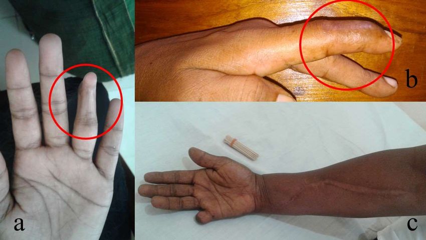
Figure 1.
Figure 1. Various long-term local
Various long-term local effects of the
effects of the bites
bites by
by Merrem’s
Merrem’s hump-nosed
hump-nosed pit pit viper
viper (Hypnale
(Hypnale
hypnale) in
hypnale) in Sri
Sri Lanka:
Lanka: (a)
(a) aa contracture
contracture deformity
deformity involving the distal
involving the distal interphalangeal
interphalangeal joint
joint of
of the
the ring
ring
finger in
finger in left
left hand
hand (red
(red circle);
circle); (b)
(b) aa contracture
contracture deformity
deformity involving
involving thethe distal
distal interphalangeal
interphalangeal jointjoint
of the
of the index
index finger
finger in
in left
left hand
hand (red
(red circle);
circle); and
and (c)
(c) amputation
amputation ofof the
the right
right little
little finger
finger due
due toto local
local
necrosis with
necrosis withthethefasciotomy
fasciotomydue duetotocompartment
compartment syndrome
syndrome of of
thethe right
right forearm.
forearm. (All(All photographs
photographs are
are published
published withwith permission
permission of patients).
of the the patients).
2.4. Blindness due
2.4. Blindness due to
to Primary
Primary Venom
Venom Effects
Effects
Venom ophthalmiafrom
Venom ophthalmia fromAfrican
Africanandand Asian
Asian spitting
spitting cobrascobras is uncommonly
is uncommonly reportedreported
in humans in
humans [33]. In the reported cases, painful conjunctivitis usually resolves in days. However, if
[33]. In the reported cases, painful conjunctivitis usually resolves in days. However, if left untreated,left
untreated, corneal ulceration
corneal ulceration and leukoma
and leukoma may leadmay lead to permanent
to permanent blindness,
blindness, especially
especially in cases
in cases of Najaof
Naja nigricollis
nigricollis [33,34].
[33,34].
3. Chronic Kidney Disease
A range of snakes have been reported to cause snakebite associated acute kidney injury,
including Russell’s viper (Daboia russelii and D. siamensis) [35–38] carpet vipers (Echis spp.) [39], the
lance-headed pit-vipers (Bothrops sp.) [40,41] and rattlesnakes (Crotalus sp.) [20,42], as well as some
Australasian elapids such as brown snakes (Pseudonaja sp.) [43], taipans (Oxyuranus sp.) [44] and tiger
snakes (Notechis sp.) [45]. The mechanism of acute kidney injury in snakabite remains unclear, but is
most likely due to secondary effects including hypotension, thrombotic microangiopathy,Toxins 2019, 11, 193 4 of 13
3. Chronic Kidney Disease
A range of snakes have been reported to cause snakebite associated acute kidney injury, including
Russell’s viper (Daboia russelii and D. siamensis) [35–38] carpet vipers (Echis spp.) [39], the lance-headed
pit-vipers (Bothrops sp.) [40,41] and rattlesnakes (Crotalus sp.) [20,42], as well as some Australasian
elapids such as brown snakes (Pseudonaja sp.) [43], taipans (Oxyuranus sp.) [44] and tiger snakes
(Notechis sp.) [45]. The mechanism of acute kidney injury in snakabite remains unclear, but is most
likely due to secondary effects including hypotension, thrombotic microangiopathy, immunological
reactions, although direct nephrotoxicity is still considered a possible mechanism [3,6]. In most cases,
the acute kidney injury resolves after treatment with antivenom and supportive care, with or without
dialysis. Progression to chronic kidney disease has been reported in a few studies [7,46–49].
In an observational study of 54 patients from Sri Lanka with acute kidney injury following
snake envenoming, 34 regained normal renal function after one year and 20 (37%) developed chronic
kidney disease [7]. Of the patients who developed chronic kidney disease, five had end stage kidney
disease, four had stage 4 and eleven had stage 3 chronic kidney disease. The duration of renal
replacement therapy during the acute kidney injury and persistence of high creatinine concentrations
after the acute stage had resolved, were the best predictors of chronic kidney disease. Glomerular
sclerosis and interstitial lymphocytic infiltration, tubular atrophy and scarring were the commonest
histopathological changes on renal biopsy. The major limitation of this study was that no patients had
baseline creatinine concentrations done prior to the snakebite, hence the possibility of chronic kidney
disease prior to the snakebite cannot be excluded. In addition, the biting species was only confirmed
in half of the cases. The study is therefore likely to have significantly over-estimated the frequency of
chronic kidney injury following snakebite.
An observational study from India reported 42 patients with acute kidney injury, including
five shown to have acute interstitial nephritis on renal biopsy. Of the 42, four developed chronic kidney
disease stage 3 to 5D, 4–10 months after the bite [48]. All four of them had severe acute kidney injury
following the bite with a prolonged hospital admission. In the same cohort, of eight patients who
had histological findings of acute tubular necrosis during the acute stage, all recovered early and
regained normal renal function. The absence of baseline creatinine, lack of snake authentication and
lack of uniformity of the follow-up periods were major limitations of the study. Although the study
suggested that the acute interstitial nephritis following snake envenoming was more likely to progress
into chronic kidney disease, the absence of information prior to the bite means that it was unclear
whether the patient was not already predisposed to or had pre-existing chronic kidney disease.
A cohort study from India of 60 patients who had snakebite associated acute kidney injury that
required dialysis, found persistent renal dysfunction, proteinuria, and/or hypertension in 24 (40%)
patients, after a mean follow up period of 45 months [46]. Of these, three patients progressed to
end-stage renal disease. This study provides a better estimate of the frequency of chronic kidney
injury, although it reports this only for those with a severe acute kidney injury at the time of the
bite. In another study from India of 100 patients who developed acute kidney injury following snake
envenoming, eight patients were found to have “chronic renal failure” after six months [47].
In a 10-year follow-up of a cohort of 37 children in India with acute kidney injury, there were
three patients in which the acute kidney injury followed a snakebite. They had either proteinuria or
hypertension, proteinuria and increased estimated glomerular filtration rate [49].
The above studies suggest that, in some patients with acute kidney injury following snake
envenoming, there is a risk of persistent renal dysfunction over months to years, which may ultimately
progress to chronic renal failure. However, there are a number of inherent limitations in the above
studies, including no information on renal function prior to the snakebite (i.e., pre-existing chronic
kidney disease), authentication of the snake species, lack of uniformity in the follow-up periods,
lack of standardisation of the measure of the clinical severity of the initial acute kidney injury and
limited details of the interventions used during the acute stage. It is therefore difficult to determine the
prevalence, predictors and time scale of chronic kidney disease that is a result of snake envenoming.Toxins 2019, 11, 193 5 of 13
4. Neurological Effects
4.1. Neuromuscular Paralysis
Neuromuscular paralysis is one of the major systemic effects of elapid snake envenoming,
including bites by kraits (Genus: Bungarus), cobras (Genus: Naja), taipans (Genus: Oxyuranus), tiger
snakes (Genus: Notechis) and some vipers, such as Russell’s viper (Daboia russelii) [5]. Snake venom
neurotoxins primarily affect the neuromuscular junction and disrupt transmission across the
neuromuscular junction. Clinically, this manifests as a rapidly progressing, flaccid paralysis that
initially involves extraocular and facial muscles, gradually descending to bulbar, neck, respiratory
and limb muscles. Of the two major groups of neurotoxins, pre-synaptic neurotoxins enter the motor
nerve terminal and lead to a depletion of synaptic vesicles followed by destruction of the motor nerve
terminal. Recovery is via natural repair of the motor nerve terminal, which initiates over 3–5 days
but may take several more days for complete repair to occur [50,51]. Snakes such as kraits, taipans
and tiger snakes have venoms rich in pre-synaptic neurotoxins. In contrast, post-synaptic or alpha
neurotoxins (three-finger toxins) antagonise the nicotinic acetylcholine receptors in the motor end plate
and lead to a “curare-like” neuromuscular block, which is reversible in comparison to pre-synaptic
neurotoxins [52]. Many elapid snakes have alpha neurotoxins in their venom [53]. Recently, this group
of toxins have been shown to be clinically less important [52].
Most observational studies have shown that the neuromuscular paralysis in snake envenoming
completely resolves within several days [19,54,55], based on clinically observed neurological
features. In a cohort of 33 patients with authenticated Indian krait (Bungarus caeruleus) bites in
Sri Lanka, which measured serial single-fibre electromyography, patients continued to have sub-clinical
neurotransmission anomalies after clinically apparent paralysis resolved [56]. These electromyographic
abnormalities consisted of increased neuromuscular jitter and increased neuromuscular blocks.
These were severe in the acute period and then gradually resolved. Mild neurotransmission
abnormalities were still present at six weeks after the krait bite, but were absent after six months.
No venom was detected in patients’ blood after the first dose of antivenom. This suggested that the
neurotransmission abnormalities are irreversible after pre-synaptic neurotoxin mediated damage,
and take several weeks to fully recover, rather than there being delayed venom effects.
Another study recruited 26 patients who had a neurotoxic snakebite in the previous year.
The study found abnormal nerve conduction parameters in all patients, including prolongation
of sensory, motor and F-wave latencies and reduction of conduction velocities, when compared to
22 control subjects [57]. None of the patients had any neuromuscular transmission abnormalities.
The study concluded a possible sub-clinical demyelinating type polyneuropathy due to neurotoxic
envenoming. However, this study identified the patients after the snakebite and therefore, lacked
species-authentication, details of the clinical severity of the neuromuscular paralysis and venom
concentrations during the acute stage. This limits any conclusion in associating the neurophysiological
anomalies with the snake envenoming.
There is a report of persistent unilateral ptosis due to paralysis of the frontalis muscle in a patient
bitten by a European adder (Vipera berus) [58]. The patient had severe swelling over the frontalis muscle
for several days, so it is unclear whether the weakness of the frontalis was due to direct muscle injury
or not.
4.2. Neurological Effects Secondary to Hypoxic or Ischemic Events
Permanent neurological injury from hypoxic encephalopathy is an important long-term effect
of snake envenoming. Respiratory paralysis or cardiac arrest can both result in hypoxia and
multiorgan failure. In many cases, this results in an early death, but some patients survive with
significant neurological impairment. A single case report described the persistence of cerebellar
ataxia in a patient who developed severe neuromuscular paralysis following a suspected Indian krait
(Bungarus caeruleus) bite [59]. The ataxia was clinically apparent after the patient had recoveredToxins 2019, 11, 193 6 of 13
from the severe neuromuscular paralysis, two weeks after the bite. The report did not provide
sufficient details to exclude pre-existing ataxia. The patient was lost to follow up after two months and
the possible pathophysiological mechanisms are difficult to determine. Ischaemic stroke leading to
leukoencephalopathy and Parkinson’s-like features that lasted beyond 10 weeks following an unknown
snakebite has been reported from India. The patient’s Parkinson’s-like features responded well to
levodopa and carbidopa combined treatment [60].
A 15-year-old male from India developed hypoxic encephalopathy following an Indian krait bite,
and had paraplegia and persistent cortical blindness four years after the bite [61].
4.3. Blindness
Blindness and visual impairment are rarely reported following snakebites and are most commonly
associated with secondary effects of envenoming. Cortical blindness has been reported in a patient
with a Russell’s viper bite (Daboia russelii), due to an ischaemic stroke [62]. Cortical blindness has
also been reported following cerebral hypoxia in a patient who had a cardiac arrest following cobra
(Naja sp.) envenoming [63] and in a patient who developed respiratory arrest following a krait
(Bungarus sp.) bite [61]. There are two reports of optic atrophy due to central retinal artery occlusion
following suspected viper bites in India [64]. Both the patients had delayed presentations. Optic neuritis
secondary to haemorrhages following a bite by Vipera lebetina (now Macrovipera lebetina) has been
reported from Israel [65]. Direct venom injury from spitting cobras may also result in blindness
(see above).
4.4. Neurological Effects following Intracranial Haemorrhage
Intracranial haemorrhage can occur in envenomings by snakes that cause venom induced
consumption coagulopathy, including many vipers and Australasian elapids. In the majority of cases,
intracranial haemorrhage in combination with severe coagulopathy is fatal, but some patients may
survive with permanent neurological sequelae. Eight patients with Bothrops spp. envenoming from
Ecuador were reported to have cerebrovascular events, and seven had intracranial haemorrhages [66].
Three patients survived and all had permanent neurological effects. Six patients in Australia
developed intracranial haemorrhages from venom induced consumption coagulopathy following
elapid envenomings, mainly brown snakes (Pseudonaja spp.). The only survivor was a 72-year old
female who had a permanent left arm and leg hemiplegia with inattention [67]. Persistent central
monoparesis of the left leg following intracerebral haemorrhage in a suspected bite by a carpet viper
(Echis sp.) has also been reported [68].
A retrobulbar haematoma that caused raised intraocular pressure resulting in bilateral corneal
opacifications has been reported in a 10-year-old child who had coagulopathy following a suspected
viper (Genus: Echis) envenoming in Nigeria [69]. A 15-year-old Nigerian boy bitten by a carpet viper
developed retrobulbar haemorrhage resulting in bilateral optic atrophy [70].
4.5. Reduced Parasympathetic Activity
Decreased parasympathetic activity following Malayan krait (Bungarus candidus) envenoming has
been reported previously in three patients not treated with antivenom [71]. These patients developed
hypertension, mydriasis and tachycardia acutely in conjunction with severe neuromuscular paralysis.
While the hypertension resolved, mydriasis and tachycardia persisted for up to two years. The possible
mechanisms of the autonomic effects in Malayan krait bite are poorly understood.
4.6. Anosmia and Changes in Taste Sensation
Changes in smell (including loss of smell—anosmia) as well as taste have been reported following
Australian elapid bites, mainly for black snakes (Pseudechis) [72,73]. In most cases, patients report
a horrible taste sensation, or change in taste/smell that persists for months to years. PersistentToxins 2019, 11, 193 7 of 13
anosmia due to olfactory bulb atrophy has been reported in another confirmed case of a Mulga snake
(Pseudechis australis) bite [74].
5. Endocrine Effects—Hypopituitarism
Clinically detectable endocrine effects are rarely reported during the acute stage of snake
envenoming. Acute hypopituitarism in Burmese Russell’s viper (Daboia siamensis) [75], Russell’s
viper in Sri Lanka (D. russelii) [76] and Addisonian crisis in Russell’s viper in India (D. russelii) [77]
have been reported. The accepted pathophysiology of hypopituitarism in these cases is a haemorrhagic
infarction in the pituitary resulting from venom induced consumption coagulopathy [8]. This results
from the combination of the consumption coagulopathy and vascular injury from haemorrhagic toxins.
Acute hypopituitarism manifests as hypotension and hypoglycaemia [75], and appears to persist,
based on 11 of 12 survivors of D. siamensis viper envenoming [78]. The involvement of the anterior
pituitary is commoner than the posterior pituitary [8].
In some snakebite survivors who had no clinically detectable hypopituitarism during the acute
stage, chronic/delayed hypopituitarism may clinically manifest later as deficiency of cortisol, growth
hormone, thyroxine and testosterone (in males) [8,75,79]. Two studies have summarised 36 previous
cases on hypopituitarism in snake envenoming [8,80]. Almost all reported cases of chronic/delayed
hypopituitarism are due to envenoming by D. siamensis and D. russelii, the majority from India.
The clinical presentation includes fatigue, loss of libido, secondary amenorrhea, infertility, weight
loss, hypoglycaemia and features of hypothyroidism such as facial puffiness, dry skin and cold
intolerance [8,80,81]. In a large proportion of patients, necrosis of the pituitary is seen as an empty
sella on magnetic resonance imaging [79,80,82] and may rarely present as psychosis [83].
The time to diagnosis of hypopituitarism varies from 2 weeks to 10 years [84,85]. In most studies,
hypopituitarism diagnosed in a patient was linked to a snakebite that occurred several years prior, of
which no reliable data existed of the acute episode. In a more recent cohort study from India, 60 patients
had baseline hormonal profiles immediately after the snakebite and were prospectively followed for
six months [82]. Of them, six patients developed asymptomatic anterior hypopituitarism during the
acute period following the bite. All of them had deficiencies in growth hormone, gonadotrophin,
thyroid hormones and secondary adrenal insufficiency. However, none of the other 54 patients
developed hypopituitarism in the acute period or at six months. This clearly indicates that the
initial insult to the anterior pituitary occurs during the acute stage of envenoming, while the patients
are asymptomatic. However, the damage is irreversible, and hypopituitarism persists with subsequent
progression to clinically detectable hypopituitarism.
Most of the above studies used less reliable methods of case-authentication, or assumed the
identity of the biting species, which might have limited the generalisation of these findings.
6. Psychological Effects
Psychological sequalae are important, but greatly under reported effects of snake envenoming.
These effects are unlikely to be direct venom effects, but rather the effects triggered by the traumatic
experience of a snakebite and the severe socioeconomic consequences associated with snakebite.
In a cross-sectional study done in Nigeria, depression was prevalent in 25% of 187 snakebite patients
who were receiving treatment in the hospital [86]. The depression was associated with more severe
complications in snakebite, being worried about family welfare, time and financial loss and previous
experiences with snakebite.
In a study from Sri Lanka that included both quantitative and qualitative arms, 88 patients
12–48 months after the snakebite had significantly higher rates of depressive symptoms, post-traumatic
stress disorder and somatisation symptoms, compared to the matched controls who had no history
of snakebite [11]. In this study, 54% of patients had depressive symptoms as opposed to 13% in
the controls. The qualitative study found various unexplainable elements of somatisation such as
blindness, tooth decay, body aches, headaches, tiredness and weakness in snakebite survivors.Toxins 2019, 11, 193 8 of 13
Following this study, a randomised controlled trial of a brief psychological intervention was
undertaken in Sri Lanka. The intervention included psychological first aid, psychoeducation and
cognitive behavioural therapy, which was associated with a reduction in psychiatric symptoms and
disability in snakebite victims, compared to controls [87]. However, the intervention was not effective
in preventing depression or post-traumatic stress disorder.
7. Knowledge Gaps
Snakebite is underreported because most affected people are poor, rural, in politically
disadvantaged communities, even within their own countries [1,88]. Modern medicine is still not
available for many snakebite victims in the rural tropics. For these reasons, the epidemiology, clinical
effects, consequences and socioeconomic impact of snakebite are still poorly understood [89]. Many of
those treated in hospitals for the snakebite are likely to not attend follow-up. Even in countries such
as Sri Lanka, in which the vast majority of patients seek western medicine as the first choice for
snakebite [90], patients seek treatment from the local indigenous doctor for persisting symptoms,
without being directed to rehabilitation programs for their musculoskeletal disabilities [9]. Even in
developed settings, long-term issues related to envenoming in snakebite victims are poorly addressed
or reported [28]. The few existing studies of the long-term effects of snake envenoming are based on
selected patient groups for follow-up, hence do not provide the true picture of the burden. Some studies
have described long-term effects of snake envenoming by relating a disability to a previous snakebite,
based on the patients’ interpretations, which might be biased. The community-based studies on the
long-term effects of snake envenoming are useful in understanding the burden of long-term effects of
snake envenoming in general. However, they are unable to answer the clinical questions due to the
inherent deficiencies associated with recall bias, so there is less-reliable information on the acute stage
of envenoming.
The range of clinical effects and their severity in snake envenoming are unique for individual
snake species. Therefore, accurate species identification is essential in clinical and epidemiological
studies. This can be done by either identification of the snake specimen by a herpetologist or specific
venom detection enzyme-linked immunosorbent assay (ELISA) [91]. Most of the studies that describe
the long-term effects of snakebite did not have accurate species authentication, which has limited
the interpretation of the results. Cohort studies that document the acute stage of envenoming must
have accurate case-authentication and active follow-up of patients on regular intervals to provide
a detailed picture of the epidemiology and the clinical consequences of the long-term effects of snake
envenoming. In regions where the facilities are available, implementation of institutional databases
that record the details of the acute envenoming as well as the details of the follow-up visits of the
discharged patients would be useful.
From the available studies, it appears that the socioeconomic burden resulting from the physical
and psychological consequences of delayed and long-term effects of snake envenoming is enormous.
Management guidelines of snakebites are largely focused on the acute management of snakebite.
At present, the attention paid to following up snakebite patients for psychological effects is non-existent.
In reality, even for minor effects of local necrosis, proper follow-up for physical therapy and exercises
does not happen due to patient and health-system factors, and hence end up as physical disabilities
due to contractures, which are preventable [10]. In the future, management protocols for snakebite
must address the issues related to long-term effects of snake envenoming.
8. Methods
We carried out a search in MEDLINE from 1946 and EMBASE from 1947 to 14 October 2018 and
included clinical studies in English on snakebite that describe long-term or chronic effects. We used
the search terms “snake envenoming”, “snake envenomation”, “snake bite”, and “ophitoxaemia” in
combination with the terms “chronic”, “long term”, “delayed”, “prolonged”, “disability”, “persistent”,
“permanent”, “morbidity”, and “recurrence”. We then searched the reference lists of retrieved articlesToxins 2019, 11, 193 9 of 13
for additional publications relevant to the topic. This search yielded 404 abstracts. After removing
duplicates and non-clinical studies, we identified a total of 88 abstracts for further study and the full
articles of these were reviewed. From these, we excluded 37 studies that were not presenting primary
data and the remaining 51 studies were included in this study (Figure 2). For this review, we classified
conditions that last or appear more than six weeks following envenoming as long term or delayed
effects of envenoming. We identified different themes/effects of long-term consequences of the snake
Toxins 2019, 11, x FOR PEER REVIEW 9 of 13
envenoming based on the content of the included articles.
Figure 2. Selection of studies for the review.
Figure 2. Selection of studies for the review.
Author Contributions:
Author Contributions: A.S.,
A.S., S.S.
S.S. and
and G.K.I.
G.K.I. conceived
conceived the
the paper;
paper; S. W. performed
S.W. performed the
the literature
literature search;
search; S.W.
S.W. and
and
A.S. analysed
A.S. analysed data;
data; and
and S.W.,
S.W, A.S., G.K.I. and
A.S., G.K.I. and S.S.
S.S. wrote
wrote the
the paper.
paper.
Funding: This study was funded by a National Health and Medical Research Council—Australia (NHMRC)
Funding: This study was funded by a National Health and Medical Research Council—Australia (NHMRC)
Centres for Research Excellence Grant ID: 1110343.
Centres for Research Excellence Grant ID: 1110343.
Conflicts of Interest: The authors declare no conflict of interest.
Conflicts of Interest: The authors declare no conflict of interest.
References
References
1. Harrison, R.A.; Hargreaves, A.; Wagstaff, S.C.; Faragher, B.; Lalloo, D.G. Snake envenoming: a disease of
1. Harrison, R.A.; Hargreaves, A.; Wagstaff, S.C.; Faragher, B.; Lalloo, D.G. Snake envenoming: a disease of
poverty. PLoS Negl. Trop. Dis. 2009, 3, e569. [CrossRef]
poverty. PLoS Negl. Trop. Dis. 2009, 3, e569.
2. Kasturiratne, A.; Wickremasinghe, A.R.; De Silva, N.; Gunawardena, N.K. The Global Burden of Snakebite:
2. Kasturiratne, A.; Wickremasinghe, A.R.; Silva, N. De; Gunawardena, N.K. The Global Burden of Snakebite :
A Literature Analysis and Modelling Based on Regional Estimates of Envenoming and Deaths. PLoS Med.
A Literature Analysis and Modelling Based on Regional Estimates of Envenoming and Deaths. PLoS Med.
2008, 5, e218. [CrossRef]
2008, 5, e218.
3. Isbister, G.K. Snakebite doesn’t cause disseminated intravascular coagulation: coagulopathy and thrombotic
3. Isbister, G.K. Snakebite
microangiopathy doesn’t cause
in snake envenoming. disseminated
Semin. intravascular
Thromb. Hemost. 2010, 36, coagulation: coagulopathy and
444–451. [CrossRef]
4. thrombotic microangiopathy in snake envenoming. Semin. Thromb. Hemost. 2010, 36, 444–451.
Gutiérrez, J.M.; Ponce-Soto, L.A.; Marangoni, S.; Lomonte, B.; Alberto Ponce-Soto, L.; Marangoni, S.;
4. Gutiérrez, B.
Lomonte, J.M.; Ponce-Soto,
Systemic L.A.; myotoxicity
and local Marangoni, induced
S.; Lomonte, B.; Alberto
by snake venomPonce-Soto, L.; Marangoni,A2:
group II phospholipases S.;
Lomonte, B. Systemic and local myotoxicity induced by snake venom group II phospholipases
comparison between crotoxin, crotoxin B and a Lys49 PLA2 homologue. Toxicon 2008, 51, 80–92. [CrossRef] A2:
5. comparisonU.K.;
Ranawaka, between crotoxin,
Lalloo, D.G.; crotoxin B and
Silva, H.J. a Lys49 PLA2 in
De Neurotoxicity homologue. ToxiconLimits
Snakebite—The 2008, 51, 80–92.
of Our Knowledge.
5. Ranawaka, U.K.; Lalloo, D.G.; Silva, H.J. De
PLoS Negl. Trop. Dis. 2013, 7, e2302. [CrossRef] Neurotoxicity in Snakebite—The Limits of Our Knowledge.
PLoS Negl. Trop. Dis. 2013, 7, e2302.
6. Sitprija, V. Snakebite nephropathy. Nephrology 2006, 11, 442–448.
7. Herath, H.M.N.J.; Wazil, A.W.M.; Abeysekara, D.T.D.J.; Jeewani, N.D.C.; Weerakoon, K.G.A.D.;
Ratnatunga, N.V.I.; Bandara, E.H.C.K.; Kularatne, S.A.M. Chronic kidney disease in snake envenomed
patients with acute kidney injury in Sri Lanka: A descriptive study. Postgrad. Med. J. 2012, 88, 138–142.
8. Antonypillai, C.N.; Wass, J.A.H.; Warrell, D.A.; Rajaratnam, H.N. Hypopituitarism following envenomingToxins 2019, 11, 193 10 of 13
6. Sitprija, V. Snakebite nephropathy. Nephrology 2006, 11, 442–448. [CrossRef] [PubMed]
7. Herath, H.M.N.J.; Wazil, A.W.M.; Abeysekara, D.T.D.J.; Jeewani, N.D.C.; Weerakoon, K.G.A.D.; Ratnatunga, N.V.I.;
Bandara, E.H.C.K.; Kularatne, S.A.M. Chronic kidney disease in snake envenomed patients with acute
kidney injury in Sri Lanka: A descriptive study. Postgrad. Med. J. 2012, 88, 138–142. [CrossRef] [PubMed]
8. Antonypillai, C.N.; Wass, J.A.H.; Warrell, D.A.; Rajaratnam, H.N. Hypopituitarism following envenoming
by Russell’s Vipers (Daboia siamensis and D. russelii) resembling Sheehan’s syndrome: First case report
from Sri Lanka, a review of the literature and recommendations for endocrine management. QJM Int. J. Med.
2011, 104, 97–108. [CrossRef]
9. Jayawardana, S.; Gnanathasan, A.; Arambepola, C.; Chang, T. Chronic Musculoskeletal Disabilities following
Snake Envenoming in Sri Lanka: A Population-Based Study. PLoS Negl. Trop. Dis. 2016, 10, e0005103. [CrossRef]
10. Jayawardana, S.; Arambepola, C.; Chang, T.; Gnanathasan, A. Long-term health complications following
snake envenoming. J. Multidiscip. Healthc. 2018, 11, 279–285. [CrossRef] [PubMed]
11. Williams, S.S.; Wijesinghe, C.A.; Jayamanne, S.F.; Buckley, N.A.; Dawson, A.H.; Lalloo, D.G.; de Silva, H.J.
Delayed psychological morbidity associated with snakebite envenoming. PLoS Neglected Trop. Dis. 2011,
5, e1255. [CrossRef] [PubMed]
12. Anz, A.W.; Schweppe, M.; Halvorson, J.; Bushnell, B.; Sternberg, M.; Koman, L.A. Management of Venomous
Snakebite Injury to the Extremities. J. Am. Acad. Orthop. Surg. 2010, 18, 749–759. [CrossRef] [PubMed]
13. Mao, Y.C.; Liu, P.Y.; Chiang, L.C.; Lai, C.S.; Lai, K.L.; Ho, C.H.; Wang, T.H.; Yang, C.C. Naja atra snakebite
in Taiwan. Clin. Toxicol. 2018, 56, 273–280. [CrossRef] [PubMed]
14. Hsu, C.P.; Chuang, J.F.; Hsu, Y.P.; Wang, S.Y.; Fu, C.Y.; Yuan, K.C.; Chen, C.H.; Kang, S.C.; Liao, C.H.
Predictors of the development of post-snakebite compartment syndrome. Scand. J. Trauma. Resusc. Emerg. Med.
2015, 23, 97. [CrossRef] [PubMed]
15. Wagener, M.; Naidoo, M.; Aldous, C. Wound infection secondary to snakebite. South African Med. J. 2017,
107, 315. [CrossRef]
16. Abubakar, S.B.; Habib, A.G.; Mathew, J. Amputation and disability following snakebite in Nigeria. Trop. Doct.
2010, 40, 114–116. [CrossRef]
17. Wong, O.F.; Lam, T.S.K.; Fung, H.T.; Choy, C.H. Five-year experience with Chinese cobra (Naja atra)-related
injuries in two acute hospitals in Hong Kong. Hong Kong Med. J. 2010, 16, 36–43. [PubMed]
18. Wang, W.; Chen, Q.F.; Yin, R.X.; Zhu, J.J.; Li, Q.B.; Chang, H.H.; Wu, Y.B.; Michelson, E. Clinical features
and treatment experience: A review of 292 Chinese cobra snakebites. Environ. Toxicol. Pharmacol. 2014, 37,
648–655. [CrossRef]
19. Kularatne, S.A.M.; Budagoda, B.D.S.S.; Gawarammana, I.B.; Kularatne, W.K.S. Epidemiology, clinical profile
and management issues of cobra (Naja naja) bites in Sri Lanka: first authenticated case series. Trans. R. Soc.
Trop. Med. Hyg. 2009, 103, 924–930. [CrossRef]
20. Milani Júnior, R.; Jorge, M.T.; de Campos, F.P.; Martins, F.P.; Bousso, A.; Cardoso, J.L.; Ribeiro, L.A.; Fan, H.W.;
França, F.O.; Sano-Martins, I.S.; et al. Snake bites by the Jararacuçu (Bothrops jararacussu): clinicopathological
studies of 29 proven cases in São Paulo State, Brazil. QJM Mon. J. Assoc. Physicians 1997, 90, 323–334.
[CrossRef]
21. Kallel, H.; Mayence, C.; Houcke, S.; Mathien, C.; Mehdaoui, H.; Gutiérrez, J.M.; Megarbane, B.; Hommel, D.;
Resiere, D. Severe snakebite envenomation in French Guiana: When antivenom is not available. Toxicon
2018, 146, 87–90. [CrossRef] [PubMed]
22. Roriz, K.R.P.S.; Zaqueo, K.D.; Setubal, S.S.; Katsuragawa, T.H.; da Silva, R.R.; Fernandes, C.F.C.;
Cardoso, L.A.P.; Rodrigues, M.M.D.S.; Soares, A.M.; Stábeli, R.G.; et al. Epidemiological study of snakebite
cases in Brazilian western Amazonia. Rev. Soc. Bras. Med. Trop. 2018, 51, 338–346. [CrossRef]
23. Ribeiro, L.A.; Jorge, M.T.; Lebrão, M.L. Prognostic factors for local necrosis in Bothrops jararaca (Brazilian pit
viper) bites. Trans. R. Soc. Trop. Med. Hyg. 2001, 95, 630–634. [CrossRef]
24. Maduwage, K.; Isbister, G.K.; Silva, A.; Bowatta, S.; Mendis, S.; Gawarammana, I. Epidemiology and
clinical effects of hump-nosed pit viper (Genus: Hypnale) envenoming in Sri Lanka. Toxicon 2013, 61, 11–15.
[CrossRef]
25. Ariaratnam, C.A.; Thuraisingam, V.; Kularatne, S.A.M.; Sheriff, M.H.R.; Theakston, R.D.G.; de Silva, A.;
Warrell, D.A. Frequent and potentially fatal envenoming by hump-nosed pit vipers (Hypnale hypnale and
H. nepa) in Sri Lanka: lack of effective antivenom. Trans. R. Soc. Trop. Med. Hyg. 2008, 102, 1120–1126.
[CrossRef]Toxins 2019, 11, 193 11 of 13
26. Mello, L.F.B.; Barcelos, M.G.; Meohas, W.; Pinto, L.W.; Melo, P.A.; Nogueira Neto, N.C.; Smith, J. Chronic
ulceration of the leg following extensive scarring due to a snake bite complicated by squamous cell carcinoma.
Skeletal Radiol. 2000, 29, 298–301. [CrossRef] [PubMed]
27. World Health Organisation. Guidelines for the Prevention and Clinical Management of Snakebite in Africa; World
Health Organisation: Brazzaville, Congo, 2010.
28. Spano, S.J.; Vohra, R.; Macias, F. Long-term complications of rattlesnake bites: A telephone survey from
central California. Wilderness Environ. Med. 2014, 25, 210–213. [CrossRef] [PubMed]
29. Kleggetveit, I.P.; Skulberg, P.K.; E, J. Complex regional pain syndrome following viper bite. Scand. J. Pain
2016, 10, 15–18. [CrossRef]
30. Seo, Y.H.; Park, M.R.; Yoo, S.H. Development of complex regional pain syndrome after a snake bite: A case
report. Korean J. Pain 2014, 27, 68–71. [CrossRef] [PubMed]
31. Aktug Ergan, S.; Yoleri, O.; Yavasi, S.; Olmez, N.; Memis, A. Complex Regional Pain Syndrome Caused By
Snake Bite: A Case Report. Türkiye Fiziksel Tip ve Rehabilitasyon Dergisi 2012, 58, 72–74.
32. Wongtongkam, N.; Wilde, H.; Sitthi-Amorn, C.; Ratanabanangkoon, K. A study of 225 Malayan pit viper
bites in Thailand. Mil. Med. 2005, 170, 342–348. [CrossRef]
33. Chu, E.R.; Weinstein, S.A.; White, J.; Warrell, D.A. Venom ophthalmia caused by venoms of spitting elapid
and other snakes: Report of ten cases with review of epidemiology, clinical features, pathophysiology and
management. Toxicon 2010, 56, 259–272. [CrossRef] [PubMed]
34. Warrell, D.A.; Ormerod, L. Snake Venom Ophthalmia and Blindness Caused by the Spitting Cobra
(Naja Nigricollis) in Nigeria. Am. J. Trop. Med. Hyg. 1976, 25, 525–529. [CrossRef] [PubMed]
35. Kularatne, S.A.M. Epidemiology and clinical picture of the Russell’s viper (Daboia russelli russelli) bite in
Anuradhapura, Sri Lanka: A prospective study of 336 patients. Southeast Asian J. Trop. Med. Public Health
2000, 34, 855–862.
36. Warrell, D.A.; Phillips, R.E. Bites by Russell’s viper (Vipera russelli siamensis) in Burma: haemostatic,
vascular, and renal disturbances and response to treatment. Lancet 1985, 2, 427–433.
37. Phillips, R.E.; Theakston, R.D.G.; Warrell, D.A.; Galigedara, Y.; Aloysius, D.J. Paralysis, Rhabdomyolysis
and Haemolysis Caused by Bites of Russell’s Viper (Vipera russelli pulchella) in Sri Lanka: Failure of Indian
(Haffkine) Antivenom. Q. J. Med. 1988, 68, 691–716.
38. Hung, D.-Z.; Wu, M.-L.; Deng, J.-F.; Lin-Shiau, S.-Y. Russell’s viper snakebite in Taiwan: differences from
other Asian countries. Toxicon 2002, 40, 1291–1298. [CrossRef]
39. Al-homrany, M. Acute Renal Failure Following Snake Bite: Case Report and Review. Saudi J. Kidney
Dis. Transpl. 1996, 7, 309–312.
40. Alves, E.C.; Gonc, J.D.A.; Sousa, D.D.B.; De Oliveira, S.; Nascimento, F.; Santos, S.; Moura, M.; Wen, F.H.;
Monteiro, W.M.; Carlos, L.; et al. Predicting acute renal failure in Bothrops snakebite patients in a tertiary
reference center, Western Brazilian Amazon. PLoS ONE 2018, 13, 1–16. [CrossRef]
41. Albuquerque, P.L.M.M.; Silva Junior, G.B.; Jacinto, C.N.; Lima, J.B.; Lima, C.B.; Amaral, Y.S.; Veras, M.D.S.B.;
Mota, R.M.S.; Daher, E.F. Acute kidney injury after snakebite accident treated in a Brazilian tertiary care centre.
Nephrology 2014, 19, 764–770. [CrossRef]
42. Pinho, F.M.O.; Zanetta, M.T.D.; Brdmann, E.A. Acute renal failure after Crotalus durissus snakebite:
A prospective survey on 100 patients. Kidney Int. 2005, 67, 659–667. [CrossRef] [PubMed]
43. Allen, G.E.; Brown, S.G.A.; Buckley, N.A.; O’Leary, M.A.; Page, C.B.; Currie, B.J.; White, J.; Isbister, G.K.
Clinical effects and antivenom dosing in brown snake (Pseudonaja spp.) envenoming–Australian snakebite
project (ASP-14). PLoS ONE 2012, 7, e53188. [CrossRef] [PubMed]
44. Johnston, C.I.; Ryan, N.M.; O’Leary, M.A.; Brown, S.G.A.; Isbister, G.K. Australian taipan (Oxyuranus spp.)
envenoming: clinical effects and potential benefits of early antivenom therapy—Australian Snakebite Project
(ASP-25). Clin. Toxicol. 2017, 55, 115–122. [CrossRef] [PubMed]
45. Isbister, G.K.; O’Leary, M.A.; Elliott, M.; Brown, S.G.A. Tiger snake (Notechis spp) envenoming: Australian
Snakebite Project (ASP-13). Med. J. Aust. 2012, 197, 173–177. [CrossRef] [PubMed]
46. Waikhom, R.; Sircar, D.; Patil, K.; Bennikal, M.; Gupta, S.D.; Pandey, R. Long-term renal outcome of snake
bite and acute kidney injury: A single-center experience. Ren. Fail. 2012, 34, 271–274. [CrossRef] [PubMed]
47. Pulimaddi, R.; Parveda, A.R.; Brahmanpally, B.; Kalakanda, P.M.; Ramakrishna, K.; Chinnapaka, V.R.D.
Incidence & prognosis of acute kidney injury in individuals of snakebite in a tertiary care hospital in India.
Indian J. Med. Res. 2017, 145, 163–174.Toxins 2019, 11, 193 12 of 13
48. Golay, V.; Roychowdhary, A.; Pandey, R.; Singh, A.; Pasari, A.; Abraham, A. Single Center Experience of
a Rare Presentation. Saudi J. Kidney Dis. Transplant. 2012, 23, 1262–1267.
49. Sinha, R.; Nandi, M.; Tullus, K.; Marks, S.D.; Taraphder, A. Ten-year follow-up of children after acute renal
failure from a developing country. Nephrol. Dial. Transplant. 2009, 24, 829–833. [CrossRef] [PubMed]
50. Prasarnpun, S.; Walsh, J.; Awad, S.S.; Harris, J.B. Envenoming bites by kraits: the biological basis of
treatment-resistant neuromuscular paralysis. Brain 2005, 128, 2987–2996. [CrossRef] [PubMed]
51. Harris, J.B.; Scott-Davey, T. Secreted phospholipases A2 of snake venoms: effects on the peripheral
neuromuscular system with comments on the role of phospholipases A2 in disorders of the CNS and
their uses in industry. Toxins (Basel) 2013, 1, 2533–2571. [CrossRef] [PubMed]
52. Silva, A.; Cristofori-Armstrong, B.; Rash, L.D.; Hodgson, W.C.; Isbister, G.K. Defining the role of post-synaptic
α-neurotoxins in paralysis due to snake envenoming in humans. Cell. Mol. Life Sci. 2018, 75, 4465–4478. [CrossRef]
53. Tasoulis, T.; Isbister, G. A Review and Database of Snake Venom Proteomes. Toxins (Basel) 2017, 9, 290.
[CrossRef]
54. Kularatne, S.A.M. Common krait (Bungarus caeruleus) bite in Anuradhapura, Sri Lanka: a prospective clinical
study, 1996–98. Postgrad. Med. J. 2002, 78, 276–280. [CrossRef]
55. Connolly, S.; Trevett, A.J.; Nwokolo, N.C.; Lalloo, D.G.; Naraqi, S.; Mantle, D.; Schofield, I.S.; Fawcett, P.R.W.;
Harris, J.B.; Warrell, D.A. Neuromuscular effects of Papuan Taipan snake venom. Ann. Neurol. 1995, 38,
916–920. [CrossRef]
56. Silva, A.; Maduwage, K.; Sedgwick, M.; Pilapitiya, S.; Weerawansa, P.; Dahanayaka, N.J.; Buckley, N.A.;
Johnston, C.; Siribaddana, S.; Isbister, G.K. Neuromuscular Effects of Common Krait (Bungarus caeruleus)
Envenoming in Sri Lanka. PLoS Negl. Trop. Dis. 2016, 10, e0004368. [CrossRef] [PubMed]
57. Bell, D.J.; Wijegunasinghe, D.; Samarakoon, S.; Palipana, H.; Gunasekera, S.; de Silva, H.A.; Lalloo, D.G.;
Ranawaka, U.K.; de Silva, H.J. Neurophysiological findings in patients 1 year after snake bite induced
neurotoxicity in Sri Lanka. Trans. R. Soc. Trop. Med. Hyg. 2010, 104, 351–356. [CrossRef]
58. Weinelt, W.; Sattler, R.W.; Mebs, D. Persistent paresis of the facialis muscle after European adder (Vipera berus)
bite on the forehead. Toxicon 2002, 40, 1627–1629. [CrossRef]
59. Awasthi, R.; Narang, S.; Chowdhury, P.P. Cerebellar ataxia following snake bite. J. Assoc. Physicians India
2010, 58, 391–393.
60. Chaudhary, S.C.; Sawlani, K.K.; Malhotra, H.S.; Singh, J. Snake bite-induced leucoencephalopathy. BMJ Case
Rep. 2013, 2012–2014. [CrossRef]
61. Samanta, S.K.; Mahapatra, N.C.; Fariduddin, K.; Mazumdar, D.B.; Mandal, K. Cortical blindness and
paraplegia following hypoxic ischemic encephalopathy as a complication of common krait bite. Nepal J.
Ophthalmol. 2011, 3, 206–209. [CrossRef]
62. De Silva, U.; Sarathchandra, C.; Senanayake, H.; Pilapitiya, S.; Siribaddana, S.; Silva, A. Hyponatraemia and
seizures in Merrem’s hump-nosed pit viper (Hypnale hypnale) envenoming: a case report. J. Med. Case Rep.
2018, 2–4.
63. Dhaliwal, U. Cortical blindness: an unusual sequela of snake bite. Indian J. Ophthalmol. 1999, 47, 191–192.
[PubMed]
64. Jalali, S.; Padhi, T.R.; Bansal, R.; Sahoo, K.; Basu, S.; Mathai, A. Visual loss with inner retinal dysfunction,
after snake bite: Two case reports. Doc. Ophthalmol. 2013, 127, 155–163. [CrossRef] [PubMed]
65. Guttmann-Friedmann, A. Blindness After Snake-Bite. Br. J. Ophthalmol. 1956, 40, 57–59. [CrossRef] [PubMed]
66. Mosquera, A.; Idrovo, L.; Tafur, A.; Del Brutto, H. Stroke following Bothrops spp. snakebite. Neurology 2003,
60, 1577–1580. [CrossRef]
67. Berling, I.; Brown, S.G.A.; Miteff, F.; Levi, C.; Isbister, G.K. Intracranial haemorrhages associated with venom
induced consumption coagulopathy in Australian snakebites (ASP-21). Toxicon 2015, 102, 8–13. [CrossRef]
68. Bartholdi, D.; Selic, C.; Meier, J.; Jung, H. Viper snakebite causing symptomatic intracerebral haemorrhage.
J. Neurol. 2004, 251, 889–891. [CrossRef]
69. Adepoju, F.; Katibi, O.; Ernest, S.; Monsudi, K.; Olorunsola, B. Blindness and scalp haematoma in a child
following a snakebite. Afr. Health Sci. 2015, 15, 1041.
70. Mustapha, S.K.; Mubi, B.M.; Askira, B.H. Bilateral blindness following snakebite. Trop. Doct. 2010, 40,
117–118. [CrossRef] [PubMed]
71. Laothong, C.; Sitprija, V. Decreased parasympathetic activities in Malayan krait (Bungarus candidus)
envenoming. Toxicon 2001, 39, 1353–1357. [CrossRef]Toxins 2019, 11, 193 13 of 13
72. Isbister, G.K.; Hooper, M.R.; Dowsett, R.; Maw, G.; Murray, L.; White, J. Collett’s snake (Pseudechis colletti)
envenoming in snake handlers. QJM 2006, 99, 109–115. [CrossRef] [PubMed]
73. Pearn, J.; McGuire, B.; McGuire, L.; Richardson, P. The envenomation syndrome caused by the Australian
Red-bellied Black Snake Pseudechis porphyriacus. Toxicon 2000, 38, 1715–1729. [CrossRef]
74. Sethi, M.; Cook, M.; Winkel, K.D. Persistent anosmia and olfactory bulb atrophy after mulga
(Pseudechis australis) snakebite. J. Clin. Neurosci. 2016, 29, 199–201. [CrossRef]
75. Warrell, D.A.; Phillips, R.E.; Moore, R.A.; Burke, C.W. Acute and chronic pitutary failure resembling
Sheehan’s syndrome following bites by Russell’s viper in Burma. Lancet 1987, 330, 763–767.
76. Jeevagan, V.; Katulanda, P.; Gnanathasan, C.A.; Warrell, D. A Acute pituitary insufficiency and hypokalaemia
following envenoming by Russell’s viper (Daboia russelii) in Sri Lanka: Exploring the pathophysiological
mechanisms. Toxicon 2013, 63, 78–82. [CrossRef] [PubMed]
77. Senthilkumaran, S.; Menezes, R.G.; Hussain, S.A.; Luis, S.A.; Thirumalaikolundusubramanian, P. Russell’s
Viper Envenomation-Associated Addisonian Crisis. Wilderness Environ. Med. 2018, 1–4. [CrossRef] [PubMed]
78. Proby, C.; Tha, A.; Thet, W.; Hla, M.; Burrin, J.M.; Joplin, G.F. Immediate and long-term effects on hormone
levels following bites by the Burmese Russell’s viper. Q. J. Med. 1990, 75, 399–411. [PubMed]
79. Srinivasan, K.G.; Srividya, S.; Usha Nandhini, K.P.; Ramprabananth, S. Chronic pituitary failure resembling
Sheehan’s syndrome following a bite of Russell’s viper: A case report. Neuroradiol. J. 2010, 23, 38–41.
[CrossRef]
80. Shivaprasad, C.; Aiswarya, Y.; Sridevi, A.; Anupam, B.; Amit, G.; Rakesh, B.; Annie, P.A.; Anish, K. Delayed
hypopituitarism following Russell’s viper envenomation: a case series and literature review. Pituitary 2019,
22, 4–12. [CrossRef] [PubMed]
81. Prabhakar, A.; Gupta, V.; Bhansali, A.; Vyas, S.; Khandelwal, N. Hypopitutarism secondary to snake
envenomation. Neurol. India 2013, 61, 310–311.
82. Nagaraju, N.B.; Bhalla, A.; Sharma, N.; Mokta, J.; Singh, S.; Gupta, P.; Rai, A.; Subbiah, S.; Bhansali, A.;
Dutta, P. Pituitary dysfunction in survivors of Russell’s viper snake bite envenomation: A prospective study.
Neurol. India 2018, 66, 1351–1358.
83. Ratnakaran, B.; Punnoose, V.; Das, S.; Kartha, A. Psychosis in secondary empty sella syndrome following
a Russell’s viper bite. Indian J. Psychol. Med. 2016, 38, 254–256. [CrossRef] [PubMed]
84. Golay, V.; Roychowdhary, A.; Dasgupta, S.; Pandey, R. Hypopituitarism in patients with vasculotoxic snake
bite envenomation related acute kidney injury: A prospective study on the prevalence and outcomes of this
complication. Pituitary 2014, 17, 125–131. [CrossRef] [PubMed]
85. Bandyopadhyay, S.K.; Bandyopadhyay, R.; Cutts, A.; Pal, S.K. Hypopituitarism following poisonous
viperbite. J. Indian Med. Assoc. 2013, 110, 120–122.
86. Muhammed, A.; Dalhat, M.M.; Joseph, B.O.; Ahmed, A.; Nguku, P.; Poggensee, G.; Adeiza, M.; Yahya, G.I.;
Hamza, M.; Habib, Z.G.; et al. Predictors of depression among patients receiving treatment for snakebite
in General Hospital, Kaltungo, Gombe State, Nigeria: August 2015. Int. J. Ment. Health Syst. 2017, 11, 1–7.
[CrossRef]
87. Wijesinghe, C.; Williams, S.; Kasturiratne, A.; Dolawaththa, N.; Wimalaratne, P.; Wijewickrema, B.;
Jayamanne, S.; Isbister, G.; Dawson, A.; Lalloo, D.; et al. A Randomized Controlled Trial of a Brief Intervention
for Delayed Psychological Effects in Snakebite Victims. PLoS Negl. Trop. Dis. 2015, 9, e0003989. [CrossRef]
88. Williams, D.; Gutiérrez, J.M.; Harrison, R.; Warrell, D.A.; White, J.; Winkel, K.D.; Gopalakrishnakone, P.
The Global Snake Bite Initiative: An antidote for snake bite. Lancet 2010, 375, 89–91. [CrossRef]
89. Isbister, G.K.; Silva, A. Addressing the global challenge of snake envenoming. Lancet 2018, 6736, 1–2. [CrossRef]
90. Silva, A.; Marikar, F.; Murugananthan, A.; Agampodi, S. Awareness and perceptions on prevention, first aid
and treatment of snakebites among Sri Lankan farmers: A knowledge practice mismatch? J. Occup. Med.
Toxicol. 2014, 9, 20. [CrossRef]
91. Isbister, G.K. Snake antivenom research: the importance of case definition. Emerg. Med. J. 2005, 22, 399–400.
[CrossRef] [PubMed]
© 2019 by the authors. Licensee MDPI, Basel, Switzerland. This article is an open access
article distributed under the terms and conditions of the Creative Commons Attribution
(CC BY) license (http://creativecommons.org/licenses/by/4.0/).You can also read