Development of electrospun keratin/ coenzyme Q10/poly vinyl alcohol nanofibrous scaffold containing mupirocin as potential dressing for infected ...
←
→
Page content transcription
If your browser does not render page correctly, please read the page content below
Amajuoyi et al. Future Journal of Pharmaceutical Sciences
https://doi.org/10.1186/s43094-020-00043-z
                                                                         (2020) 6:25
                                                                                                                                 Future Journal of
                                                                                                                           Pharmaceutical Sciences

 RESEARCH                                                                                                                                           Open Access

Development of electrospun keratin/
coenzyme Q10/poly vinyl alcohol
nanofibrous scaffold containing mupirocin
as potential dressing for infected wounds
Joyce N. Amajuoyi1, Margaret O. Ilomuanya1,2* , Yaa Asantewaa-Osei3, Chukwuemeka P. Azubuike1,
Samson O. Adeosun4 and Cecilia I. Igwilo1

  Abstract
  Background: Nanostructured wound dressings produced by electrospinning biocompatible polymers possess
  great potential because they resemble the natural extracellular matrix and support cell adhesion, proliferation,
  and differentiation. This study seeks to fabricate mupirocin, keratin, and coenzyme Q10 (Co Q10)-loaded PVA
  electrospun scaffolds intended for wound healing application and to characterize their morphology, physical
  properties, antibacterial activity, and biocompatibility. Polyvinyl alcohol (PVA) (10% w/v), various concentrations of
  keratin/Co Q10 fibrous scaffolds (electrospun at a voltage of 50 kV, flow rate of 4 mL/h), and 2% mupirocin was
  designed and fabricated to activate keratinocytes in the wound bed, stimulate cell proliferation, and increase
  antimicrobial penetration.
  Results: The diameters of the scaffolds were observed to be in the nanoparticulate range 2.11 ± 0.20 to 3.27 ±
  0.10 nm. By 30 min, all the scaffolds had more than 50% of the cumulative concentration of mupirocin released
  with 51.06 ± 2.104% to 74.66 ± 1.72% of mupirocin released. At 1 h, 80% of the mupirocin in the scaffold was
  seen to have diffused out of the scaffold. Release of mupirocin was modulated; an initial burst release was
  followed by sustained release over 2 h. Electrospun keratin/Co Q10/PVA scaffold containing mupirocin showed
  excellent antimicrobial activity against all the clinical isolates of 2586, Staphylococcus aureus 2590, 2583, 2587,
  2555. All the electrospun scaffolds showed higher cell viability values than the control at 48 and 72 h, with the
  optimized CoQ10 scaffold concentration being 0.05% w/w.
  Conclusion: Electrospun nanofibers combining the biocompatibility potential of PVA with the bioactive nature of
  keratin (0.01% w/w) and CoQ10 (0.5% w/w) and the antibacterial property of mupirocin as a new potential for
  proper wound care was successfully developed. The cell line studies on this electrospun scaffold (PKCM 3)
  showed their ability to support the growth of keratinocytes hence the potential of developed scaffolds as a
  wound dressing. In vivo studies to further investigate the applications of the electrospun keratin/Co Q10/PVA
  nanofibrous scaffold as a wound dressing is however required.
  Keywords: Wound dressing, Keratin, Coenzyme Q10, Polyvinyl alcohol, Mupirocin

* Correspondence: milomuanya@unilag.edu.ng; milomuanya@popcouncil.org
1
 Department of Pharmaceutics and Pharmaceutical Technology, Faculty of
Pharmacy, University of Lagos, PMB 12003, Surulere, Lagos, Nigeria
2
 Population Council, Center for Biomedical Research, Rockefeller University,
New York 10065, USA
Full list of author information is available at the end of the article

                                       © The Author(s). 2020 Open Access This article is licensed under a Creative Commons Attribution 4.0 International License,
                                       which permits use, sharing, adaptation, distribution and reproduction in any medium or format, as long as you give
                                       appropriate credit to the original author(s) and the source, provide a link to the Creative Commons licence, and indicate if
                                       changes were made. The images or other third party material in this article are included in the article's Creative Commons
                                       licence, unless indicated otherwise in a credit line to the material. If material is not included in the article's Creative Commons
                                       licence and your intended use is not permitted by statutory regulation or exceeds the permitted use, you will need to obtain
                                       permission directly from the copyright holder. To view a copy of this licence, visit http://creativecommons.org/licenses/by/4.0/.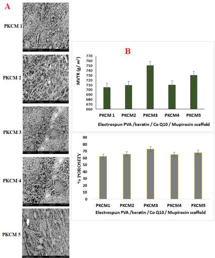
Amajuoyi et al. Future Journal of Pharmaceutical Sciences   (2020) 6:25                                                   Page 2 of 13

Background                                                                   Mupirocin is a bactericidal antimicrobial agent which
Chronic wound deviates from the normal sequence of                        has been used extensively to promote pathogen free
physiologic interactions involved in wound healing [1,                    wound bed [9]. It acts by competitive inhibitor of bacter-
2]. It therefore is necessary to tailor interventions to                  ial isoleucyl-tRNA synthetase, thereby impeding protein
match both causative pathophysiology and prevailing de-                   and RNA synthesis, which ultimately leads to bacterial
ficiencies at the wound site to promote healing [1].                      death [9]. No systemic absorption of mupirocin or its
Wound dressings play a vital role in the management of                    major metabolite, monic acid, has been detected in short
wounds through the protection of the wound from the                       courses of topical administration and penetration into
external environment, the provision of moisture, and fa-                  the deeper epidermal and dermal layers is enhanced in
cilitation of the wound healing process [3]. Varying dos-                 traumatized skin and under occlusive dressings [9].
age forms for wound healing include antibacterial-based                      Co-enzyme Q10 (CoQ10), also known as ubiquinone,
creams, ointments, and gauze dressings [3]. Antibacterial                 is both a component in the electron transport chain and,
agents are incorporated into such wound dressings to                      when reduced to ubiquinol, it acts as an antioxidant. It
lessen the risk of bacterial infection during healing                     substantially improves mitochondrial functions in dia-
process. To overcome the challenges and bridge the gap                    betic wounds and plays an important role in diabetic
posed by the high cost of skin substitutes, wound man-                    wound by reducing the production of reactive oxygen
agement approaches which utilize nanoscale biomate-                       species (ROS). This activity is very significant because of
rials are advocated. Nanoscale biomaterials have                          the ability ROS to disturb cellular signaling involved in
semblance to the extracellular matrix (ECM), both in                      wound healing, slowing the healing process [10]. Topical
structure and function, which affords them excellent                      application of skin products containing Co Q10 can sig-
capability to influence cellular pathways to tissue regen-                nificantly increase the levels of ubiquinone on both the
eration, through provision of conducive platforms for                     skin surface and deeper layers of the epidermis. Subse-
cell attachment, differentiation, and proliferation [4].                  quent elevation of ubiquinol levels demonstrated after
Electrospun fibers meet all these criteria hence their ad-                such topical therapy indicated metabolic transformation
vantage as wound dressing materials. Electrospinning                      of ubiquinone. Incubation of cultured human keratino-
ensures that fibers possess high surface area to volume                   cytes with Co Q10 showed significant augmentation of
ratio necessary for large surface applications. Fiber func-               energy metabolism [11].
tionalization and ease of materials combination is easily                    This study seeks to fabricate mupirocin, keratin, and
achieved when wound dressing materials are electrospun                    CoQ10-loaded PVA electrospun scaffolds intended for
hence the advantage of electrospun fibers over trad-                      wound healing application and to characterize their
itional wound dressings fabricated via weaving fibers to                  morphology, physical properties, antibacterial activity,
obtained cotton or gauze wound dressings [2, 4].                          and biocompatibility. A novel nanofibrous co-spun mat
   A wound dressing material of desired functionality                     containing keratin, mupirocin, and CoQ10 will offer
should possess a bio-inspired architecture that resembles                 greater chances of skin repair efficiency than any of the
the natural ECM organization in order to support cell                     wound dressing materials currently in use for the man-
adhesion, proliferation, and differentiation; the delivery                agement of chronic wounds.
of drugs and growth factors to accelerate tissue growth;
and the controlled biodegradation of the scaffold to                      Method
avoid further traumas during dressing detachment and                      Materials
peeling [4]. This is what thus makes electrospun fibers                   Mupirocin analytical standard (5,9-Anhydro-2,3,4,8-tet-
excellent wound dressing materials.                                       radeoxy-8-[[3-(2-hydroxy-1-methylpropyl)oxiranyl]-
   Polyvinyl alcohol (PVA) is a popular candidate for tis-                methyl]-3-methyl-[2E,8[2S,3S(1S,2S)]]-L-talonon-2-eno-
sue engineering due to its hydrophilicity and biocom-                     nic acid 8-carboxyoctyl ester, BRL 4910A, Pseudomonic
patibility which promote a conducive environment for                      acid. CAS number 12650-69-0 Sigma–Aldrich St. Louis,
skin regeneration. It however lacks adequate functional                   USA), mupirocin powder utilized as active pharmaceut-
groups and cell recognition sites and to improve its cell                 ical ingredient (CAS number 12650-69-0 Shaanxi Top
affinity, it needs to be used in combination with bio-                    Pharmaceutical Chemical Co. Limited, Shaanxi, China),
active composites to enhance cell adhesion and faster                     polyvinyl alcohol (PVA with Degree of hydrolysis DH =
skin repair [5]. A good candidate for this purpose is                     95; degree of polymerization = 1700. Hebei Addtie Bio-
keratin, due to its good bioactive potentials which sup-                  technology Co. Limited China), keratin (Well-green
port cell viability, cell attachment, and proliferation [6,               Technology Co. Limited, Shaanxi, China). Acetic acid
7]. Keratin has the ability to activate keratinocytes in the              (Merck KGaA, Darmstadt, Germany), ethylene glycol
wound bed, stimulating them to proliferate and migrate                    diglycidyl ether (EGDE) from (Merck KGaA, Darmstadt,
leading to eventual wound closure [8].                                    Germany). CoQ10 (Ubiquinone 50 CAS number 303-98-
Amajuoyi et al. Future Journal of Pharmaceutical Sciences         (2020) 6:25                                                  Page 3 of 13

0 Sigma–Aldrich St. Louis, USA), phosphate buffer saline                        Mechanical characterization
(PBS Merck KGaA, Darmstadt, Germany), Mueller–Hinton                            The mechanical strength, i.e., ultimate tensile strength
Agar (MHA-Merck KGaA, Darmstadt, Germany), OxoidTM                              (UTS) and ductility of the nanofibrous mats, was deter-
antimicrobial susceptibility disc for erythromycin and cipro-                   mined using Universal Tensile Machine (UTM -
floxacin (ThermoFisher Scientific USA), resistant clinical iso-                 Instron- series 3369, USA), fitted with 50 kN load cell at
lates of Staphylococcus aureus 2586, Staphylococcus aureus                      25 ± 2 °C. The UTS is a material’s maximum resistance
2590, Staphylococcus aureus 2583, Staphylococcus aureus                         to fracture. It is equivalent to the maximum load that
2587, methicillin-resistant Staphylococcus aureus 2555                          can be carried by one square inch of cross-sectional area
(MRSA), and Escherichia coli 1808. Water used in all the                        when the load is applied as simple tension. Ductility
tests was Milli-Q water (Millipore, USA). All other chemical                    measured the ability of a material to be drawn or plastic-
reagents and solvents were of analytical grade and were used                    ally deformed without fracture.
for this research without further purification.                                   The cross-linked scaffolds were cut into dog bone-
                                                                                shaped sections (50 mm × 13.5 mm) to control failure lo-
                                                                                cation and tested in ambient temperature of 20 °C and hu-
Electrospun scaffolds design and fabrication                                    midity of 60%. The sections of each of the electro spun
Using the method of Qiang et al. [5] with slight                                mats (dimensions 13.5 cm) were mounted between two
modification, five electrospinning solutions were each                          clamps and stretched at rate of 50 mm/min with an ap-
prepared by dissolving 10% w/w PVA in 18 mL of                                  plied load range of 50 N and gauge length of 50 mm [12].
the distilled water/acetic acid solvent system. The                             UTS and ductility determinations were done in triplicate.
mixture was transferred into a 200 mL conical flask
and magnetically stirred at 300 rpm at a temperature                            Morphological characterization
of 80 °C for 2 h and then reduced to 50 °C for 1 h,                             Scanning electron microscopy
followed by the addition of keratin dissolved in 10                             The surface morphology of the fabricated electrospun
mL of distilled water (Table 1). After 30 min, mupir-                           keratin/Co Q10/PVA nanofibrous scaffolds was deter-
ocin and/or Co Q10 (Table 1) were each separately                               mined using field emission scanning electron micros-
dissolved in 2 mL of the water-acetic acid solvent and                          copy (FEG-SEM) via a Phenom World Eindhoven
added to the PVA solution, followed by 0.1 mL of                                Phenom ProX scanning electron microscope at an accel-
EGDE for cross linking of the polymer. The mixture                              erating voltage of 15 kV. The scaffolds were cut into
was stirred at 300 rpm for 30 min at 60 °C to ensure                            small pieces of 5 mm × 5 mm. Before observation, each
complete dissolution. The solutions were kept for 24                            sample fibers were sputtered coated with thin layer of
h to ensure homogeneity and then subjected to elec-                             gold using a quorum-Q150R Plus E. The 5 mm × 5 mm
trospinning. Electrospinning was carried out after                              cut samples were then mounted over the studs using a
process optimization under defined constant ambient                             carbon tape and analyzed at 15 KV. All the various sam-
condition (temperature: 25 ± 1.56 °C and relative hu-                           ples were analyzed in triplicate.
midity 40%). In each electrospinning solution, PKCM
series (Table 1) was loaded into a 50 mL plastic syr-                           Moisture vapor transmission rate
inge with an internal diameter of 0.8 mm, length of                             A 40 mm diameter sample of each electrospun scaffold
20 mm, and fitted with a gauge 19G needle. The so-                              of the series was fixed over a calibrated cylindrical vial
lutions were fed at 4.0 mL/h, and this was achieved                             (Smith Scientific glassware Edenbridge, Kent UK) of 35.7
by fall under gravity. The collector was placed at 15                           mm inner diameter, containing 20 mL of distilled water.
cm from the needle tip to the collector. Electrospun                            The test sample container was weighed (W1) before the
fiber mats were collected by applying a voltage at 50                           start of the test and the container was then transferred
kV in the needle. The electrospun scaffolds were                                to an incubator for 24 h (temperature 37.0 ± 0.5 °C and
stored in airtight containers and suitable samples                              RH 20%). At the end of the test period, the container
withdrawn intermittently for analysis.                                          was reweighed (W2) to observe any changes in weight

Table 1 Composition of the solutions for electrospinning
Electrospun                     Electrospun PVA/keratin/Co Q10/Mupirocin scaffold formulation
component
                                PKCM1                       PKCM2                      PKCM3                  PKCM4              PKCM5
PVA (% w/w)                     10                          10                         10                     10                 10
Keratin (% w/w)                 0.01                        0.1                        0.01                   0.1                0.05
CoQ10 (% w/w)                   0.05                        –                          0.05                   0.1                0.15
Mupirocin (% w/w)               –                           2                          2                      2                  2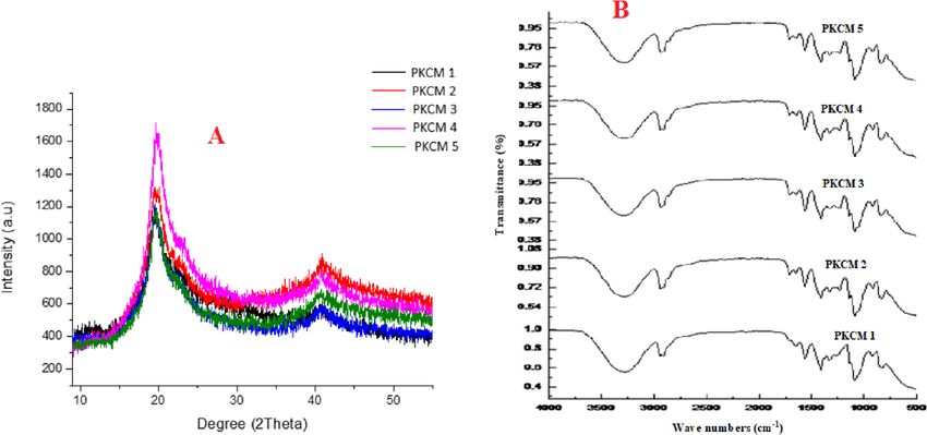
Amajuoyi et al. Future Journal of Pharmaceutical Sciences   (2020) 6:25                                                              Page 4 of 13

due to water absorption. The moisture vapor transmis-                     incubated at 37 ± 0.5 °C and stirred at 100 rpm using a
sion rate (MVTR) was obtained from Eq. (1).                               magnetic stirrer. Aliquots of each sample (0.5 mL) were
                                                                          taken from the release medium at specific intervals (0, 5,
    X ¼ ðW 1−W 2Þ  1000  24=T                                ð1Þ        15, 30, and 60, 75 min) and replaced with 0.5 mL with
                                                                          fresh buffer solution maintain sink condition [14]. The
  where X is MVTR (g/m2/24 h); W1 is the initial mass
                                                                          solution was subsequently filtered using nylon mem-
of the container, sample, and liquid in grams, W2 is the
                                                                          brane of 0.45 μm porosity. The resultant drug solution
mass of the container, sample, and liquid in grams after
                                                                          was analyzed by high-performance liquid chromatog-
the test duration; and T is the test period in hours [3].
                                                                          raphy using C18 column (250 × 4.6 mm, 5 μm) at 25 °C
                                                                          and the mobile phase combination of ammonium acet-
Determination of the porosity of the electrospun scaffolds
                                                                          ate (0.05 M adjusted to pH 5.0 with acetic acid) and
The porosity of the scaffolds was determined using the
                                                                          acetonitrile 60:40 (v/v). The injection volume was 20 μL
“liquid displacement method” [13] with ethanol used as
                                                                          and the flow rate 1.0 mL/min; the ultraviolet detection
the displacement liquid. The weight of the scaffold used
                                                                          was set at 228 nm. The reported values are average of
was noted as “w.” The scaffold was then immersed in 5
                                                                          five readings ± SD [14]. LOD release for mupirocin re-
mL of ethanol and volume of ethanol (V1) was noted.
                                                                          lease was linear between 1 and 100 μg/mL (R2 = 0.998).
After 5 min, the volume of ethanol present in the
ethanol-impregnated scaffold (V2) and the volume of
ethanol after removal of the ethanol-impregnated scaf-                    Biological characterization
fold (V3) was used to calculate porosity of the fiber as in               Skin irritation
Eq. (2). All measurements were achieved using a cali-                     Twenty-four 3-month-old male and female rats with body
brated cylindrical vial (Smith Scientific glassware Eden-                 weight 230–240 g were purchased form Tosab® Laborator-
bridge, Kent UK) and performed in triplicate.                             ies Ogbomoso Osun State. They were individually housed
                                                                          and free access to standard chow and drinking water.
                                 V 1−V 3
    Porosity of the fiber ¼                                    ð2Þ        They were allowed to acclimatize to the environment for
                                 V 2−V 3
                                                                          7 days prior to the start of the experiments and were
                                                                          maintained on a 12-h light/dark cycle. This study was car-
Chemical characterization                                                 ried out in strict accordance with the recommendations in
X-ray diffraction                                                         the Guide for the Care and Use of Laboratory Animals of
The X-ray diffraction patterns of the electrospun inning                  the National Institutes of Health [15, 16]. This study uti-
fibers were obtained using an X-ray diffractometer (D/                    lized the Animal research: reporting in vivo experiments:
MAX-2500X, Rigaku, Tokyo, Japan) with Cu Kα charac-                       the ARRIVE guidelines in documenting the study [16].
teristic radiation (wavelength λ = 0.154 nm at 40 kV,                     The protocol was approved by Health research ethics
150 mA, and a scan speed of 4° per minute in the 2θ                       committee (CMUL/HREC/09/19/607) (Table 2).
range of 5°–80°) [7].                                                        The skin irritation test was performed on the electro-
                                                                          spun PKCM nanofibers in healthy albino rats weighing
Fourier transform infrared spectroscopy                                   averagely 238 g ± 1.45 g. The rats were randomly
The infra-red spectra of the electrospun scaffolds were
obtained using a Fourier transform infrared spectrom-
                                                                          Table 2 Scale for assessing skin irritancy reactions
eter (Cary 630 FTIR Spectrometer; Agilent Technologies,
                                                                          Reaction   Description                                             Score
Santa Clara, CA, USA). The spectral range was 4000
cm−1 to 400 cm−1. Then, 0.5 g of nanofibrous mats were                    Erythema No erythema                                               3

dried in a vacuum oven (45 °C) and subsequently placed                               Slight erythema (not very noticeable)                   2
over the diamond crystal for analysis. Twenty scans were                             Moderate erythema (noticeable,but not covering the      1
recorded for each spectrum. Smoothing was done where                                 entire test area)
necessary to reduce the noise, without loss of any peak.                             Severe erythema (noticeable and covering the entire     0
                                                                                     test area)
The absorptions peaks/bands were studied to know if
there was any chemical interaction occurring as a result                  Edema      No oedema (no swelling within the entire test area)     3
of the co-formulation.                                                               Slight edema (1 swollen spot, having an area not        2
                                                                                     more than 0.5 mm2)
In vitro release of mupirocin from PKCM scaffolds                                    Moderate edema                                          1
                                                                                     (2–3 swollen spots, each having an area not more
Using a modified in situ glass vial system, a patch of                               than 0.5 mm2)
PKCM scaffold (4 cm × 4 cm) was immersed into 50 mL
                                                                                     Severe edema (up to half of the test area is swollen)   0
of phosphate buffer solution (pH 7.4). The samples were
Amajuoyi et al. Future Journal of Pharmaceutical Sciences   (2020) 6:25                                                    Page 5 of 13

divided into six groups (three rats in a group). Exposure                 Cytotoxicity and cell proliferation assay
of skin to the test materials was accomplished by means                   The stock solutions were prepared by dissolving 26.5 mg
of a patch test technique using two intact sites on the                   of each of the scaffolds in 4 mL of phosphate buffer pH
back of each of the eighteen albino rats. The skin was                    7.4. and sterilized by filtration using of 0.20 μm pore size
clipped free of hair a day prior to testing. A square patch               filter and subsequently stored in the refrigerator until
of the five electrospun scaffolds, with the dimension of                  needed for the tests. The CellTiter-Glo® 2.0 assay kit
2.5 cm × 2.5 cm were applied to the test site on the cor-                 (Promega) was used to determine cell viability, as de-
responding test group and covered with gauze of same                      scribed by the manufacturer. Briefly, 1.0 × 10⁴ keratino-
dimension.                                                                cyte (HaCaTs) cells were seeded in a luminometer
  Mupirocin ointment was applied to group 6 and cov-                      compatible 96-well plate and allowed to grow and ad-
ered with gauze. PKCM1, PKCM2, PKCM3, PKCM4,                              here overnight at 37 °C in a 5% CO2 culture incubator.
and PKCM5 were tested for the presence of erythema or                     Treatments were performed by adding a 1:1 scaffold so-
oedema. Group 1 rats served as the negative control                       lution/media mix to the cells for 24 h and 72 h respect-
group and the test areas of the skin were covered with                    ively. The plate and its contents were equilibrated to 25
polyethylene sleeve only. Group 6 rats were the positive                  ± 1 °C for approximately 30 min and then a volume of
control group and were treated with 2% mupirocin oint-                    CellTiter-Glo 2.0 reagent equal to the volume of cell cul-
ment. This was followed by the occlusion of the entire                    ture medium present in each well was added. The con-
trunk using a polyethylene sleeve. The entire covering                    tents were subsequently mixed for 2 min using an
was removed after 24 h and the site assessed and scored                   orbital shaker to induce cell lysis and then followed by
for erythema and edema. Following the administration                      plate incubation at room temperature for 10 min to
of the test samples and controls (24, 48, or 72 h). At the                stabilize the luminescent signal and finally the lumines-
end of the study, all the animals were humanely eutha-                    cence was recorded using luminometer (MODULUS,
nized via exposure to carbon dioxide gas in a sealed                      Promega). The luminescent signal is proportional to the
chamber.                                                                  amount of ATP in the sample, which indicates the pres-
                                                                          ence of metabolically active cells.
                                                                             Cell viability is expressed in Eq. (3).
Antibacterial activity assessment
This was done by disk diffusion method using Muel-                                                 sample luminescence
                                                                              %Cell viability ¼                           100      ð3Þ
ler–Hinton Agar (MHA) as the medium. Using sterile                                                average luminescence x
swaps, the bacterial culture suspensions of resistant
clinical isolates of Staphylococcus aureus 2586,
Staphylococcus aureus 2590, Staphylococcus aureus                         Antioxidant activity of the of the PKCM scaffolds using
2583, Staphylococcus aureus 2587, methicillin-resistant                   DPPH assay
Staphylococcus aureus 2555 (MRSA), and Escherichia                        The capacity of CoQ10 molecule (Ubiquinone) to act as
coli 1808 standardized to a turbidity of 0.5 McFarland                    free radical scavenging antioxidant after it has been in-
standard were spread on different plates of MHA. In                       corporated in the electrospun scaffolds was determined
vitro antibacterial activity of PKCM electrospun nano-                    using DPPH assay. Spectrophotometric determination of
fibrous scaffold against each bacterial strain was de-                    the free radical scavenging activity was evaluated using a
termined via disc diffusion test [14] using 150 mm                        method described by Ilomuanya et al. [17]. The radical
diameter Mueller-Hinton (Carolina Biological Supply                       scavenging activity of the scaffolds against 1,1 diphenyl1-
Co. Burlington, NC) agar plates. PKCM electrospun                         1-picryl-1-hydrazyl (DPPH) radical via UV-Vis absorb-
scaffolds were cut into 10 mm diameter and placed                         ance at 517 nm was determined utilizing ascorbic acid
on the inoculated test organisms and then observed                        as standard. Further, 100 mg of the electrospun scaffolds
for any zone of inhibition around the scaffolds. Clear                    was weighed out and the antioxidant content extracted
inhibition zones around the PCKM electrospun nano-                        using 50 mL absolute ethanol in a separating funnel to
fibrous discs indicate that the scaffolds possess anti-                   obtain the test sample. To a methanolic solution of
microbial activity. OxoidTM antimicrobial susceptibil-                    DPPH (100 mmol/L 2 mL), varying concentrations (5–
ity disc for erythromycin and ciprofloxacin were used                     25 mg/mL) of the test sample was further dissolved in 3
as control for antibacterial assessment. Each assay was                   mL ethanol and assayed. When DPPH reacts with an
performed in triplicate and incubated in an inverted                      antioxidant compound, which can donate hydrogen, it is
position at 37 ± 1 °C for 18 h and the zones of in-                       reduced. The changes in color (from deep violet to light
hibition was measured. The protocol was approved by                       yellow) after 100 min of reaction was determined using a
Health research ethics committee (CMUL/HREC/09/                           UV-Visible Spectrophotometer (Unico S2150UV NJ,
19/607).                                                                  USA) with absorbance recorded at 517 nm. A
Amajuoyi et al. Future Journal of Pharmaceutical Sciences (2020) 6:25 Page 6 of 13
commercial antioxidant Citrix® Topix Pharmaceuticals, b). PKCM3 scaffold containing CoQ10 0.05% had high-
Inc. NY, USA was also evaluated as antioxidant control est ductility value of 81.2%, while PKCM4 containing
and compared with the fabricated scaffolds. % Scaven- 0.1% CoQ10 had the lowest value of 44.2%. Variations in
ging activity was calculated using Eq. 4. ductility of electrospun PKCM electrospun scaffolds are
shown in Fig. 1b. Research has confirmed that a high
Absorbance517control −Absorbance517 sample
%Scavenging activity ¼ 100 elastic material will possess a high value of elongation at
Absorbance517control
ð4Þ break [18]. PKCM3 scaffold showed the best capacity to
deform before breakage with a ductility of 81.2%.
PKCM1 shows 78.7% response to deformation which is
Statistical analysis second to PKCM3. PKCM2, PKCM4, and PKCM5 reveal
The data was presented as mean ± standard deviation of ductility values of 57.9%, 44.2%, and 70.7%. The reduced
more than three experimental values for every variable ductility values could be attributed to clustering of parti-
and analyzed by one-way ANOVA followed by Dunnett’s cles making each component to act as a rigid constituent
test using GraphPad Prism version 5. P value of less than which obstructs the mobility of crazes during tensile
0.05 was considered statistically significant. loading [18].
Results Morphological characterization
Mechanical characterization Scanning electron microscopy, moisture vapor transmission
PKCM2 scaffold which did not contain CoQ10 had least rate, and porosity of the electrospun scaffolds
tensile strength of 0.03 MPa, while PKCM4 containing All the electrospun scaffolds developed has sizes in the
0.1% CoQ10 had the highest value of 0.20 MPa (Fig. 1a, nanometer range. Different morphological features of
Fig. 1 a Ultimate tensile strength and (B)% Ductility of the electrospun PVA/keratin Co Q10 mupirocin scaffoldsAmajuoyi et al. Future Journal of Pharmaceutical Sciences (2020) 6:25 Page 7 of 13 electrospun fibers are shown in Fig. 2a. The diameters of (MVTR) 700–1200 g/m2 [19]. The developed PKCM the scaffolds were observed to be in the nano particulate scaffolds exhibited MVTR in the range 709 ± 1.75–758 range. The values were 3.27 ± 0.1 nm, 2.61 ± 0.3 nm, ± 1.75 g/m2 for PKCM1 and PKCM3. The highest 2.36 ± 0.1 nm, 2.32 ± 0.2 nm, and 2.11 ± 0.2 nm for MVTR was exhibited by PKCM 3, and PKCM 1 having PKCM1, PKCM2, PKCM3, PKCM4, and PKCM5 re- the lowest values all within the desired limits to prevent spectively. It is observed that fibers of sample PKCM1 excessive hydration of the wound bed. The porosity of (Fig. 2a) have varying diameters with few beads and rela- the samples is 62.6 ± 3.2%, 66 ± 4.3%, 73.3 ± 2.8, 65.3 ± tively smooth surfaces. Fibers of sample PKCM2 (Fig. 4.5%, and 68.1 ± 2.6% for PKCM1, PKCM2, PKCM3, 2a) have near uniform diameter with a smooth surface PKCM4, and PKCM5 respectively. This shows that the while fibers of samples PKCM3, PKCM4, and PKCM5 difference in the concentration of the keratin loaded in have uniform densely knit scaffolds with near uniform the fibers may influence fiber porosity. diameters. The formation of uniform densely knit scaf- folds could be attributed to surface tension of electro- Chemical characterization spinning solutions. Wound dressings should be able to X-ray diffraction protect the surface of the wound from drying out; Diffraction patterns of reinforced PKCM1, PKCM2, healthy skin possess moisture vapor transmission rate PKCM3, PKCM4, and PKCM5 scaffolds are shown in Fig. 2 a Scanning electron micrographs of electrospun scaffolds. b Moisture vapor transmission rate and c percentage porosity of the electrospun scaffolds
Amajuoyi et al. Future Journal of Pharmaceutical Sciences     (2020) 6:25                                                         Page 8 of 13

Fig. 3a. Two characteristic peaks existing at 2θ= 19.7°                     C-O stretching indicative of PVA. The FTIR spectra
and 40.8° are related to the semi-crystalline nature of                     PKCM1 to 5 showed characteristic bands at 1712 cm−1
PVA and as such, the scaffolds possess similar diffraction                  corresponding to C=O stretching, at 2859 and 2929
patterns. The variations in the additives used in the                       cm−1 corresponding to C−H deformation, at 1150 cm−1
preparation of the precursor solutions for the scaffolds                    corresponding to the C−O−C stretching vibration, and
culminated in the formation of different peak intensities                   at 1052 cm−1 corresponding to the C−C skeleton which
and sizes. PKCM4 possesses the highest intensity in the                     are indicative of the presence of mupirocin [20]. The
stronger diffraction angle (2θ= 19.7°) with the narrowest                   spectrum shows no indication of chemical interactions
peak, thus indicating the highest crystallinity. This                       in all the PKCM spectra hence mupirocin and keratin
means that the cross-linking agent performed optimally                      were successfully incorporated into the scaffolds during
at the ratio of the additives used in the formulation of                    electrospinning.
PKCM4. This also collaborates with the observation that
PKCM4 scaffold possessed the greatest mechanical                            In vitro release of mupirocin from PKCM scaffolds
strength than the other scaffolds produced in this study.                   The ability of the scaffold to effectively modulate the re-
This is evident in the highest magnitude of tensile                         lease of mupirocin is critical to the success of the devel-
strength exhibited by this sample compared to other                         oped scaffold. The electrospun scaffolds must be seen to
electrospun scaffolds (Fig. 1a).                                            have the ability to release the active ingredient at the site
                                                                            of action. The drug release of mupirocin for the PKCM
Fourier transform infrared spectroscopy                                     scaffolds was obtained at r2 = 0.998 at wavelength of 228
Figure 3b shows the Fourier transform infrared spectros-                    nm. There was an initial burst release of mupirocin from
copy (FTIR) spectra in the region 4000–600 cm−1 of the                      the scaffolds. PKCM 4 had 27.25 ± 0.19% release at 5
scaffold formulation. FTIR shows that the large bands                       min. At 30 min, all the scaffolds had more than 50% of
observed between 3500 and 3200 cm−1 are linked to the                       the cumulative concentration of mupirocin released with
stretching O-H from the intermolecular and intramo-                         51.06 ± 2.104%, 70.31 ± 1.99%, 74.11 ± 2.36%, and 74.66
lecular hydrogen bonds. Characteristic bands for α-helix,                   ± 1.72% of mupirocin released respectively for PKCM 2,
β-sheet, and random coil conformations in the amide I                       PKCM 3, PKCM 4, and PKCM 5. At 1 h, 80% of the
(C=O stretch vibrations of the peptide linkages 1700–                       mupirocin in the scaffold was seen to have diffused out
1600 cm−1) and amide II (N-H bending and from the C-                        of the scaffold (Fig. 4), hence buttressing the presence of
N stretching vibration 1560–1500 cm−1) regions [20] in-                     reversible chemical linkages in the scaffolds that are not
dicative of the presence of keratin. There is a 3000–2800                   interfering with the release of the API mupirocin hence
cm−1 vibration band due to stretching C-H from alkyl                        allowing the drug to be available at the site of action
groups, and 1750–1690 cm−1 peaks showing C=O and                            when it is applied on an actual wound.

 Fig. 3 a X-ray diffraction curves and b Fourier transform infrared spectra for PKCM1, PKCM2, PKCM3, PKCM4, and PKCM5 scaffolds
Amajuoyi et al. Future Journal of Pharmaceutical Sciences (2020) 6:25 Page 9 of 13
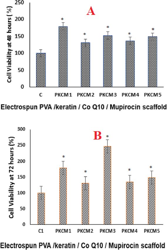
Biological characterization Cytotoxicity and cell proliferation assay
Skin irritation Biocompatible polymers and their products must be
There was no skin irritation; all the scaffolds had a score of a less toxic nature with assured cell viability. By
of 3 for both edema and erythema. Hence, the scaffolds ISO 10993-5 standards, anything that reduces cell via-
did not cause any allergic reactions or hypersensitivity bility by more than 30% can be cytotoxic to the
reactions. membranes. All the electrospun fabricated PKMC
scaffolds were found to have viable cell percentage
above 100%. The cell line studies on the electrospun
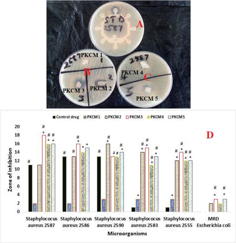
Antibacterial activity assessment scaffolds showed their ability to support the growth
For antimicrobial assessment, the prepared scaffolds of keratinocytes as indicated by higher values for %
were tested against six microorganisms, resistant clinical viability (Fig. 5a, b). All the electrospun scaffolds
isolates of Staphylococcus aureus 2586, Staphylococcus showed higher cell viability values than the control at
aureus 2590, Staphylococcus aureus 2583, Staphylococcus 48 and 72 h, with the CoQ10 containing scaffolds
aureus 2587, Staphylococcus aureus 2555, and MRD PKCM1: 180%, 179%; PKCM 3: 152%, 247%; PKCM
Escherichia coli 1808 using the agar diffusion method to 4: 136%, 135%; and PKCM5: 152%, 148%, had higher
assess their antimicrobial activity. The growth of E. coli cell viability values than PKCM2 (130%), which did
was not inhibited by either the control drug (erythro- not contain CoQ10 (Fig. 6).
mycin) or the PKCM1 electrospun keratin/Co Q10/PVA
scaffold which did not contain any mupirocin. Electro-
spun keratin/Co Q10/PVA scaffold containing mupiro- Antioxidant activity of the of the PKCM scaffolds using
cin PKCM2, PKCM3, PKCM4, and PKCM5 showed DPPH assay
antimicrobial activity against all the clinical isolates of Coenzyme Q10 (CoQ10) is an effective antioxidant of
Staphylococcus aureus (Fig. 5a–d). Oxoid™ antimicrobial the cellular membranes hence its inclusion in the elec-
susceptibility disc for erythromycin was seen to have ac- trospun PKCM scaffold development. There was a
tivity against Staphylococcus aureus 2587 and Staphylo- concentration-dependent increase in DPPH free radical
coccus aureus 2586. There was no activity against multi- scavenging with increasing scaffold concentration of
drug resistant bacteria with the Oxoid™ antimicrobial CoQ10 (Fig. 7). The percentage DPPH free radical scav-
susceptibility disc for the control drugs, compared to the enging was not influenced by the concentration of
scaffolds which showed some activity. CoQ10 in the scaffolds. PKCM5 exhibited the highest
The strongest activity was observed with PKCM3 with percentage DPPH free radical scavenging activity 65.8 ±
zone of inhibition 18.5 mm against Staphylococcus aureus 1.02. This result was not statistically significantly differ-
2587. There was a statistically significant difference between ent from the values obtained for PKCM1, PKCM3, and
all the scaffolds containing mupirocin and control drug with PKCM4 as shown in Fig. 7. The DPPH scavenging activ-
PKCM 2, 3, 4, and 5 having excellent activity against ity of the scaffolds also showed that the electrospinning
Staphylococcus aureus, hence the fabricated scaffold could be did not adversely affect/ diminish the antioxidant activity
more effective for treatment of infected wounds. of the scaffold.
Fig. 4 Cumulative release of mupirocin from electrospun PVA/keratin/Co Q10 mupirocin scaffoldsAmajuoyi et al. Future Journal of Pharmaceutical Sciences (2020) 6:25 Page 10 of 13 Fig. 5 Evaluation of antibacterial activity of the electrospun keratin/Co Q10/PVA nanofibrous scaffold. a Control drug erythromycin. b Microbial plates showing PKCM1, PKCM2, and PKCM3. c Microbial plates showing PKCM3 and PKCM4. d Zone of inhibition exhibited by various microorganisms when challenged with control drug and electrospun keratin/Co Q10/PVA nanofibrous scaffolds. * The difference in mean is significant (P < 0.05) with respect to control. # The difference in mean is significant (P < 0.05) with respect to PKCM1 Discussion composites like keratin to enhance cell adhesion and fas- Electrospun nanofibres have found its place in wound ter skin repair [8, 21]. This new developed electrospun management. Polyvinyl alcohol (PVA) is a popular poly- nanofibers combine the biocompatibility potential of mer used because of its biocompatibility and its ability PVA, with the bioactive nature of keratin and CoQ10 to provide a conducive environment for skin regener- and the antibacterial property of mupirocin as a new po- ation. This polymer lacks other functional groups and tential for proper wound care. cell recognition sites for functionality at the cellular level The presence of CoQ10 in PKCM3, PKCM4, and is therefore used in combination with bioactive PKCM5 lowered the surface tension thereby enhancing
Amajuoyi et al. Future Journal of Pharmaceutical Sciences          (2020) 6:25                                                    Page 11 of 13

                                                                                 it from excess hydration which may impede wound heal-
                                                                                 ing. Normal skin has moisture vapor transmission rate
                                                                                 (MVTR) of 700–1200 g/m2 [19], an observed MVTR of in
                                                                                 the range 705–750 g/m2. Twenty-four hours for that sam-
                                                                                 ples under study is indicative of the potential of the scaf-
                                                                                 folds to provide an optimal moist environment locally and
                                                                                 hence prevent excessive dehydration while allowing con-
                                                                                 trolled evaporation of wound exudates to promote wound
                                                                                 healing. The mechanical studies are carried out to check
                                                                                 the strength of the synthesized electrospun scaffolds,
                                                                                 which is essential for its biomedical application. The most
                                                                                 important criterion for such application is that the sam-
                                                                                 ples should be strong enough to avoid the damage. A
                                                                                 wound dressing scaffold should have enough mechanical
                                                                                 strength to withstand handling both during packaging and
                                                                                 wound dressing application. Figure 1a shows the highest
                                                                                 mechanical load the electrospun scaffolds can withstand
                                                                                 prior to fracture under tension. The scaffolds showed dif-
                                                                                 ferent UTS indicating variations in strengthening effects
                                                                                 imparted by the crosslinking agent, ethylene glycol diglyci-
                                                                                 dylether (EDGE), used in this study. The PKCM4 scaffold
                                                                                 had the highest UTS at maximum magnitude of 0.2 MPa.
                                                                                 This could be attributed to promoted wettability and bet-
                                                                                 ter matrix/fiber adhesion at the concentrations of the vari-
                                                                                 ous components used in the formulation of PKCM4
                                                                                 scaffold. It has been reported that effective stress transfer
                                                                                 between matrix and fiber occurs in composites of en-
                                                                                 hanced wettability [25]. PKCM5 (UTS of 0.15 MPa) is the
 Fig. 6 a Forth-eight hours and b 72 hours cell viability of the                 second strongest electrospun composite after PKCM4.
 control and the electrospun. Treatments were compared with
 control *P < 0.05
                                                                                 Decline in UTS could be attributed to agglomeration and
                                                                                 sediment effects associated with filler contents which may
                                                                                 ultimately lead to poor wetting between the fillers and
the morphological features of the fibers. No crystallization                     matrix; this, according to Pan et al. [26], may result in for-
of drugs was detected on the polymer surface of the elec-                        mation of micro-cracks. PKCM2 possesses the least UTS
trospun scaffolds and this suggests that there was a homo-                       of 0.03 MPa; since PKCM2 is the only formulation with-
geneous dispersion of mupirocin in the electrospun fibers                        out CoQ10, it suggests that the presence of CoQ10 in-
[22–24]. It is desirable that a wound healing material                           creased the wettability. The native skin has tensile
could prevent dehydration of the wound, but also protect                         strength and ductility of 5–30 MPa and 35–115%

 Fig. 7 Scavenging of DPPH activity of the CoQ10 in the PKCM electrospun scaffolds utilizing(n = 3 ± SD)
Amajuoyi et al. Future Journal of Pharmaceutical Sciences (2020) 6:25 Page 12 of 13
respectively [5]. Though the electrospun scaffolds all have hence, the presence of CoQ10 offered protection to the
ductility values that fall within the range of the values for cells due to the antioxidant properties [30], leading to
the native skin, the corresponding mechanical strengths growth of the keratinocytes used in the study.
are below the range. The best capacity to deform before
breakage was exhibited by PKCM3, with a ductility of Conclusion
81.2%. When combined with the value obtained for UTS, Electrospun nanofibers combining the biocompatibility
PKCM3 possessed adequate mechanical strength and a potential of PVA, with the bioactive nature of keratin
low enough tensile strength (0.1 MPa) to act as a wound and CoQ10 and the antibacterial property of mupirocin
dressing [5]. It has however been reported that scaffolds as a new potential for proper wound care, were success-
with low tensile strength offer more advantages for cell fully developed. The PKCM3 scaffolds displayed
culture because they have closer semblance with the colla- optimum porosity and ductility and MVTR, important
gen, elastin and other soft tissues [5]. Furthermore, the for absorbing excess exudates and maintaining a moist
challenge that low tensile strength can pose can be over- environment on the wound surface. Electrospun keratin/
come by either using them as primary dressings in direct Co Q10/PVA scaffold containing mupirocin PKCM3
contact with the wound or by electrospinning them dir- showed excellent antimicrobial activity against all the
ectly on the wound site, using a handheld electrospinning clinical isolates of 2586, Staphylococcus aureus 2590,
device [4, 26, 27]. PKCM3 showed the highest values in % 2583, 2587, 2555. The cell line studies on the PKCM3
porosity and moisture vapor transmission rate, hence opti- showed their ability to support the growth of keratino-
mizing it ability to deter wound bed dehydration while cytes hence the potential of developed scaffolds as a
controlling wound exudate flow thus keeping the wound wound dressing. In vivo studies to further investigate the
area oxygenated and protected from reinfection [19]. applications of the electrospun keratin/Co Q10/PVA
Mupirocin is an effective antibacterial agent and is nanofibrous scaffold as a wound dressing is however
often used in clinical practice as topical ointment to required.
treat a wide variety of topical wounds. Excellent activity
Abbreviations
against Staphylococcus aureus, by the fabricated scaffold PKCM: Polyvinyl alcohol (PVA) and keratin / Co Q10 fibrous scaffolds;
containing mupirocin, will be effective for treatment of ECM: Extra-cellular matrix; MVTR: Moisture vapor transmission rate;
infected wounds. PKCM5 showed the highest cumulative EDGE: Ethylene glycol diglycidylether
drug release while PKCM2 had the lowest (Fig. 4). Acknowledgement
PKCM5 contains CoQ10 0.15%, while there is no The content is solely the responsibility of the authors. The authors
CoQ10 in PKCM2. The improved drug release may be acknowledge the technical effort of Mr Olaleye from the Department of
Metallurgical and Materials Engineering, Faculty of Engineering, University of
attributed to the lowering of the surface tension and im- Lagos during the electrospinning procedure.
proved wettability of the PKCM5 scaffold due to the
presence of CoQ10. PKCM3 and PKCM4 contain 0.05% Authors’ contributions
MOI conceived the study. MOI and JNA and helped design and coordinate
and 0.1% of CoQ10 respectively and showed similar drug the study. JNA, YAO, and SOA carried out the experimental studies. MOI,
release patterns. CoQ10 can enhance mitochondrial ac- CPA, and CI drafted the manuscript. All authors have read and approved the
tivity by virtue of being a cofactor in the complexes I–III final manuscript.
in the mitochondrial electron transport chain [10, 28]. Funding
The higher cell viability values shown by the CoQ10 Not applicable.
electrospun scaffolds can be attributed to increase in cel-
Availability of data and materials
lular energy available for cell growth stimulated by the
All data and material are available upon request.
presence of CoQ10. Topically applied Co Q10 has been
shown to have beneficial effects on mitochondrial mem- Ethics approval and consent to participate
brane potential of UV-stressed keratinocytes, suggesting Ethical approval was obtained from the Health Research and Ethics committee
of the College of Medicine University of Lagos. All the experiments accorded
that topically applied ubiquinone reaches the vital layers with the Institution Guidelines and were approved by College of Medicine
of the skin and promotes energy metabolism thereby be- University of Lagos Health Research Ethical Committee CMUL/HREC/09/19/607.
ing transformed into ubiquinol [10]. Increase in the anti-
Consent for publication
oxidant content of the scaffolds was also attributed to Not applicable.
the presence of CoQ10 in the fibers. This is in conson-
ance with Bardania et al. [29], who studied the antioxi- Competing interests
The authors declare that they have no competing interests.
dant and antimicrobial activity of silver ion
nanoparticles for wound healing applications [29–31]. Author details
1
The DPPH scavenging activity of CoQ10 within the scaf- Department of Pharmaceutics and Pharmaceutical Technology, Faculty of
Pharmacy, University of Lagos, PMB 12003, Surulere, Lagos, Nigeria.
folds was not affected by electrospinning process and 2
Population Council, Center for Biomedical Research, Rockefeller University,
did not affect the antioxidant activity of the scaffolds; New York 10065, USA. 3Department of Pharmaceutics and PharmaceuticalAmajuoyi et al. Future Journal of Pharmaceutical Sciences (2020) 6:25 Page 13 of 13
Technology, Faculty of Pharmacy, Kwame Nkrumah University of Science and evaluation. Dhaka Univ. J. Pharm. Sci. 17(2):213–219 https://doi.org/10.3329/
Technology, Kumasi, Ghana. 4Department of Metallurgical and Materials dujps.v17i2.39178
Engineering, Faculty of Engineering, University of Lagos, PMB 12003, 18. Balaji S, Kumar R, Sripriya R, Kakkar P, Ramesh DV, Reddy PNK, Sehgal PK
Surulere, Lagos, Nigeria. (2012) Preparation and comparative characterization of keratin–chitosan and
keratin–gelatin composite scaffolds for tissue engineering applications.
Received: 17 March 2020 Accepted: 1 June 2020 Mater Sci Eng C 32(4):975–982 https://:doi.org/10.1016/j.msec.2012.02.023
19. Xu R, Xia H, He W, Li Z, Zhao J, Liu B, Wang Y, Lei Q, Kong Y, Bai Y, Yao Z,
Yan R, Li H, Zhan R, Yang S, Luo G, Wu J (2016) Controlled water vapor
transmission rate promotes wound healing via wound re-epithelialization
References and contraction enhancement. Scientific reports 6:24596 https://doi.org/10.
1. Frykberg RG, Banks J (2015) Challenges in the treatment of chronic wounds. 1038/srep24596
Advances in Wound Care 4(9):560–582 https://doi.org/10.1089/wound.2015.0635 20. Kong J, Yu S (2007) Fourier transform infrared spectroscopic analysis of
2. Han G, Ceilley R. (2017) Chronic wound healing: a review of current protein secondary structures. Acta Biochim Biophys Sin 39(8):549–559
management and treatments. Advances in Therapy. Springer Healthcare. 34, https://doi.org/10.1111/j.1745-7270.2007.00320.x
599–610 https://doi.org/10.1007/s12325-017-0478-y. 21. Hill P, Brantley H, Van Dyke M (2010) Some properties of keratin
3. Ilomuanya MO, Adeyinka O, Aghaizu C, Cardoso-Daodu I, Akhimien T, Ajayi biomaterials: Kerateines. Biomaterials 31(4):585–593 https://doi.org/10.1016/j.
T et al., (2019) Co-formulation and characterization of gentamicin-loaded biomaterials.2009.09.076
alkyl acrylate cross polymer hydrogel infused with ethanol extract of 22. Minoo S, Arab-Sorkhi S, Vatani H, Bagheri-Pebdeni A (2015) New wound
Tetracarpidium conophorum impregnated on gauze sponge for wound dressing polymeric nanofiber containing green tea extract prepared by
dressing Wound Healing Southern Africa 12(1):22-28 https://journals.co.za/ electrospinning method. Fibers and Polymers 16(8):1742–1750 https://doi.
content/journal/10520/EJC-17b07e7ea5 org/10.1007/s12221-015-5297-7
4. Dong Y, Zheng Y, Zhang K et al (2020) Electrospun nanofibrous materials 23. Sill TJ, von Recum AH (2008) Electrospinning: applications in drug delivery
for wound healing. Adv. Fiber Mater. https://doi.org/10.1007/s42765-020- and tissue engineering. Biomaterials 29(13):1989–2006 https://doi.org/10.
00034-y 1016/j.biomaterials.2008.01.011
5. Qiang Z, Du Q, Zhao Y, Chen F, Wang Z, Zhang Y, Ni H, Deng H, Li Y, Chen 24. Jiajia X, Wu T, Dai Y, Xia Y (2019) Electrospinning and electrospun
Y (2017) Graphene oxide-modified electrospun polyvinyl alcohol nanofibers: methods, materials, and applications. Chem Rev 119(8):5298–
nanofibrous scaffolds with potential as skin wound dressings. RSC Advances 5415 https://doi.org/10.1021/acs.chemrev.8b00593
7(46):28826–28836 https://doi.org/10.1039/c7ra03997b 25. Zhaoqian L, Xiaodong Z, Chonghua P (2011) Effect of sisal fiber surface
6. Yanfang W, Li P, Xiang P, Lu J, Yuan J, Shen J (2016) Electrospun treatment on proper-ties of sisal fiber reinforced polylactide composites Int.
polyurethane/keratin/AgNP bio composite mats for biocompatible and J. Polym. Sci Article ID 803428:7 https://doi.org/10.1155/2011/803428
antibacterial wound dressings. J. Mater. Chem. B 4:635 https://doi.org/10. 26. Pan MZ, Zhou DG, Bousmina M, Zhang SY (2009) Effects of wheat straw
1039/C5TB02358K fiber content and characteristics and coupling agent concentration on the
7. Marek K, Sulejczak D, Czuwara J, Kosson P, Misicka A, Lipkowski AW, mechanical properties of wheat straw fiber-polypropylene composites. J.
Rudnicka L (2017) The role of allogenic keratin-derived dressing in wound Appl. Polym. Sci. 113:1000–1007. https://doi.org/10.1002/app.29789
healing in a mouse model. Wound Repair Regen 25(1):62–74 https://doi. 27. Ilomuanya MO, Adebona AC, Wang W, Sowemimo AA, Eziegbo C, Silva BO,
org/10.1111/wrr.12500 Adeosun SO, Joubert E, De Beer D (2020) Development and characterization
of collagen-based electrospun scaffolds containing silver sulphadiazine and
8. Kelly R (2017) Keratins in wound healing. In: Wound Healing Biomaterials,
Aspalathus linearis extract for potential wound healing applications. SN
vol 2. Elsevier Inc, pp 353–365 https://doi.org/10.1016/B978-1-78242-456-7.
Applied Sciences 2:881 https://doi.org/10.1007/s42452-020-2701-8
00017-9
28. Zaki NM (2016) Strategies for oral delivery and mitochondrial targeting of
9. Poovelikunnel TG, Gethin G, Humphreys H (2015) Mupirocin resistance:
CoQ10. Drug Delivery 23(6):1868–1881. https://doi.org/10.3109/10717544.
clinical implications and potential alternatives for the eradication of MRSA. J
2014.993747
Antimicrob Chemother 70(10):2681–2692 https://doi.org/10.1093/jac/dkv169
29. Bardania H, Mahmoudi R, Bagheri H et al (2020) Facile preparation of a
10. Zhigang M, Wu JH, Dong T, Wu MX (2016) Additive enhancement of
novel biogenic silver-loaded Nanofilm with intrinsic anti-bacterial and
wound healing in diabetic mice by low level light and topical CoQ10.
oxidant scavenging activities for wound healing. Sci Rep 10:6129 https://doi.
Scientific Reports 6 https://doi.org/10.1038/srep20084
org/10.1038/s41598-020-63032-5
11. Anja K, Achterberg V, Smuda C, Mielke H, Sperling G, Dunckelmann K,
30. Lagares MA, da Silva GC, Cortes SF, Luz SB, de Resende AC, Alves NC,
Vogelsang A et al (2015) Topical treatment with coenzyme Q10-containing
Wenceslau RR, Stahlberg R (2020) Does coenzyme Q10 exert antioxidant
formulas improves skin’s Q10 level and provides antioxidative effects.
effect on frozen equine sperm? J Equine Vet Sci 88:102964 https://doi.org/
BioFactors 41(6):383–390 https://doi.org/10.1002/biof.1239
10.1016/j.jevs.2020.102964
12. Waghmare VS, Wadke PR, Dyawanapelly S, Deshpande A, Jain R, Dandekar P
31. Li Z, Zhou X, Pei C. (2011) Effect of sisal fiber surface treatment on
(2018) Starch based nanofibrous scaffolds for wound healing applications.
properties of sisal fiber reinforced polylactide composites. International
Bioactive Materials 3(3):255–266 https://doi.org/10.1016/j.bioactmat.2017.11.006
Journal of Polymer Science Article ID 803428 https://doi.org/10.1155/2011/
13. Zheng L, Li H, Yu H, Kang G, Xu T, Yu J, Li X, Xu H (2018) Modified liquid-
803428
liquid displacement porometry and its applications in Pd-based composite
membranes. Membranes, 2018; 8(2), 29. https://doi.org/10.3390/
membranes8020029 Publisher’s Note
14. David SR, Malek N, Mahadi AH, Chakravarthi S, Rajabalaya R (2018) Springer Nature remains neutral with regard to jurisdictional claims in
Development of controlled release silicone adhesive-based mupirocin patch published maps and institutional affiliations.
demonstrates antibacterial activity on live rat skin against Staphylococcus
aureus. Drug Des Devel Ther 12:481–494 https://doi.org/10.2147/DDDT.
S146549
15. National Institutes of Health guide for the care and use of laboratory
animals (NIH Publication No. 8023, revised in 1978) https://grants.nih.gov/
grants/olaw/guide-for-the-care-and-use-of-laboratory-animals.pdf
16. Kilkenny C, Browne W, Cuthill IC, Emerson M, Altman DG (2010) NC3Rs
Reporting guidelines working group. Animal research: reporting in vivo
experiments: the ARRIVE guidelines. Br J Pharmacol 160(7):1577–1579
https://doi.org/10.1111/j.1476-5381.2010.00872.x
17. Ilomuanya MO, Akhimien T, Aghaizu C, Adeyinka O, Ajayi T (2018)
Polyherbal antioxidant topical preparation comprising ethanol extract of
Tetracarpidium conophorum and Ocimum gratissimum: formulation andYou can also read