The R22X Mutation of the SDHD Gene in Hereditary Paraganglioma Abolishes the Enzymatic Activity of Complex II in the Mitochondrial Respiratory ...
←
→
Page content transcription
If your browser does not render page correctly, please read the page content below
Am. J. Hum. Genet. 69:1186–1197, 2001

The R22X Mutation of the SDHD Gene in Hereditary Paraganglioma
Abolishes the Enzymatic Activity of Complex II in the Mitochondrial
Respiratory Chain and Activates the Hypoxia Pathway
Anne-Paule Gimenez-Roqueplo,1 Judith Favier,3 Pierre Rustin,4 Jean-Jacques Mourad,5
Pierre-François Plouin,2 Pierre Corvol,2,3 Agnès Rötig,4 and Xavier Jeunemaitre1,3
1
Département de Génétique Moléculaire and 2Service d’Hypertension Artérielle, Hôpital Européen Georges Pompidou, Assistance Publique/
Hôpitaux de Paris, 3INSERM U36, Collège de France, 4INSERM U393, Hôpital des Enfants Malades, and 5Service de Médecine Interne,
Hôpital Saint-Michel, Paris

Hereditary paragangliomas are usually benign tumors of the autonomic nervous system that are composed of cells
derived from the primitive neural crest. Even though three genes (SDHD, SDHC, and SDHB), which encode three
protein subunits of cytochrome b of complex II in the mitochondrial respiratory chain, have been identified, the
molecular mechanisms leading to tumorigenesis are unknown. We studied a family in which the father and his
eldest son had bilateral neck paragangliomas, whereas the second son had a left carotid-body paraganglioma and
an ectopic mediastinal pheochromocytoma. A nonsense mutation (R22X) in the SDHD gene was found in these
three affected subjects. Loss of heterozygosity was observed for the maternal chromosome 11q21-q25 within the
tumor but not in peripheral leukocytes. Assessment of the activity of respiratory-chain enzymes showed a complete
and selective loss of complex II enzymatic activity in the inherited pheochromocytoma, that was not detected in
six sporadic pheochromocytomas. In situ hybridization and immunohistochemistry experiments showed a high
level of expression of markers of the angiogenic pathway. Real-time quantitative reverse transcriptase (RT)–PCR
measurements confirmed that vascular endothelial growth factor and endothelial PAS domain protein 1 mRNA
levels were significantly higher (three- and sixfold, respectively) than those observed in three sporadic benign
pheochromocytomas. Thus, inactivation of the SDHD gene in hereditary paraganglioma is associated with a com-
plete loss of mitochondrial complex II activity and with a high expression of angiogenic factors.

Introduction                                                               transmitted through fathers, whereas no disease phe-
                                                                           notype is observed if transmission is maternal, an ob-
Paragangliomas are highly vascularized benign tumors                       servation consistent with maternal genomic imprinting.
derived from neuroectodermal cells. They are prefer-                       The third, not maternally imprinted, susceptibility gene
entially localized in the carotid body. They may be as-                    was located in 1q21-q23 (PGL3) in one German family
sociated with other neural-crest–derived tumors, such as                   (Niemann et al. 1999). It has recently been demonstrated
pheochromocytomas. In ∼30% of published cases, para-                       that PGL1 corresponds to the SDHD gene, which en-
gangliomas are inherited. Individuals with familial para-                  codes a mitochondrial respiratory-chain protein of com-
gangliomas (MIM 168000, MIM 601650, and MIM                                plex II called “cybS” (small subunit of cytochrome b in
605373) have a more severe presentation (early age at                      succinate-ubiquinone oxidoreductase) (Baysal et al.
onset and tumors at multiple sites) than do those with                     2000). In addition to five nonsense mutations, the au-
sporadic disease. Genetic linkage analyses in several                      thors reported a loss of the maternal allele in tumor
large families have identified loci associated with para-                  DNA, suggesting that SDHD is a tumor-suppressor gene
ganglioma on 11q23 (PGL1; Heutink et al. 1992) and                         that requires two events for inactivation, as hypothesized
11q13.1 (PGL2; Mariman et al. 1995). Transmission is                       by Knudson (1986). Then, several germline SDHD mu-
autosomal dominant with incomplete penetrance when                         tations were reported in families with paraganglioma
                                                                           (Badenhop et al. 2001; Milunsky et al. 2001) and in
  Received September 6, 2001; accepted for publication September
                                                                           families with pheochromocytoma (Astuti et al. 2001a).
14, 2001; electronically published October 16, 2001.                       The SDHC gene encoding the large subunit of cyto-
  Address for correspondence and reprints: Dr. Anne-Paule Gimenez-         chrome b in succinate-ubiquinone oxidoreductase was
Roqueplo, Département de Génétique Moléculaire, Hôpital Européen     reported to correspond to the PGL3 gene (Niemann and
Georges Pompidou, 20-40, rue Leblanc, 75015 Paris, France. E-mail:
                                                                           Müller 2000; Niemann et al. 2001). More recently, in-
Anne-Paule.GIMENEZ@hop.egp.ap-hop-paris.fr
  䉷 2001 by The American Society of Human Genetics. All rights reserved.   activating SDHB gene mutations were also detected in
0002-9297/2001/6906-0004$02.00                                             two kindreds (Astuti et al. 2001b).

1186
Gimenez-Roqueplo et al.: SDHD Mutation and Paraganglioma Angiogenesis                                                                1187

   Complex II or succinate-ubiquinone reductase (EC
1.3.99.1) is an important enzymatic complex crucial for
both the tricarboxylic acid cycle and the aerobic res-
piratory chains of mitochondria (Saraste 1999). It con-
tains succinate dehydrogenase, with subunits that en-
able this enzyme to bind directly to the inner
mitochondrial membrane. These four nuclear-encoded
subunits are composed of two hydrophilic proteins—a
flavoprotein (SDHA [70 kD]) and an iron-sulfur protein
(SDHB [27 kD]) that form the enzymatic core of the
complex—and two hydrophobic integral membrane
protein subunits—the large (cybL or SDHC [15 kD])
and small (cybS or SDHD [12 kD]) subunits of cyto-
chrome b that anchor the complex. Mitochondria can
act as O2 sensors by increasing the generation of reactive
oxygen species, which are required for hypoxia-induc-
ible factor 1 DNA–binding activity and subsequent in-
creases in the synthesis of mRNA that encodes eryth-
ropoietin, vascular endothelial growth factor (VEGF),
and glycolytic enzymes (Chandel et al. 1998). Because
the carotid body contains O2 chemoreceptors, it has
been suggested that chronic hypoxic stimulation could
account for the high frequency of sporadic occurrence
carotid-body paragangliomas in individuals who live at
high altitudes (Pacheco-Ojeda et al. 1988) and for the
involvement of the SDHD protein in the pathogenesis
of hereditary paraganglioma (Baysal et al. 2000).
   In the present study, we investigated the function of
a new nonsense mutation in the SDHD gene discovered
in a family with familial paraganglioma. We used mo-             Figure 1          Analysis of mutations in the SDHD gene. A, Schematic
lecular genetics at the germline and somatic levels and          representation of the affected kindred. Paragangliomas are represented
                                                                 by a hatched box and pheochromocytoma by a blackened box. B,
in vitro measures of enzymatic activity of the mito-             Sequence analysis of exon 2 of SDHD in DNA extracted from the
chondrial respiratory chain to determine the nature of           peripheral blood of one affected subject (II:2) and from an unaffected
the genetic defect and to assay its effects on the activity      relative (I:2). C, Amplification of exon 2 from the blood of all family
of complex II. We have performed in situ hybridization,          members and its cleavage with BstBI. The BstBI restriction fragments,
immunohistochemistry, and real-time quantitative RT-             obtained after digestion at 65⬚C for 3 h, were separated by electro-
                                                                 phoresis on a 3% agarose gel. The normal maternal allele gave two
PCR to evaluate the expression of genes involved in the          bands after enzymatic digestion (159 and 131 bp), whereas the paternal
angiogenic pathway in tumoral tissues.                           mutated allele gave a single undigested band (290 bp). A three-band
                                                                 heterozygous pattern (290, 159, and 131 bp) was observed for patients
                                                                 I:1, II:1, and II:2, whereas the unaffected subjects (I:2, III:1 and III:2)
Subjects and Methods                                             exhibit a two-band homozygous profile. ND p not digested.

Patients                                                         were drawn for DNA analysis of each affected and un-
                                                                 affected subject.
   Genetic counseling was offered to a French family that
had a history of hereditary paraganglioma. The pedigree          Somatic and Constitutive DNA Analysis
of this family is presented in figure 1A. Subject I:1 un-
derwent surgery for bilateral carotid-body tumors. His             DNA was extracted from leukocytes by the classical
wife (subject I:2) was unaffected by tumors. The first           phenol/chloroform extraction protocol. After removal
son (subject II:1) had bilateral neck paragangliomas, and        by surgery, the unfixed ectopic pheochromocytoma from
the second (subject II:2) had a unilateral glomus tumor          subject II:2 was immediately frozen by immersion in liq-
with an ectopic mediastinal pheochromocytoma. Clini-             uid nitrogen. DNA from pheochromocytoma material
cal examination of the second son’s two children (sub-           was extracted using a commercially available kit (RNA/
jects III:1 and III:2) revealed no signs of tumor. Written       DNA System, Qiagen). The four exons of the SDHD
informed consent was obtained before blood samples               gene (GenBank accession number AB 026906) were am-
1188                                                                              Am. J. Hum. Genet. 69:1186–1197, 2001

plified by PCR with primers and a procedure that has        sine triphosphate, to fully activate the succinate dehy-
been described elsewhere (Baysal et al. 2000). PCR re-      drogenase; and 1 mM potassium cyanide, to avoid cy-
actions were performed with 3 mM MgCl2 and 0.1 U            tochrome c reoxydation by the cytochrome c oxidase.
TaqGold DNA polymerase (Applied Biosystems). The            The addition of 40 mM oxidized cytochrome c started
resulting PCR products were directly sequenced using        the reaction. Isolated complex II (EC 1.3.5.1) was mea-
an ABI 3700 fluorescence sequencer (Applied Biosys-         sured as the succinate quinone dichlorophenol indo-
tems). Genotyping of the R22X mutation within the           phenol (DCPIP) in 1 ml of medium A added with 1 mM
family was performed by forward and reverse sequenc-        DCPIP, and the reaction was started by adding 50 mM
ing using 2F6610, 5-CCCCAGTGAAATAGATGCT-                   decylubiquinone. Under these conditions, electron flow
ATC-3 (forward) and 5-AGCAGCAGCGATGGAGA-                  from succinate to quinone requires the functional and
GAA-3 (reverse) primers. The R22X mutation was             structural integrity of the four subunits (A–D) of com-
confirmed by BstBI restriction-enzyme digestion at 65⬚C     plex II. The activity of SDH itself (subunit A plus B) was
for 3 h. The digested amplicons were visualized after       measured by substituting 800 mM phenazine methosul-
electrophoresis in a 3% ethidium bromide–stained aga-       fate (PMS) to decylubiquinone in the presence of 1 mM
rose gel. The undigested amplicon size was 290 bp; the      thenoyltrifluoroacetone, which inhibits electron flow
wild-type allele digested by BstBI yielded two bands (159   from subunit C to the quinone-acceptor site of complex
and 131 bp).                                                II. Under these conditions, electron flow from succinate
   We investigated whether the PGL1 locus cosegre-          to PMS needs only the functional integrity of subunits
gated with the disease, using a set of seven fluorescent    A and B of the succinate dehydrogenase (EC 1.3.99.1).
polymorphic microsatellite 11q markers (D11S987,
D11S1314, D11S901, D11S937, D11S4175, D11S898,              Immunohistochemistry and In Situ Hybridization
and D11S908) provided by the ABI Prism Linkage Map-
                                                               Carotid-body paragangliomas (subjects I:1 and II:2)
ping Set, version 2 (Applied Biosystems). These markers
                                                            and the ectopic pheochromocytoma (subject II:2) were
overlap a 10-cM region between 11q13 (PGL2 locus)
                                                            fixed in formalin and embedded in paraffin. Sections
and 11q23 (PGL1 locus). Loss of heterozygosity
                                                            (5–7 mm) were cut and mounted on silane-coated slides.
(LOH) at the PGL1 locus was investigated with a
                                                            In situ hybridization was performed, as described else-
second set of fluorescent oligonucleotides (D11S1339,
                                                            where (Sibony et al. 1995), using [35S]-labeled ribo-
D11S1343, D11S5011, D11S5019, D11S1347,
                                                            probes produced by in vitro transcription of the follow-
D11S908, D11S4111, D11S4104, D11S925, D11S934,
                                                            ing cDNA fragments inserted into pCRII: endothelial
D11S4131, D11S4125, and D11S968) (Life Technolo-
                                                            PAS domain protein (EPAS) 1 (nucleotides [nt]
gies). Amplification conditions were as follows: initial
                                                            512–985), VEGF (nt 1–576), VEGFR-1 (nt 1–1610),
denaturation at 95⬚C for 10 min, then 35 cycles with 1
                                                            VEGFR-2 (nt 57–1387), and Tie2 (nt 52–975). Immu-
min of denaturation at 95⬚C, 1 min of annealing at 55⬚C,
                                                            nohistochemistry experiments were performed, as de-
and 1 min of extension at 72⬚C, followed by a final
                                                            scribed elsewhere (Favier et al. 1999), using an antity-
extension at 72⬚C for 30 min. The amplicons were sub-
                                                            rosine hydroxylase antibody (Institut Jacques Boye) at
jected to electrophoresis and were analyzed with an ABI
                                                            a dilution of 1:1,500, a specific antineurone enolase an-
3700 instrument.
                                                            tibody used at a dilution of 1:1,000 (gift of N. Lamandé)
                                                            (Legault-Demare et al. 1981) and an anti-HIF1a anti-
Enzyme Assays                                               body used at a dilution of 1:1,000 (gift from D. Richard
                                                            and J. Pouyssegur) (Richard et al. 2000).
   The activities of succinate cytochrome c reductase
(SCCR) were measured spectrophometrically in tissue
                                                            Real-Time Quantitative RT-PCR
homogenates. After we had checked for the absence of
germline and somatic mutation of the SDHD gene, ex-            Total RNA of frozen inherited pheochromocytoma of
periments were performed using six sporadic pheo-           subject II:2 and of three sporadic pheochromocytomas
chromocytomas as controls. The activity of the respi-       were extracted with the RNeasy kit (Qiagen), followed
ratory-chain complexes was measured, as described           by RNAse-free DNAse treatment. The three sporadic
elsewhere (Rustin et al. 1994). SSCR, reflecting the com-   pheochromocytomas were used as controls. Direct se-
bined activity of complexes II and III, was measured in     quencing of DNA extracted from these tumors showed
medium A, which consists of 10 mM phosphate buffer          no SDHD mutation, and enzyme assays showed normal
(pH 7.8), 10 mM succinate, 1 mg/ml bovine serum al-         SSCR activities. Reverse transcription of 1 mg total RNA
bumin, 3 mM rotenone (to avoid oxaloacetate produc-         was performed in a total volume of 50 ml with 3 mmol/
tion by the malate dehydrogenase, an activity that de-      liter MgCl2, 2.5 mmol/liter dNTP mix, 75 mmol/liter
pends on the recycling of reduced nicotinamide adenine      KCl, 50 mmol/liter Tris-HCl (pH 8.3), 8.3 mmol/liter
dinucleotide by the respiratory chain), 200 mM adeno-       DTT, 2.5 mmol/liter random hexamer, 1 U/ml RNAsin,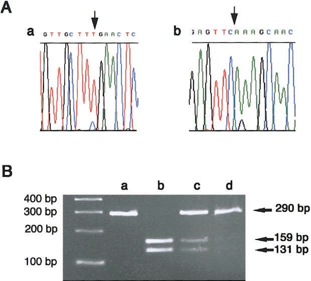
Gimenez-Roqueplo et al.: SDHD Mutation and Paraganglioma Angiogenesis                                                           1189

and 400 U Moloney murine leukemia virus (MMLV)                   and thoracic computed tomography scan. This corre-
RT. Samples were incubated at 65⬚C for 15 min, 37⬚C              sponded to a 29-g catecholamine-secreting mediastinal
for 1 h, and 99⬚C for 3 min. Quantitation of EPAS1 and           paraganglioma, which was surgically removed and had
VEGF cDNAs was performed with a SYBR Green assay,                not recurred after 18 mo of follow-up.
using an ABI Prism 7700 sequence detector (Applied
Biosystems). Quantitation of b-actin and 18S ribosomal           Germline and Somatic Analysis of the SDHD Locus
RNA control cDNAs was also performed with a real-
time TaqMan PCR assay. For amplification of EPAS1                   Linkage analysis showed a complete father-to-son pa-
(GenBank accession number NM 001430), primer se-                 ternal allele transmission for the seven markers of the
quences are 5-GCGCTAGACTCCGAGAACAT-3                           chromosome 11q13-q23 region, with no recombination
(forward) and 5-TGGCCACTTACTACCTGACCCTT-                        event. Direct sequencing of the four exons of the SDHD
3 (reverse). For amplification of VEGF (GenBank ac-             gene from peripheral DNA of affected subjects showed
cession number NM 003376), primer sequences are                  a CrT nt change in exon 2, creating a premature stop
5-CTACCTCCACCATGCCAAGTG-3 (forward) and                        codon in the sequence encoding the signal peptide
5-TGATTCTGCCCTCCTCCTTCT-3 (reverse). For                       (R22X) of the mature protein (fig. 1B). This heterozy-
amplification of b-actin and 18S ribosomal RNA prod-             gous mutation which was confirmed by BstBI restriction
ucts, primers and probe were purchased directly from             enzymatic digestion (fig. 1C), cosegregated with the
Applied Biosystems. Thermocycling conditions were 2              disease and was not found in 178 normal control
min at 50⬚C and 10 min of initial denaturation at 95⬚C,          chromosomes.
which was followed by 45 cycles of two-step PCR that                DNA was extracted from the ectopic pheochromo-
consisted of 15 s at 95⬚C and 1 min at 60⬚C. Duplicate           cytoma tissue and the SDHD gene sequenced. Within
experiments were performed to compare five dilutions             the tumor, a loss of the maternal allele was observed for
(1, 10⫺1, 10⫺2, 10⫺3, and 10⫺4) of each target cDNA              exon 2 of the SDHD gene (fig. 2A). The deletion of the
(EPAS1 or VEGF) and five dilutions (1, 10⫺1, 10⫺2, 10⫺3,         maternal allele was confirmed by the amplification of
and 10⫺4) of each endogenous control (b-actin or 18S             exon 2 and its digestion by BstBI. Conversely, biallelic
ribosomal RNA) for the inherited pheochromocytoma
and the three sporadic pheochromocytomas. Relative
quantitation of EPAS1 and VEGF gene expression was
analyzed as a target-to-control expression ratio, using
the standard curve method (Higuchi et al, 1993). The
statistical significance of the differential expression be-
tween inherited and sporadic pheochromocytomas was
assessed by a Mann-Whitney nonparametric test (Sta-
view 5.0., SAS Institute).

Results

Clinical Features of the Family
   In this family, tumors were transmitted through the
paternal line (fig. 1A). The father developed bilateral
neck paragangliomas in his sixties. Glomus carotid tu-
mors were diagnosed in his two sons when they were
!40 years of age. The clinical history of subject II:2 is
remarkable. He underwent surgery in 1998 to remove
a left-carotid–body paraganglioma, which was suspected
on the basis of self-examination and confirmed by ca-            Figure 2        Loss of maternal allele at the SDHD gene. A, Electro-
                                                                 phoretograms corresponding to the forward (a) and reverse (b) se-
rotid angiogram. Six months after surgery, a blood pres-         quences of exon 2 of SDHD from the pheochromocytoma of subject
sure increase (to 150/100 mm Hg) was observed, to-               II:2. The small peak of C probably results from minor contamination
gether with a fivefold increase in urinary metanephrines         from nontumoral DNA (blood vessel and/or leukocytes). B, Amplifi-
(19.4 nmol/24 h). Abdominal computed tomography                  cation of exon 2 and cleavage with BstBI: undigested control (lane a),
                                                                 digested profiles of maternal peripheral DNA (lane b), germline DNA
and whole-body scintigraphy with metaiodobenzyl gua-
                                                                 (lane c), and pheochromocytoma DNA (lane d) of subject II:2. The
nidine detected no tumor. An ectopic pheochromocy-               normal maternal allele shows two bands (159 and 131 bp) after di-
toma, 50 mm in diameter, was finally located in the              gestion and the undigested paternal mutated allele shows one band
mediastinum behind the heart by octreotide scintigraphy          (290 bp).
1190 Am. J. Hum. Genet. 69:1186–1197, 2001
expression was observed in leukocytes. In tumor DNA,
the maternal allele was lost (fig. 2B). To determine the
extent of the LOH, 13 regional markers were genotyped
in the peripheral and tumor DNA of patient II:2 and
were compared with maternal and paternal peripheral
DNA (fig. 3). Ten of these markers were informative.
An LOH was observed between markers D11S1343 and
D11S968, demonstrating a terminal deletion 11q21-q25
of the maternal chromosome
Enzyme-Function Studies
We assessed the activity of respiratory-chain enzymes
in the pheochromocytoma developed by subject II:2 and
in six sporadic pheochromocytomas that were removed
and conserved in the same conditions and for which no
mutation of the SDHD gene was found. SCCR activity
(complex II plus III) was first measured in the control
pheochromocytomas (fig. 4A). Cytochrome c was re- Figure 3 Loss of maternal allele at chromosome 11q21-25. The
duced by succinate, and this reduction was readily in- LOH was typed by microsatellite analysis, using constitutive and tu-
mor DNA from patient II:2. The positions of the markers tested are
hibited by malonate, a competitive inhibitor of succinate indicated on an ideogram of chromosome 11. The maternal allele lost
dehydrogenase (fig. 4C). After the addition of lauryl mal- is indicated by a line and homozygosity by NI. * p noninformative
toside that increases the accessibility of duroquinol and marker.
cytochrome c to complex III, the addition of duroquinol
triggered a rapid reduction of cytochrome c. This re-
Gene Expression Studies
duction was essentially inhibited by antimycin, making
it possible to estimate complex III activity (QCCR: an- Because the oxidation of mitochondrial cytochromes
timycin-sensitive quinol cytochrome c reductase activity is known to be a sensor of hypoxia in the glomus carotid
or complex III) in the analyzed sample. Similar mea- body and in cells in general, we thought that complex
surements were made for the pheochromocytoma sam- II mitochondrial chain inactivation might change the ex-
ple from subject II:2, which showed a complete loss of pression of genes sensitive to hypoxia and/or encoding
SCCR activity and an increase in complex III activity angiogenic factors in paragangliomas. The expression of
(fig. 4A). This resulted in the QCCR:SCCR ratio 500 eight genes was studied using in situ hybridization and
times higher than that for the control pheochromocy- immunohistochemistry (table 1) in carotid-body para-
tomas (fig. 4A). Cytochrome oxidase activity was nor- ganglioma (fig. 5) and in pheochromocytoma from sub-
mal in all samples analyzed (data not shown). ject II:2 (fig. 6). We detected mRNA encoding the tran-
We then measured the activity of isolated complex II scription factor EPAS1 (HIF2a) (fig. 5A) in vascular
(SQDR) by using quinone as an electron acceptor, in the endothelial cells and also in vascular smooth muscle
presence of cyanide. This activity was abolished in the cells. We observed very large amounts of this transcript
pheochromocytoma sample from subject II:2 but was within the cell clusters characteristic of paragangliomas
detected in control pheochromocytomas (fig. 4B). Fi- (“Zellballen”), in chief cells (type I) of all specimens.
nally, succinate dehydrogenase activity (SPDR) was mea- Immunostaining with a polyclonal antibody revealed
sured by following the reduction of phenazine metho- that its homolog, HIF1a, was also detected in these cells
sulfate (PMSF) by succinate in the presence of (fig. 5B). Several putative target genes of these tran-
thenoyltrifluoroacetone, which blocks electron transfer scription factors were also highly expressed in these
between the SDH (subunits A plus B) and the respiratory tissues. A very strong immunoreactivity was observed
chain. It was also undetectable in the pheochromocy- for the neurone-specific enolase (NSE), which revealed
toma sample from subject II:2 (fig. 4B). These results the Zellballen architecture (fig. 5D and 5I) of all para-
demonstrate that this inactivating mutation of the gangliomas, as well as in the cells of the pheochromo-
SDHD gene in patients with paraganglioma is associated cytoma. This result was expected, because NSE is con-
with the complete and selective loss of complex II elec- sidered an indicator of neuroendocrine differentiation
tron transfer activity and with the loss of the succinate (Schmechel et al. 1978). The tyrosine hydroxylase (TH)
dehydrogenase–mediated step of the Krebs cycle. This protein was also detected in these cells in all paragan-
complete loss of activity is consistent with somatic loss gliomas (fig. 5C) and was particularly strong in the pheo-
of the wild-type maternal allele. chromocytoma. We observed a high expression of theGimenez-Roqueplo et al.: SDHD Mutation and Paraganglioma Angiogenesis 1191
Figure 4 Mitochondrial enzyme function studies. A, Spectrophotometric assay of respiratory-chain enzyme activities in tumors with (first
trace) and without (second trace) mutations in SDHD. The values given on the traces are nmol/min/mg protein. Mean Ⳳ 1 SD is indicated in
brackets. LM p lauryl maltoside; DUQH2 p decylubiquinol. B, Enzyme activities in subject II:2 (inherited pheo) and controls (sporadic pheos).
SCCR p malonate-sensitive succinate cytochrome c reductase activity (complexes II⫹III). SQDR p succinate quinone dichlorophenolindophenol
reductase (complex II). SPDR p succinate phenazine methosulfate dichlorophenolindophenol reductase (succinate dehydrogenase). C, Schematic
representation of the principal electron paths through the various subunits of complex II. The acceptor sites for quinones and phenazine
methosulfate are indicated as well as the thenoyltrifluoroacetone inhibition site. Aa p antimycin; C p SDHC; CIII p complex III; CIV p
complex IV; D p SDHD; DCPIP p dichlorophenolindophenol; DUQ p decylubiquinone; DUQH2 p decylubiquinol; FAD p flavine adenosine;
䡵⫺
Fp p flavoprotein; Ip p iron protein; KCN p potassium cyanide; PMS p phenazine methosulfate; Q p ubiquinone; Q p semiubiquinol;
䡵 ⫺
QH2 p ubiquinol. Q, Q , and QH2 are the various forms of the ubiquinone present in the mitochondrial membrane.
VEGF gene in chief cells (fig. 5E and 5J) and of that combinations (EPAS1 vs. b-actin, EPAS1 vs. 18S, VEGF
encoding its receptor, VEGF-R1 (fig. 5F and 5K) in en- vs. b-actin, and VEGF vs. 18S).
dothelial cells from large vessels and capillaries sur-
rounding the Zellballen structures (fig. 5K). Higher levels Discussion
of VEGF-R1 than of VEGF-R2 were detected (fig. 5G),
particularly in large blood vessels, in which VEGF-R2 The mitochondrial respiratory chain is a protein com-
was hardly detectable. The Tie2 receptor hybridization plex of potential interest in oncogenesis, especially after
signal was at the threshold of detection in vascular en- the discovery that the SDHD gene corresponds to the
dothelial cells (fig. 5H). PGL1 locus (Baysal et al. 2000). The present study not
Interestingly on in situ hybridization experiments, the only confirms the role of the SDHD gene in hereditary
level of expression of VEGF and of EPAS1 mRNA (fig. paraganglioma but also shows that the induced defect
6) was higher in the three carotid-body paragangliomas in the complex II mitochondrial chain is associated with
and in the pheochromocytoma of the family in the pre- the overproduction of angiogenic factors, which may in
sent study than those observed in sporadic benign pheo- turn stimulate angiogenesis and therefore tumor growth.
chromocytomas. To confirm these observations, we used Three different loci have been implicated in familial
real-time quantitative RT-PCR to compare the level of paraganglioma. The inheritance pattern of these genes
EPAS1 and VEGF transcripts in pheochromocytoma is usually consistent with autosomal dominant trans-
from subject II:2 and three sporadic pheochromocyto- mission with maternal genomic imprinting (Van der
ma controls who had no SDHD mutation and a normal Mey et al. 1989). Indeed, the children of affected fathers
SDH activity. As indicated in figure 7, there was, on developed tumors, whereas the children of affected
average, a sixfold increase in the level of EPAS1 mRNAs mothers remained tumor free. Several authors have sug-
and a threefold increase of VEGF mRNAs whatever the gested that an epigenetic modification, such as the meth-
endogenous RNA control used. The statistical signifi- ylation of CpG islets on parental chromosomes might
cance of these results was demonstrated using a Mann- confer a reversible difference on the maternal and pa-
Whitney statistical test: z p ⫺2.910, P p .0036 for all ternal chromosomes during gametogenesis (Van der1192 Am. J. Hum. Genet. 69:1186–1197, 2001
Table 1
Semiquantitative Assessment of In Situ Hybridization and Immunohistochemistry Observations
IN SITU HYBRIDIZATION IMMUNOHISTOCHEMISTRY
SUBJECT AND TUMOR EPAS1 VEGF VEGF-R1 VEGF-R2 Tie2 HIF1a NSE TH
I:1
Right paraganglioma ⫹⫹⫹ ⫹⫹⫹ ⫹⫹⫹ ⫹⫹⫹ ⫹ ⫹⫹ ⫹⫹ ⫹⫹
Left paraganglioma ⫹⫹ ⫹⫹⫹⫹ ⫹⫹⫹ ⫹⫹⫹ ⫹ ⫹ ⫹⫹⫹ ⫹
II:2
Paraganglioma ⫹⫹⫹ ⫹⫹⫹ ⫹⫹⫹⫹ ⫹⫹⫹ ⫹ ⫹⫹ ⫹⫹⫹ ⫹⫹
Pheochromocytoma ⫹⫹⫹ ⫹⫹⫹ ⫹⫹⫹⫹ ⫹⫹⫹ ⫹ ⫹⫹ ⫹⫹ ⫹⫹⫹⫹
NOTE.—Relative intensities of the markers studied are represented for the three carotid-body paragangliomas
(in subjects I:1 and II:2) and the pheochromocytoma (in subject II:2). Labeling intensity was evaluated twice
in a blinded manner and was graded using a 5-scale system: ⫺ p no signal; ⫹ p weak; ⫹⫹ p moderate; ⫹⫹⫹
p strong; ⫹⫹⫹⫹ p very strong.
Mey et al. 1989; Milunsky et al. 1997). This modifi- SDHA gene encoding the flavoprotein of complex II,
cation could affect a tissue-specific promoter (Pfeifer, and the enzymatic defect was partial (Bourgeron et al.
2000). This hypothesis is consistent with biallelic ex- 1995). No associated tumor was detected in this family
pression of the SDHD gene in tumor tissue. Nonethe- (A.R. and P.R., unpublished data). In Caenorhabditis
less, our data provide no evidence for maternal imprint- elegans, a mutant of mev-1 gene that is homologous to
ing but instead suggest an LOH in tumor tissue, the human SDHC gene has been found to be hypersen-
consistent with Knudson’s classical two-hit hypothesis sitive to high oxygen concentrations. In mev-1 mutant
for the induction of tumor formation (Knudson 1986). animals, the ability of complex II to catalyze electron
Conversely, we were able to detect biallelic expression transport from succinate to ubiquinone is compromised,
of the SDHD gene and normal complex II activity in indirectly causing an increase in superoxide levels and
adrenal tissue in two other sporadic pheochromocyto- premature aging (Ishii et al. 1998). Surprisingly enough,
mas (data not shown). succinate dehydrogenase (subunits A and B), assumed
Tumor prognosis probably differs with the function to be not inserted in the mitochondrial membrane, was
and number of deleted genes. A high frequency of 11q found still active despite the loss of the anchoring sub-
loss in familial paragangliomas, similar to the loss in units in mev-1 mutant animals. These observations con-
the family described here, was recently reported else- trast with numerous cases where the absence (or even
where (Dannenberg et al. 2001). Several of the genes the mutation) of one subunit in a given respiratory-
surrounding SDHD on human chromosome 11q21- chain complex results in the destabilization of the com-
q25, may be involved in tumorigenesis (Koreth et al. plex and the subsequent proteolysis of the noninserted
1999; Baysal et al. 2001). One of them is the PPP2R1B subunits (Birch-Machin et al. 1996; Bruno et al. 1999;
gene which is separated from SDHD by only 250 kb Clark et al. 1999). Our studies of human pheochrom-
on the physical map. This putative tumor-suppressor ocytomas did not confirm the observation made in the
gene, which encodes the b isoform of the A subunit of nematode. The defect in one of the anchoring subunits
serine/threonine protein phosphatase 2A, has been im- is predicted to result in the noninsertion of the SDH
plicated in human lung and colon cancer (Wang et al. and presumably to its proteolysis. Accordingly, muta-
1998). Therefore, it will be important to study the phe- tion in the SDHD gene of subject II:2 resulted in the
notype-genotype relationships in several families with complete loss of both the catalytic activity of succinate
paraganglioma to determine whether the size and lo- dehydrogenase (subunits A plus B) and electron flow
cation of the somatic deletions are associated with a from succinate to the ubiquinone pool, which requires
higher risk of malignancy in paraganglioma and spo- all complex II subunits. The complete abolition of com-
radic pheochromocytoma induced by SDHD mutations plex II activity resulting from a heterozygous consti-
(Gimm et al. 2000). tutive mutation of the SDHD gene associated with a
It has been suggested that mitochondria are the pri- somatic maternal LOH, as described in the pheochrom-
mary site of oxygen sensing in the carotid body (Prab- ocytoma of this family, has not been reported before.
hakar. 2000). Mitochondria produce cellular energy by This dramatic disturbance of the mitochondria res-
a process of oxidative phosphorylation involving five piratory chain accounts for the hypoxic status of the
respiratory-enzyme complexes. Complex II transfers cells and probably explains the high level of expression
electrons from succinate to the ubiquinone pool. Iso- of genes encoding hypoxia induced factors. Jyung et al.
lated complex II deficiency has been reported in Leigh (2000) reported the detection of positive immunohis-
syndrome. The causative mutation was identified in the tochemical staining for VEGF in 65% of sporadic glo-Gimenez-Roqueplo et al.: SDHD Mutation and Paraganglioma Angiogenesis 1193 Figure 5 Gene-expression patterns in right carotid-body paraganglioma of patient I:1, as shown by in situ hybridization or immunostaining. EPAS1 mRNA is observed in both endothelial cells and tumor cells (A). The protein of the homolog transcription factor HIF1a is also present (B). Tyrosine hydroxylase (C), NSE (D and I) and VEGF (E and J) are present at a very high level in tumor cells within the Zellballen clusters, whereas VEGF receptors R1 (F and K) and R2 (G) transcripts are restricted to endothelial cells. Note the absence of VEGF-R2 in the wall of the central large vessel (*) and the almost undetectable signal with the Tie2 probe (H). High magnifications clearly reveal the expression of NSE (I) and VEGF (J) within the Zellballen structures (arrowheads), surrounded by a VEGF-R1 positive capillary network (K, arrows). The in situ hybridization signals are visualized either under dark (A and E–H; signal visible as white dots) or bright field illumination (J and K;labeling detected by black dots). Immunostaining (B, D, and I) is revealed by a brown coloration. Scale bars p 200 mm (A–H); scale bars p 100 mm (I–K). mus paraganglioma studied. We have confirmed and (EPAS1/HIF2a and HIF1a) and genes encoding some extended these results in inherited paragangliomas by of the targets of these factors. The hybridization signal describing the expression pattern of several angiogenic for EPAS1 and for VEGF and its receptor VEGF-R1 markers that may be involved in tissue adaptation to was particularly strong in these tumors. We made two hypoxia. We have shown, in these tumors, the strong important observations that suggest a direct link be- expression of hypoxia-inducible transcription factors tween the high expression of genes encoding angiogenic
1194                                                                                                   Am. J. Hum. Genet. 69:1186–1197, 2001

Figure 6        EPAS1 mRNA expression; comparison between patient II:2 pheochromocytoma and a sporadic benign pheochromocytoma. The
expression of EPAS1 within tumor cells is higher in the inherited pheochromocytoma (A and B) than in a sporadic pheochromocytoma (C and
D). In contrast, the intensity of EPAS1 signal does not vary between the two samples in endothelial cells (black arrows). The in situ hybridization
signals are observed under dark-field illumination for low magnifications (A and C [white dots]) and under bright field illumination for
enlargements (B, D [black dots]). White arrows p blood vessels; ⫹⫹⫹ p very strong signal; ⫹ p weak signal. Scale bars p 100 mm.

factors and the loss of SDHD function. First, VEGF-R1                      which revealed a higher expression of both genes in
mRNA, which encodes the VEGF receptor induced in                           patient II:2 pheochromocytoma than in three sporadic
hypoxic conditions (Gerber et al. 1997), was found to                      pheochromocytomas, with no SDHD mutation and a
be more abundant than VEGF-R2 mRNA. Second, the                            normal SDH activity. The mutation in the SDHD gene
level of EPAS1 and VEGF labeling observed in the three                     is therefore probably involved in induction of the hy-
paraganglioma tumors and in the pheochromocytoma                           poxia/angiogenesis response and possibly in tumor de-
of this family was stronger than that observed in spo-                     velopment. However, it is unclear whether the activation
radic benign pheochromocytomas. These observations                         of hypoxia pathways is or is not sufficient to induce
were confirmed by quantitative RT-PCR experiments                          tumorigenesis by itself. It remains possible that other
Gimenez-Roqueplo et al.: SDHD Mutation and Paraganglioma Angiogenesis                                                                  1195

Figure 7         Real-time quantitative RT-PCR experiments. The level of EPAS1 and VEGF transcripts were compared between the SDHD
mutated pheochromocytoma (inherited Pheo) with three sporadic pheochromocytomas (sporadic Pheo 1, 2, and 3). Comparison of quantitative
DNA data obtained with EPAS1 as target and b-actin (A) or 18S ribosomal RNA (B) as endogenous control and of data obtained with VEGF
as target and b-actin (C) or 18S ribosomal RNA (D) as endogenous controls. The circle represents the mean of 10 calculated measurements
(ratio of target gene on reference gene for each of five log of concentration in duplicate). The superior bar shows the maximal value, and the
inferior bar shows the minimal value.

functions, downstream from SDHD, are impaired in                         might in turn facilitate or trigger tumorigenesis in para-
these cells and/or that genes other than SDHD are also                   ganglial tissues.
affected (e.g., PPP2R1B).
   Recent identification of mutations in the SDHD,
SDHC and SDHB genes as responsible for familial par-
                                                                         Acknowledgments
aganglioma has added to the molecular tools available                       We thank Dr. Jean-Marie Gasc, for helpful discussions of
for genetic counseling, which was previously based on                    the manuscript, Dr. N. Lamandé, Dr. D. Richards, and Dr. J.
indirect testing using microsatellite markers from chro-                 Pouyssegur, for antibodies. The English text was edited by Dr.
mosome 11q13 and 11q23 (Oosterwijk et al. 1996; Bik-                     J. Sappa. This study was supported by INSERM and by PHRC
hazi et al. 1999; Petropoulos et al. 2000). Genetic di-                  grant AOM 95201, for the COMETE Network.
agnosis can now be made by direct sequencing to search
for a causative mutation in these three genes, even in                   Electronic-Database Information
families with few affected members. The discovery of a                   Accession numbers and URLs for data in this article are as
genomic mutation in a patient with paraganglioma is                      follows:
important for the genetic counseling of his or her family,
including predictive DNA testing mutations in all rel-                   GenBank, http://www.ncbi.nlm.nih.gov/Genbank (for the se-
atives and screening for paraganglioma at a presymp-                       quence of SDHD)
tomatic stage. Indeed, the early detection of paragan-                   Online Mendelian Inheritance in Man (OMIM), http://
glioma reduces the incidence of morbidity and mortality                    www.ncbi.nlm.nih.gov/Omim/ (for reviews of familial
(Petropoulos et al. 2000).                                                 paragangliomas)
   In conclusion, this study shows that somatic loss of
maternal allele of the PGL1 locus, together with in-                     References
herited paternal mutation of the SDHD gene, is re-                       Astuti D, Douglas F, Lennard TWJ, Aligianis IA, Woodward
sponsible for a complete loss of activity of the complex                   ER, Evans GR, Eng C, Latif F, Maher ER (2001a) Germline
II of the mitochondrial respiratory chain and is asso-                     SDHD mutation in familial phaeochromocytoma. Lancet
ciated with a stimulation of angiogenic factors that                       357:1181–1182
1196 Am. J. Hum. Genet. 69:1186–1197, 2001
Astuti D, Latif F, Dallol A, Dahia PL, Douglas F, George E, Gerber HP, Condorelli F, Park J, Ferrara N (1997) Differential
Skoldberg Husebye ES, Eng C, Maher ER (2001b) Gene transcriptional regulation of the two vascular endothelial
mutations in the succinate dehydrogenase subunit SDHB growth factor receptor genes. Flt-1, but not Flk-1/KDR, is
cause susceptibilty to familial pheochromocytoma and to up regulated by hypoxia. J Biol Chem 272:23659–23667
familial paraganglioma. Am J Hum Genet 69:49–54 Gimm O, Armanios M, Dziema H, Neumann HP, Eng C
Badenhop RF, Cherian S, Lord RS, Baysal BE, Taschner PE, (2000) Somatic and occult germ line mutations in SDHD,
Schofield PR (2001) Novel mutations in the SDHD gene in a mitochondrial complex II gene, in nonfamilial pheochrom-
pedigrees with familial carotid body paraganglioma and ocytoma. Cancer Res 60:6822–6825
sensorineural hearing loss. Genes Chromosomes Cancer 31: Heutink P, Van der Mey AG, Sandkuijl LA, Van Gils AP, Bar-
255–263 doel A, Breedveld GJ, Van Vliet M, Van Ommen GJ, Cor-
Baysal BE, Ferrell RE, Willett-Brozick JE, Lawrence EC, Mys- nelisse CJ, Oostra BA, Weber JL, Devilee P (1992) A gene
siorek D, Bosch A, Van Der Mey A, Taschner PEM, Rubin- subject to genomic imprinting and responsible for hereditary
stein WS, Myers EN, Richard CW 3d, Cornelisse CJ, Devilee paragangliomas maps to chromosome 11q23-qter. Hum
P, Devlin B (2000) Mutations in SDHD, a mitochondrial Mol Genet 1:7–10
complex II gene, in hereditary paraganglioma. Science 287: Higuchi R, Fockler C, Dollinger G, Watson R (1993) Kinetic
848–851 PCR analysis: real-time monitoring of DNA amplification
Baysal BE, Willett-Brozick JE, Tascner PEM, Dauwerse JG, reactions. Biotechnology 11:1026–1030
Devilee P, Devlin B (2001) A high-resolution integrated map Ishii N, Fujii M, Hartman PS, Tsuda M, Yasuda K, Senoo-
spanning the SDHD gene at 11q23: a 1.1-Mb BAC contig, Matsuda N, Yanase S, Ayusawa D, Suzuki K (1998) A mu-
a partial transcript map and 15 new repeat polymorphisms tation in succinate dehydrogenase cytochrome b causes ox-
in a tumor suppressor region. Eur J Hum Genet 9:121–129 idative stress and ageing in nematodes. Nature 394:694–697
Bikhazi PH, Roeder E, Attaie A, Lalwani AK (1999) Familial Jyung RW, LeClair EE, Bernat RA, Kang TS, Ung F, McKenna
paragangliomas: the emerging impact of molecular genetics MJ, Tuan RS (2000) Expression of angiogenic growth fac-
on evaluation and management. Am J Otol 20:639–643 tors in paragangliomas. Laryngoscope 110:161–167
Birch-Machin M, Marsac C, Ponsot G, Parfait B, Taylor RW, Knudson AG (1986) Genetics of human cancer. Annu Rev
Rustin P, Munnich A (1996). Biochemical investigations and Genet 20:231–251
immunoblot analysis of two unrelated patients with an iso- Koreth J, Bakkenist CJ, McGee JO (1999) Chromosomes, 11q
lated deficiency in complex II of the respiratory chain. and cancer: a review. J Pathol 187:28–38
Biochem Biophys Res Commun 220:57–62 Legault-Demare L, Lamande N, Zeitoun Y, Gros F, Scarna H,
Bourgeron T, Rustin P, Chretien D, Birch-Machin M, Bour- Keller A, Lando D, Cousin MA (1981) Transition between
geois M, Viegas-Pequignot E, Munnich A, Rotig A (1995) isozymic forms of enolase during in vitro differentiation of
Mutation of a nuclear succinate dehydrogenase gene results neuroblastoma cells-II. Neurochem Int 3:301–310
in mitochondrial respiratory chain deficiency. Nat Genet 11:
Mariman ECM, Van Beersum SEC, Cremers CWRJ, Struycken
144–149
PM, Ropers HH (1995) Fine mapping of a putatively im-
Bruno C, Martinuzzi A, Tang Y, Andreu AL, Palloti F, Bonilla
printed gene for familial non-chromaffin paragangliomas to
E, Shanske S, Fu J, Sue CM, Angelini C, DiMauro S, Man-
chromosome 11q13.1: evidence for genetic heterogeneity.
fredi G (1999) A stop-codon mutation in the human mtDNA
Hum Genet 95:56–62
cytochrome c oxidase I gene disrupts the functional structure
Milunsky JM, DeStefano A, Huang XL, Baldwin C, Michels
of complex IV. Am J Hum Genet 65:611–620
V, Jako G, Milunsky A (1997) Familial paragangliomas:
Chandel NS, Maltepe E, Goldwasser E, Mathieu CE, Simon
linkage to chromosome 11q23 and clinical implications. Am
MC, Schumacker PT (1998) Mitochondrial reactive oxygen
J Med Genet 72:66–70
species trigger hypoxia-induced transcription. Proc Natl
Acad Sci USA 95:11715–11720 Milunsky JM, Maher TA, Michels VV, Milunsky A (2001)
Clark KM, Taylor RW, Johnson MA, Chinnery PF, Chrza- Novel mutations and the emergence of a common mutation
nowska-Lightowlers ZM, Andrews RM, Nelson IP, Wood in the SDHD gene causing familial paraganglioma. Am J
NW, Lamont PJ, Hanna MG, Lightowlers RN, Turnbull DM Med Genet 100:311–314
(1999) An mtDNA mutation in the initial codon of the cy- Niemann S, Steinberger D, Müller U (1999) PGL3, a third,
tochrome c oxidase subunit II gene results in lower levels not maternally imprinted locus in autosomal dominant par-
of the protein and a mitochondrial encephalomyopathy. Am aganglioma. Neurogenetics 2:167–170
J Hum Genet 64:1330–1339 Niemann S, Müller U (2000) Mutations in SDHC cause au-
Dannenberg H, Krijger RR, Zhao J, Speel EJM, Saremaslani tosomal dominant paraganglioma, type 3. Nat Genet 26:
P, Dinjens WNM, Mooi WJ, Roth J, Heitz PU, Komminoth 268–270
P (2001) Differential loss of chromosome 11q in familial Niemann S, Becker-Follmann J, Nürnberg G, Rüschendorf F,
and sporadic parasympathetic paragangliomas detected Sieweke N, Hügens-Penzel M, Traupe H, Wienker TF, Reis
by comparative genomic hybridization. Am J Pathol 158: A, Müller U (2001) Assignment of PGL3 to chromosome
1937–1942 1(q21-q23) in a family with autosomal dominant non-chro-
Favier J, Kempf H, Corvol P, Gasc JM (1999) Cloning and maffin paraganglioma. Am J Med Genet 98:32–36
expression pattern of EPAS1 in the chicken embryo: co- Oosterwijk J, Jansen J, Van Schothorst E, Oosterhof A, Devilee
localization with tyrosine hydroxylase. FEBS Lett 462:19– P, Bakker E, Zoeteweij M, Van der Mey A (1996) First
24 experiences with genetic counselling based on predictiveGimenez-Roqueplo et al.: SDHD Mutation and Paraganglioma Angiogenesis 1197 DNA diagnosis in hereditary glomus tumours (paragan- lar investigations in respiratory chain deficiencies. Clin Chim gliomas). J Med Genet 33:379–383 Acta 228:35–51 Pacheco-Ojeda L, Durango L, Rodriguez C, Vivar N (1988) Saraste M (1999) Oxidative phosphorylation at the fin de Carotid body at high altitudes. World J Surg 12:856–860 siecle. Science 283:1488–1493 Petropoulos AE, Luetje CM, Camarata PJ, Whittaker CK, Lee Schmechel D, Marangos PJ, Brightman M (1978) Neurone- G, Baysal BE (2000) Genetic analysis in the diagnosis of specific enolase is a molecular marker for peripheral and familial paragangliomas. Laryngoscope 110:1225–1229 central neuroendocrine cells. Nature 276:834–836 Pfeifer K (2000) Mechanisms of genomic imprinting. Am J Sibony M, Commo F, Callard P, Gasc JM (1995) Enhancement Hum Genet 67:777–787 of mRNA in situ hybridization signal by microwave heating. Prabhakar NR (2000) Oxygen sensing by the carotid body Lab Invest 73:586–591 chemoreceptors. J Appl Physiol 88:2287–2295 Van Der Mey AGL, Maaswinkel-Moy PD, Cornelisse CJ, Richard DE, Berra E, Pouyssegur J (2000) Nonhypoxic Schmidt PH, Van de Kamp JJP (1989) Genomic imprinting pathway mediates the induction of hypoxia-inducible fac- in hereditary glomus tumours: evidence for new genetic the- tor 1 a in vascular smooth muscle cells. J Biol Chem 275: ory. Lancet 2:1291–1294 26765–26771 Wang SS, Esplin ED, Li JL, Huang L, Gazdar A, Minna J, Rustin P, Chretien D, Bourgeron T, Gerard B, Rotig A, Sau- Evans GA (1998) Alterations of the PPP2R1B gene in human dubray JM, Munnich A (1994) Biochemical and molecu- lung and colon cancer. Science 282:284–287
You can also read