Jasmonate-independent regulation of digestive enzyme activity in the carnivorous butterwort Pinguicula Tina
←
→
Page content transcription
If your browser does not render page correctly, please read the page content below

Journal of Experimental Botany, Vol. 71, No. 12 pp. 3749–3758, 2020
doi:10.1093/jxb/eraa159 Advance Access Publication 27 March 2020
This paper is available online free of all access charges (see https://academic.oup.com/jxb/pages/openaccess for further details)

RESEARCH PAPER

Jasmonate-independent regulation of digestive enzyme
activity in the carnivorous butterwort Pinguicula × Tina
Ondřej Kocáb1, Jana Jakšová1, Ondřej Novák2, Ivan Petřík2, René Lenobel3, , Ivo Chamrád3, and
Andrej Pavlovič1,*,

                                                                                                                                                          Downloaded from https://academic.oup.com/jxb/article/71/12/3749/5812645 by guest on 23 September 2020
1
  Department of Biophysics, Centre of the Region Haná for Biotechnological and Agricultural Research, Faculty of Science, Palacký
University, Šlechtitelů 27, Olomouc CZ-783 71, Czech Republic
2
  Laboratory of Growth Regulators, Institute of Experimental Botany, The Czech Academy of Sciences and Faculty of Science, Palacký
University, Šlechtitelů 27, Olomouc CZ-783 71, Czech Republic
3
  Department of Protein Biochemistry and Proteomics, Centre of the Region Haná for Biotechnological and Agricultural Research,
Faculty of Science, Palacký University, Šlechtitelů 27, Olomouc CZ-783 71, Czech Republic.

* Correspondence: andrej.pavlovic@upol.cz

Received 12 December 2019; Editorial decision 23 March 2020; Accepted 25 March 2020

Editor: John Lunn, Max Planck Institute of Molecular Plant Physiology, Germany

Abstract
Carnivorous plants within the order Caryophyllales use jasmonates, a class of phytohormone, in the regulation of
digestive enzyme activities. We used the carnivorous butterwort Pinguicula × Tina from the order Lamiales to in-
vestigate whether jasmonate signaling is a universal and ubiquitous signaling pathway that exists outside the order
Caryophyllales. We measured the electrical signals, enzyme activities, and phytohormone tissue levels in response to
prey capture. Mass spectrometry was used to identify proteins in the digestive secretion. We identified eight enzymes
in the digestive secretion, many of which were previously found in other genera of carnivorous plants. Among them,
alpha-amylase is unique in carnivorous plants. Enzymatic activities increased in response to prey capture; however,
the tissue content of jasmonic acid and its isoleucine conjugate remained rather low in contrast to the jasmonate re-
sponse to wounding. Enzyme activities did not increase in response to the exogenous application of jasmonic acid or
coronatine. Whereas similar digestive enzymes were co-opted from plant defense mechanisms among carnivorous
plants, the mode of their regulation differs. The butterwort has not co-opted jasmonate signaling for the induction of
enzyme activities in response to prey capture. Moreover, the presence of alpha-amylase in digestive fluid of P. × Tina,
which has not been found in other genera of carnivorous plants, might indicate that non-defense-related genes have
also been co-opted for carnivory.

Keywords: Butterwort, carnivorous plant, digestive enzymes, electrical signals, jasmonic acid, Pinguicula, protease, variation
potential.

Introduction
The carnivorous plants have evolved specialized leaves or leaf                     least 10 times in several orders of flowering plants (Albert et al.,
parts that function as traps for prey capture and digestion to ob-                 1992; Givnish et al., 2015; Fleischmann et al., 2018). Whereas
tain scarce nutrients.This adaptation to low nutrient content in                   the mechanisms of prey capture have been studied in great
the soil has independently evolved by convergent evolution at                      detail during the past two centuries, the process of digestion

© The Author(s) 2020. Published by Oxford University Press on behalf of the Society for Experimental Biology.
This is an Open Access article distributed under the terms of the Creative Commons Attribution License (http://creativecommons.org/licenses/by/4.0/),
which permits unrestricted reuse, distribution, and reproduction in any medium, provided the original work is properly cited.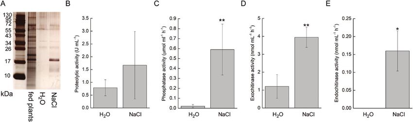
3750 | Kocáb et al.

was almost completely unknown. The first endogenous en-               digestive glands of Pinguicula share a special characteristic with
zyme in carnivorous plants was described only at the begin-           the Venus flytrap and sundew in that they do not secrete en-
ning of this century in the pitcher plant (genus Nepenthes),          zymes until stimulated by the presence of prey (Darwin, 1875).
which resolved the longstanding question of whether prey              It has been postulated that the glands of Pinguicula undergo a
digestion is mediated by symbiotic microorganisms or plant-           type of total autophagy and are simply a sac of enzymes that
derived enzymes (Athauda et al., 2004). In the decade that            are discharged in response to prey capture, in contrast to the
followed, the development of mass spectrometry techniques             jasmonate-mediated expression/secretion of digestive enzymes
enabled the discovery of over 20 other digestive enzymes in           in the carnivorous plants within the order Caryophyllales. The
different species of carnivorous plants, which are surprisingly       initial event associated with the onset of secretion is the rapid
very similar across distantly related taxa (Eilenberg et al., 2006;   movement of chloride ions followed by water across the glands,
Hatano and Hamada, 2008; 2012; Rottloff et al., 2011; 2016;           flushing out the stored enzymes from the cell walls of the
Lee et al., 2016; Fukushima et al., 2017; Krausko et al., 2017).      glands (Heslop-Harrison and Knox, 1971; Heslop-Harrison
Yet, the mechanism by which the secretion of these digestive          and Heslop-Harrison, 1980). Contrary to this eccrine hypoth-
enzymes is regulated by stimuli from prey remained unknown            esis,Vassilyev and Muravnik (1988a , b) showed that the glands

                                                                                                                                                      Downloaded from https://academic.oup.com/jxb/article/71/12/3749/5812645 by guest on 23 September 2020
until Escalanté-Pérez et al. (2011) found that a phytohormone         remain highly active during the entire secretion process and
from the group of jasmonates, 12-oxo-phytodienoic acid                additional digestive enzymes are synthesized and secreted into
(OPDA), was responsible for activation of the digestive process       the digestive fluid after stimulation, much in common with the
in Venus flytrap (Dionaea muscipula). An increased level of the       process in Venus flytrap. In this study, we aimed to shed light
true bioactive compound in jasmonate signaling, the isoleucine        on this discrepancy in the view of jasmonate signaling within
conjugate of jasmonic acid (JA-Ile), was later found in Venus         a less-studied genus of carnivorous plant, Pinguicula. We were
flytrap, sundew plant (Drosera capensis), and the pitcher plant       interested in whether the jasmonate signaling pathway was
Nepenthes alata in response to prey capture (Nakamura et al.,         co-opted for plant carnivory outside the order Caryophyllales.
2013; Libiaková et al., 2014; Yilamujiang et al., 2016; Krausko       We measured electrical activity, analyzed the composition of
et al., 2017; Pavlovič et al., 2017). The binding of JA-Ile to        the digestive fluid and its enzymatic activity in response to
CORONATINE INSENSITIVE1 (COI1) protein as part of a                   prey capture, and assessed endogenous phytohormone content.
co-receptor complex mediates the ubiquitin-dependent deg-             We did not find any evidence that butterworts use jasmonate
radation of JASMONATE ZIM-DOMAIN (JAZ) repressors,                    signaling for the induction of enzyme activities.
resulting in the activation of jasmonate-dependent gene ex-
pression (Chini et al., 2007; Thines et al., 2007; Fonseca et al.,
2009; Sheard et al., 2010). In ordinary plants, JA-Ile is respon-     Materials and methods
sible for the activation of defense mechanisms after herbivore
attack or wounding, and it was postulated that the carnivorous        Plant material and experimental setup
plants co-opted the jasmonate signaling pathway for prey cap-         We used a horticultural hybrid of Pinguicula × Tina (Pinguicula agnata
ture (Pavlovič and Saganová 2015; Bemm et al., 2016; Pavlovič         × Pinguicula zecheri) purchased from Gartneriet Lammehave (Ringe,
                                                                      Denmark) and Drosera capensis in our experiments (Fig. 1A). Pinguicula ×
and Mithöfer, 2019). Unfortunately, the studies to date have
                                                                      Tina is famous for being vigorous and easy to grow, with many flowers,
generally been confined to three genera of carnivorous plants         and producing large leaves with a sufficient amount of digestive fluid for
(Drosera, Dionaea, and Nepenthes), which all are within the order     analyses. Plants were grown at the Department of Biophysics of Palacký
Caryophyllales (or, according to some authors, the separate           University in Olomouc, Czech Republic, under standard greenhouse
order Nepenthales; Fleischmann et al., 2018) and are mono-            conditions. Plants were grown in plastic pots filled with well-drained
                                                                      peat moss, placed in a tray filled with distilled water to a depth of 1–2 cm.
phyletic. Therefore, it remains unclear whether the jasmonate
                                                                      During the experiments the plants were placed in a growth chamber
signaling pathway is a universal and ubiquitous signaling             maintained at 21–22 °C and 100 μmol m−2 s−1 photosynthetically active
pathway in other phylogenetic lineages of carnivorous plants.         radiation, with a 16/8 h light/dark period.
   In this study, we focused on carnivorous plants of the genus          Fruit flies (Drosophila melanogaster) were used as a model prey. Flies were
Pinguicula (butterworts), which belongs to the order Lamiales         cultured from eggs in a carbohydrate-rich medium and were provided
                                                                      by the Department of Genetics, Faculty of Natural Sciences Comenius
and is distantly related to the Venus flytrap, sundew, and
                                                                      University in Bratislava, Slovakia. Before the experiments, adult flies were
pitcher plant (Albert et al., 1992; Givnish, 2015). Most species      cooled in a refrigerator at 4 °C for 15 minutes to facilitate manipulation.
of Pinguicula have a basal rosette of compact leaves that are         Ten fruit flies were placed on one leaf surface or one flower stalk of plant
more or less broadly ovate, and only a few species (Pinguicula        under study (Fig. 1B, C). After 2 h and 24 h, 10 control and 10 fed leaves
heterophylla, Pinguicula gypsicola) have filiform upright leaves.     were cut off the plant using a scalpel, and leaf blades were submerged one
                                                                      at a time in 4 ml of 50 mM sodium acetate buffer solution (pH 5.0) for
Some species can bend their leaf edges slightly in response to
                                                                      3 min to collect the exudates. Because of seasonal blooming, the limited
prey capture, while others have no such ability (Fleischmann          number and low biomass of flower stalks available were collected only
and Roccia, 2018). The leaves are covered by two types of             after 24 h.
glands. The stalked glands produce sticky mucilage and serve             In the experiments with jasmonates (see below), plants were sprayed
mainly for prey capture, but with the capacity to produce their       with 1 mM jasmonic acid or 100 µM coronatine in 0.001% Tween 20.
                                                                      Control plants were sprayed with 0.001% Tween 20 only. The leaf exud-
own digestive enzymes. The sessile glands are the main site for
                                                                      ates were collected after 24 h as described above. For this experiment,
the production of digestive enzymes (Heslop-Harrison and              sundew plants (D. capensis) were used as a positive control, as this species
Knox, 1971; Heslop-Harrison and Heslop-Harrison, 1980,                is known to increase digestive enzyme synthesis in response to the ex-
1981; Legendre, 2000; Heslop-Harrison, 2004). Both types of           ogenous application of jasmonates (Krausko et al., 2017). For experiments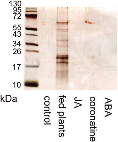
Jasmonates were not co-opted for carnivory in Pinguicula | 3751

                                                                                                                                                            Downloaded from https://academic.oup.com/jxb/article/71/12/3749/5812645 by guest on 23 September 2020
Fig. 1. Butterwort Pinguicula × Tina. (A) Whole plant. (B) Flower stalk covered with digestive glands. (C) Leaf covered with stalked and sessile glands.

with hypertonic NaCl solution, 20 µl drops of 5% NaCl or distilled water       to terminate the reaction. Absorbance was measured at 410 nm with a
(as a control) were applied on the glandular leaf surface and collected        Specord 250 Plus double-beam spectrophotometer (Analytik Jena). The
using a pipette after 15 min. This time point was chosen as sufficiently       calibration curve was determined using 4-nitrophenol and the activities
long to induce the flow of water from the glands but too short for the         were expressed in µmol ml−1 h−1.
synthesis and secretion of digestive enzymes de novo (based on our ex-            Amylase activity was measured using an amylase assay kit (Sigma-
perience with Venus flytrap; Jakšová et al., 2020; Pavlovič et al., 2020).     Aldrich). Ethylidene-pNP-G7 was used as a substrate, which upon
For wounding experiments, a leaf was wounded with a needle once for            cleavage by amylase generates 4-nitrophenyl. A 20 µl aliquot of collected
electrical signal measurement or 10–15 times for phytohormone analyses         digestive fluid was added to a 96-well plate and adjusted to 50 μl with
(see below).                                                                   the amylase assay buffer. Then 100 µl of substrate was added, the reaction
                                                                               mixture was incubated at 25 °C, and absorbance at 405 nm was meas-
                                                                               ured every 15 min for 2 h using a SynergyMx microplate reader (BioTek
Extracellular recording of electrical signals                                  Instruments, Winooski,VT, USA). Positive control (amylase enzyme) and
Changes in the surface potential were measured by using non-polarizable        4-nitrophenyl standard at different concentrations were incubated under
Ag–AgCl surface electrodes (Scanlab Systems, Prague, Czech Republic)           the same conditions on the same microplate.
moistened with a drop of conductive EV gel (Hellada, Prague, Czech                Chitinase activities were measured using a fluorimetric chitinase
Republic) that is commonly used in electrocardiography. The electrode          assay kit (Sigma Aldrich). We used 4-methylumbelliferyl N-acetyl-β-d-
was attached on the abaxial side of the leaf either beneath an applied fly     glucosaminide, 4-methylumbelliferyl N,N′-diacetyl-β-d-chitobioside,
(D. melanogaster) or 1 cm from a wounding site that was made with a            and 4-methylumbelliferyl β-d-N,N′,N″-triacetylchitotriose for the de-
needle. The electrical signals were recorded by a non-invasive device in-      tection of β-N-acetylglucosaminidase (exochitinase), chitobiosidase, and
side a Faraday cage according to Ilík et al. (2010).The reference electrode    endochitinase activities, respectively, according to the manufacturer’s
was taped to the side of the plastic pot containing the plant, submerged       instructions. A 10 μl aliquot of collected digestive fluid was incubated
in 1–2 cm of water in a dish beneath the pot. The electrodes were con-         with 90 μl of substrate working solution at 37 °C and after 2 h the re-
nected to an amplifier [gain 1–1000, noise 2–3 μV, bandwidth (–3 dB)           action was stopped by the addition of 200 μl of sodium carbonate pro-
105 Hz, response time 10 μs, input impedance 1012 Ω]. The signals from         vided in the kit.The fluorescence of liberated 4-methylumbelliferone was
the amplifier were transferred to an analogue–digital PC data converter        measured in alkaline pH using a SynergyMx microplate reader (BioTek
(eight analogue inputs, 12-bit converter, ±10 V, PCA-7228AL, supplied          Instruments, Winooski, VT, USA) with excitation at 360 nm and emis-
by TEDIA, Plzeň, Czech Republic), collected every 6 ms.                        sion at 450 nm. Chitinase from Trichoderma viride (positive control) and
                                                                               4-methylumbelliferone standard at different concentrations were incu-
                                                                               bated under the same conditions on the same microplate.
Measurements of enzyme activities                                                 All enzyme activities were measured on pooled samples from 10 leaves
The proteolytic activity of digestive fluid was determined by incubating       from 3 plants to have sufficiently concentrated samples within the limit
150 µl of the collected sample of digestive fluid with 150 µl of 2% (w/v)      of detection.
bovine serum albumin in 200 mM glycine–HCl (pH 3.0) at 37 °C for
2 h. The reaction was stopped by the addition of 450 µl of 5% (w/v) tri-
chloroacetic acid (TCA). Samples were incubated on ice for 10 min and          SDS-PAGE electrophoresis
then centrifuged at 20 000 g for 10 min at 4 °C. The amount of released        Digestive fluid collected for the enzyme assays was subjected to SDS-
non-TCA-precipitable peptides was used as a measure of proteolytic ac-         PAGE. The samples were heated and denatured for 30 min at 70 °C
tivity, which was determined by comparing the absorbance of the super-         and then mixed with modified Laemmli sample buffer to a final con-
natant at 280 nm with that of a blank sample with a Specord 250 Plus           centration of 50 mM Tris–HCl (pH 6.8), 2% SDS, 10% glycerol, 1%
double-beam spectrophotometer (Analytik Jena). One unit of proteolytic         β-mercaptoethanol, 12.5 mM EDTA, and 0.02% bromophenol blue.The
activity is defined as an increase of 0.001 min–1 in the absorbance at         same volume of digestive fluid was electrophoresed in 10% (v/v) SDS-
280 nm (Matušíková et al., 2005).                                              polyacrylamide gel (Schägger, 2006). The proteins in the gels were visu-
   We used 5 mM 4-nitrophenyl phosphate (Sigma-Aldrich) in 50 mM               alized by silver staining (ProteoSilver; Sigma Aldrich).
acetate buffer (pH 5) to estimate the activity of acid phosphatases. A 50 µl
sample of the collected digestive fluid was added to 500 µl of the acetate
buffer and mixed with 400 µl of the substrate. As a control, 400 µl of         Proteomic analysis of digestive fluid
substrate solution was added to 550 µl of buffer. Mixed samples were in-       Freshly collected digested fluid from fed plants was divided into
cubated at 25 °C for 2 h. Thereafter, 160 µl of 1.0 M NaOH was added           1 ml aliquots, which were subsequently frozen in liquid nitrogen
3752 | Kocáb et al.
and lyophilized overnight. The dry residue corresponding to one ali- Republic) per sample to validate the LC-MS/MS method. The extracts
quot was adjusted to 100 µl with 10× cOmplete™ Protease Inhibitor were purified using Oasis® HLB columns (30 mg 1 ml–1, Waters) and
Cocktail (Roche, Switzerland) in 100 mM NaCl, and proteins were hormones were eluted with 80% methanol. The eluent was evaporated
precipitated using the TCA/acetone method. Briefly, the protein to dryness under a stream of nitrogen. Phytohormone levels were de-
sample was thoroughly mixed with 8 volumes of ice-cold acetone and termined by ultra-high performance liquid chromatography-electrospray
1 volume of TCA, and the resulting solution was kept at –20 °C for tandem mass spectrometry (UPLC-MS/MS) using an Acquity UPLC®
1 h. The protein pellet was recovered by centrifugation at 20 000 g I-Class System (Waters, Milford, MA, USA) equipped with an Acquity
and 4 °C for 10 min, rinsed twice with 2 volumes of ice-cold acetone UPLC CSH® C18 column (100 × 2.1 mm; 1.7 µm; Waters) coupled to
(Kim et al., 2006), dissolved in Laemmli sample buffer, and separated a Xevo™ TQ-S MS triple quadrupole mass spectrometer equipped with
by SDS-PAGE (Laemmli, 1970). The resolved proteins were stained electrospray ionization technique (Waters MS Technologies, Manchester,
with colloidal Coomassie (Candiano et al., 2004) and digested in-gel UK). Three independent technical measurements were performed on
with raffinose-modified trypsin (Šebela et al., 2006) as described else- 5–15 biological replicates.
where (Shevchenko et al., 2006). Peptides were cleaned on home-made
C18 StageTips (Rappsilber et al., 2008), and mass spectrometry (MS)
analysis was done on a UHR-QTOF maXis tandem mass spectrom- Statistical analyses
eter (Bruker Daltonik, Bremen, Germany) coupled to a RSLCnano Throughout this paper, data are presented as means ±SD. To evaluate
nanoflow capillary liquid chromatography system (Dionex, Thermo the significance of differences between the control and treated plants,
Downloaded from https://academic.oup.com/jxb/article/71/12/3749/5812645 by guest on 23 September 2020
Fisher Scientific, Sunnyvale, CA, USA) via online nanoESI source two-tailed Student’s t-tests was used (Origin 2015, Northampton, MA,
(Bruker Daltonik, Bremen, Germany). The specific settings of the USA). Before the statistical tests, the data were analyzed for normality
chromatography system and the mass analyzer were identical to those and homogeneity of variance. When non-homogeneity was present,
described previously (Simerský et al., 2017). the t-test was used with the appropriate corrected degrees of freedom
The acquired MS data were either processed by classical MASCOT (Welch’s t-test).
searches against a selected database or subjected to de novo sequencing.
In the first case, the precursor and fragmentation data were ex-
tracted from raw data using DataAnalysis v 4.3 x64 (Bruker Daltonik,
Bremen, Germany), exported into MGF files, and uploaded to Protein Results
Scape v. 2.1 (Bruker Daltonik, Bremen, Germany). Peptide and pro-
tein searches were performed employing the MASCOT algorithm Electrical signaling
(v2.2.07, in-house server; Matrix Science, London, UK) against an
The presence of live prey did not elicit any electrical signal in
order Lamiales-specific protein database (NCBI; 325 526 sequences;
downloaded 23 October 2017) that was supplemented with common the leaf for the duration of measurements (6 h). By contrast,
protein contaminants. The following parameters were used for each wounding with a needle elicited depolarization of membrane
MASCOT search: MS and MS/MS tolerance were set at ±25 ppm and potential, a typical variation potential (VP) with amplitude
±0.03 Da, respectively; trypsin was selected as the protease and two 15–50 mV and duration 200–400 s (negative voltage shift re-
missed cleavages were allowed; carbamidomethylation of cysteine was
corded extracellularly, representing intracellular depolarization;
included as a fixed modification; and N-terminal protein acetylation
and methionine oxidation were selected as variable modifications. Fig. 2).
A positively identified protein had to fulfil the following parameters:
contain at least one peptide with identity score calculated by the
Insect prey-induced enzyme activity
MASCOT algorithm (a cut-off score required for the other assigned
peptides was 25 with P-value of 0.05); pass over a protein cut-off score The activities of proteases, acid phosphatases, amylases, and
of 30. For de novo sequencing, the DeNovoGUI interface (v1.16.0;
Muth et al., 2014) containing the Novor (Ma, 2015), DirecTag (Tabb exochitinases increased 2 h after prey feeding (Fig. 3A–D).
et al., 2008), PepNovo (Frank and Pevzner, 2005), and pNovo (Chi After 24 h, all measured enzyme activities in leaf exudates
et al., 2010) algorithms was applied to generate full-length peptide were significantly increased (Fig. 3A–F). Surprisingly, the
sequences directly from raw data. The same settings were adopted as flower stalk exudates also had increased proteolytic, acid phos-
for the MASCOT searches described above. To assign all obtained de phatase, amylase, and exochitinase activities 24 h after feeding
novo peptide sequences, a local pBLAST search was carried out against
a compiled list of proteins identified in digestive fluids from all carniv- (Fig. 3A–D).
orous plant species that have been examined to date. The BLAST hits
were filtered by similarity with following requirements: an alignment
length of at least five amino acids; cut-off values for overall identity
and positivity were set at 75%. After manual quality control of pep-
tide spectra, all assigned de novo peptides were considered as positive
identifications.
Quantification of phytohormones
At 2 h and 24 h after prey feeding or wounding with a needle, leaves
were collected from control and fed plants and immediately (within
10 s) frozen in liquid nitrogen and stored at –80 °C until analysis.
Quantification of jasmonic acid (JA), JA-Ile, cis-12-oxo-phytodienoic
acid (cis-OPDA), abscisic acid (ABA), salicylic acid (SA), and indole-3-
acetic acid (IAA) was performed according to the modified method de-
scribed by Floková et al. (2014). Briefly, frozen plant material (20 mg) Fig. 2. Extracellular membrane potential in response to prey (Drosophila
was homogenized and extracted using 1 ml of ice-cold 10% methanol/ melanogaster) applied on the trap surface (upper trace) and wounding
H2O (v/v). A cocktail of stable isotope-labeled standards was added as (lower trace) in Pinguicula × Tina. Arrows indicate the time point of stimulus
follows: 10 pmol of [2H6]JA, [2H2]JA-Ile, [2H5]OPDA, [2H6]ABA, and application. These representative records are shown from five independent
[13C6]-IAA, and 20 pmol of [2H4]SA (all from Olchemim Ltd, Czech measurements.Jasmonates were not co-opted for carnivory in Pinguicula | 3753
Downloaded from https://academic.oup.com/jxb/article/71/12/3749/5812645 by guest on 23 September 2020
Fig. 3. Enzyme activities in Pinguicula × Tina in response to insect prey feeding and jasmonic acid (JA) application. Enzyme activities were measured
2 h and 24 h after feeding in leaf exudates, 24 h after feeding in flower stalk exudates, and 24 h after the application of 1 mM JA on the trap surface.
(A) Proteolytic activity. (B) Phosphatase activity. (C) Amylase activity. (D) Exochitinase activity. (E) Chitobiosidase activity. (F) Endochitinase activity. Data
are mean ±SD (n=5–6). Significant differences between control and fed plants, and between control and JA-applied plants, were evaluated by Student’s
t-test: *P3754 | Kocáb et al.
Table 1. Proteins identified by mass spectrometry analysis in Pinguicula × Tina digestive fluid collected 24 h after feeding on fruit flies
MS data processing Identification characteristics
method
MASCOT search Detected sequence Assigned protein Accessiona MASCOT score Peptides/PSMs/SCb
LAASILR Peroxidase 10-like KZV23101.1 33.5 1/3/2.1
AVADIVINHR Alpha-amylase EPS60632.1 38.9 1/1/2.8
GILQAAVQGELWR Alpha-amylase KZV28895.1 39.9 1/4/3.4
De novo sequencing Detected sequence Homologous protein Accessiona De novo score pBLAST identity/
pBLAST search positivityc
TVPMVLNGAGLLNMGPPHMK Nepenthesin II BAD07475.1 30.28 77/88
WESSLNWVLCMK Asp protease GAV80475.1 30.93 75/75
HQMLVALQYYCNR Cysteine protease BAW35427.1 32.63 83/83
MVQGGSGKVAQQTLAAN Desiccation-related protein BAW35440.1 31.03 75/100
Downloaded from https://academic.oup.com/jxb/article/71/12/3749/5812645 by guest on 23 September 2020
GRLMVAGLGGLGMKER Cinnamyl alcohol dehydro- - 35.07d 87/87d
PNKFGVGLGGLGLMQR genase 35.08d 100/100d
MPVDFNVTATFHLQ Leu-rich repeat protein - 33.77 75/100
NrLRR1
SLNLNSLRGNVK Peroxidase BAM28609.1 32.28 100/100
YYFNLNYPEGFTK Beta-xylosidase AAX92967.1 40.02 85/85
TLLSDLVNSTTAMMK Peroxidase - 34.23 77/100
ARMTNMRNKVQQVQQNMPR GDSL esterase/lipase XP_004232991.1 30.64 77/77
AQKRNWVQQWQR Endonuclease 2 - 32.59 100/100
a
NCBI database accession. b PSMs, peptide-spectrum matches; SC, sequence coverage in %. c pBLAST identity and positivity in %. d Characteristics
for two independent peptide hits acquired for the respective protein.
Fig. 4. Tissue levels of phytohormones in Pinguicula × Tina in response to feeding and wounding. (A) Jasmonic acid (JA). (B) Isoleucine conjugate of
jasmonic acid (JA-Ile). (C) cis-12-oxophytodienoic acid (cis-OPDA). (D) Abscisic acid (ABA). (E) Salicylic acid (SA). (F) Indole-3-acetic acid (IAA). Data
are mean ±SD (n=5–15). Significant differences between control (time 0) and fed plants after 2 h and 24 h were evaluated by Student’s t-test: *PJasmonates were not co-opted for carnivory in Pinguicula | 3755
in this process (for review, see Pavlovič and Mithöfer, 2019). plants. This is consistent with the uptake of nitrogen from prey
The carnivorous plants with active trapping mechanisms (e.g. captured by the flower stalk, as was previously documented in
Drosera and Dionaea spp.) rely on both stimuli (Libiaková et al., Pinguicula vulgaris and Pinguicula villosa (Hanslin and Karlsson,
2014; Bemm et al., 2016; Krausko et al., 2017; Jakšová et al., 1996). After prey capture, MS revealed the presence of enzymes
2020).The carnivorous plants with passive trapping mechanisms in leaf exudate well known from other non-related genera
(Nepenthes and Sarracenia spp.) rely solely on chemical stimuli of carnivorous plants, such as cysteine and aspartic proteases,
(Gallie and Chang, 1997; Yilamujiang et al., 2016; Saganová endonuclease, and peroxidase (Hatano and Hamada, 2008,
et al., 2018). The presence of chitin, protein, or different ions 2012; Schulze et al., 2012; Lee et al., 2016; Rottloff et al., 2016;
(e.g. NH4+) has been shown to be effective in induction pro- Fukushima et al., 2017; Krausko et al., 2017). We propose the
cesses (Libiaková et al., 2014;Yilamujiang et al., 2016; Saganová names ‘pinguiculain’ for cysteine protease and ‘pinguiculasin’
et al., 2018; Jakšová et al., 2020). All these stimuli increase the for aspartic protease, following the nomenclature of Takahashi
level of jasmonates, which transcriptionally activate the genes et al. (2009, 2012). This finding supports the hypothesis that
encoding digestive enzymes (Bemm et al., 2016; Yilamujiang carnivorous plants with independent origins repeatedly
et al., 2016; Pavlovič et al., 2017; Jakšová et al., 2020). However, co-opted the same plant defense protein lineages to acquire
Downloaded from https://academic.oup.com/jxb/article/71/12/3749/5812645 by guest on 23 September 2020
all these studies were confined to carnivorous plants within the digestive physiology (Fukushima et al., 2017). However, we also
order Caryophyllales. found one unique enzyme that has not been identified in the
In this study, we showed that the butterwort (Pinguicula × Tina, secretome of carnivorous plants before: alpha-amylase. Amylase
order Lamiales) had increased enzyme activities in digestive is an enzyme that catalyzes the hydrolysis of starch, a poly-
fluid from leaves in response to prey capture. Interestingly, the saccharide produced by most green plants as an energy store.
enzyme activities were also increased in flower stalk exudate, Our finding is consistent with the work of Heslop-Harrison
which indicates that the flower stalk of Pinguicula is an additional and Knox (1971), which demonstrated amylase activity in the
carnivorous organ—a unique adaptation among carnivorous digestive glands of Pinguicula. The flat leaves of Pinguicula often
trap significant amounts of plant material (Darwin, 1875) and
amylases may help to digest this alternative source of carbon,
implying that Pinguicula is a true mixotroph. When grown in
axenic culture, plants of Pinguicula lusitanica showed significant
increases in the numbers of leaves and flowers when fed with
pine pollen (Harder and Zemlin, 1968). This ‘vegetarianism’ of
carnivorous plants within the order Lamiales is not rare; on the
contrary, it is very common in the genus Utricularia (Peroutka
et al., 2008, Koller-Peroutka et al., 2015) and probably also
in Genlisea (Plachno and Wolowski, 2008). The presence of
alpha-amylase in digestive fluid might represent another ex-
ample of non-defense-related genes that have been co-opted
for the syndrome of carnivory. The class V β-1,3-glucanases
in Nepenthes and Dionaea, like alpha-amylase, are involved in
embryo and pollen development and germination rather than
defense responses (Michalko et al., 2017). The detection of
phosphatase and chitinase activities (Fig. 3) but the absence of
Fig. 5. Effect of application of 100 µM coronatine on enzyme activities Fig. 6. Silver-stained SDS-PAGE of digestive fluid released in response
in leaf exudates of Pinguicula × Tina and Drosera capensis. (A) Protease to different stimuli in Pinguicula × Tina. The same volume of digestive fluid
activity. (B) Phosphatase activity. Data are mean ±SD (n=5). Significant 24 h after different treatments was electrophoresed and the proteins were
differences between control and coronatine-treated plants were evaluated separated in 10% (v/v) SDS-polyacrylamide gel and silver stained. ABA,
by Student’s t-test: *P3756 | Kocáb et al.
Fig. 7. Silver-stained SDS-PAGE and enzyme activities in the digestive fluid released in response to salt application in Pinguicula × Tina. (A) Protein profile
Downloaded from https://academic.oup.com/jxb/article/71/12/3749/5812645 by guest on 23 September 2020
resolved by SDS-PAGE. The same volume of digestive fluids 15 minutes after H2O and 5% NaCl application and 24 h after feeding was electrophoresed
and the proteins were separated in 10% (v/v) SDS-polyacrylamide gel and silver stained. (B) Proteolytic activity. (C) Phosphatase activity. (D) Exochitinase
activity. (E) Endochitinase activity. Data are mean ±SD (n=3–6). Significant differences between H2O and 5% NaCl treated plants were evaluated by
Student’s t-test: *PJasmonates were not co-opted for carnivory in Pinguicula | 3757
de novo after stimulation, as occurs in Venus flytrap. Based on our Athauda SB, Matsumoto K, Rajapakshe S, et al. 2004. Enzymic and
structural characterization of nepenthesin, a unique member of a novel sub-
experiments with NaCl, it seems that at least phosphatases are family of aspartic proteinases. Biochemical Journal 381, 295–306.
pre-synthesized and are only flushed away from the cell walls Bemm F, Becker D, Larisch C, et al. 2016. Venus flytrap carnivorous
of the digestive glands by chloride ion movement (Heslop- lifestyle builds on herbivore defense strategies. Genome Research 26,
Harrison and Heslop-Harrison, 1980). Indeed, a cytochemical 812–825.
study of the leaf gland enzymes in Pinguicula showed the pres- Böhm J, Scherzer S, Krol E, et al. 2016. The Venus flytrap Dionaea
muscipula counts prey-induced action potentials to induce sodium uptake.
ence of phosphatases in the spongy radial walls of the head of Current Biology 26, 286–295.
the unstimulated gland, which are clearly flushed away by the Candiano G, Bruschi M, Musante L, Santucci L, Ghiggeri GM,
flux of water (Heslop-Harrison and Knox, 1971). However, Carnemolla B, Orecchia P, Zardi L, Righetti PG. 2004. Blue silver: a
some of the enzymes need to be synthesized de novo (e.g. pro- very sensitive colloidal Coomassie G-250 staining for proteome analysis.
Electrophoresis 25, 1327–1333.
teases and amylases). The signal that triggers the expression of
Chi H, Sun RX, Yang B, et al. 2010. pNovo: de novo peptide sequencing
these enzymes remains unknown. and identification using HCD spectra. Journal of Proteome Research 9,
2713–2724.
Chini A, Fonseca S, Fernández G, et al. 2007. The JAZ family of repres-
Conclusions
Downloaded from https://academic.oup.com/jxb/article/71/12/3749/5812645 by guest on 23 September 2020
sors is the missing link in jasmonate signalling. Nature 448, 666–671.
Darwin C. 1875. Insectivorous plants. London: John Murray.
Our study clearly shows that whereas the proteomic com-
Eilenberg H, Pnini-Cohen S, Schuster S, Movtchan A, Zilberstein A.
position of digestive fluid is similar among different orders of 2006. Isolation and characterization of chitinase genes from pitchers of the
carnivorous plants (with the exception of alpha-amylase), the carnivorous plant Nepenthes khasiana. Journal of Experimental Botany 57,
mode of their regulation may differ. This finding is consistent 2775–2784.
with the study of Nishimura et al. (2013), who found S-like Escalante-Pérez M, Krol E, Stange A, Geiger D, Al-Rasheid KA,
Hause B, Neher E, Hedrich R. 2011. A special pair of phytohormones
RNases in three genera of carnivorous plants from two orders, controls excitability, slow closure, and external stomach formation in the
but with different regulation of their expression. Although the Venus flytrap. Proceedings of the National Academy of Sciences, USA 108,
genus Pinguicula shows strongly enhanced enzyme secretion in 15492–15497.
response to prey capture, jasmonates are not involved in this Fleischman A, Roccia A. 2018. Systematics and evolution of
Lentibulariaceae: I. Pinguicula. In: Ellison AM, Adamec L, eds. Carnivorous
process. The hypothesis that the digestive enzymes are pre- plants. Physiology, ecology, and evolution. Oxford: Oxford University Press,
synthesized and only flushed away by water outflow cannot be 70–80.
accepted entirely. The type of signal that is involved after prey Fleischmann A, Schlauer J, Smith SA, Givnish TJ. 2018. Evolution
capture in Pinguicula remains unknown. of carnivory in angiosperms. In: Ellison AM, Adamec L, eds. Carnivorous
plants. Physiology, ecology, and evolution. Oxford: Oxford University Press,
22–41.
Supplementary data Floková K, Tarkowská D, Miersch O, Strnad M, Wasternack C,
Novák O. 2014. UHPLC-MS/MS based target profiling of stress-induced
phytohormones. Phytochemistry 105, 147–157.
Supplementary data are available at JXB online.
Fonseca S, Chini A, Hamberg M, Adie B, Porzel A, Kramell R,
Fig. S1. Protein profile of the digestive fluid from Pinguicula Miersch O, Wasternack C, Solano R. 2009. (+)-7-iso-Jasmonyl-l-
× Tina in response to feeding. isoleucine is the endogenous bioactive jasmonate. Nature Chemical Biology
Table S1. Proteins identified in the digestive fluid of 5, 344–350.
Pinguicula × Tina. Frank A, Pevzner P. 2005. PepNovo: de novo peptide sequencing via
probabilistic network modeling. Analytical Chemistry 77, 964–973.
Fukushima K, Fang X, Alvarez-Ponce D, et al. 2017. Genome of the
pitcher plant Cephalotus reveals genetic changes associated with carnivory.
Acknowledgements Nature Ecology Evolution 1, 0059.
This work was supported by Internal Grant of Palacký University Gallie DR, Chang SC. 1997. Signal transduction in the carnivorous plant
(IGA_PrF_2019_030 and IGA_PrF_2019_020) and grant no. CZ.02. Sarracenia purpurea. Plant Physiology 115, 1461–1471.
1.01/0.0/0.0/16_019/0000827 (Plants as a Tool for Sustainable Global Givnish TJ. 2015. New evidence on the origin of carnivorous plants.
Development) from the Operational Programme Research, Development Proceedings of the National Academy of Sciences, USA 112, 10–11.
and Education, Ministry of Education Youth and Sports, Czech Republic. Hanslin HM, Karlsson PS. 1996. Nitrogen uptake from prey and substrate
as affected by prey capture level and plant reproductive status in four car-
We thank Tanya Renner (Pennsylvania State University, USA) for critical
nivorous plant species. Oecologia 106, 370–375.
reading of the early version of the manuscript.
Harder R, Zemlin I. 1968. Blütenbildung von Pinguicula lusitanica in vitro
durch Fütterung mit pollen. Planta 78, 72–78.
Hatano N, Hamada T. 2008. Proteome analysis of pitcher fluid of the
Author contributions carnivorous plant Nepenthes alata. Journal of Proteome Research 7,
AP designed the study and measured electrical signals; JJ, IP, and ON did 809–816.
phytohormone analysis; OK measured enzyme activities and SDS-PAGE; Hatano N, Hamada T. 2012. Proteomic analysis of secreted protein in-
duced by a component of prey in pitcher fluid of the carnivorous plant
IC and RL analysed the composition of digestive fluid; OK and AP wrote
Nepenthes alata. Journal of Proteomics 75, 4844–4852.
the manuscript; AP and ON provided materials and financial support.
Heslop-Harrison Y. 2004. Pinguicula L. Journal of Ecology 92, 1071–1118.
Heslop-Harrison Y, Heslop-Harrison J. 1980. Chloride ion movement
and enzyme secretion from the digestive glands of Pinguicula. Annals of
References Botany 45, 729–731.
Albert VA, Williams SE, Chase MW. 1992. Carnivorous plants: phylogeny Heslop-Harrison Y, Heslop-Harrison J. 1981. The digestive glands of
and structural evolution. Science 257, 1491–1495. Pinguicula: structure and cytochemistry. Annals of Botany 47, 293–319.3758 | Kocáb et al.
Heslop-Harrison Y, Knox RB. 1971. A cytochemical study of the leaf- Pavlovič A, Libiaková M, Bokor B, Jakšová J, Petřík I, Novák O,
gland enzymes of insectivorous plants of the genus Pinguicula. Planta 96, Baluška F. 2020. Anaesthesia with diethyl ether impairs jasmonate
183–211. signalling in the carnivorous plant Venus flytrap (Dionaea muscipula). Annals
Ilík P, Hlaváčková V, Krchňák P, Nauš J. 2010. A low-noise multichannel of Botany 125, 173–183.
device for the monitoring of systemic electrical signal propagation in plants. Pavlovič A, Mithöfer A. 2019. Jasmonate signalling in carnivorous plants:
Biologia Plantarum 54, 185–190. copycat of plant defence mechanisms. Journal of Experimental Botany 70,
Iriti M, Faoro F. 2008. Abscisic acid is involved in chitosan-induced resist- 3379–3389.
ance to tobacco necrosis virus (TNV). Plant Physiology and Biochemistry Pavlovič A, Saganová M. 2015. A novel insight into the cost–benefit model
46, 1106–1111. for the evolution of botanical carnivory. Annals of Botany 115, 1075–1092.
Iriti M, Valentina Picchi V, Rossoni M, Gomarasca S, Ludwig N, Peroutka M, Adlassnig W, Volgger M, Lendl T, Url WG, Lichtscheidl IK.
Gargano M, Faoro F. 2009. Chitosan antitranspirant activity is due to ab- 2008. Utricularia: a vegetarian carnivorous plant? Plant Ecology 199,
scisic acid-dependent stomatal closure. Environmental and Experimental 153–162.
Botany 66, 493–500. Plachno BJ Wolowski K. 2008. Algae commensal community in Genlisea
Jakšová J, Libiaková M, Bokor B, Petřík I, Novák O, Pavlovič A. 2020. traps. Acta Societatis Botanicorum Poloniae 77, 77–86.
Taste for protein: chemical signal from prey activates jasmonate signalling Rappsilber J, Mann M, Ishihama Y. 2008. Protocol for micro-purification,
in the carnivorous plant Venus flytrap (Dionaea muscipula Ellis). Plant enrichment, pre-fractionation and storage of peptides for proteomics using
Physiology and Biochemistry 146, 90–97. StageTips. Nature Protocols 2, 1896–1906.
Downloaded from https://academic.oup.com/jxb/article/71/12/3749/5812645 by guest on 23 September 2020
Kim SC, Chen Y, Mirza S, Xu Y, Lee J, Liu P, Zhao Y. 2006. A clean, Rottloff S, Miguel S, Biteau F, et al. 2016. Proteome analysis of digestive
more efficient method for in-solution digestion of protein mixtures without fluids in Nepenthes pitchers. Annals of Botany 117, 479–495.
detergent or urea. Journal of Proteome Research 5, 3446–3452.
Rottloff S, Stieber R, Maischak H, Turini FG, Heubl G, Mithöfer A.
Koller-Peroutka M, Lendl T, Watzka M, Adlassnig W. 2015. Capture 2011. Functional characterization of a class III acid endochitinase from
of algae promotes growth and propagation in aquatic Utricularia. Annals of the traps of the carnivorous pitcher plant genus, Nepenthes. Journal of
Botany 115, 227–236. Experimental Botany 62, 4639–4647.
Krausko M, Perutka Z, Šebela M, Šamajová O, Šamaj J, Novák O, Saganová M, Bokor B, Stolárik T, Pavlovič A. 2018. Regulation of en-
Pavlovič A. 2017. The role of electrical and jasmonate signalling in the rec- zyme activities in carnivorous pitcher plants of the genus Nepenthes. Planta
ognition of captured prey in the carnivorous sundew plant Drosera capensis. 248, 451–464.
New Phytologist 213, 1818–1835.
Schägger H. 2006. Tricine-SDS-PAGE. Nature Protocols 1, 16–22.
Laemmli UK. 1970. Cleavage of structural proteins during the assembly of
Schulze WX, Sanggaard KW, Kreuzer I, et al. 2012. The protein com-
the head of bacteriophage T4. Nature 227, 680–685.
position of the digestive fluid from the Venus flytrap sheds light on prey
Legendre L. 2000. The genus Pinguicula L. (Lentibulariaceae): an overview. digestion mechanisms. Molecular and Cellular Proteomics 11, 1306–1319.
Acta Botanica Gallica 147, 77–95.
Šebela M, Štosová T, Havlis J, Wielsch N, Thomas H, Zdráhal Z,
Lee L, Zhang Y, Ozar B, Sensen CW, Schriemer DC. 2016. Shevchenko A. 2006. Thermostable trypsin conjugates for high-
Carnivorous nutrition in pitcher plants (Nepenthes spp.) via an unusual throughput proteomics: synthesis and performance evaluation. Proteomics
complement of endogenous enzymes. Journal of Proteome Research 15, 6, 2959–2963.
3108–3117. Sheard LB, Tan X, Mao H, et al. 2010. Jasmonate perception by inositol-
Libiaková M, Floková K, Novák O, Slováková L, Pavlovič A. 2014. phosphate-potentiated COI1-JAZ co-receptor. Nature 468, 400–405.
Abundance of cysteine endopeptidase dionain in digestive fluid of Venus Shevchenko A, Tomas H, Havlis J, Olsen JV, Mann M. 2006. In-gel di-
flytrap (Dionaea muscipula Ellis) is regulated by different stimuli from prey gestion for mass spectrometric characterization of proteins and proteomes.
through jasmonates. PLoS One 9, e104424. Nature Protocols 1, 2856–2860.
Ma B. 2015. Novor: real-time peptide de novo sequencing software. Simerský R, Chamrád I, Kania J, Strnad M, Šebela M, Lenobel R.
Journal of the American Society for Mass Spectrometry 26, 1885–1894. 2017. Chemical proteomic analysis of 6-benzylaminopurine molecular part-
Matušíková I, Salaj J, Moravcíková J, Mlynárová L, Nap JP, ners in wheat grains. Plant Cell Reports 36, 1561–1570.
Libantová J. 2005. Tentacles of in vitro-grown round-leaf sundew (Drosera Tabb DL, Ma ZQ, Martin DB, Ham AJ, Chambers MC. 2008. DirecTag:
rotundifolia L.) show induction of chitinase activity upon mimicking the pres- accurate sequence tags from peptide MS/MS through statistical scoring.
ence of prey. Planta 222, 1020–1027. Journal of Proteome Research 7, 3838–3846.
Michalko J, Renner T, Mészáros P, Socha P, Moravčíková J, Takahashi K, Matsumoto K, Nishi W, Muramatsu M, Kubota K,
Blehová A, Libantová J, Polóniová Z, Matušíková I. 2017. Molecular Shibata C, Athauda SBP. 2009. Comparative studies on the acid pro-
characterization and evolution of carnivorous sundew (Drosera rotundifolia teinase activities in the digestive fluids of Nepenthes, Cephalotus, Dionaea,
L.) class V β-1,3-glucanase. Planta 245, 77–91. and Drosera. Carnivorous Plant Newsletter 38, 75–82.
Muth T, Weilnböck L, Rapp E, Huber CG, Martens L, Vaudel M, Takahashi K, Nishii W, Shibata C. 2012. The digestive fluid of Drosera
Barsnes H. 2014. DeNovoGUI: an open source graphical user interface indica contains a cysteine endopeptidase (“Droserain”) similar to dionain
for de novo sequencing of tandem mass spectra. Journal of Proteome from Dionaea muscipula. Carnivorous Plant Newsletter 41, 132–134.
Research 13, 1143–1146. Thines B, Katsir L, Melotto M, et al. 2007. JAZ repressor proteins are targets
Nakamura Y, Reichelt M, Mayer VE, Mithöfer A. 2013. Jasmonates of the SCF (COI1) complex during jasmonate signalling. Nature 448, 661–665.
trigger prey-induced formation of ‘outer stomach’ in carnivorous sundew Vassilyev AE, Muravnik LE. 1988a. The ultrastructure of the digestive
plants. Proceedingsof the Royal Society B: Biological Sciences 280, glands in Pinguicula vulgaris L. (Lentibulariaceae) relative to their function.
20130228. I. The changes during maturation. Annals of Botany 62, 329–341.
Nishimura E, Kawahara M, Kodaira R, Kume M, Arai N, Nishikawa J, Vassilyev AE, Muravnik LE. 1988b. The ultrastructure of the digestive
Ohyama T. 2013. S-like ribonuclease gene expression in carnivorous glands in Pinguicula vulgaris L. (Lentibulariaceae) relative to their function. II.
plants. Planta 238, 955–967. The changes on stimulation. Annals of Botany 61, 343–351.
Pavlovič A, Jakšová J, Novák O. 2017. Triggering a false alarm: wounding Yilamujiang A, Reichelt M, Mithöfer A. 2016. Slow food: insect prey and
mimics prey capture in the carnivorous Venus flytrap (Dionaea muscipula). chitin induce phytohormone accumulation and gene expression in carniv-
New Phytologist 216, 927–938. orous Nepenthes plants. Annals of Botany 118: 369–735.You can also read