High Contiguity De Novo Genome Sequence Assembly of Trifoliate Yam (Dioscorea dumetorum) Using Long Read Sequencing - Uni Bielefeld
←
→
Page content transcription
If your browser does not render page correctly, please read the page content below
Article

High Contiguity De Novo Genome Sequence
Assembly of Trifoliate Yam (Dioscorea dumetorum)
Using Long Read Sequencing
Christian Siadjeu 1,2,†, Boas Pucker 2,3,†, Prisca Viehöver 2, Dirk C. Albach 1 and Bernd Weisshaar 2,*
 1 Institute for Biology and Environmental Sciences, Biodiversity and Evolution of Plants,
   Carl‐von‐Ossietzky University Oldenburg, Carl‐von‐Ossietzky Str. 9‐11, 26111 Oldenburg, Germany;
   christian.siadjeu@uol.de (C.S.); dirk.albach@uol.de (D.C.A.)
 2 Genetics and Genomics of Plants, Faculty of Biology, Center for Biotechnology (CeBiTec),

   Bielefeld University, Sequenz 1, 33615 Bielefeld, Germany; bpucker@cebitec.uni‐bielefeld.de (B.P.);
   viehoeve@cebitec.uni‐bielefeld.de (P.V.)
 3 Molecular Genetics and Physiology of Plants, Faculty of Biology and Biotechnology,

   Ruhr‐University Bochum, Universitätsstraße 150, 44801 Bochum, Germany
 * Correspondence: bernd.weisshaar@uni‐bielefeld.de; Tel.: +49‐521‐106‐8720
 † These authors contributed equally to this work.

 Received: 31 January 2020; Accepted: 29 February 2020; Published: 4 March 2020

 Abstract: Trifoliate yam (Dioscorea dumetorum) is one example of an orphan crop, not traded
 internationally. Post‐harvest hardening of the tubers of this species starts within 24 h after
 harvesting and renders the tubers inedible. Genomic resources are required for D. dumetorum to
 improve breeding for non‐hardening varieties as well as for other traits. We sequenced the D.
 dumetorum genome and generated the corresponding annotation. The two haplophases of this
 highly heterozygous genome were separated to a large extent. The assembly represents 485 Mbp of
 the genome with an N50 of over 3.2 Mbp. A total of 35,269 protein‐encoding gene models as well as
 9941 non‐coding RNA genes were predicted, and functional annotations were assigned.

 Keywords: yam; D. dumetorum; nanopore sequencing; genome assembly; comparative genomics;
 read depth

1. Introduction
     The yam species Dioscorea dumetorum (Trifoliate yam) belongs to the genus Dioscorea comprising
about 600 described species. The genus is widely distributed throughout the tropics [1] and includes
important root crops that offer staple food for over 300 million people. Eight Dioscorea species are
commonly consumed in West and Central Africa, of which D. dumetorum has the highest nutritional
value [2]. Tubers of D. dumetorum are protein‐rich (9.6%) with a fairly balanced essential amino acids
composition [3]. The provitamin A and carotenoid contents of the tubers of deep yellow genotypes
are equivalent to those of yellow corn maize lines selected for increased concentrations of
provitamin A [4]. The deep yellow yam tubers are used in antidiabetic treatments in Nigeria [5],
probably due to the presence of dioscoretine, which is a bioactive compound with hypoglycaemic
properties [6]. Yet, D. dumetorum constitutes an underutilized and neglected crop species despite its
great potential for nutritional, agricultural, and pharmaceutical purposes.
     Unlike other yam species, the agricultural value of D. dumetorum is limited by post‐harvest
hardening, which starts within 24 h after harvest and renders tubers inedible. Previous research
showed that among 32 D. dumetorum cultivars tested, one cultivar was not affected by the hardening

Genes 2020, 11, 274; doi:10.3390/genes11030274                                    www.mdpi.com/journal/genes
Genes 2020, 11, 274 2 of 11
phenomenon [7]. This discovery provides a starting point for a breeding program of D. dumetorum
against the post‐harvest hardening phenomenon. Dioscorea cultivars are obligate outcrossing plants
that display highly heterozygous genomes. Thus, methods of genetic analysis routinely applied in
inbreeding species such as linkage analysis using the segregation patterns of an F2 generation or
recombinant inbred lines are inapplicable to yam [8]. Furthermore, the development of
marker‐assisted selection requires the establishment of marker assays and dense genetic linkage
maps. Thus, access to a complete and well‐annotated genome sequence is one essential step towards
the implementation of comprehensive genetic, genomic, and population genomic approaches for D.
dumetorum breeding. So far, a genome sequence assembly for Dioscorea rotundata (Guinea yam) [8]
and a reference genetic map for Dioscorea alata (Greater yam) [9] have been released. However, these
two species belong to the same section of Dioscorea (D. sect. Enantiophyllum) but are distant from D.
dumetorum (D. sect. Lasiophyton) in phylogenetic analyses [10,11]. They also differ in chromosome
number [8,12,13] making it unlikely that genetic maps can be directly transferred to D. dumetorum.
Here, we report long read sequencing and de novo genome sequence assembly of the cultivar D.
dumetorum Ibo sweet 3 that does not display post‐harvest hardening.
2. Materials and Methods
2.1. Sampling and Sequencing
The D. dumetorum accession Ibo sweet 3 that does not display post‐harvest hardening had been
collected in the South‐West region of Cameroon in 2013 [7]. Tubers of this accession were transferred
to Oldenburg (Germany) and the corresponding plants were cultivated in a greenhouse at 25 °C. The
haploid genome size of the Ibo sweet 3 genotype had been estimated to be 322 Mbp through flow
cytometry [14].
High molecular weight DNA was extracted from 1g of leaf tissue using a CTAB‐based method
modified from [15]. After grinding the sample in liquid nitrogen, the powder was suspended in 5
mL CTAB1 (100 mM Tris‐HCl pH 8.0, 20 mM EDTA, 1.4 M NaCl, 2% CTAB, 0.25% PVP) buffer
supplemented with 300 μL β‐mercaptoethanol. The suspension was incubated at 75 °C for 30 min
and inverted every 5 min. Next, 5 mL dichloromethane was added and the solutions were gently
mixed by inverting. The sample was centrifuged at 12,000 g at 20 °C for 30 min. The clear
supernatant was mixed with 10 mL CTAB2 (50 mM Tris‐HCl pH 8.0, 10 mM EDTA, 1% CTAB,
0.125% PVP) in a new reaction tube by inverting. Next, a centrifugation was performed at 12,000 g at
20 °C for 30 min. After discarding the supernatant, 1 mL NaCl (1 M) was added to re‐suspend the
sediment by gently flicking the tube. By adding an equivalent amount of 1mL isopropanol and
careful mixing, the DNA was precipitated again and the sample was centrifuged as described above.
After washing the sediment with 1 mL of 70% ethanol, 200 μL TE buffer (10 mM Tris pH 8.0, 0.1 mM
EDTA) containing 2 mg DNAse‐free RNaseA were added. Re‐suspension and RNA degradation
were achieved by incubation overnight at room temperature. DNA quality and quantity were
assessed via NanoDrop2000 (Thermo Fisher Scientific, Waltham, MA, USA) measurement, agarose
gel electrophoresis, and Qubit (Invitrogen, Carlsbad, CA, USA) measurement. The short read
eliminator (SRE) kit (Circulomics, Baltimore, MD, USA) was used to enrich long DNA fragments
following the suppliers’ instructions. Results were validated via Qubit measurement.
Library preparation was performed with 1 μg of high molecular weight DNA following the
SQK‐LSK109 protocol (Oxford Nanopore Technologies, ONT, Oxford, UK). Sequencing was
performed on four R9.4.1 flow cells on a GridION. Flow cells were treated with nuclease flush (20 μL
DNaseI (New England Biolabs (NEB), Ipswich, MA, USA) and 380 μL nuclease flush buffer) once the
number of active pores dropped below 200, to allow successive sequencing of multiple libraries on
an individual flow cell. Live base calling was performed on the GridION by Guppy v3.0 (ONT).
A total of 200 ng high molecular weight gDNA was fragmented by sonication using a Bioruptor
(Diagenode) and subsequently used for Illumina library preparation. End‐repaired fragments were
size selected by AmpureXp Beads (Beckmann‐Coulter) to an average size of 650 bp. After A‐tailing
and adaptor ligation fragments that carried adaptors on both ends were enriched by eight cycles ofGenes 2020, 11, 274 3 of 11
PCR (TruSeq Nano DNA Sample Kit; Illumina, San Diego, CA, USA). The final library was
quantified using PicoGreen (Quant‐iT, Invitrogen, CA, USA) on a FLUOstar plate reader (BMG
labtech, Ortenberg, Germany) and quality checked by HS‐Chips on a 2100 Bioanalyzer (Agilent
Technologies, Santa Clara, CA, USA). The PE library was sequenced in 2 × 250 nt mode on an
Illumina HiSeq‐1500 instrument.
2.2. Genome Assembly and Polishing
Genome size prediction was performed with GenomeScope [16], findGSE [17], and gce [18]
based on k‐mer histograms generated by JellyFish v2 [19] as previously described [20] for different
k‐mer size values. In addition, MGSE [20] was run on an Illumina read mapping with single copy
BUSCOs as reference regions for the haploid coverage calculation. Smudgeplot [21] was run on the
same k‐mer histograms (also for different k‐mer size values) as the genome size estimations to
estimate the ploidy.
Canu v1.8 [22] was deployed for the genome assembly. Raw ONT reads were provided as input
to Canu for correction and trimming. Subsequently, Canu assembled the genome sequence from the
resulting polished reads. The following optimized parameters were used “’genomeSize = 350 m’,
‘corOutCoverage = 200’ ‘correctedErrorRate = 0.12’ batOptions = ‐dg 3 ‐db 3 ‐dr 1 ‐ca 500 ‐cp 50’
‘minReadLength = 10000’ ‘minOverlapLength = 5000’ ‘corMhapFilterThreshold = 0.0000000002’
‘ovlMerThreshold = 500’ ‘corMhapOptions = ‐‐threshold 0.85 –num‐hashes 512 –num‐min‐matches 3
–ordered‐sketch‐size 1000 –ordered‐kmer‐size 14 –min‐olap‐length 5000 –repeat‐idf‐scale 50’”. The
selected parameters were optimized for the assembly of a heterozygous genome sequence and our
data set. The value for the genome size, estimated to be 322 Mbp, was increased to 350 Mbp to
increase the number of reads utilized for the assembly process. A total of 66.7 Gbp of ONT reads
with an N50 of 23 kbp was used for assembly, correction, and trimming.
ONT reads were mapped back to the assembled sequence with minimap2 v2.17 [23], using the
settings recommended for ONT reads. Next, the contigs were polished with racon v.1.4.7 [24] with
‐m 8 ‐x ‐6 ‐g ‐8 as recommended prior to the polishing step with medaka. Two runs of medaka
v.0.10.0 (https://github.com/nanoporetech/medaka) polishing were performed with default
parameters (‐m r941_min_high) using ONT reads. Illumina short reads were aligned to the medaka
consensus sequence using BWA‐MEM v. 0.7.17 [25]. This alignment was subjected to Pilon v1.23 [26]
for final polishing in three iterative rounds with default parameters for the correction of all variant
types and −mindepth 4.
Downstream processing was based on a previously described workflow [27] and was
performed by customized Python scripts for purging of contigs shorter than 100 kbp and calculation
of assembly statistics (https://github.com/bpucker/yam). In general, sequences were kept if matching
a white list (D. rotundata) and discarded if matching a black list (bacterial/fungal genome sequences).
Sequences with perfect matches against the genome sequences of plants that were sequenced in the
lab in parallel (Arabidopsis thaliana, Beta vulgaris, and Vitis vinifera) were discarded as well. Contigs
with less than 3‐fold average coverage in an Illumina short read mapping were compared against nt
via BLASTn with an e‐value cut‐off at 10‐10 to identify and remove additional bacterial and fungal
sequences.
For sorting (“scaffolding” according to linkage groups) the D. dumetorum assembly we
employed D. rotundata pseudochromosomes. D. rotundata pseudochromosome sequences longer
than 100 kbp were split into chunks of 1000 bp and subject to a BLASTn search against the D.
dumetorum assembly with a word size of 12. Hits were considered if the similarity was at least 70%
and if at least 70% of the query length were covered by the alignment. To avoid ambiguous hits
against close paralogs or between repeat units, BLAST hits were excluded if the second hit exceeded
90% of the score of the top hit. The known order of all chunks on the D. rotundata sequence was
considered as a “pseudo genetic map” to arrange the D. dumetorum contigs via ALLMAPS v0.9.14
[28].Genes 2020, 11, 274 4 of 11
2.3. Genome Sequence Annotation
Hints for gene prediction were generated by aligning D. rotundata transcript sequences (TDr96
v1.0) [8] as previously described [29]. BUSCO v3 [30] was applied to generate a species‐specific set of
AUGUSTUS gene prediction parameter files. For comparison of annotation results, the D. rotundata
genome assembly GCA_002260605.1 [8] was retrieved from NCBI. Gene prediction hints of D.
dumetorum and dedicated parameters were subjected to AUGUSTUS v.3.3 [31] for gene prediction
with previously described settings [29]. Various approaches involving AUGUSTUS parameter files
for rice and maize genome sequences provided by AUGUSTUS, as well as running the gene
prediction on a sequence with repeats masked by RepeatMasker v4.0.8 [32] with default parameters,
were evaluated. BUSCO was applied repeatedly to assess the completeness of the gene predictions.
The best results for D. dumetorum genome sequence annotation were obtained by using an
unmasked assembly sequence and by applying yam specific AUGUSTUS gene prediction parameter
files generated via BUSCO as previously described [30,33]. Predicted genes were filtered based on
sequence similarity to entries in several databases (UniProt/SwissProt, Araport11, Brachypodium
distachyon v3.0, Elaeis guineensis v5.1, GCF_000005425.2, GCF_000413155.1, Musa acuminata Pahang
v2). Predicted peptide sequences were compared to these databases via BLASTp [34] using an
e‐value cut‐off of 10‐5. Scores of resulting BLASTp hits were normalized to the score when searched
against the set of predicted peptides. Only predicted sequences with at least 0.25 score ratio and 0.25
query length covered by the best alignment were kept. Representative transcript and peptide
sequences were identified per gene to encode the longest possible peptide as previously established
[29,35]. GO terms were assigned via InterProScan5 [36]. Reciprocal best BLAST hits against
Araport11 [35] were identified based on a previously developed script [27]. Remaining sequences
were annotated via best BLAST hits against Araport11 with an e‐value cut‐off at 0.0001. The
Araport11 annotation was transferred to predicted sequences.
Prediction of non‐protein coding RNA genes like tRNA and rRNA genes was performed based
on tRNAscan‐SE v2.0.3 [37,38] and INFERNAL (cmscan) v1.1.2 [39] based on Rfam13 [40].
RepeatModeler v2 [41] was deployed with default settings for the identification of repeat family
consensus sequences.
2.4. Assembly and Annotation Assessment
The percentage of phase‐separated and merged regions in the genome assembly was assessed
with the focus on predicted genes. Based on Illumina and ONT read mappings, the average coverage
depth per gene was calculated. The distribution of these average values per gene allowed the
classification of genes as phase‐separated (haploid read depth) or merged (diploid read depth). As
previous studies revealed that Illumina short reads have a higher resolution for such coverage
analysis [42], we focused on the Illumina read data set for these analyses. Sequence variants were
detected based on this read mapping as previously described [43]. The number of heterozygous
variants per gene was calculated and compared between the groups of putatively phase‐separated
and merged genes. Predicted peptide sequences were compared against the annotation of other
species including A. thaliana and D. rotundata via OrthoFinder v2 [44].
Sequence reads and assembled sequences are available at ENA under the project ID ERP118030
(see File S1 for details). The assembly described in this manuscript is available under
GCA_902712375. Additional annotation files including the contigs assigned to organelle genomes
are available as a data publication from the institutional repository of Bielefeld University at
https://doi.org/10.4119/unibi/2941469.
Alleles covered by the fraction of phase‐separated gene models were matched based on
reciprocal best BLAST hits of the coding sequences (CDSs) following a previously described
approach [27]. Alleles were considered a valid pair that represents a single gene if the second best
match displayed 99% or less of the score of the best match. A customized Python script for this allele
assignment is available on github (https://github.com/bpucker/yam).Genes 2020, 11, 274 5 of 11
3. Results
In total, we generated 66.7 Gbp of ONT reads data representing about 207× coverage of the
estimated 322 Mbp haploid D. dumetorum genome. Read length N50 of the raw ONT data set was 23
kbp and increased to 38 kbp through correction, trimming, and filtering. Additionally, 30 Gbp of
Illumina short read data (about 100× coverage) were generated. After polishing, the final assembly
represents 485 Mbp of the highly heterozygous D. dumetorum genome with an N50 of 3.2 Mbp (Table
1). Substantial improvement of the initial assembly through various polishing steps was indicated by
the increasing number of recovered BUSCOs (File S2). The final assembly displayed more BUSCOs
(92.30% out of 1440 included in the embryophyta data set, see File S2 for details on the various
BUSCO classes) compared to the publicly available genome sequence assembly of D. rotundata (v0.1)
for that we detected 81.70% BUSCOs with identical parameters. Since there is no genetic map
available for D. dumetorum, we transferred linkage group assignments from D. rotundata to our
assembly. In total, 206 contigs comprising 330 Mbp were assigned to a linkage group, while 718
contigs remained unplaced with a total sequence of 155 Mbp (File S3). One plastid and six
mitochondrial contigs were identified based on sequence similarity to D. rotundata organelle genome
sequences (see https://doi.org/10.4119/unibi/2941469); the assignment was confirmed by very high
coverage in the read mapping. Our D. dumetorum plastid sequence turned out to be almost identical
to the data recently provided for the D. dumetorum plastome [11].
Haploid genome size estimations based on k‐mer distributions of the Illumina sequence reads
ranged from 215 Mbp (gce) over 254 Mbp (GenomeScope) to 350 Mbp (findGSE, MGSE) (File S4).
The differences between the estimates might be influenced by the repeat content of the D. dumetorum
genome (see below).
Table 1. Statistics of selected versions of the Dioscorea dumetorum genome assembly (see File S5 for a
full table).
Initial Assembly Racon1 Medaka2 Pilon3 Final
Number of contigs 1172 1172 1215 1215 924
Max. contig length (bp) 20,187,448 20,424,333 17,910,017 17,878,854 17,878,854
Assembly size (bp) 501,985,705 508,061,170 507,215,754 506,184,192 485,115,345
Assembly size without N (bp) 501,985,705 508,061,170 507,215,754 506,184,192 485,115,345
GC content 37.74% 37.66% 37.87% 37.59% 37.57%
N50 (bp) 3,896,882 3,930,287 2,598,889 2,593,751 3,190,870
N90 (bp) 136,614 138,199 137,206 136,754 156,407
BUSCO (complete) 85.70% 89.80% 91.90% 92.30% 92.30%
Different gene prediction approaches were evaluated (File S6) leading to a final set of 35,269
protein‐encoding gene models. The average gene model spans 4.3 kbp, comprises six exons and
encodes 455 amino acids (see File S6 for details). The gene prediction dataset for D. dumetorum is
further supported by the identification of 6475 single copy orthologs between D. dumetorum and D.
rotundata as well as additional orthogroups (File S7). Based on these single copy orthologs, the
similarity of D. dumetorum and D. rotundata sequences was determined to be mostly above 80% (File
S8). If the phase‐separated allelic gene models were considered (Figure 1), 3352 additional single
copy orthologs were detected. Functional annotation was assigned to 23,835 gene models (File S9).
Additionally, 9941 non‐coding RNA gene models were predicted including 784 putative tRNA
genes (see https://doi.org/10.4119/unibi/2941469). Finally, and in addition to gene models encoding
proteins and various RNA types, we identified 1129 repeat consensus sequences with a combined
length of 1.3 Mbp (File S10). The maximal repeat consensus length is 17.4 kbp, while the N50 is only
2.5 kbp.Genes 2020, 11, 274 6 of 11
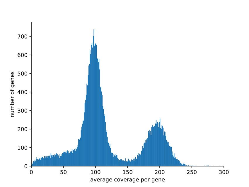
a b
Figure 1. (a) Distribution of the average sequencing read depth per gene model. Predicted gene
models were classified into phase‐separated and merged based on the average read depth value
deduced from the analysis presented here. The haploid read depth with Illumina short reads ranges
from 50‐fold to 150‐fold. (b) Number of heterozygous sequence variants in phase separated and
merged genes. The high proportion of heterozygous variants in merged gene models is due to the
mapping of reads originating from two different alleles to the same region of the assembly.
Average read mapping depth per gene was analyzed to distinguish genes annotated in
separated haplophases as well as merged sequences, respectively (Figure 1, File S11). About 64% of
all predicted protein‐encoding gene models were in the expected range of the haploid read mapping
depth between 50‐fold and 150‐fold and about 27% are merged with a read depth between 150‐fold
and 250‐fold. Only 6% of all genes show an average read depth below 50‐fold and only 1% show an
average coverage higher than 250‐fold. It should be noted that the gene models annotated in the
phase separated part will cover in general two alleles per gene. A total of 22,885 gene models,
representing the 64% in the range of the haploid read mapping depth, were sorted into allelic pairs
which was successful for 8492 genes. The findings presented above can be explained by a diploid
genome. An analysis with Smudgeplot indicated hints for a tetraploid genome from analysis with a
k‐mer size of 19, while the other three investigated k‐mer sizes supported a diploid genome (File
S12).
4. Discussion
The release of genome sequences of many model and crop plants has provided new
opportunities for gene identification and studies of genome evolution, both ultimately serving the
process of plant breeding [45] by allowing discovery of genes responsible for important agronomic
traits and the development of molecular markers associated with these traits. Here, we present the
first genome sequence for D. dumetorum, an important crop for Central and Western Africa, and the
second genome sequence for the genus. Our assembly offers a great opportunity to understand the
evolution of yam and to elucidate some biological constraints inherent to yam including a long
growth cycle, poor to non‐flowering, polyploidy, vegetative propagation, and a heterozygous
genetic background [46]. Yam improvement has been challenging due to these factors preventing the
genetic study of important traits in yam [47].
Oxford Nanopore Technology sequencing has proven to be a reliable and affordable technology
for sequencing genomes thus replacing Illumina technique for de novo genome sequencing due to
substantially higher assembly continuity [42,48]. Large fractions of the genome sequence were
separated into phases, while regions with lower heterozygosity are merged into one representative
sequence. Coverage analysis with Illumina read mapping allowed to classify predicted gene models
as ‘phase‐separated’ or ‘merged’ based on an average coverage around 100‐fold or around 200‐fold,Genes 2020, 11, 274 7 of 11
respectively. While this distinction is possible at the gene model level, whole contigs cannot be
classified this way. Several Mbp long contigs comprise alternating phase‐separated and merged
regions. Therefore, it is likely that the contigs represent a mixture of both haplophases with the risk
of switching between phases at each merged region. Since the haplophases cannot be resolved
continuously through low heterozygosity regions, purging of contigs to reduce the assembly into a
representation of the haploid genome might be advantageous for some applications in the future.
The bimodal coverage distribution (Figure 1a) supports the assumption that D. dumetorum Ibo sweet
3 has a diploid genome. This is supported by Smudgeplot for three out of four k‐mer sizes tested
while the shortest k‐mer size used (19) finds indications for tetraploidy. Since a high ploidy would
result in more distinct coverage peaks as observed for a genome with up to pentaploid parts [42], we
assume that the genome is diploid. The weak hint for tetraploidy might be due to a whole genome
duplication event early in the diversification of the genus. The N50 of 3.2 Mbp is in the expected
range for a long read assembly of a highly heterozygous plant species which contains some quite
repetitive sequences as others reported similar values before [49]. Due to regions of merged
haplophases the total assembly size of 485 Mbp is smaller than expected for a fully phase‐separated
“diploid” genome sequence based on the haploid genome size estimation of 322 Mbp.
We noticed an increase in the number of BUSCOs through several polishing rounds. Initial
assemblies of long reads can contain numerous short insertions and deletions as these are the major
error type in ONT reads [50]. As a result, the identification of CDSs and deduced open reading
frames is hindered through apparent disruptions of some CDS. Through the applied polishing steps,
the number of such apparent frame shifts is reduced thus leading to an increase of detected BUSCOs.
D. dumetorum has 36 chromosomes [12], so with 924 contigs we are far from chromosome‐level
resolution but considerably better than the other genome assembly published in the genus, that of D.
rotundata with 40 chromosomes [8]. Knuth [51] circumscribed D. dumetorum and D. rotundata in two
distant sections D. sect. Lasiophyton and D. sect. Enantiophlyllum, respectively. Additionally,
phylogenetically the two species are quite distantly related with a last common ancestor about 30
million years ago [11,52]. Comparing our predicted peptides to the D. rotundata peptide set [8], we
identified about 9800 single copy orthologs (6475 in the whole set of 35,269 gene models plus 3352
with a relation of one gene in D. rotundata and two phase‐separated alleles in D. dumetorum) which
could elucidate the evolutionary history of those species. The total number of predicted
protein‐encoding gene models was determined to be 35,269, but this number includes two copies of
about 11,300 gene models (see Figure 1) as these are represented by two alleles each. The CDS‐based
pairing we performed detected about 8500 of the theoretical maximum of 11,300 cases which is a
good success rate given the fact that close paralogs and also hemizygous genome regions contribute
to the detected number of phase‐separated gene models. If phase‐separated gene models (alleles) are
excluded, a number of about 24,000 genes would result for D. dumetorum. This fits to the range
detected in other plant genomes [53,54]. The BUSCO results support this interpretation with about
40% of BUSCOs that occur with exactly two copies. Therefore, the true number of protein‐encoding
genes of a haploid D. dumetorum (Trifoliate yam) genome could be around 25,000, also considering
that the BUSCO analysis indicated by 5.8% missing BUSCOs that still a small fraction of the genome
is not represented in the current assembly. This gene number fits well to gene numbers of higher
plants based on all available annotations at NCBI/EBI [54]. The average length of genes and the
number of encoded amino acids are in the same range as previously observed for other plant species
from diverse taxonomic groups [33,55].
It should be noted that the assignment of D. dumetorum sequences to the D. rotundata
pseudochromosomes and indirectly the respective linkage groups contain the risk of incorrect
assignments. However, although D. rotundata and D. dumetorum are evolutionary separated, D.
rotundata is the most closely related species with genetic and genomic resources.
Our draft genome has the potential to provide the basis for new ways to breed with D.
dumetorum, for example avoiding the post‐harvest hardening phenomenon, which begins within 24
h after harvest and makes it necessary to process the tubers within this time to allow consumption
[2]. The family Dioscoreaceae consists of more than 800 species [56] and the post‐harvest hardeningGenes 2020, 11, 274 8 of 11
phenomenon has only been reported from D. dumetorum [57], outlining the singularity of this species
among yam species. We predicted a large number of genes, which will include putative genes
controlling the post‐harvest hardening on D. dumetorum and many useful bioactive compounds
detected in this yam species, which is considered the most nutritious and valuable from a
phytomedical point of view [58]. Ongoing work will try to identify these genes and polymorphisms
for making them available for subsequent breeding.
In summary, we present the first de novo nuclear genome sequence assembly of D. dumetorum
with very good contiguity and partially separated phases. Our assembly has no ambiguous bases
and offers a well applicable gene model annotation. This assembly unraveled the genomic structure
of D. dumetorum to a large extent and will serve as a reference genome sequence for yam breeding by
helping to identify and develop molecular markers associated with relevant agronomic traits, and to
understand the evolutionary history of D. dumetorum and yam species in general.
Supplementary Materials: The following are available online at www.mdpi.com/xxx/s1, File S1: Sequencing
overview with ENA identifiers of runs. File S2: Results of BUSCO analysis of different assembly versions. File
S3: AGP file describing contig assignment to D. rotundata pseudochromosomes. File S4: Genome size estimation
overview using four different tools. File S5: General statistics of different assembly versions. File S6:
Comparison of results from different gene prediction approaches. File S7: Orthogroups of predicted peptides of
D. rotundata and D. dumetorum. File S8: Similarity of D. dumetorum and D. rotundata based on single copy
orthologs. File S9: Functional annotation of predicted genes in the D. dumetorum genome sequence. File S10:
Consensus sequences of repeat elements detected in the D. dumetorum genome sequence. File S11: Average
short read mapping coverage of predicted genes in the D. dumetorum genome sequence. File S12: Results from
Smudgeplot analyses.
Author Contributions: C.S., B.P., D.C.A., and B.W. designed the study. C.S. collected the sample. B.P.
performed DNA extraction, ONT sequencing, and genome assembly. P.V. performed Illumina sequencing. C.S.
and B.P. processed the assembly. B.P. performed gene prediction and evaluation. C.S. and B.P. wrote the initial
draft. B.W. and D.C.A. revised the manuscript. All authors read and approved the final version of the
manuscript.
Funding: This research was partly funded by the German Academic Exchange Service (DAAD, No. 57299294)
with a fellowship to CS.
Acknowledgments: We thank the Appropriate Development for Africa foundation (ADAF) for the yam
collection in Cameroon and for permission to study the plants in Germany in the framework of our mutual
protocol agreement. We also thank the German Society of Botany (DBG) for supporting the research stay of CS
in Bielefeld. We acknowledge support for the Article Processing Charge by the Deutsche
Forschungsgemeinschaft and the Open Access Publication Fund of Bielefeld University.
Conflicts of Interest: The authors declare no conflict of interest.
References
1. Viruel, J.; Forest, F.; Paun, O.; Chase, M.W.; Devey, D.; Couto, R.S.; Segarra‐Moragues, J.G.; Catalan, P.;
Wilkin, P. A nuclear Xdh phylogenetic analysis of yams (Dioscorea Dioscoreaceae) congruent with plastid
trees reveals a new Neotropical lineage. Bot. J. Linn. Soc. 2018, 187, 232–246.
2. Sefa‐Dedeh, S.; Afoakwa, E.O. Biochemical and textural changes in trifoliate yam Dioscorea dumetorum
tubers after harvest. Food Chem. 2002, 79, 27–40.
3. Alozie, Y.E.; Akpanabiatu, M.; Eyong, E.U.; Umoh, I.B.; Alozie, G. Amino Acid Composition of Dioscorea
dumetorum Varities. Pak. J. Nutr. 2009, 8, 103–105.
4. Ferede, R.; Maziya‐Dixon, B.; Alamu, O.E.; Asiedu, R. Identification and quantification of major
carotenoids of deep yellow‐fleshed yam (tropical Dioscorea dumetorum). J. Food Agric. Environ. 2010, 8,
160–166.
5. Nimenibo‐Uadia, R.; Oriakhi, A.; Proximate, Mineral and Phytochemical Composition of Dioscorea
dumetorium Pax. J. Appl. Sci. Environ. Manag. 2017, 21, 771–774.
6. Iwu, M.M.; Okunji, C.O.; Ohiaeri, G.O.; Akah, P.; Corley, D.; Tempesta, M.S. Hypoglycaemic activity of
dioscoretine from tubers of Dioscorea dumetorum in normal and alloxan diabetic rabbits. Plant. Med. 1990,
56, 264–267.Genes 2020, 11, 274 9 of 11
7. Siadjeu, C.; Panyoo, E.A.; Toukam, G.M.S.; Bell, J.M.; Nono, B.; Medoua, G.N. Influence of Cultivar on the
Postharvest Hardening of Trifoliate Yam (Dioscorea dumetorum) Tubers. Advances in Agriculture 2016,
2658983, doi:10.1155/2016/2658983.
8. Tamiru, M.; Natsume, S.; Takagi, H.; White, B.; Yaegashi, H.; Shimizu, M.; Yoshida, K.; Uemura, A.;
Oikawa, K.; Abe, A.; et al. Genome sequencing of the staple food crop white Guinea yam enables the
development of a molecular marker for sex determination. BMC Biol. 2017, 15, 86.
9. Cormier, F.; Lawac, F.; Maledon, E.; Gravillon, M.C.; Nudol, E.; Mournet, P.; Vignes, H.; Chaïr, H.; Arnau,
G. A reference high‐density genetic map of greater yam (Dioscorea alata L.). Theor. Appl. Genet. 2019, 132,
1733–1744, doi:10.1007/s00122‐019‐03311‐6.
10. Ngo Ngwe, M.F.; Omokolo, D.N.; Joly, S. Evolution and Phylogenetic Diversity of Yam Species (Dioscorea
spp.): Implication for Conservation and Agricultural Practices. PLoS ONE 2015, 10, e0145364,
doi:10.1371/journal.pone.0145364.
11. Magwe‐Tindo, J.; Wieringa, J.J.; Sonke, B.; Zapfack, L.; Vigouroux, Y.; Couvreur, T.L.P.; Scarcelli, N.
Complete plastome sequences of 14 African yam species (Dioscorea spp.). Mitochondrial DNA Part
B‐Resour. 2019, 4, 74–76, doi:10.1080/23802359.2018.1536466.
12. Miege, J. Nombres chromosomiques et répartition géographique de quelques plantes tropicales et
équatoriales. Revue de Cytologie et de Biologie Végétales 1954, 15, 312–348.
13. Hui‐Chen, C.; Mei‐Chen, C.; Ping‐Ping, L.; Chih‐Tsun, T.; Fang‐Ping, D. A cytotaxonomic study on
Chinese Dioscorea, L.—The chromosome numbers and their relation to the origin and evolution of the
genus. J. Syst. Evol. 1985, 23, 11–18.
14. Siadjeu, C.; Mayland‐Quellhorst, E.; Albach, D.C. Genetic diversity and population structure of trifoliate
yam (Dioscorea dumetorum Kunth) in Cameroon revealed by genotyping‐by‐sequencing (GBS). BMC
Plant Biol. 2018, 18, 359.
15. Rosso, M.G.; Li, Y.; Strizhov, N.; Reiss, B.; Dekker, K.; Weisshaar, B. An Arabidopsis thaliana T‐DNA
mutagenised population (GABI‐Kat) for flanking sequence tag based reverse genetics. Plant Mol. Biol. 2003,
53, 247–259.
16. Vurture, G.W.; Sedlazeck, F.J.; Nattestad, M.; Underwood, C.J.; Fang, H.; Gurtowski, J.; Schatz, M.C.
GenomeScope: Fast reference‐free genome profiling from short reads. Bioinformatics 2017, 33, 2202–2204,
doi:10.1093/bioinformatics/btx153.
17. Sun, H.; Ding, J.; Piednoel, M.; Schneeberger, K. findGSE: Estimating genome size variation within human
and Arabidopsis using k‐mer frequencies. Bioinformatics 2018, 34, 550–557,
doi:10.1093/bioinformatics/btx637.
18. Liu, B.; Shi, Y.; Yuan, J.; Hu, X.; Zhang, H.; Li, N.; Li, Z.; Chen, Y.; Mu, D.; Fan, W. Estimation of genomic
characteristics by analyzing k‐mer frequency in de novo genome projects. arXiv 2013,
arXiv:preprint/1308.2012.
19. Marcais, G.; Kingsford, C. A fast, lock‐free approach for efficient parallel counting of occurrences of
k‐mers. Bioinformatics 2011, 27, 764–770, doi:10.1093/bioinformatics/btr011.
20. Pucker, B. Mapping‐based genome size estimation. bioRxiv 2019, doi:10.1101/607390.
21. Ranallo‐Benavidez, T.R.; Jaron, K.S.; Schatz, M.C. GenomeScope 2.0 and Smudgeplots: Reference‐free
profiling of polyploid genomes. bioRxiv 2019, 747568, doi:10.1101/747568.
22. Koren, S.; Walenz, B.P.; Berlin, K.; Miller, J.R.; Bergman, N.H.; Phillippy, A.M. Canu: Scalable and accurate
long‐read assembly via adaptive k‐mer weighting and repeat separation. Genome Res. 2017, 27, 722–736.
23. Li, H. Minimap2: Pairwise alignment for nucleotide sequences. Bioinformatics 2018, 34, 3094–3100.
24. Vaser, R.; Sović, I.; Nagarajan, N.; Šikić, M. Fast and accurate de novo genome assembly from long
uncorrected reads. Genome Res. 2017, 27, 737–746.
25. Li, H. Aligning sequence reads, clone sequences and assembly contigs with BWA‐MEM. arXiv 2013,
arXiv:preprint/1303.3997
26. Walker, B.J.; Abeel, T.; Shea, T.; Priest, M.; Abouelliel, A.; Sakthikumar, S.; Cuomo, C.A.; Zeng, Q.;
Wortman, J.; Young, S.K.; et al. Pilon: An integrated tool for comprehensive microbial variant detection
and genome assembly improvement. PLoS ONE 2014, 9, e112963.
27. Pucker, B.; Holtgräwe, D.; Rosleff Sörensen, T.; Stracke, R.; Viehöver, P.; Weisshaar, B. A De Novo Genome
Sequence Assembly of the Arabidopsis thaliana Accession Niederzenz‐1 Displays Presence/Absence
Variation and Strong Synteny. PLoS ONE 2016, 11, e0164321.Genes 2020, 11, 274 10 of 11
28. Tang, H.; Zhang, X.; Miao, C.; Zhang, J.; Ming, R.; Schnable, J.C.; Schnable, P.S.; Lyons, E.; Lu, J.
ALLMAPS: Robust scaffold ordering based on multiple maps. Genome Biol. 2015, 16, 3,
doi:10.1186/s13059‐014‐0573‐1.
29. Pucker, B.; Holtgräwe, D.; Weisshaar, B. Consideration of non‐canonical splice sites improves gene
prediction on the Arabidopsis thaliana Niederzenz‐1 genome sequence. BMC Res. Notes 2017, 10, 667.
30. Simão, F.A.; Waterhouse, R.M.; Ioannidis, P.; Kriventseva, E.V.; Zdobnov, E.M. BUSCO: Assessing
genome assembly and annotation completeness with single‐copy orthologs. Bioinformatics 2015, 31, 3210–
3212.
31. Keller, O.; Kollmar, M.; Stanke, M.; Waack, S. A novel hybrid gene prediction method employing protein
multiple sequence alignments. Bioinformatics 2011, 27, 757–763.
32. Smit, A.F.A.; Hubley, R.; Green, P. RepeatMasker Open‐4.0. Availabe online:
http://www.repeatmasker.org (accessed on 2nd December 2018).
33. Pucker, B.; Feng, T.; Brockhington, S. Next generation sequencing to investigate genomic diversity in
Caryophyllales. bioRxiv 2019, doi:10.1101/646133.
34. Altschul, S.F.; Gish, W.; Miller, W.; Myers, E.W.; Lipman, D.J. Basic local alignment search tool. J. Mol. Biol.
1990, 215, 403–410.
35. Cheng, C.Y.; Krishnakumar, V.; Chan, A.; Thibaud‐Nissen, F.; Schobel, S.; Town, C.D. Araport11: A
complete reannotation of the Arabidopsis thaliana reference genome. Plant J. 2017, 89, 789–804.
36. Jones, P.; Binns, D.; Chang, H.Y.; Fraser, M.; Li, W.; McAnulla, C.; McWilliam, H.; Maslen, J.; Mitchell, A.;
Nuka, G.; et al. InterProScan 5: Genome‐scale protein function classification. Bioinformatics 2014, 30, 1236–
1240, doi:10.1093/bioinformatics/btu031.
37. Lowe, T.M.; Eddy, S.R. tRNAscan‐SE: A program for improved detection of transfer RNA genes in
genomic sequence. Nucleic Acids Res. 1997, 25, 955–964.
38. Chan, P.P.; Lowe, T.M. tRNAscan‐SE: Searching for tRNA Genes in Genomic Sequences. In Gene
Prediction: Methods and Protocols; Kollmar, M., Ed.; Springer: Berlin/Heidelberg, Germany, 2019; Volume
1962, pp. 1–14.
39. Nawrocki, E.P.; Eddy, S.R. Infernal 1.1: 100‐fold faster RNA homology searches. Bioinformatics 2013, 29,
2933–2935.
40. Kalvari, I.; Argasinska, J.; Quinones‐Olvera, N.; Nawrocki, E.P.; Rivas, E.; Eddy, S.R.; Bateman, A.; Finn,
R.D.; Petrov, A.I. Rfam 13.0: Shifting to a genome‐centric resource for non‐coding RNA families. Nucleic
Acids Res. 2018, 46, D335–D342.
41. Flynn, J.M.; Hubley, R.; Goubert, C.; Rosen, J.; Clark, A.G.; Feschotte, C.; Smit, A.F. RepeatModeler2:
Automated genomic discovery of transposable element families. bioRxiv 2019, doi:10.1101/856591.
42. Pucker, B.; Ruckert, C.; Stracke, R.; Viehover, P.; Kalinowski, J.; Weisshaar, B. Twenty‐Five Years of
Propagation in Suspension Cell Culture Results in Substantial Alterations of the Arabidopsis Thaliana
Genome. Genes 2019, 10, 671, doi:10.3390/genes10090671.
43. Baasner, J.S.; Howard, D.; Pucker, B. Influence of neighboring small sequence variants on functional
impact prediction. bioRxiv 2019, doi:10.1101/596718.
44. Emms, D.M.; Kelly, S. OrthoFinder: Phylogenetic orthology inference for comparative genomics. Genome
Biol. 2019, 20, 238, doi:10.1186/s13059‐019‐1832‐y.
45. Ruggieri, V.; Alexiou, K.G.; Morata, J.; Argyris, J.; Pujol, M.; Yano, R.; Nonaka, S.; Ezura, H.; Latrasse, D.;
Boualem, A.; et al. An improved assembly and annotation of the melon (Cucumis melo L.) reference
genome. Sci. Rep. 2018, 8, 8088, doi:10.1038/s41598‐018‐26416‐2.
46. Mignouna, H.D.; Abang, M.M.; Asiedu, R. Harnessing modern biotechnology for tropical tuber crop
improvement: Yam (Dioscorea spp.) molecular breeding. Afr. J. Biotechnol. 2003, 2, 12.
47. Mignouna, H.D.; Abang, M.M.; Asiedu, R. Genomics of Yams, a Common Source of Food and Medicine in
the Tropics. In Genomics of Tropical Crop Plants; Moore, P.H., Ming, R., Eds.; Springer: New York, NY, USA,
2008; pp. 549–570, doi:10.1007/978‐0‐387‐71219‐2_23.
48. Michael, T.P.; Jupe, F.; Bemm, F.; Motley, S.T.; Sandoval, J.P.; Lanz, C.; Loudet, O.; Weigel, D.; Ecker, J.R.
High contiguity Arabidopsis thaliana genome assembly with a single nanopore flow cell. Nat. Commun.
2018, 9, 541.
49. Paajanen, P.; Kettleborough, G.; Lopez‐Girona, E.; Giolai, M.; Heavens, D.; Baker, D.; Lister, A.;
Cugliandolo, F.; Wilde, G.; Hein, I.; et al. A critical comparison of technologies for a plant genome
sequencing project. Gigascience 2019, 8, giy163, doi:10.1093/gigascience/giy163.Genes 2020, 11, 274 11 of 11
50. Salmela, L.; Walve, R.; Rivals, E.; Ukkonen, E. Accurate self‐correction of errors in long reads using de
Bruijn graphs. Bioinformatics 2017, 33, 799–806, doi:10.1093/bioinformatics/btw321.
51. Knuth, R. Dioscoreaceae. In Das Pflanzenreich; Engelr, A., Ed.; Engelmann, W.: Leipzig, Germany, 1924.
52. Viruel, J.; Segarra‐Moragues, J.G.; Raz, L.; Forest, F.; Wilkin, P.; Sanmartin, I.; Catalan, P. Late
Cretaceous‐Early Eocene origin of yams (Dioscorea, Dioscoreaceae) in the Laurasian Palaearctic and their
subsequent Oligocene‐Miocene diversification. J. Biogeogr. 2016, 43, 750–762, doi:10.1111/jbi.12678.
53. Wendel, J.F.; Jackson, S.A.; Meyers, B.C.; Wing, R.A. Evolution of plant genome architecture. Genome Biol.
2016, 17, 37, doi:10.1186/s13059‐016‐0908‐1.
54. Pucker, B.; Brockington, S.F. Genome‐wide analyses supported by RNA‐Seq reveal non‐canonical splice
sites in plant genomes. BMC Genomics 2018, 19, 980.
55. Pucker, B.; Holtgräwe, D.; Stadermann, K.B.; Frey, K.; Huettel, B.; Reinhardt, R.; Weisshaar, B. A
chromosome‐level sequence assembly reveals the structure of the Arabidopsis thaliana Nd‐1 genome and
its gene set. PLoS ONE 2019, 14, e0216233.
56. Barton, H. Yams: Origins and Development. Encyclopaedia of Global Archaeology 2014, 7943–7947.
57. Treche, S.; Delpeuch, F. Physiologie Vegetale—Mise en evidence de lʹapparition dʹun epaississement
membranaire dans le parenchyme des tubercules de Dioscorea dumetorum au cours de la conservation.
Comptes Rendus de lʹAcadémie des Sciences. Série D: Sciences Naturelles 1979, 288, 67–70.
58. Price, E.J.; Wilkin, P.; Sarasan, V.; Fraser, P.D. Metabolite profiling of Dioscorea (yam) species reveals
underutilised biodiversity and renewable sources for high‐value compounds. Sci. Rep. 2016, 6, 29136.
© 2020 by the authors. Licensee MDPI, Basel, Switzerland. This article is an open access
article distributed under the terms and conditions of the Creative Commons Attribution
(CC BY) license (http://creativecommons.org/licenses/by/4.0/).You can also read