Detection and genotypes of Toxoplasma gondii DNA in feces of domestic cats in Colombia - Parasite
←
→
Page content transcription
If your browser does not render page correctly, please read the page content below
Parasite 27, 25 (2020)
Ó A. Zamora-Vélez et al., published by EDP Sciences, 2020
https://doi.org/10.1051/parasite/2020023
                                                                                                                            Available online at:
                                                                                                                           www.parasite-journal.org

    RESEARCH ARTICLE                                                                                                                OPEN        ACCESS

Detection and genotypes of Toxoplasma gondii DNA in feces
of domestic cats in Colombia
Alejandro Zamora-Vélez1,*, Jessica Triviño1, Sebastián Cuadrado-Ríos2, Fabiana Lora-Suarez1,
and Jorge Enrique Gómez-Marín1
1
    Grupo de Estudio en Parasitología y Micología Molecular (GEPAMOL), Centro de Investigaciones Biomédicas, Facultad Ciencias de la
    Salud, Universidad del Quindío, 630004 Armenia, Colombia
2
    Grupo de Biodiversidad y Conservación Genética, Instituto de Genética, Universidad Nacional de Colombia, 111321 Bogotá, Colombia

             Received 18 July 2019, Accepted 3 April 2020, Published online 17 April 2020

             Abstract – The high prevalence of Toxoplasma gondii in the human population in Colombia has been linked to the
             existence of a high density of urban stray cats, exposing the whole population to a high density of oocysts. The goal of
             this study was to determine the DNA prevalence of T. gondii by conventional PCR and to phylogenetically analyze
             ROP18 sequences from positive samples in domestic cat (Felis catus) fecal samples in the city of Armenia, Quindío.
             Fecal samples from 140 cats were collected from 10 districts around the city. Samples were concentrated using
             Ritchie’s method and analyzed through optical microscopy. Concentrates were used for DNA extraction followed
             by nested PCR amplification for T. gondii gene B1. PCR for ROP18 was performed on all B1 positive samples;
             the ROP18 sequences obtained were related to the Archetype I Brazilian and Chinese strains. No oocysts were detected
             by optical microscopy; however, 17.8% (25/140) B1 and 24% (6/25) ROP18 PCR-positive samples were detected.
             Phylogenetic analyses showed that isolates clustered into a single group. We assessed whether associations existed
             between T. gondii positive fecal samples and survey variables such as cat healthcare and socioeconomic characteristics
             of owners, but no statistically significant associations were found. The presence of T. gondii in cat feces is an important
             factor contributing to the high prevalence in the human population of this city.

             Key words: Toxoplasma gondii, Cats, PCR, Prevalence, ROP18.

             Résumé – Détection d’ADN et génotypes de Toxoplasma gondii dans les fèces de chats domestiques en
             Colombie. La forte prévalence de Toxoplasma gondii dans la population humaine en Colombie a été liée à
             l’existence d’une forte densité de chats errants urbains, exposant l’ensemble de la population à une forte densité
             d’oocystes. Le but de ce travail était de déterminer la prévalence de l’ADN de T. gondii par PCR conventionnelle et
             d’analyser phylogénétiquement les séquences ROP18 d’échantillons positifs dans des échantillons fécaux de chat
             domestique (Felis catus) dans la ville d’Armenia, Quindío. Des échantillons fécaux de 140 chats ont été collectés
             dans 10 districts de la ville. Les échantillons ont été concentrés en utilisant la méthode de Ritchie et analysés par
             microscopie optique. Des concentrés ont été utilisés pour l’extraction d’ADN suivie d’une amplification par PCR
             nichée pour le gène B1 de T. gondii. La PCR pour ROP18 a été réalisée sur tous les échantillons positifs pour B1 ;
             les séquences ROP18 obtenues étaient apparentées aux souches Archétype I brésiliennes et chinoises. Aucun oocyste
             n’a été détecté par microscopie optique mais les échantillons étaient positifs par PCR pour 17,8 % (25/140) pour B1
             et 24 % (6/25) pour ROP18. Les analyses phylogénétiques ont montré que les isolats formaient un seul groupe.
             Nous avons évalué s’il existait des associations entre des échantillons fécaux positifs à T. gondii et des variables
             d’enquête telles que les soins de santé des chats et les caractéristiques socioéconomiques des propriétaires, mais
             aucune association statistiquement significative n’a été trouvée. La présence de T. gondii dans les excréments de
             chats est un facteur important contribuant à la forte prévalence dans la population humaine de cette ville.

Introduction                                                                     humans via warm-blooded animals. Wild and domestic felids
                                                                                 are the only known definitive hosts with the ability to shed
   Toxoplasma gondii is an obligate intracellular parasite with                  oocysts in their feces. Common pathways of infection include
worldwide distribution inducing toxoplasmosis and infecting                      oocyst-contaminated water, soil, and food; tissue cysts in
                                                                                 undercooked or raw meat; and congenital transmission
*Corresponding author: oazamorav@uqvirtual.edu.co                                [13, 20, 37, 43]. Oocysts are the environmentally resistant form
    This is an Open Access article distributed under the terms of the Creative Commons Attribution License (https://creativecommons.org/licenses/by/4.0),
                   which permits unrestricted use, distribution, and reproduction in any medium, provided the original work is properly cited.
2 A. Zamora-Vélez et al.: Parasite 2020, 27, 25
of the parasite and play a key role in transmission to new hosts the domestic cats eat; water source for domestic cats to drink;
and ecosystems, generating the need to study humans alongside last deworming (recently: 30 days ago or less; not long since:
domestic and wild animal populations [43]. The large number 30–90 days; long ago: more than 90 days); and hunting habits.
of oocysts shed during primary-infection by felids could lead
to extensive environmental contamination, which can infect a
high number of intermediate species, such as humans, mice Microscopic analysis
or birds. [26]. High rainfall rates can facilitate survival of
oocysts for months, explaining why regions with higher preci- The cat feces samples were processed with the Ritchie tech-
pitation show higher prevalence compared to arid regions, nique [35], approximately 5 g of fecal sample from each cat
which show far lower rates of infection in the population living were diluted in 15 mL of 0.9% sterile saline solution and cen-
in these areas [1, 16, 25]. trifuged for 5 min at 1600 g. This washing procedure was
In Colombia, the prevalence in the human population varies repeated three times. We discarded the supernatant, and resus-
between 30% and 60% [6], and this high prevalence has been pended the pellet using 5 mL of sterile saline solution. Then, we
linked to the existence of a high density of urban stray cats, added 5 mL of 10% formalin solution and 3 mL of 99% diethyl
exposing people to an elevated density of oocysts [10]. This ether (Sigma, USA) to each tube. We sealed and rigorously
high density of free-ranging domestic cats can explain why shook the tube to bring the diethyl ether into contact with all
cat ownership in homes does not increase the risk for T. gondii parts of the sediment and performed a new centrifugation at
in surveys in some cities in Colombia [29]. A study in Armenia 1000 g for 2 min. After this, 30 lL of sediment were scanned
city in 1998 found 89.3% seroprevalence in 28 domestic cats in triplicate using Olympus microscopy with 40 objective lens
and detected a 66.6% shedding prevalence of T. gondii-like using 1% parasitological lugol (Químicos Albor, Colombia) as
oocysts by microscopy in fecal samples from 18 cats [30]. In a contrast visual. The microorganisms were identified by three
2006, another study found a seroprevalence of 84.8% in 33 observers and confirmed with the collaboration of Dr. Fidel
stray cats, but no oocysts were identified by microscopy in Angel Nuñez from the Tropical Medicine Institute Pedro Kouri,
feces collected from the rectum from these cats [10]. Although Havana, Cuba.
training helps with identification of T. gondii-like oocysts, mor-
phological structure along cannot confirm that the oocysts visu-
alized are actually T. gondii, as Hammondia oocysts for Toxoplasma gondii DNA extraction procedure
example look identical [11]. Consequently, molecular detec- and PCR detection
tion-based methods, like PCR, can be an alternative and com-
The resultant pellet from the Ritchie technique was used to
plementary method to microscopy to identify cats infected
obtain DNA from cat feces. The pellets were washed four times
with T. gondii. As a consequence, the objective of this study
in 2 mL tubes with PBS and centrifuged at 4500 g for 10 min.
was to determine the prevalence of T. gondii DNA in cat fecal
The supernatant was discarded and 600 lL of DNAzol (Invit-
samples by conventional PCR and to analyze the ROP18 gene
rogen, USA), 10 lL of isoamyl alcohol (Fisher Scientific,
from positive samples in Armenia, Quindío, Colombia.
USA) and 0.3 g of zirconium silicate beads with 0.5 mm diam-
eter (BioSpec, USA) were added. Afterwards, the tubes were
shaken five times in a Mini Bead Beater (BioSpec, USA) to
Methodology maximum speed for 1 minute and 1 minute in ice [41]. Later,
Sample a Wizard Genomic DNA extraction kit (Promega, USA) was
used for nuclear lysis and purification, following the manufac-
Armenia is the capital of the department of Quindío in turer’s protocol. In order to detect DNA, the T. gondii B1 repet-
Colombia’s central mountain range, at an altitude of 1480 m itive fragment (GenBank accession number AF179871) was
above sea level. Armenia has 301,226 inhabitants, according used in an amplification nested PCR method [5]. All B1 posi-
to the projections of the Administrative Department for tive samples were sequenced to confirm that the amplified pro-
National Statistics (DANE, 2018). Armenia is divided into 10 duct was in fact T. gondii, because it is possible to amplify
administrative zones, called communes. The number of cats DNA from other organisms [27]. As has been previously
to sample was estimated according to the rabies vaccination described [31, 41, 45], the primers for the first PCR were Toxo
program statistics reported in Armenia, with a population of N1 50 –GGAACTGCATCCGTTCATGAG–30 and Toxo C1 50 –
15,015 owned cats [28]. Therefore, considering inferences with TCTTTAAAGCGTTCGTGGTC–30 to obtain a 193-bp frag-
prevalence with 95% confidence, an accepted error margin of ment. The second PCR was performed with the primers Toxo
8%, and using an expected prevalence of 66.6% based on pre- N2 50 –TGCATAGGTTGCCAGTCACTG–30 and Toxo C2
vious studies, at least 132 owned cats needed to be sampled 50 –GGCGACCAATCTGCGAATACACC–30 to obtain a 96-
according to Epiinfo v.7.2.3.1 software. The residences were bp fragment. The first amplification protocol consisted of one
selected at random in the 10 communes of the city of Armenia. initial stage of denaturation for 5 min at 94 °C, followed by
40 cycles of amplification, and 1 cycle consisting of 1 min at
Questionnaire 94 °C for DNA denaturation, 1 min of annealing at 53 °C,
and 1 min of extension at 72 °C. Subsequently, an additional
The owners were interviewed about the following aspects: step of 10 min of final extension at 72 °C was performed.
number of stray cats observed 500 m around the residence; The second PCR with the product of the first amplification,
commune; age and sex of domestic cats; kind of food that consisted of one initial stage of denaturation for 5 min atA. Zamora-Vélez et al.: Parasite 2020, 27, 25 3
94 °C, followed by 14 cycles of amplification, and 1 cycle con- alignment, as regions dominated by gaps, insertions or deletions
sisting of 1 min at 94 °C for DNA denaturation, 1 min of usually contain phylogenetically useful information, and the
annealing at 53 °C, and extension at 72 °C for 30 s. Then, an percentage of identity obtained in BLAST suggested that our
additional step of 10 min of final extension at 72 °C was carried sequences were sufficiently informative.
out. The ROP18 T. gondii sequence (GenBank accession num- The evolutionary model and partition scheme that best sui-
ber JX045319.1) was detected in B1 PCR-positive samples. To ted the aligned sequences were determined using PARTITION-
detect this sequence, we used forward primer ROP18S 50 – FINDER v 1.1.1 [22], according to the corrected Akaike
GACCGTCTTTCAAGAGGAGGA–30 and reverse primer information criterion (AICc). The optimal partitioning was the
ROP18R 50 –ACGCTGGTGAGAGGTGCAC–30 to obtain a minimum partitioning scheme, and the resulting model was
514-bp fragment. The amplified protocol consisted of one initial K80 [39]. These parameters were implemented in the Bayesian
stage of denaturation for 3 min at 94 °C followed by 35 cycles analyses executed in BEAST 1.8.3 [8]. We assumed a constant
of 30 s at 94 °C for DNA denaturation, 45 s at 60 °C for anneal- population size to estimate the patterns of speciation across the
ing, and 30 s at 72 °C for extension. Then, 5 min at 72 °C for phylogeny, and a strict clock; we kept priors at the default. The
final extension was performed. All primers were synthetized by number of Markov chain Monte Carlo (MCMC) iterations was
Invitrogen Corporation (USA). Finally, a 1.5% agarose elec- set to 100 million, with 25% of trees obtained as burning,
trophoresis gel was used to analyze PCR products. The positive retaining trees each 1000 steps. Two independent chains were
control was DNA from the T. gondii control RH strain, and the run and we checked effective sample size (ESS) in Tracer
negative control was distilled water in the presence of primers. v1.6 [8]. The maximum clade credibility (MCC) tree was esti-
mated in TreeAnnotator [8], discarding 25% of initial trees. We
Statistical analysis also investigated alternative phylogenetic grouping, construct-
ing an unrooted network (split network) with the Neighbor-
As the dependent variable was binary (presence vs. absence Net method implemented in SplitsTree4 [18]. We estimated a
of T. gondii DNA in fecal samples), a Chi-squared test was haplotype network, with equal weighting on transversions/tran-
used to assess relationships with other variables including sitions, without considering gaps/missing and removing invari-
demographic, behavioral and cat care. A p value < 0.05 for able sites, and the median-joining network algorithm [3]
the significance level was employed. OR values were calculated implemented in PopART [24].
with a 95% confidence interval. The Statgraphics Centurion
v.17 software was used.
Results
Sequencing and phylogenetic analyses Prevalence of Toxoplasma gondii by microscopy
and PCR in cat fecal samples
PCR-positive products for ROP18 were gel-purified from
low-melt agarose gels, followed by recovery using a Wizard Between November 2014 and August 2015, in the commu-
PCR SV and PCR clean up system kit (Promega, USA). nes selected, 521 residences were visited. In 217 of these houses
Sequencing was done under BigDyeÒ terminator cycling condi- (41.6%), there was at least one domestic cat, and in 140 of the
tions by using the normal automatic service by Macrogen 217 houses (64.5% of visited houses with at least one domestic
(South Korea) in a 3730XL DNA sequencer with the same pri- cat), it was possible to collect fecal samples with permission
mers as the PCR amplifications. BLAST (http://blast.ncbi.nml. from the owners. We found via optical microscopy, Ancy-
nih.gov/Blast.cgi) search on the GenBank database with all lostoma spp. in 6/140 samples (4.3%), Toxocara spp. in 6/140
ROP18 sequences was performed, verifying that they belong samples (4.3%), Toxascaris spp. in 3/140 samples (2.1%), and
to T. gondii, as well as to identify closely related genotyped Hymenolepis spp. in 4/140 samples (2.8%) (Fig. 1). Toxoplasma
strains. We explored strain relatedness in more detail through gondii-like oocysts were not observed.
phylogenetic analyses, including ROP18 sequences available Of the 140 collected samples, 25 (17.85%) were positive for
in GenBank and ToxoDB. In a previous study, T. gondii T. gondii DNA by B1 PCR and confirmed through sequencing
ROP16 sequences from human and meat samples obtained in according to the results obtained from Macrogen (South Korea).
the same study area showed a high degree of genetic divergence Prevalence among the communes varied between 0% and
and clustered with highly virulent strains, and we expected the 37.8% (Table 1), but these differences did not attain statistical
ROP18 sequences we obtained to show the same pattern within significance (v2 test p = 0.27).
the phylogeny [2]. Sequences were aligned with T. gondii se- In all, 10/56 positive samples (17.8%) were from male cats
quences available in GenBank and ToxoDB (Supp. Table 1), and 15/78 (19.2%) were from female cats. Also, 6/45 positive
restricting the number of sequences per strain to one (except samples (13.3%) were from young cats (7 years old), and 2/15 (13.3%) were
with ocular toxoplasmosis in the same study area [38]. Align- from cats without identify age data (Table 2). The 7 years used
ment was performed with MAFFT v7.187 [21], using the auto as an age cut-off was a reference from Hand et al., 2000 [17].
routine and default settings. Most of downloaded sequences There were no significant associations between the data
corresponded to the complete gene (1663–1671 bp), depending obtained from the questionnaire and the prevalence of T. gondii
on the strain [15], and sequences obtained here represented only DNA-positive samples through Chi squared and OR tests
a partial region. Therefore, we decided not to trim the resulting (Table 3).4 A. Zamora-Vélez et al.: Parasite 2020, 27, 25
Table 1. Distribution of the samples collected in Armenia, Quindío,
Colombia during 2014–2015 by commune and positive samples
from domestic cat feces using B1 PCR to detect T. gondii DNA.
Commune Number of Number of % positive Confidence
samples positive samples samples interval (95%)
1 20 2 10 [7.6–12.3]
2 17 1 5.9 [4.6–7.1]
3 17 3 17.6 [15.1–20.0]
4 14 4 28.5 [24.4–32.5]
5 19 7 36.8 [33.7–39.8]
6 10 1 10 [5.4–14.5]
7 18 3 16.6 [13.9–19.3]
8 12 1 8.3 [5.1–11.4]
9 8 3 37.8 [31.8–43.7]
10 5 0 0 0
Table 2. Comparison of PCR T. gondii prevalence in cat feces
according sex and age (years) in the 140 cats from which the samples
were taken in 10 communes from Armenia.
Cats (140) B1 PCR
(N; %) Positive samples/N (%)
Gender (140; 100%)
Female (78; 55.7%) 15/78 (19.2%)
Male (56; 40%) 10/56 (17.8%)
Non-determined (6; 4.3%) 0 (0%)
Age in years (140; 100%)
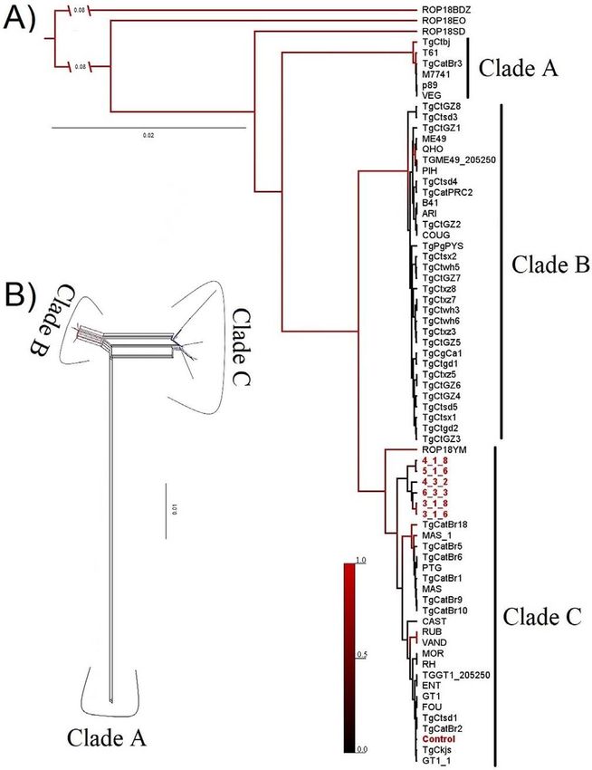
Figure 1. (A) Phylogenetic relationships among ROP18 sequences
1 (45; 32.14%) 6/45 (13.3%)
from several strains of T. gondii as reported in GenBank, and
> 1 y 7 (68; 48.5%) 15/68 (22%)
sequences found in cats in Armenia in present study are shown with
> 7 (12; 8.5%) 2 /12 (16.6%)
numbers (6_3_3; 5_1_6; 3_1_6; 3_1_8; 4_3_2; 4_1_8) and in red
Non-determined (15; 10.7%) 2/15 (13.3%)
font. The phylogeny was inferred by Bayesian analysis in BEAST.
Branch lengths indicate genetic distance, and posterior probabilities
Note: The 7 years used as an age cut-off was referenced from Hand
of nodes are represented by the size of the branches that separate
et al. [17].
from the node. Major clades labeled are supported by absolute
support (PP < 0.99). (B) Neighbor-net phylogenetic network based
on the rhoptry protein 18 gene from T. gondii strains. Potential
reticulation between strains is denoted by the network patterns
Major clades were also identified by the unrooted network
connecting strains.
(Fig. 1B). The haplotype network constructed (Supp. Fig. 2)
showed that three ROP18 sequences obtained from cat feces
Phylogenetic analysis of ROP18 sequences grouped into a single haplotype, which also contained the
Archetype I (RH and GT1) and Chinese III (TgCkjs and TgCts-
We assayed ROP18 amplification on B1 PCR-positive sam- d1) strains, as well as Brazilian strains. Additionally, the
ples and found 6/25 (24%) ROP18-positive samples. The Baye- remaining three sequences were found to represent unique hap-
sian tree retained (Fig. 1A) clarified the phylogenetic position of lotypes, separated by one to three mutation steps from the afore-
T. gondii isolates obtained from cats in the present study, rela- mentioned haplotype.
tive to the other T. gondii strains analyzed. Our sequences clus-
tered into a single clade with Brazilian and Chinese strains
(Fig. 1, Clade C). Despite overall support for most of the nodes, Discussion
we obtained strong support (PP < 0.99) for three major clades:
the first clade (A) consisted of Archetype III strains, a second Oocyst shedding by the domestic cat (Felis catus) is epi-
clade (B) grouped the Chinese strains I and II [6] and the demiologically important and shedding needs to be investigated
Archetype II strains [27], and a third clade (C) consisted of because (1) it is suspected that oocyst density is correlated with
Colombian, Brazilian and Chinese strains (III, according to high prevalence in some communities, (2) oocysts can lead to
Gao et al. [17]), and a genotype previously identified and outbreaks of acute human toxoplasmosis, and (3) they are prob-
named Archetype I [16]. All six ROP18 sequences obtained ably significantly responsible for infection in animals, which
in the present study grouped into clade C, which included could be animals for human consumption [40]. Likewise, felids
ROP18 sequences from mouse-virulent strains (GT1 and RH) are highly susceptible and a single bradyzoite from cyst tissue
and ocular toxoplasmosis strains (ROP18YM) (Supp. Fig. 1). can be enough to produce infection [9].A. Zamora-Vélez et al.: Parasite 2020, 27, 25 5
Table 3. Relationship between demographic, behavioral and cat care variables and presence of T. gondii DNA in fecal samples.
Risk factor PCR B1 positive PCR B1 negative OR. 95% CI p
Cat age (< 1 year vs. 1 year) 6 vs. 19 45 vs. 70 0.4 [0.1 – 1.3] 0.1
Cat sex (male vs. female) 10 vs. 15 66 vs. 49 1.1 [0.4 – 2.6] 0.8
Food (dry food vs. mix) 14 vs. 11 72 vs. 43 1.3 [0.5 – 3.1] 0.5
Faucet water for drink (yes vs. no) 22 vs. 3 101 vs. 14 1 [0.2 – 3.8] 0.9
Cats go outdoors (yes vs. no) 16 vs. 9 61 vs. 54 1.5 [0.6 – 3.8] 0.3
Hunting activities (yes vs. no) 15 vs. 10 47 vs. 68 2.1 [0.8 – 5.2] 0.08
Deworming at least once (yes vs. no) 6 vs. 19 87 vs. 28 0.9 [0.3 – 2.6] 0.9
Stray cats located around the house of the cat owners 24 vs. 1 111 vs. 4 0.8 [0 – 8] 0.9
In this study, it was not possible to detect T. gondii oocysts results can be used for genetic analysis of the strains circulating
using the optical microscopy technique in domestic cat feces. in a geographic region. We did not find statistically significant
The same occurred in an earlier study in Armenia [10]. One relationships between the presence of T. gondii DNA and the
possible explanation for the lack of evidence of oocysts in variables obtained from the questionnaire. However, other stud-
microscopy examination is that, at a given point in time, only ies [23, 42] have indicated that cats were probably exposed to
1% of cats could be shedding oocysts [10] and the power of the infection from an early age and later, many times through-
the sample that we calculated could fail to detect such a low out their lives, through predation and hunting [43].
prevalence of oocyst shedding. The low frequency of oocysts Lower prevalence of T. gondii in cat feces in some coun-
observed by optical microscopy could also be explained by tries could be related to greater access of domestic cats to
the fact that we evaluated owned domestic cats that often eat human food with a low risk of infection [43]. In contrast, high
inside houses. The situation could have been different if we prevalence could be explained by the access of domestic cats to
had tested wild cats with a natural pray diet. Furthermore, it undercooked meat provided by their human owners or by hunt-
is important to note that identification may be difficult due to ing [44].
the morphological similarities between T. gondii and other coc- Previous studies on the genetic diversity of T. gondii strains
cidian parasites that can be shed in cat feces. Additionally, infecting cats in Colombia have shown that parasite populations
oocyst shedding by cats occurs over short periods of time, with are highly diverse [10]. Phylogenetic and haplotype analyses
primary infection mainly in juvenile cats [7, 19]. Other gas- including ROP18 sequences obtained here also found high
trointestinal parasites were found, showing that the frequency genetic diversity, despite being obtained from closely located
of domestic cat feces samples with helminths was lower com- sampling sites. Interestingly, their phylogenetic relationships
pared to previous reports in the same city [12]. This could indi- confirmed the presence of virulent ROP18 alleles circulating
cate improved deworming habits by cat owners in the city of in cats, as reported for patients with ocular toxoplasmosis in
Armenia. the same city [38] (Supp. Fig. 1). However, fine-scale genotyp-
In contrast with the absence of oocysts via microscopy, ing is imperative to reach definitive conclusions, facilitating the
PCR detected 17% of cats with the presence of T. gondii evaluation of the virulence of cat T. gondii strains, and their
DNA in cat feces samples. The prevalence of T. gondii, accord- relationships with ocular toxoplasmosis strains. The clades
ing to the DNA frequency, in this study was 17.85% (25/140). found with recent phylogenetic analyses based on a concate-
This is low in comparison with other studies, like the one per- nated matrix that included the ROP18 gene [15, 33, 34]. In con-
formed in Portugal, where 20.5% prevalence was found in clusion, this study confirmed a lack of microscopic detection of
domestic cats feces samples using the B1 sequence in PCR oocysts in domestic cats in Armenia (Colombia), but that it is
[14]. However, our study found a higher prevalence compared possible to detect and analyze genetic diversity by PCR analy-
to that observed in a Korean study, where prevalence was 4.5% sis. In this case, the ROP18 gene was found to be an indicator
[20], and a study in Switzerland, where prevalence was 0.4% of virulence of T. gondii found in cat feces samples.
[4]. This is the first study in Colombia detecting T. gondii
DNA from cats feces samples, but the frequency of positive
samples may be lower compared to previous studies with
oocyst prevalence reported at 66.6% [30] and seroprevalence
Conflict of interest
at 84.8%–89.3% [10, 30]. These differences could be associated The authors declare that they have no conflict of interest.
with the methodology and cats sampled. These PCR results
must be interpreted with caution since the T. gondii PCR-posi-
tives are not always related to oocysts. This is because the pres- Supplementary materials
ence of T. gondii DNA could be due to the presence of
bradyzoites of infected prey consumed by a cat [32]. In addi- Supplementary materials are available at https://www.
tion, the PCR methods overestimate exposure to infective par- parasite-journal.org/10.1051/parasite/2020023/olm
asites because they detect all populations of infectious and non- Supplementary Figure 1. Polymorphic nucleotide sites at
infectious parasites alive or dead [36]. The utility of PCR detec- the ROP18 polymorphic region for 6 isolates of T. gondii from
tion on cat feces samples is that it offers information on the cat feces (plus RH control strain), 4 isolates of T. gondii from
maximum occurrence and, after sequencing for PCR products, ocular toxoplasmosis [38], and three sequences representing6 A. Zamora-Vélez et al.: Parasite 2020, 27, 25
three archetypal lineages detected for T. gondii strains (GT1, Kwok OCH, Smith T, Su C. 2006. Prevalence of Toxoplasma
ME49 and VEG). Consensus sequence was determined by gondii in cats from Colombia, South America and genetic
the percentage of nucleotides common to all sequences. Sites characterization of T. gondii isolates. Veterinary Parasitology,
145, 45–50.
in grey indicate similarity with the consensus sequence.
11. Dumètre A, Dardé ML. 2003. How to detect Toxoplasma gondii
Supplementary Figure 2. Median-joining network for oocysts in environmental samples? FEMS Microbiology
T. gondii haplotypes of the rhoptry protein 18 gene. The size Reviews, 27, 651–661.
of the circles represents the haplotype frequency and the colors 12. Echeverry DM, Giraldo MI, Castaño JC. 2012. Prevalencia de
represent the country to which the strain belongs. The number helmintos intestinales en gatos domésticos del departamento del
of transversal lines connecting haplotypes represents the Quindío, Colombia. Biomédica, 32, 430–436.
number of substitutions between them. Sequences found in cats 13. Elmore SA, Jones JL, Conrad PA, Patton S, Lindsay DS, Dubey
in the present study are shown with numbers (6_3_3; 5_1_6; JP. 2010. Toxoplasma gondii: epidemiology, feline clinical
3_1_6; 3_1_8; 4_3_2; 4_1_8). aspects, and prevention. Trends in Parasitology, 26, 190–196.
Supplementary Table 1. Accession numbers and country of 14. Esteves F, Aguiar D, Rosado J, Costa ML, de Sousa B, Antunes
T. gondii ROP18 gene sequences downloaded from the F, Matos O. 2014. Toxoplasma gondii prevalence in cats from
Lisbon and in pigs from centre and south of Portugal. Veterinary
GenBank database, and later included in our phylogenetic Parasitology, 200, 8–12.
analyses. 15. Gao J, Xie Y, Xu Z, Chen H, Hide G, Yang T, Shen J, Lai D.
2017. Genetic analyses of Chinese isolates of Toxoplasma
Acknowledgements. We thank the Biomedical Research Center gondii reveal a new genotype with high virulence to murine
and members of the GEPAMOL group in the “Universidad del hosts. Veterinary Parasitology, 241, 52–60.
Quindío” for all their support, and Dr. Fidel Angel Nuñez from
16. Gómez-Marin JE, De-la-Torre A, Angel-Muller E, Rubio J,
the Tropical Medicine Institute Pedro Kouri, Havana, Cuba. Arenas J, Osorio E, Nuñez L, Pinzon L, Mendez-Cordoba LC,
Bustos A, De-la-Hoz I, Silva P, Beltran M, Chacon L, Marrugo
M, Manjarres C, Baquero H, Lora F, Torres E, Zuluaga OE,
References Estrada M, Moscote L, Silva MT, Rivera R, Molina A, Najera S,
Sanabria A, Ramirez ML, Alarcon C, Restrepo N, Falla A,
1. Afonso E, Thulliez P, Gilot-Fromont E. 2006. Transmission of Rodriguez T, Castaño G. 2011. First Colombian multicentric
Toxoplasma gondii in an urban population of domestic cats newborn screening for congenital toxoplasmosis. PLoS
(Felis catus). International Journal for Parasitology, 36, 1373– Neglected Tropical Diseases, 5, e1195.
1382. 17. Hand M, Thatcher C, Remillard R, Roudebush P. 2000.
2. Alvarez C, Vargas M, Herrera C, Uribe-Huertas LD, Lora F, Nutrición Clínica de los Pequeños Animales, 4th edn. Mark
Gómez-Marín JE. 2015. Striking divergence in Toxoplasma Morris: Colombia.
ROP16 nucleotide sequences from human and meat samples. 18. Huson DH, Bryant D. 2006. Application of phylogenetic
Journal of Infectious Diseases, 211, 1–8. networks in evolutionary studies. Molecular Biology and
3. Bandelt HJ, Forster P, Röhl A. 1999. Median-joining networks Evolution, 23, 254–267.
for inferring intraspecific phylogenies. Molecular Biology and 19. Jones JL, Dubey JP. 2012. Foodborne toxoplasmosis. Clinical
Evolution, 16(1), 37–48. Infectious Diseases, 55, 845–851.
4. Berger-Schoch AE, Herrmann DC, Schares G, Müller N, Bernet 20. Jung BK, Lee SE, Lim H, Cho J, Kim DG, Song H, Kim MJ,
D, Gottstein B, Frey CF. 2011. Prevalence and genotypes of Shin EH, Chai JY. 2015. Toxoplasma gondii B1 gene detection
Toxoplasma gondii in feline faeces (oocysts) and meat from in feces of stray cats around Seoul, Korea and genotype analysis
sheep, cattle and pigs in Switzerland. Veterinary Parasitology, of two laboratory-passaged isolates. Korean Journal of Para-
177, 290–297. sitology, 53, 259–263.
5. Burg JL, Grover CM, Pouletty P, Boothroyd JC. 1989. Direct 21. Katoh K, Standley DM. 2013. MAFFT multiple sequence
and sensitive detection of a pathogenic direct and sensitive alignment software version 7: Improvements in per-
detection of a pathogenic protozoan, Toxoplasma gondii, by formance and usability. Molecular Biology and Evolution, 30,
polymerase chain reaction. Journal of Clinical Microbiology, 772–780.
27, 1787–1792. 22. Lanfear R, Calcott B, Ho SYW, Guindon S. 2012. Partition-
6. Cañón-Franco WA, López-Orozco N, Gómez-Marín JE, Dubey Finder: Combined selection of partitioning schemes and
JP. 2014. An overview of seventy years of research (1944– substitution models for phylogenetic analyses. Molecular
2014) on toxoplasmosis in Colombia, South America. Parasites Biology and Evolution, 29, 1695–1701.
& Vectors, 7, 427. 23. Lee SE, Kim JY, Kim YA, Cho SH, Ahn HJ, Woo HM, Lee
7. Dabritz HA, Miller MA, Atwill ER, Gardner IA, Leutenegger WJ, Nam HW. 2010. Prevalence of Toxoplasma gondii
CM, Melli AC, Conrad PA. 2007. Detection of Toxoplasma infection in stray and household cats in regions of Seoul,
gondii-like oocysts in cat feces and estimates of the environ- Korea. Korean Journal of Parasitology, 48, 267–270.
mental oocyst burden. Journal of the American Veterinary 24. Leigh JW, Bryant D. 2015. POPART: full-feature software for
Medical Association, 231, 1676–1684. haplotype network construction. Methods in Ecology and
8. Drummond AJ, Rambaut A. 2007. BEAST: Bayesian evolu- Evolution, 6, 1110–1116.
tionary analysis by sampling trees. BMC Evolutionary Biology, 25. Lélu M, Villena I, Dardé M, Aubert D, Geers R, Dupuis E,
7, 214. Marnef F. 2012. Quantitative estimation of the viability of
9. Dubey JP. 2009. History of the discovery of the life cycle of Toxoplasma gondii oocysts in soil. Applied and Environmental
Toxoplasma gondii. International Journal for Parasitology, 39, Microbiology, 78, 5127–5132.
877–882. 26. Lilly EL, Wortham CD. 2013. High prevalence of Toxoplasma
10. Dubey JP, Cortes-Vecino JA, Vargas-Duarte JJ, Sundar N, gondii oocyst shedding in stray and pet cats (Felis catus) in
Velmurugan GV, Bandini LM, Polo LJ, Zambrano L, Mora LE, Virginia, United States. Parasites & Vectors, 6, 266.A. Zamora-Vélez et al.: Parasite 2020, 27, 25 7
27. Liu Q, Wang ZD, Huang SY, Zhu XQ. 2015. Diagnosis of 2018. Assessing viability and infectivity of foodborne and
toxoplasmosis and typing of Toxoplasma gondii. Parasites and waterborne stages (cysts/oocysts) of Giardia duodenalis,
Vectors, 8, 1–14. Cryptosporidium spp., and Toxoplasma gondii: a review of
28. Subdirección de salud ambiental. 2018. Reporte de vacunación methods. Parasite, 25, 14.
antirrábica de perros y gatos Colombia 2017. MINSALUD, 37. Saevik BK, Krontveit RI, Eggen KP, Malmberg N, Thoresen SI,
Gobierno de Colombia. Prestrud KW. 2015. Toxoplasma gondii seroprevalence in pet
29. Montoya-de-Londono MT, Castano-Osorio JC, Gomez-Marin JE. cats in Norway and risk factors for seropositivity. Journal of
1997. A maternal screening program for congenital toxoplasmosis Feline Medicine and Surgery, 17(12), 1049–1056.
in Quindio, Colombia and application of mathematical models to 38. Sánchez V, Gómez-Marín JE. 2014. Characterization of ROP18
estimate incidences using age-stratified data. American Journal of alleles in human toxoplasmosis. Parasitology International, 63,
Tropical Medicine and Hygiene, 57, 180–186. 463–469.
30. Montoya-Londoño MT, Loango-Chamorro N, Sierra-Infante M, 39. Stadler T. 2009. On incomplete sampling under birth–death
Castaño-Osorio J. 1998. Infección por Toxoplasma gondii en models and connections to the sampling-based coalescent.
gatos de dos barrios del sur de Armenia y su importancia en la Journal of Theoretical Biology, 261, 58–66.
toxoplasmosis humana. Colbaquin Actualidades Clínicas y 40. Tenter AM, Heckeroth AR, Weiss LM. 2000. Toxoplasma
Biotecnológicas, 12, 18–23. gondii: from animals to humans. International Journal for
31. Ponce N, Gomez-Marin JE. 2003. Estandarización y validación Parasitology, 30, 1217–1258.
clínica de la prueba de reacción en cadena de la polimerasa 41. Triviño-Valencia J, Lora F, Zuluaga JD, Gomez-Marin JE.
(PCR) para diagnóstico de toxoplasmosis cerebral en pacientes 2016. Detection by PCR of pathogenic protozoa in raw and
infectados por el VIH. Infectio, 7, 8–14. drinkable water samples in Colombia. Parasitology Research,
32. Poulle M, Josse-Dupuis É, Villena I, Aubert D. 2016. Detection 115, 1789–1797.
of Toxoplasma gondii DNA by qPCR in the feces of a cat that 42. Vanwormer E, Conrad PA, Miller MA, Melli AC, Carpenter TE,
recently ingested infected prey does not necessarily imply Mazet JAK. 2013. Toxoplasma gondii, source to sea: Higher
oocyst shedding. Parasite, 23, 29. contribution of domestic felids to terrestrial parasite loading
33. Ramos S, Fátima H, Pena DJ, Gonçalves A, Maria L, De Faria J, despite lower infection prevalence. EcoHealth, 10, 277–289.
Gonzales B, Oliveira S, Maria S, Ramos S, Amélia N. 2018. 43. Vanwormer E, Fritz H, Shapiro K, Mazet JAK, Conrad PA.
Characterization of Toxoplasma gondii isolates from herds of 2013. Molecules to modeling : Toxoplasma gondii oocysts at the
sheep in southern Brazil reveals the archetypal type II genotype human – animal – environment interface. Comparative Immunol-
and new non- archetypal genotypes. Parasitology International, ogy, Microbiology and Infectious Diseases, 36, 217–231.
67, 59–63. 44. Wastling JM, Nicoll S, Buxton D. 1993. Comparison of two
34. Rêgo WMF, Costa JGL, Baraviera RCA, Pinto LV, Bessa GL, gene amplification methods for the detection of Tomplasma
Lopes REN, Vitor RWA. 2017. Association of ROP18 and gondii in experimentally infected sheep. Journal of Medical
ROP5 was e ffi cient as a marker of virulence in atypical isolates Microbiology, 38, 360–365.
of Toxoplasma gondii obtained from pigs and goats in Piauí, 45. Zamora-Vélez A, Cuadrado-Ríos S, Triviño-Valencia J. 2016.
Brazil. Veterinary Parasitology, 247, 19–25. Genetic diversity and phylogeny of Toxoplasma gondii based
35. Ritchie LS. 1948. An ether sedimentation technique for routine on B1 partial sequences from colombia and other countries.
stool examinations. Bulletin of U.S. Army Medical Department, Revista de la Asociación Colombiana de Ciencias Biológicas,
8, 326. 28, 8–15.
36. Rousseau A, La Carbona S, Dumètre A, Robertson LJ, Gargala
G, Escotte-Binet S, Favennec L, Villena I, Gérard C, Aubert D.
Cite this article as: Zamora-Vélez A, Triviño J, Cuadrado-Ríos S, Lora-Suarez F & Enrique Gómez-Marín J. 2020. Detection and
genotypes of Toxoplasma gondii DNA in feces of domestic cats in Colombia. Parasite 27, 25.
An international open-access, peer-reviewed, online journal publishing high quality papers
on all aspects of human and animal parasitology
Reviews, articles and short notes may be submitted. Fields include, but are not limited to: general, medical and veterinary parasitology;
morphology, including ultrastructure; parasite systematics, including entomology, acarology, helminthology and protistology, and molecular
analyses; molecular biology and biochemistry; immunology of parasitic diseases; host-parasite relationships; ecology and life history of
parasites; epidemiology; therapeutics; new diagnostic tools.
All papers in Parasite are published in English. Manuscripts should have a broad interest and must not have been published or submitted
elsewhere. No limit is imposed on the length of manuscripts.
Parasite (open-access) continues Parasite (print and online editions, 1994-2012) and Annales de Parasitologie Humaine et Comparée
(1923-1993) and is the official journal of the Société Française de Parasitologie.
Editor-in-Chief: Submit your manuscript at
Jean-Lou Justine, Paris http://parasite.edmgr.com/You can also read