Designing P. aeruginosa synthetic phages with reduced genomes
←
→
Page content transcription
If your browser does not render page correctly, please read the page content below
www.nature.com/scientificreports
OPEN Designing P. aeruginosa synthetic
phages with reduced genomes
Diana P. Pires1*, Rodrigo Monteiro1, Dalila Mil‑Homens2, Arsénio Fialho2,3, Timothy K. Lu4 &
Joana Azeredo1*
In the era where antibiotic resistance is considered one of the major worldwide concerns,
bacteriophages have emerged as a promising therapeutic approach to deal with this problem.
Genetically engineered bacteriophages can enable enhanced anti-bacterial functionalities, but
require cloning additional genes into the phage genomes, which might be challenging due to the DNA
encapsulation capacity of a phage. To tackle this issue, we designed and assembled for the first time
synthetic phages with smaller genomes by knocking out up to 48% of the genes encoding hypothetical
proteins from the genome of the newly isolated Pseudomonas aeruginosa phage vB_PaeP_PE3. The
antibacterial efficacy of the wild-type and the synthetic phages was assessed in vitro as well as in vivo
using a Galleria mellonella infection model. Overall, both in vitro and in vivo studies revealed that the
knock-outs made in phage genome do not impair the antibacterial properties of the synthetic phages,
indicating that this could be a good strategy to clear space from phage genomes in order to enable
the introduction of other genes of interest that can potentiate the future treatment of P. aeruginosa
infections.
Antibiotic resistance is rising to high levels all around the world, being currently considered as one of the major
threats to human health1. This fact has triggered an increasing interest on alternative therapeutic approaches
including phage t herapy2. Bacteriophages have been broadly explored for a wide range of applications but despite
all the efforts that have been made to bring phages into the clinical settings, phage therapy still faces some chal-
lenges such as limited host ranges, bacterial resistance to phages, side effects of bacterial lysis, as well as safety, sta-
bility and delivery issues3–5. Phage engineering holds a great potential to generate phage variants with improved
antibacterial properties, which might contribute to the enhancement of phage therapy. This is usually achieved by
cloning additional genes into the phage g enomes6, which might be difficult without removing unnecessary phage-
encoded genes. Considering that a very large proportion of phage genomes encode for hypothetical proteins
with unknown f unctions7, a possible approach to get some space for genome engineering would be the precise
removal of those genes from viral genomes. In addition, for safety purposes, it has been advised that phages
carrying genes encoding toxins, virulence factors or antibiotic resistance should be avoided from therapeutic
applications8. Therefore, it would be better for therapeutic purposes to have phages carrying a minimum number
of genes with unknown functions, as we still don’t know if those genes can be potentially hazardous. Although
transcriptomic studies on phage-bacteria interaction are bringing important clues to understand the role of the
hypothetical proteins encoded on phage genomes9–11, their functional analysis is still challenging and can be time
consuming. Here, we envisioned that synthetic phages carrying a minimal number of genes essential for their
replication and host killing, would be more easily accepted for therapy and a minimal genome concept would
also facilitate the integration of extra functions into the phage genome to improve its performance.
The recent advances in genetic engineering and sequencing technologies have led to a fast development of
phage-engineering tools that can now be applied in a high throughput fashion to design and build synthetic
phages with desirable f eatures5,6. Taking advantage of the yeast-based phage-engineering p latform12, we aimed to
create synthetic phages with reduced genomes, minimizing the number of genes encoding hypothetical proteins.
Since P. aeruginosa is currently considered by the World Health Organization as one of the top priority bacterial
pathogens urgently requiring new treatments13, in this study we isolated a new P. aeruginosa phage and used it
as a model to design our synthetic phages. From our knowledge, this is the first study focused on minimizing
phage genomes and understanding its impact on phages’ performance.
1
CEB ‑ Centre of Biological Engineering, Universidade Do Minho, Campus de Gualtar, Braga, Portugal. 2Institute for
Bioengineering and Biosciences (iBB), Instituto Superior Técnico, Lisboa, Portugal. 3Department of Bioengineering,
Instituto Superior Técnico, Universidade de Lisboa, Lisboa, Portugal. 4Department of Electrical Engineering and
Computer Science and Department of Biological Engineering, Synthetic Biology Center, Massachusetts Institute
of Technology, 77 Massachusetts Avenue, Cambridge, MA 02139, USA. *email: priscilapires@deb.uminho.pt;
jazeredo@deb.uminho.pt
Scientific Reports | (2021) 11:2164 | https://doi.org/10.1038/s41598-021-81580-2 1
Vol.:(0123456789)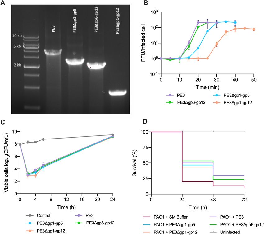
www.nature.com/scientificreports/
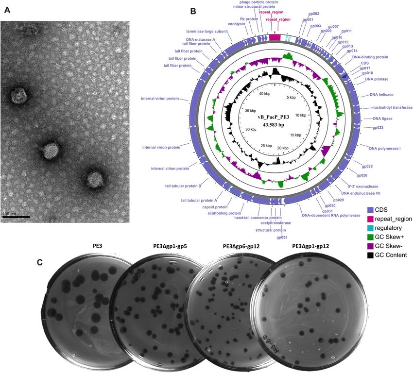
Figure 1. Phage characterization. (A) TEM observation of phage PE3. The bar indicates 100 nm. (B) Circular
view of phage PE3 genome. Created using CGView server16. (C) Phage plaques of the wild-type and synthetic
phages.
Results
Characterization of the newly isolated P. aeruginosa phage vB_PaeP_PE3. The lytic phage PE3
was isolated from a wastewater treatment plant using the P. aeruginosa PAO1 as host for the enrichment proce-
dure. After isolation and propagation, the phage was tested against a panel of 28 P. aeruginosa clinical isolates14
and revealed to be quite specific, being able to infect 7 of them (Table S1). The morphological characterization
by TEM (Fig. 1A) revealed that phage PE3 has a short and noncontractile t ail15.
Phage PE3 was further characterized by whole genome sequencing. The data obtained from sequencing
revealed that PE3 genome consists of a 43,583 bp double-stranded DNA with a GC content of 62.3% and two
479 bp direct terminal repeats (DTRs). Phage PE3 encodes 55 predicted CDSs with lengths ranging between 120
to 4014 bp (Fig. 1B)16. Based on BLASTP analysis, 30 of the predicted CDSs have assigned functions while the
other 25 encode hypothetical proteins (Table S2). The genome sequencing analysis together with TEM revealed
that this phage has the defining characteristics of the Autographiviridae family, which was until 2019 considered
a subfamily of Podoviridae17. This family includes phages similar to T7 with genomes composed of linear ter-
minally redundant dsDNA encoding a RNA p olymerase17. The genome analysis of phage PE3 further revealed
that this phage is most probably virulent as it does not encode genes known to be associated with lysogeny. In
addition, no tRNAs were detected and, based on BLASTN search, we found that phage vB_PaeP_PE3 shares
high homology with phages vB_PaeP_PAO1_1-15pyo18 and LUZ199.
Scientific Reports | (2021) 11:2164 | https://doi.org/10.1038/s41598-021-81580-2 2
Vol:.(1234567890)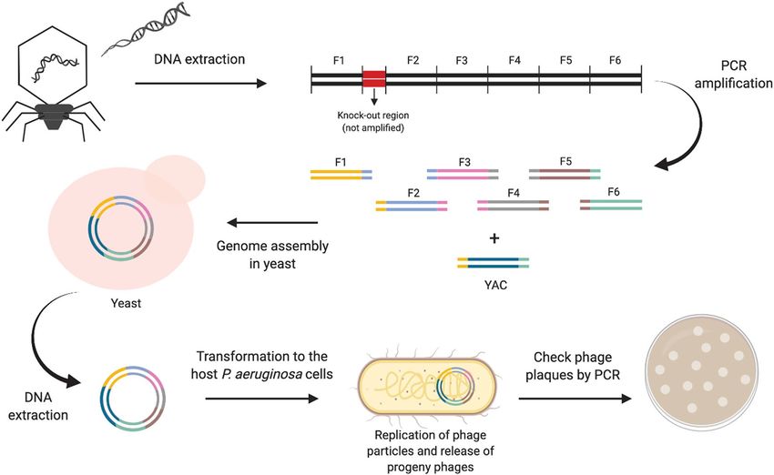
www.nature.com/scientificreports/
Figure 2. Workflow to build synthetic phages with reduced genomes. Phage DNA is used as template to
generate overlapping PCR products covering the entire phage genome with the exception of the knock-
out region. The PCR products are co-transformed into yeast cells along with the linearized YAC, where the
phage genome is assembled. The phage genome captured into the YAC is then extracted from yeast cells and
transformed to the host P. aeruginosa cells to generate infectious phage particles. This figure was created with
BioRender.com and exported under a paid subscription.
Efficient assembly of synthetic phages with reduced genomes. A large part of the genes encoded
in phage genomes do not have assigned functions, which raises the question about whether these genes may
play an essential role in phage replication and bacterial killing, or if they are unnecessary and can therefore
be removed from phage genomes without a detrimental effect on their antibacterial activity. Taking this into
consideration, we wondered about the possibility to build synthetic phages with reduced genomes by remov-
ing genes encoding hypothetical proteins that are not essential for phage viability and replication. At the first
part of the annotated sequence of phage PE3 it is possible to find two modules of genes encoding hypothetical
proteins separated by regulatory elements: the first module is between gp1 and gp5 and the second between gp6
and gp12. Based on this information, we aimed to understand if by removing each of these regions of genes,
the phage would still be viable. Using the yeast-based phage-engineering p latform12, we built two fully syn-
thetic phages, vB_PaeP_PE3Δgp1-gp5 (short name PE3Δgp1–gp5) and vB_PaeP_PE3Δgp6–gp12 (short name
PE3Δgp6–gp12), each of them encoding knock-outs between gp1–gp5 and gp6–gp12, respectively. This was
achieved by amplifying the whole phage genome of phage PE3, with the exception of the knock-out regions,
by PCR in overlapping fragments using specific sets of primers (Table S3). The six PCR products spanning the
phage genome and the linearized YAC carrying homologous “arms” with the extremities of phage genome were
successfully assembled in yeast as a consequence of the gap repair system that joins each fragment to the adja-
cent, resulting in a full phage genome captured in the YAC (Fig. 2)12. The yeast transformants were then screened
by yeast colony PCR to confirm the correct assembly of the phages using a set of primers located upstream and
downstream the knock-out regions (Table S4), which will result in smaller PCR products comparatively to the
wild-type phage. To understand if these knock-outs would affect phage’s viability, the YAC-phage DNA was
extracted from yeast cells and transformed into the P. aeruginosa PAO1 host, where phage genes can be tran-
scribed and generate functional phages. In fact, phage plaques were observed after plating. The resulting plaques
were checked by PCR using the set of primers described above and revealed that the knock-outs were success-
fully performed on phage genome (Fig. 3A) and yielded viable phage particles, which were then propagated for
further characterization. This result clearly indicates that the first 12 genes encoded in the genome of phage PE3
are non-essential for phage viability and propagation.
Since we succeeded in separately removing 5 and 7 genes from PE3 genome, we decided to design and
synthesize a third synthetic phage by removing the first 12 genes encoding hypothetical proteins (gp1–gp12)
corresponding to a knock-out of 3194 bp. Using the same phage-engineering strategy, the construct was assem-
bled in yeast and the chimeric phage vB_PaeP_PE3Δgp1–gp12 (short name PE3Δgp1–gp12) was successfully
obtained (Fig. 3A). Although several phage-engineering strategies have been developed and proved to be efficient
in editing genomes of virulent p hages19–21, none of them had been previously applied to engineer P. aeruginosa
phages. Therefore, we opted for the yeast-based phage-engineering strategy because it was already demonstrated
Scientific Reports | (2021) 11:2164 | https://doi.org/10.1038/s41598-021-81580-2 3
Vol.:(0123456789)www.nature.com/scientificreports/
Figure 3. Wild-type phage versus synthetic phages. (A) PCR-based confirmation of the deletions in the genome
of phage PE3. (B) One-step growth curves of wild-type (PE3) and synthetic phages phages (PE3Δgp1–gp5,
PE3Δgp6–gp12 and PE3Δgp1–gp12). (C) Treatment of P. aeruginosa PAO1 log-phase cultures with the wild-
type phage or synthetic phages using a MOI of ~ 5. No phage was added in the control. Error bars represent SD
from three independent experiments. (D) G. mellonella larvae were injected with either PBS (uninfected group)
or P. aeruginosa PAO1 followed by a subsequent administration of wild-type or synthetic phages at a MOI
of ~ 100 or SM Buffer in the non-treated group. Survival curves represent three independent experiments, each
with 10 larvae per treatment group.
to be a robust platform to capture and/or manipulate genomes of phages infecting different bacterial species,
including P. aeruginosa12.
Synthetic phages versus wild‑type phage: characterization. In order to better understand the
impact of the knock-outs on phage behavior, the chimeric phages were characterized and compared to the wild-
type phage. First, we noticed that the morphology of the phage plaques was slightly different from the wild-type
phage, showing a reduced size (Fig. 1C). Then, we assessed the host range of the chimeric phages using the same
panel of strains we used for the wild-type phage (Table S1). Surprisingly, not all phages displayed the same host
range. Although PE3 and PE3Δgp6–gp12 share the same host range infecting 7 out of 28 clinical isolates, the
other two chimeric phages were only able to infect 4 of the 7 strains, causing lysis from without on the others.
Next, the phage growth parameters were evaluated by performing one-step-growth curves. The results revealed
that the wild-type phage PE3 and the chimeric phage PE3Δgp6–gp12 have a similar growth curve, while phages
PE3Δgp1–gp5 and PE3Δgp1–gp12 have 5 min and 15 min longer latent periods, respectively (Fig. 3B). Regarding
the burst sizes, the values obtained for phages PE3Δgp1–gp5 (211 PFU/infected cell) and PE3Δgp6–gp12 (203
PFU/infected cell) were similar with the wild-type phage (206 PFU/infected cell). However, phage PE3Δgp1–
gp12 has a significantly lower burst size of 83 PFU/infected cell.
The genomes of the three synthetic phages were sequenced to understand if mutations were introduced in
the genomes due to PCR amplification. The sequencing results revealed three point mutations for all phages,
comparatively with wild-type phage. To confirm these results, the three regions encoding the mutations were
amplified by PCR and sequenced by Sanger. This was also performed for the wild-type phage. However, for phages
Scientific Reports | (2021) 11:2164 | https://doi.org/10.1038/s41598-021-81580-2 4
Vol:.(1234567890)www.nature.com/scientificreports/
PE3Δgp1–gp5 and PE3Δgp6–gp12 only two of the point mutations were confirmed by Sanger sequencing. One
of these mutations is shared by both phages, as they were built at the same time and some PCR products used
for yeast recombination were the same for both, and corresponds to a G → A transition on gp47, which encodes
a tail fiber protein. Phage PE3Δgp1–gp5 also carries a silent mutation C → T in the endolysin gene (gp51) while
phage PE3Δgp6–gp12 carries a C → A transversion resulting in a Ser to Tyr change in the tail fiber gene (gp46).
The sequencing of phage PE3Δgp1–gp12 revealed a silent mutation G → A in gp37, which encodes a scaffolding
protein, and two additional point mutations in genes encoding tail fiber proteins: a C → A transversion in gp46
that results in a Gln to Lys change, and a A → G transition in gp47 that results in a Asn to Asp change.
The in vitro and in vivo antibacterial efficacy of the synthetic phages is not impaired by
genome knock‑outs. In order to understand if the knock-outs performed on PE3 phage genome had some
impact on its antibacterial performance, in vitro and in vivo efficacy assays were performed using the wild-type
and the chimeric phages.
For the in vitro experiments, phages were added to log-phase bacterial cultures at a MOI of ~ 5 and the phage
infection was followed for 24 h. As observed in Fig. 3C (Table S5), all the phages revealed a similar behavior and
no significant differences were found among them (p > 0.01). Two hours after adding phages to the cultures, the
number of P. aeruginosa PAO1 viable cells was reduced by more than 5 orders-of-magnitude for all the phages,
comparatively with the control (non-infected culture) (p < 0.01). After this time point, the number of viable cells
started increasing and, 24 h after the beginning of phage infection, no differences were observed between treated
and non-treated cultures (p > 0.01), indicating the fast proliferation of bacteriophage-insensitive mutants (BIMs).
The emergence of BIMs within a short time after phage infection is very common for P. aeruginosa species and
has already been widely studied22–24. Despite this fact, the results obtained here clearly indicate that phages’
performances were not affected by genome knock-outs as no differences were observed between the wild-type
and chimeric phages.
As the in vitro assays are usually unable to mimic the complexity of the environments found in vivo, we used
a G. mellonella infection model to compare the efficacy of the wild-type versus synthetic phages. After bacterial
challenge, the larvae were injected either with phages (treated groups) or with SM Buffer (non-treated group).
One control group was used by injecting larvae with PBS and SM Buffer to monitor the killing due to injection
trauma (uninfected group). As shown in Fig. 3D, the survival rates of the larvae in the untreated group were
20%, 13% and 10% after 24, 48 and 72 h of bacterial infection, respectively (Table S6). The groups treated with
the different phages revealed significantly higher survival rates for all time points when compared with the
untreated group (p < 0.05). For instance, the treatment of larvae with the wild-type phage resulted in a survival
rate of 50% after 24 h of infection and 30% survival after 48 and 72 h. The treatment of infected larvae with the
synthetic phages resulted in survival rates similar to the wild-type phage and no significant differences were
found among the treated groups. Altogether, these data evidence that the genome knock-outs do not impair
phages performance both in vitro as well as in vivo. In addition, no cytotoxic effects were observed in the larvae
when injected only with wild-type or chimeric phages with concentrations up to 1 08 PFU/mL during the course
of 72 h (data not shown).
Discussion
A minimal cell is usually defined as a cell carrying only essential genes, but this definition can be controversial
as the essential functions can be dependent on the environmental c onditions25,26. Some studies have already
addressed this topic for a number of bacterial species by identifying sets of essential genes26,27. For instance,
based on transposon mutagenesis, Hutchison III et al.25 were able to classify bacterial genes as essential or non-
essential, which enabled a rational design and posterior synthesis of a M. mycoides minimal genome. However,
when it comes to phages, there is still a limited knowledge about which genes are essential or not, even though
transcriptomics are bringing important insights in this fi eld9–11.
Despite the fact that phage genomes are relatively small, phage particles are extremely abundant in the bio-
sphere and they display a huge genetic diversity and n ovelty28. It is therefore predicted that most of the phage-
encoded genes (about 80%) have unknown f unctions28, which might constitute a barrier for their therapeutic
application. Taking this into consideration, in this work we intended to design and generate synthetic phages
with reduced genomes in order to encode a minor number of genes with unknown functions. On the other
hand, we aimed to study whether the genomic deletions made on phages influence their performance. To address
this, three synthetic phages were built by removing three sets of genes encoding hypothetical proteins from the
genome of the newly isolated P. aeruginosa phage PE3.
Phages PE3Δgp1–gp5 and PE3Δgp6–gp12 were initially designed to understand if the deletion of each module
of genes would result in viable phage particles. As both constructs resulted in viable phages, this clearly revealed
that all the genes comprised in the two knock-out regions of the phage genome (gp1–gp5 and gp6–gp12) are
non-essential. This led us to build a third synthetic phage—PE3Δgp1–gp12—lacking both sets of genes that
were shown to be unnecessary for phage viability and replication. The characterization of the chimeric phages
comparatively to the wild-type phage revealed that the phage with the larger genomic deletion, PE3Δgp1–gp12,
had a longer latent period and a lower burst size. However, this feature doesn’t seem to influence its performance
in vitro and in vivo but elucidates that, although the set of genes from gp1 to gp12 are non-essential, some
genes might have a role in the early stage of phage infection. The gene or genes that might influence phage-host
interaction are probably located between gp1 and gp5 as the synthetic phage PE3Δgp1–gp5 also presented a
longer latent period comparatively with the wild-type phage PE3, while phage PE3Δgp6–gp12 showed the exact
same behavior as the wild-type. This similarity between phage PE3Δgp6–gp12 and the wild-type phage was also
observed when analyzing their host ranges. While these two phages share the same host range, the other two
Scientific Reports | (2021) 11:2164 | https://doi.org/10.1038/s41598-021-81580-2 5
Vol.:(0123456789)www.nature.com/scientificreports/
phages lost the ability to infect 3 of the clinical strains being only able to cause them lysis from without, which
indicates that might not be a problem of interaction with phage receptors. However, the cause for this host-
range change remains to be explained. Apart from the knock-outs, the whole genome sequencing only revealed
two point mutations in the genomes of phages PE3Δgp1–gp5 and PE3Δgp6–gp12 and three point mutations
in the genome of phage PE3Δgp1–gp12. These mutations were probably introduced by PCR reactions during
amplification of phage DNA fragments for assembly but do not seem to influence phages’ antibacterial behavior.
The introduction of mutations during PCR amplification can happen, even at higher rates. For example, in a
study developed by Smith et al. in which the genome of phage φX174 (5386 bp) was assembled from synthetic
oligonucleotides, the synthetic phage DNA showed lower infectivity than the natural phage DNA, which was
attributed to PCR-generated mutations that reached 1 per 500 bp29.
To further understand if the genomic knock-outs impaired the in vivo antibacterial performance of the
phages, we used a G. mellonella infection model. The results showed that the efficacy of the chimeric phages to
rescue larvae from infection was not affected by the genomic manipulations, suggesting that phage genomes can
be redesigned to remove genes without relevant functions. The elimination of genes with unknown functions
that proved to be unnecessary can make phages more attractive for therapeutic applications. In addition, the
deletion of unnecessary genes creates some room in phage genomes, paving the way to introduce other genes of
interest that can potentiate their antibacterial activity.
In conclusion, here we demonstrated that although phage genomes are usually very compact and well organ-
ized, they carry many non-essential genes that can be removed from its genome without a detrimental effect to
the infection. In this work we were able to remove 48% of the genes encoding hypothetical proteins, building
phages in which the vast majority of the genes have assigned functions. In this way, we proved that 12 of the
55 genes encoded in the genome of PE3 phage are non-essential. Although the yeast-based phage-engineering
platform used in this work was shown to be a reliable and efficient strategy for genome editing P. aeruginosa
phages, it might be limited to bacterial hosts amenable to genetic manipulation.
Despite the major developments in phage-engineering tools and platforms and the increasing interest in this
field5,6, there is still a limited number of reports on phage engineering, which target a narrow range of bacterial
hosts. However, we envisioned that in a near future, the new advances in synthetic biology field will enable to
quickly design and create specialized phages “a la carte” to a wide range of bacterial species. Finally, the in vivo
experiments showed that engineered phages can be safely used and might constitute an interesting approach to
treat P. aeruginosa infections in the future.
Materials and methods
Strains, plasmids and primers. Phage vB_PaeP_PE3 (short name PE3, see Table S7) was isolated in this
study. The three synthetic phages PE3Δgp1–gp5, PE3Δgp6–gp12 and PE3Δgp1–gp12 are listed in Table S7. The
host bacterial strain of PE3 phage is the reference strain P. aeruginosa PAO1 (DSM22644), obtained from the
German Collection of Microorganisms and Cell Cultures (DSMZ-Deutsche Sammlung von Mikro-organismen
und Zellkulturen.) The P. aeruginosa clinical strains used to evaluate the host range of phages belong to the lab
collection14. The Saccharomyces cerevisiae BY4741 (MATa his3Δ1 leu2Δ0 met15Δ0 ura3Δ0) and the yeast cen-
tromere vector pRS415 (ATCC 87520) with LEU2 marker were obtained from laboratory stocks. All primers
used in this study are listed in Table S4.
Culture conditions. Pseudomonas aeruginosa PAO1 was grown at 37 °C in lysogeny broth (LB) or LB agar
(LB with 1.2% (w/v) of agar). S. cerevisiae BY4741 was cultured in YPD (1% (w/v) Bacto Yeast Extract, 2% (w/v)
Bacto Peptone and 2% dextrose (w/v)) or YPD agar at 30 °C.
Phage isolation. The bacteriophage PE3 used in this study was isolated from a wastewater treatment plant
(Braga, Portugal) using P. aeruginosa PAO1 as the host strain for the enrichment procedure and following the
protocol described by Azeredo et al.30.
Phage propagation and titration. Phage production and titration were performed using the double agar
overlay technique as described previously22,30.
Transmission electron microscopy. Phage PE3 was morphologically characterized by Transmission
Electron Microscopy (TEM) using a procedure previously described31. Briefly, phage particles were collected by
centrifugation (25,000×g, 4 °C, 60 min). The sedimented particles were washed twice with tap water and cen-
trifuged again. Phages were then deposited on copper grids with carbon-coated Formvar films, stained with 2%
uranyl acetate (pH 4.0) and examined using a Jeol JEM 1400 transmission electron microscope (Tokyo, Japan).
Phage DNA isolation. The isolation of phages’ DNA was performed using an adapted version of the pro-
tocol described by Ando et al.12. 150 mL of phage lysates were centrifuged (9000×g, 4 °C, 15 min) and filtered
(0.22 μm) to remove cellular debris. Purified lysates were then mixed with 200 μL of buffer L1 [20 mg/mL RNase
A, 6 mg/mL DNase I, 0.2 mg/mL BSA, 10 mM EDTA, 100 mM Tris-HCl, 300 mM NaCl, pH 7.5] for 30 min at
37 °C. After that, 30 mL of cold buffer L2 [30% (w/v) polyethylene glycol (PEG) 6000, 3 M NaCl] were added and
incubated on ice under agitation (90 rpm) for 1 h. The suspension was then centrifuged (9000×g, 4 °C, 30 min),
the supernatant discarded, and the pellets resuspended in a total of 9 mL of buffer L3 (100 MM Tris-HCl,
100 mM NaCl, 25 mM EDTA, pH 7.5). Nine mL of buffer L4 [4% (w/v) SDS] were added, mixed gently and the
tubes were incubated at 70 °C for 20 min and then allowed to cool on ice. Afterwards, 9 mL of buffer L5 (2.55 M
Scientific Reports | (2021) 11:2164 | https://doi.org/10.1038/s41598-021-81580-2 6
Vol:.(1234567890)www.nature.com/scientificreports/
potassium acetate, pH 4.8) were added and mixed gently by inverting the tubes. This solution was centrifuged
(10,000×g, 4 °C, 1 h) and the supernatant was saved and passed through a Qiagen-tip 100 column according to
the manufacturer’s instructions. The eluted DNA was then precipitated by adding 0.7 volumes of isopropanol
and centrifuged (10,000×g, 4 °C, 30 min). The pellet was sequentially washed with 70% (v/v) ethanol and 95%
(v/v) ethanol. After completely air-dried, the pellet was resuspended in sterile water and stored at − 20 °C.
Genome sequencing. The Illumina Nextera XT library preparation kit was used for the library construc-
tion of the phage DNA samples. The generated DNA fragments (DNA libraries) were sequenced in the Illumina
MiSeq platform using 300 bp paired-end sequencing reads. The raw sequence data were then automatically
trimmed and the demultiplexed reads were assembled into a single contig using Geneious R11.
In silico analysis of phage genome. The potential coding sequences (CDSs) of phages were firstly anno-
tated using myRAST32. Sequence similarity searches were then manually performed with the translation of
each predicted CDS against the National Center for Biotechnology Information (NCBI) protein database, using
BLASTP33, in order to assign putative functions. Promoters were predicted using MEME34 and phiSITE35, while
predicted terminators were found using ARNold36. The tRNAscan-SE tool37 was used to search for tRNAs.
Preparation of PCR products for assembling phage genomes. All PCR products were prepared
using specific sets of primers (see Table S3 and S4) and the Phusion High-Fidelity DNA Polymerase (Thermo
Scientific). All the PCR fragments were excised from an agarose gel after electrophoresis and recovered using
the Zymoclean Gel DNA Recovery kit (Zymo Research). Homologous overhangs to the 5′ and 3′ ends of phage
genome were added to the primers used to amplify the yeast artificial chromosome (YAC) pRS415, in order to
enable the subsequent capture of phage genomes into the YAC. Seven PCR products (including the YAC) were
used for each yeast transformation (Table S3).
Preparation of yeast competent cells. The preparation of yeast competent cells was performed as
described by Ando et al.12 with minor modifications. Briefly, S. cerevisiae BY4741 was grown in 10 mL YPD at
30 °C for approximately 24 h. Five mL of this culture were transferred into 50 mL of YPD and incubated at 30 °C
for 5 h. Cells were then harvested by centrifugation (5000×g, RT, 5 min), washed twice with 25 mL of water and
resuspended in 1 mL of water. The cellular suspension was centrifuged again (13,000×g, RT, 30 s) and resus-
pended in 1 mL of water. One hundred microliters of this cellular suspension were used for each transformation.
Yeast transformation. Yeast transformation was performed according to a previously described protocol
with minor modifications12. All PCR products (phage DNA fragments and the linearized pRS415) were com-
bined in a tube (1 μg of each DNA fragment and 200 ng linearized pRS415 in 35 μl water), and mixed with the
transformation mixture [100 μL yeast competent cells, 240 μL 50% (w/v) PEG 3350, 36 μL 1 M LiAc, 50 μL 2 mg/
mL salmon sperm DNA]. The mixture was incubated at 42 °C for 45 min, then centrifuged at 13,000×g for 30 s
and resuspended in 200 μL of YPD. Transformants were selected on a complete synthetic defined medium with
leucine dropout (SD-Leu) [0.67% Yeast Nitrogen Base (YNB), 0.069% CSM-Leu, 2% dextrose] agar plates at
30 °C for 3 days.
Yeast DNA extraction of captured phage genomes. YAC-Phage DNA was extracted from yeast cells
as previously described by Ando et al.12.
Preparation of P. aeruginosa electrocompetent cells and bacterial transformation. Pseu-
domonas aeruginosa PAO1 electrocompetent cells were prepared according to a protocol described e lsewhere38.
Briefly, 6 mL of an overnight grown culture were distributed by 4 microcentrifuge tubes and the cells were har-
vested by centrifugation (16,000×g, RT, 1 min). Afterwards, each pellet was washed twice with 1 mL of 300 mM
sucrose. Then, the 4 bacterial pellets were resuspended and combined in a total of 100 μL of 300 mM sucrose for
each transformation.
To transform P. aeruginosa PAO1 cells, 100 μL of electrocompetent cells were mixed with extracted DNA
(YAC-phage DNA) and the mixture was transferred to a 2 mm gap electroporation cuvette. Afterwards, a pulse
was applied (25 μF, 200 Ω, 2.5 kV) and 900 μL of SOC medium were added. The cellular suspension was trans-
ferred to a tube and incubated at 37 °C for 2–3 h with agitation (200 rpm) before plaque formation assays.
Plaque formation assays. To recover the synthetic phages, approximately 200–500 μL of the cellular sus-
pension resulting from YAC-phage DNA electroporation were mixed with 3 mL of LB soft agar into an agar
plate. After overnight incubation at 37 °C, the plates were analyzed to check for the presence of phage plaques.
The resulting phage plaques were checked by PCR to confirm the correct knock-outs on the synthetic phages.
Phage host range. The host-range of the wild-type and synthetic phages was assessed using 28 P. aerugi-
nosa clinical strains14. One drop of phage suspensions was spotted on the lawns of the different bacterial isolates.
The plates were then incubated overnight at 37 °C and the susceptibility of each host to the phage was evaluated.
In the cases where lysis was observed, serial dilutions of phage suspensions were made and spotted again on the
bacterial lawns to identify possible cases of lysis from without.
Scientific Reports | (2021) 11:2164 | https://doi.org/10.1038/s41598-021-81580-2 7
Vol.:(0123456789)www.nature.com/scientificreports/
Phage growth parameters. To determine phage growth parameters, one-step growth curves were per-
formed. Briefly, 10 mL of a mid-exponential phase culture with an OD600 of 0.3 were centrifuged (7000×g, 4 °C,
5 min) and resuspended in fresh medium. Then, the phage solution was added to obtain a MOI of 0.01 and
phages were allowed to adsorb for 5 min at 37 °C under agitation (120 rpm). After that, the suspension was
centrifuged again (7000×g, 4 °C, 5 min), the pellet was resuspended in 10 mL of fresh medium and incubated at
37 °C with agitation. One sample was immediately taken before incubation and after that, samples were taken
every 5 min during the course of the experiment. Each sample was serially diluted to count the plaque formation
units (PFUs).
Phage infection of suspended cultures. A P. aeruginosa PAO1 culture grown for 16 h was diluted 1:100
in a final volume of 20 mL and incubated at 37 °C and 120 rpm until reaching an OD600nm of 0.25. At this point,
the phages were added at an MOI of 5, except for the control. The suspensions were then incubated at 37 °C with
agitation (120 rpm) and CFU counts were determined after 2, 4, 6 and 24 h, as described by Pires et al.22.
Galleria mellonella infection model. Wax moth larvae G. mellonella were reared at 25 °C in the dark-
ness, from egg to last instar larvae on natural diet (beeswax and pollen grains). Worms of the final instar larval
stage, weighing 250 ± 25 mg, were selected to be used in the experiments. The G. mellonella survival experiment
was adapted from previous studies with small c hanges39,40. Briefly, P. aeruginosa PAO1 overnight cultures were
grown in LB at 37 °C with shaking. The bacterial culture was diluted in PBS to 8–9 CFU per volume of injection
(5 μL). Ten larvae per group were randomly selected based on size. Using a hypodermic microsyringe, the larvae
were injected with 8–9 CFU suspensions via the last left side proleg, previously surface-sanitized with alcohol
at 70% (v/v). Approximately 30 min after bacterial injection, SM Buffer (5.8 g/L NaCl, 2 g/L M gSO4·7H2O,
50 mL/L 1 M Tris-HCl pH = 7.5) or phage treatment at a MOI of 100 was administered in the penultimate right
proleg. In the control group, the larvae were injected with PBS and SM Buffer to monitor the killing due to injec-
tion trauma. Cytotoxicity assays were also performed by injecting larvae only with phages with titers ranging
between 102 and 106 PFU per injection. Larvae were then incubated in petri dishes and maintained in the dark
at 37 °C. The survival was monitored every 24 h for 72 h and the larvae were considered dead when there was a
lack of mobility in response to touch. Kaplan–Meier survival curves were generated and analyzed with the log-
rank test using GraphPad Prism.
GenBank submission. The complete genome of the P. aeruginosa phage PE3 has been deposited in Gen-
Bank under the accession number MN901924.
Statistical analysis. All data of the experiments were analyzed using GraphPad Prism version 8. The data
are presented as the mean of three independent experiments and the error bars represent the SD.
Received: 8 October 2020; Accepted: 29 December 2020
References
1. Hernando-Amado, S., Coque, T. M., Baquero, F. & Martínez, J. L. Defining and combating antibiotic resistance from One Health
and Global Health perspectives. Nat. Microbiol. 4, 1432–1442 (2019).
2. Czaplewski, L. et al. Alternatives to antibiotics: a pipeline portfolio review. Lancet. Infect. Dis 16, 239–251 (2016).
3. Pires, D. P., Costa, A. R., Pinto, G., Meneses, L. & Azeredo, J. Current challenges and future opportunities of phage therapy. FEMS
Microbiol. Rev. 44, 684–700 (2020).
4. Lu, T. K. & Koeris, M. S. The next generation of bacteriophage therapy. Curr. Opin. Microbiol. 14, 524–531 (2011).
5. Kilcher, S. & Loessner, M. J. Engineering bacteriophages as versatile biologics. Trends Microbiol. 27, 355–367 (2019).
6. Pires, D. P., Cleto, S., Sillankorva, S., Azeredo, J. & Lu, T. K. Genetically engineered phages: a review of advances over the last
decade. Microbiol. Mol. Biol. Rev. 80, 523–543 (2016).
7. Hatfull, G. F. Bacteriophage genomics. Curr. Opin. Microbiol. 11, 447–453 (2008).
8. Pirnay, J. P. et al. Quality and safety requirements for sustainable phage therapy products. Pharm. Res. 32, 2173–2179 (2015).
9. Lavigne, R. et al. A multifaceted study of Pseudomonas aeruginosa shutdown by virulent podovirus LUZ19. MBio 4, e00061-e113
(2013).
10. Zhao, X. et al. Global transcriptomic analysis of interactions between Pseudomonas aeruginosa and bacteriophage PaP3. Sci. Rep.
6, 19237 (2016).
11. Blasdel, B. G., Chevallereau, A., Monot, M., Lavigne, R. & Debarbieux, L. Comparative transcriptomics analyses reveal the con-
servation of an ancestral infectious strategy in two bacteriophage genera. ISME J. 11, 1988–1996 (2017).
12. Ando, H., Lemire, S., Pires, D. P. & Lu, T. K. Engineering modular viral scaffolds for targeted bacterial population editing. Cell
Syst. 1, 187–196 (2015).
13. WHO. Global priority list of antibiotic-resistant bacteria to guide research, discovery, and development of new antibiotics (2017).
14. Pires, D., Sillankorva, S., Faustino, A. & Azeredo, J. Use of newly isolated phages for control of Pseudomonas aeruginosa PAO1 and
ATCC 10145 biofilms. Res. Microbiol. 162, 798–806 (2011).
15. Ackermann, H.-W. Phage classification and characterization. In Bacteriophages: Methods and Protocols Vol. 501 (eds Clokie, M.
R. & Kropinski, A. M.) 127–140 (Humana Press, Totowa, 2009).
16. Grant, J. R. & Stothard, P. The CGView server: a comparative genomics tool for circular genomes. Nucleic Acids Res. 36, W181–
W184 (2008).
17. Adriaenssens, E. M. et al. Taxonomy of prokaryotic viruses: 2018–2019 update from the ICTV bacterial and archaeal viruses
subcommittee. Arch. Virol. 165, 1253–1260 (2020).
18. Essoh, C. et al. Investigation of a large collection of Pseudomonas aeruginosa bacteriophages collected from a single environmental
source in Abidjan Côte d’Ivoire. PLoS ONE 10, e0130548 (2015).
Scientific Reports | (2021) 11:2164 | https://doi.org/10.1038/s41598-021-81580-2 8
Vol:.(1234567890)www.nature.com/scientificreports/
19. Martel, B. & Moineau, S. CRISPR-Cas: an efficient tool for genome engineering of virulent bacteriophages. Nucleic Acids Res. 42,
9504–9513 (2014).
20. Lemay, M.-L., Tremblay, D. M. & Moineau, S. Genome engineering of virulent lactococcal phages using CRISPR-Cas9. ACS Synth.
Biol. 6, 1351–1358 (2017).
21. Kilcher, S. et al. Cross-genus rebooting of custom-made, synthetic bacteriophage genomes in L-form bacteria. Proc. Natl. Acad.
Sci. USA 115, 567–572 (2018).
22. Pires, D. P. et al. A genotypic analysis of five P. aeruginosa strains after biofilm infection by phages targeting different cell surface
receptors. Front. Microbiol. 8, 1229 (2017).
23. Hosseinidoust, Z., Tufenkji, N. & van de Ven, T. G. M. Predation in homogeneous and heterogeneous phage environments affects
virulence determinants of P. aeruginosa. Appl. Environ. Microbiol. 79, 2862–2871 (2013).
24. Le, S. et al. Chromosomal DNA deletion confers phage resistance to P. aeruginosa. Sci. Rep. 4, 4738 (2014).
25. Hutchison, C. A. et al. Design and synthesis of a minimal bacterial genome. Science 351, aad6253–aad253 (2016).
26. Gil, R., Silva, F. J., Pereto, J. & Moya, A. Determination of the core of a minimal bacterial gene set. Microbiol. Mol. Biol. Rev. 68,
518–537 (2004).
27. Mushegian, A. R. & Koonin, E. V. A minimal gene set for cellular life derived by comparison of complete bacterial genomes. Proc.
Natl. Acad. Sci. USA 93, 10268–10273 (1996).
28. Hatfull, G. F. & Hendrix, R. W. Bacteriophages and their genomes. Curr. Opin. Virol. 1, 298–303 (2011).
29. Smith, H. O., Hutchison, C. A., Pfannkoch, C. & Venter, J. C. Generating a synthetic genome by whole genome assembly: phiX174
bacteriophage from synthetic oligonucleotides. Proc. Natl. Acad. Sci. USA 100, 15440–15445 (2003).
30. Azeredo, J., Sillankorva, S. & Pires, D. P. Pseudomonas bacteriophage isolation and production. In Pseudomonas Methods and
Protocols Vol. 1149 (eds Filloux, A. & Ramos, J.-L.) 23–32 (Humana Press, Totowa, 2014).
31. Melo, L. D. R. et al. Isolation and characterization of a new Staphylococcus epidermidis broad-spectrum bacteriophage. J. Gen.
Virol. 95, 506–515 (2014).
32. Aziz, R. K. et al. The RAST server: rapid annotations using subsystems technology. BMC Genomics 9, 75 (2008).
33. Altschul, S. F., Gish, W., Miller, W., Myers, E. W. & Lipman, D. J. Basic local alignment search tool. J. Mol. Biol. 215, 403–410 (1990).
34. Bailey, T. L. et al. MEME suite: tools for motif discovery and searching. Nucleic Acids Res. 37, W202–W208 (2009).
35. Klucar, L., Stano, M. & Hajduk, M. PhiSITE: database of gene regulation in bacteriophages. Nucleic Acids Res. 38, D366–D370
(2009).
36. Naville, M., Ghuillot-Gaudeffroy, A., Marchais, A. & Gautheret, D. ARNold: a web tool for the prediction of Rho-independent
transcription terminators. RNA Biol. 8, 11–13 (2011).
37. Schattner, P., Brooks, A. N. & Lowe, T. M. The tRNAscan-SE, snoscan and snoGPS web servers for the detection of tRNAs and
snoRNAs. Nucleic Acids Res. 33, W686–W689 (2005).
38. Choi, K.-H., Kumar, A. & Schweizer, H. P. A 10-min method for preparation of highly electrocompetent Pseudomonas aeruginosa
cells: application for DNA fragment transfer between chromosomes and plasmid transformation. J. Microbiol. Methods 64, 391–397
(2006).
39. Mil-Homens, D., Rocha, E. P. C. & Fialho, A. M. Genome-wide analysis of DNA repeats in Burkholderia cenocepacia J2315 identi-
fies a novel adhesin-like gene unique to epidemic-associated strains of the ET-12 lineage. Microbiology 156, 1084–1096 (2010).
40. Mil-Homens, D., Bernardes, N. & Fialho, A. M. The antibacterial properties of docosahexaenoic omega-3 fatty acid against the
cystic fibrosis multiresistant pathogen Burkholderia cenocepacia. FEMS Microbiol. Lett. 328, 61–69 (2012).
Acknowledgements
This study was supported by the Portuguese Foundation for Science and Technology (FCT) under the scope
of the project PTDC/SAU-PUB/29182/2017 (POCI-01-0145-FEDER-029182) and the strategic funding of
UIDB/04469/2020 unit and BioTecNorte operation (NORTE-01-0145-FEDER-000004) funded by European
Regional Development Fund under the scope of Norte2020—Programa Operacional Regional do Norte. DPP
was supported by FCT through the grant SFRH/BPD/116187/2016. DPP also acknowledges the support from
L’Oréal Portugal Medals of Honor for Women in Science 2019. Instituto for Bioengineering and Biosciences
acknowledges the funding received from FCT (UID/BIO/04565/2020) and Programa Operacional Regional de
Lisboa 2020 (Project N. 007317).
Author contributions
D.P.P. and J.A. designed the study; D.P.P., R.M. and D.M.H. performed the experiments; D.P.P. and J.A wrote the
manuscript; All authors analysed the data and discussed results.
Competing interests
The authors declare no competing interests.
Additional information
Supplementary Information The online version contains supplementary material availlable at https://doi.
org/10.1038/s41598-021-81580-2.
Correspondence and requests for materials should be addressed to D.P.P. or J.A.
Reprints and permissions information is available at www.nature.com/reprints.
Publisher’s note Springer Nature remains neutral with regard to jurisdictional claims in published maps and
institutional affiliations.
Scientific Reports | (2021) 11:2164 | https://doi.org/10.1038/s41598-021-81580-2 9
Vol.:(0123456789)www.nature.com/scientificreports/
Open Access This article is licensed under a Creative Commons Attribution 4.0 International
License, which permits use, sharing, adaptation, distribution and reproduction in any medium or
format, as long as you give appropriate credit to the original author(s) and the source, provide a link to the
Creative Commons licence, and indicate if changes were made. The images or other third party material in this
article are included in the article’s Creative Commons licence, unless indicated otherwise in a credit line to the
material. If material is not included in the article’s Creative Commons licence and your intended use is not
permitted by statutory regulation or exceeds the permitted use, you will need to obtain permission directly from
the copyright holder. To view a copy of this licence, visit http://creativecommons.org/licenses/by/4.0/.
© The Author(s) 2021
Scientific Reports | (2021) 11:2164 | https://doi.org/10.1038/s41598-021-81580-2 10
Vol:.(1234567890)You can also read