A direct comparison of next generation sequencing enrichment methods using an aortopathy gene panel-clinical diagnostics perspective
←
→
Page content transcription
If your browser does not render page correctly, please read the page content below
Wooderchak-Donahue et al. BMC Medical Genomics 2012, 5:50
http://www.biomedcentral.com/1755-8794/5/50

 RESEARCH ARTICLE                                                                                                                              Open Access

A direct comparison of next generation
sequencing enrichment methods using
an aortopathy gene panel- clinical
diagnostics perspective
Whitney L Wooderchak-Donahue1†, Brendan O’Fallon1†, Larissa V Furtado2, Jacob D Durtschi1, Parker Plant1,
Perry G Ridge1, Alan F Rope3, Angela T Yetman4 and Pinar Bayrak-Toydemir1,2,5*

  Abstract
  Background: Aortopathies are a group of disorders characterized by aneurysms, dilation, and tortuosity of the
  aorta. Because of the phenotypic overlap and genetic heterogeneity of diseases featuring aortopathy, molecular
  testing is often required for timely and correct diagnosis of affected individuals. In this setting next generation
  sequencing (NGS) offers several advantages over traditional molecular techniques.
  Methods: The purpose of our study was to compare NGS enrichment methods for a clinical assay targeting
  the nine genes known to be associated with aortopathy. RainDance emulsion PCR and SureSelect RNA-bait
  hybridization capture enrichment methods were directly compared by enriching DNA from eight samples.
  Enriched samples were barcoded, pooled, and sequenced on the Illumina HiSeq2000 platform. Depth of coverage,
  consistency of coverage across samples, and the overlap of variants identified were assessed. This data was also
  compared to whole-exome sequencing data from ten individuals.
  Results: Read depth was greater and less variable among samples that had been enriched using the RNA-bait
  hybridization capture enrichment method. In addition, samples enriched by hybridization capture had fewer
  exons with mean coverage less than 10, reducing the need for followup Sanger sequencing. Variants sets
  produced were 77% concordant, with both techniques yielding similar numbers of discordant variants.
  Conclusions: When comparing the design flexibility, performance, and cost of the targeted enrichment methods
  to whole-exome sequencing, the RNA-bait hybridization capture enrichment gene panel offers the better
  solution for interrogating the aortopathy genes in a clinical laboratory setting.
  Keywords: Aortopathy, Hybridization capture, Marfan syndrome, Next generation sequencing (NGS), Target
  enrichment, Emulsion PCR

Background                                                                            20% of thoracic aneurysms result from inherited disor-
Aortopathies are a group of disorders characterized by                                ders [2]. Syndromic connective tissue diseases with
aneurysms, dilation, and tortuosity of the aorta. Thoracic                            aortic involvment, such as Marfan syndrome (MFS;
aortic aneurysm with dissection is the most common                                    OMIM# 154700), Loeys-Dietz syndrome (LDS; OMIM#
fatal condition involving the aorta [1], and can be                                   609192), Ehlers Danlos syndrome type IV (EDS IV;
syndromic, familial nonsyndromic, or sporadic. Over                                   OMIM# 130050), and congenital contractural arachno-
                                                                                      dactyly (OMIM# 121050) each result from mutations
                                                                                      in different genes yet have broadly overlapping pheno-
* Correspondence: pinar.bayrak-toydemir@aruplab.com
†
 Equal contributors                                                                   types [3-6]. In addition, mutations in genes related to
1
 ARUP Institute for Clinical and Experimental Pathology, Salt Lake City, USA          the structure and function of the aortic wall, including
2
 Department of Pathology, University of Utah, Salt Lake City, USA                     MYH11 [7,8], ACTA2 [9], SLC2A10 [10], and NOTCH1
Full list of author information is available at the end of the article

                                         © 2012 Wooderchak-Donahue et al.; licensee BioMed Central Ltd. This is an Open Access article distributed under the terms of
                                         the Creative Commons Attribution License (http://creativecommons.org/licenses/by/2.0), which permits unrestricted use,
                                         distribution, and reproduction in any medium, provided the original work is properly cited.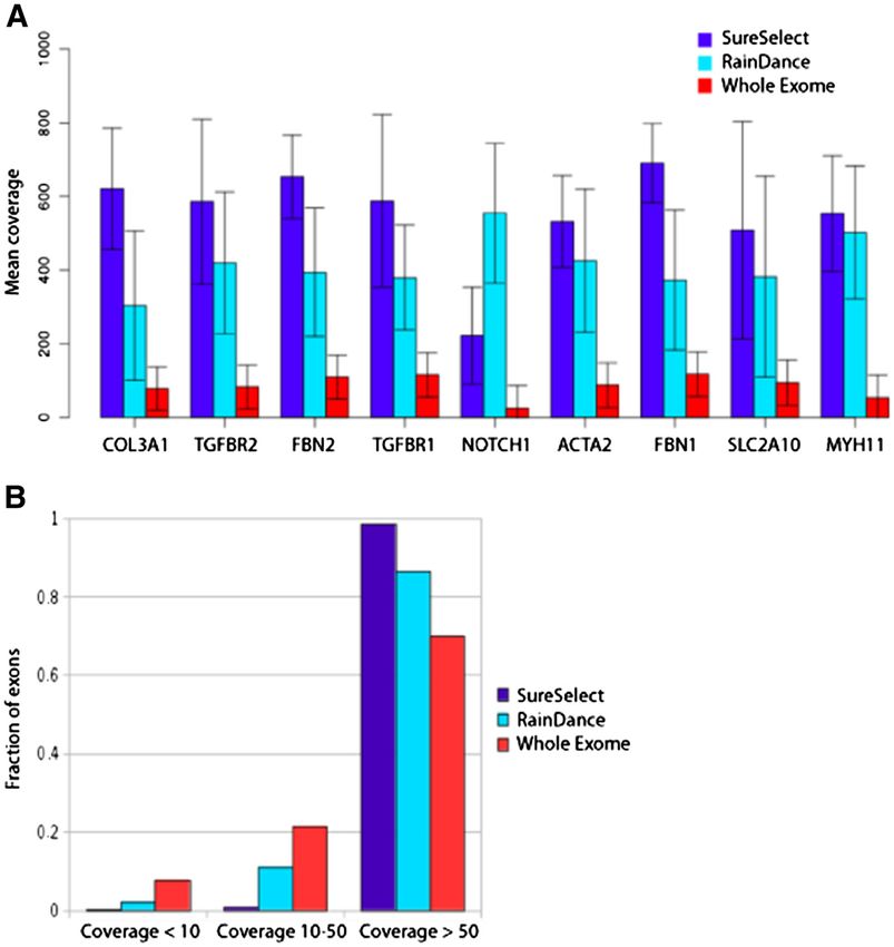
Wooderchak-Donahue et al. BMC Medical Genomics 2012, 5:50                                                     Page 2 of 10
http://www.biomedcentral.com/1755-8794/5/50

[11], have been linked to non-syndromic familial forms          the aortopathy genes was not expected was used as a
of thoracic aortic aneurysms (reviewed in [12]).                negative control. A Coriell sample with an FBN1
   Due to the overlapping phenotypes presented by these         c.1888delAAinsC, p.M717X genotype was also evaluated.
disorders genetic sequencing is often required for accurate       This study was approved by the University of Utah
diagnosis and appropriate clinical intervention. However,       and Primary Children’s Medical Center Institutional
comprehensive sequencing of the many genes involved             Review Boards (IRB#00028740). Written informed con-
is often impractical with traditional Sanger methods. In        sent for participation in the study was obtained from the
contrast, high-throughput “next-generation” sequencing          participants or their parents.
(NGS) has emerged as a new tool in the clinical laboratory
for the rapid, cost-effective detection of mutations in genes   Emulsion PCR enrichment
associated with multigenic disorders [13,14]. NGS assays        A primer library (RainDance Technologies, Boston, MA)
targeted to a panel of genomic regions associated with          was custom-designed to amplify 194 exons and exon/
known pathogenic mutations offer several advantages over        intron boundaries for nine aortopathy genes listed in
traditional sequencing methods, including lower cost and        Table 1 (~0.1 Mb). Primers were designed using Rain-
rapid assessment of many regions.                               Dance’s design parameters and Primer3 (http://primer3.
   While NGS offers a promising alternative to Sanger           sourceforge.net/). The 350 PCR amplicons ranged in size
sequencing, the clinical utility of NGS methods depends         from 201–919 bp and had a guanine cytosine (GC) con-
on the ability to accurately isolate or amplify the genomic     tent of 24-80%. Genomic DNA (1.5-3 μg) was sheared
regions of interest. Ideally, the enrichment will yield high    to 2–4 kb fragments using a Covaris S2 instrument
read depths in the targeted regions while keeping errors        (Covaris, Woburn, MA) and added to a mixture that
introduced through PCR and other sample manipula-               included all the components of the PCR reaction exclud-
tions to a minimum. Several NGS target enrichment               ing the primers. This mixture and the primer library
strategies are currently available, all with various advan-     were loaded separately onto the RDT1000 instrument,
tages depending on the size of the targeted region and          and PCR droplets containing one primer pair per droplet
the genetic targets themselves. Previous comparisons of         were generated. After amplification, emulsion PCR dro-
enrichment techniques have identified considerable differ-      plets were broken releasing the amplicons which were
ences in the depth of coverage of targeted regions as well      then purified and concatenated according to the manu-
as the number of variants identified [15-17].                   facturer’s instructions. Concatenated samples were
   In this work we compare two competing enrichment             sheared to 300 bp fragments, and Illumina adapters were
protocols, an emulsion-PCR based technique from                 added using the SPRI-TE instrument (Beckman-Coulter,
RainDance technologies [18] and a method utilizing              Danvers, MA). Barcode indexes were added using PCR,
solution-based RNA-bait hybrid capture marketed as the          and sample quality and quantity was assessed using a
SureSelect Target Enrichment Platform [19]. Using an            Bioanalyzer (Agilent Technologies, Santa Clara, CA).
aortopathy panel of nine genes (Table 1), we assess the
clinical utility of each enrichment technique. We quan-         Solution-based hybridization capture enrichment
tify the coverage of the targeted regions and compare           Custom RNA baits were designed to specifically target
the accuracy and overlap of the variants identified. To         the exons and exon/intron boundaries of the nine aorto-
determine which molecular approach is best suited for           pathy genes from Table 1 (~0.1 Mb). RNA baits were
routine clinical diagnostics for this disorder, we also         tiled at 5 × spacing and were in replicates of 10 to in-
compare read depths for the aortopathy genes generated          crease hybridization efficiency. Genomic DNA (3 μg)
from the two custom-designed gene panel enrichments             was sheared to 180 bp fragments. Illumina adapters were
to those obtained from whole-exome sequencing.                  added using SureSelect XT kit reagents (Agilent Tech-
                                                                nologies, Santa Clara, CA). Adapter ligated DNA under-
Methods                                                         went hybridization with the biotinylated RNA baits for
Samples                                                         24 hours at 65°C. Hybridized DNA targets of interest
Eight samples were used to directly compare the enrich-         were captured using streptavidin-coated magnetic beads.
ment technologies. Four samples were positive controls          DNA targets of interest were eluted and barcode/
with a known mutation in one of the aortopathy genes            indexed after a series of washes to remove the non-
(Table 2, Samples 1–4). Samples 5 and 6 were from               targeted, unbound genome. DNA quality and quantity
patients with a clinical diagnosis of aortopathy and a          was assessed (Bioanalyzer).
negative molecular analysis of FBN1, TGFβR1, and
TGFβR2 by Sanger sequencing analysis and multiplex              NGS sequencing and data analysis
ligation-dependent probe amplification (MLPA) [20]. A           Concentrations of indexed samples from the enrich-
normal, healthy individual’s DNA in which a mutation in         ments were determined by quantitative-PCR (KAPA
Wooderchak-Donahue et al. BMC Medical Genomics 2012, 5:50                                                                                     Page 3 of 10
http://www.biomedcentral.com/1755-8794/5/50

Table 1 Aortopathy panel genes
Gene name          Chromosomal            Protein                                 Disease                      No. reported         Transcript       Exons
                   locus                                                                                       mutations            size
FBN1               15q21.1                Fibrillin                               Marfan syndrome              601                  8616 bp          66
                                                                                                                   a
TGFΒR1             9q33-q34               Transforming growth factor,             Loeys-Dietz syndrome;        28                   1512 bp          9
                                          beta receptor 1                         Marfan-like syndrome
TGFΒR2             3p22                   Transforming growth factor,             Loeys-Dietz syndrome;        105a                 1779 bp          7
                                          beta receptor 2                         Marfan-like syndrome
COL3A1             2q31                   Collagen type III alpha 1               Ehlers Danlos Type IV        227                  4401 bp          51
MYH11              16p13.13-p13.12        Myosin heavy chain 11                   TAADb-patent ductus          3                    5919 bp          41
                                                                                  arteriosus
ACTA2              10q22-q24              smooth muscle actin, alpha 2            TAAD4 syndrome               19                   1134 bp          9
SLC2A10            20q13.1                solute carrier family 2 (facilitated    Arterial Tortuosity          18                   1626 bp          5
                                          glucose transporter), member 10         syndrome
NOTCH1             9q34.3                 Notch homolog 1,                        BAV-TAAD syndrome            11                   7668 bp          34
                                          translocation-associated
FBN2               5q23-q31               Fibrillin 2                             BAV-TAAD syndrome            45                   8739 bp          71
a
 Of the 28 patients with a TGFΒR1 mutation, 14 had Loeys-Dietz syndrome, 4 had thoracic aortic aneurysms, and 4 had Marfan-like syndrome. Of the 105 TGFΒR2
mutations, 32 had Loeys-Dietz syndrome, 31 had Marfan-like syndrome, and 8 had thoracic aortic aneurysms.
b
 Thoracic aortic aneurysms and dissections (TAAD); Bicuspid-Aortic Valve (BAV).

Biosystems, Woburn, MA). Samples were pooled and                                 Results
sequenced on a HiSeq2000 instrument (Illumina, San                               Sample enrichment
Diego, CA) using 2 × 100 paired-end sequencing.                                  Six of eight samples underwent successful enrichment
Sequences were aligned to the human genome reference                             using both RainDance emulsion PCR and SureSelect
(hg19) sequence using the Burrows-Wheeler Alignment                              hybridization capture (Table 2). Sample 7 (from Coriell)
tool (BWA 0.5.9) [21] with default parameters. PCR dupli-                        failed during the RDT1000 merge when the DNA dro-
cates were removed using the Samtools package [22], and                          plets failed to merge properly with the primer library
base quality score recalibration, local realignment, and                         droplets. Hybridization capture of this sample was also
variant calling were performed with the Genome Analysis                          poor and analysis was not completed due to sub-optimal
Toolkit (GaTK v1.3) [23]. Variants with coverage less than                       DNA quality (A260/A280 was not between 1.8 and 2.0).
6, with 'quality-by-depth' scores of less than two, with                         Sample 8 failed during hybridization capture due to a
variant allele frequencies of less than 0.15, or with overall                    pipetting error. Herein, the accuracy of the two enrich-
quality scores of less than 10 were discarded.                                   ment methods, depth of coverage, and variant calling
                                                                                 were compared among the six samples enriched using
Sanger sequencing                                                                both techniques.
Select variants were Sanger sequenced. Primers
sequences are available upon request. Amplicons were                             Accuracy
bi-directly sequenced using the Big DyeW Terminator                              Pathogenic mutations from four positive control samples
v3.1 cycle sequencing kit and an ABI 3730 DNA                                    were detected using both enrichment methods (Table 2,
Analyzer (Life Technologies, Carlsbad, CA). Sequences                            samples 1–4). For example, a heterozygous FBN1
were compared to reference sequences using Mutation                              c.1585C > T, p.R529X mutation was detected in sample 1
Surveyor (SoftGenetics, State College, PA).                                      using both enrichment methods and was confirmed
                                                                                 using Sanger sequencing (Additional file 1: Figure S1).
Exome sequencing
In addition to the custom targeted enrichments, we also                          Depth of coverage
examined depth of coverage for the targeted regions                              Omitting failed samples, both methods generated very
obtained through exome sequencing. Briefly, ten samples                          high coverage in the targeted regions. Pooling data from
from individuals without known aortopathies were                                 all samples and examining coverage on a gene-by-gene
sequenced on the Illumina HiSeq2000 platform after                               basis, the SureSelect enriched material yielded slightly
undergoing exome capture enrichment (SureSelect                                  higher, but also more variable, coverage (per-gene aver-
50Mb exome kit, Agilent Technologies, Santa Clara,                               age 471.7, range 189.9-592.0) compared to the emulsion
CA). Exome samples were indexed and pooled, two per                              PCR technique (per-gene average 414.8, range 303.4-
lane, using a one-to-one ratio, and were sequenced using                         554.4). Both methods yielded significantly higher aver-
2x100-base pair paired end reads.                                                age coverage per gene than whole-exome sequencing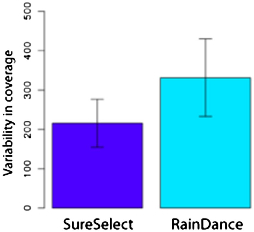
http://www.biomedcentral.com/1755-8794/5/50
Wooderchak-Donahue et al. BMC Medical Genomics 2012, 5:50
Table 2 NGS accuracy sample results
# Sample Mutations Coverage RDa Coverage SSa Mean coverage Other information
(% mutant allele) (% mutant allele)
1 FBN1 Positive c.1585C>T, p.R529X 101x (47%) 576x (60%) 1477x (RD) 508x (SS) In COL3A1, c.198A>G, p.I66M,
a novel change.
2 TGFBR1 Positive c.799A>G, p.N267D 317x (51%) 504x (58%) 462x (RD) 563x (SS)
3 TGFBR2 Positive c.1583G>A, p.R528H 183x (48%) 257x (50%) 177x (RD) 499x (SS)
4 MYH11 Positive IVS32+1C>A (splice) 71x (58%) 80x (50%) 140x (RD) 164x (SS)
5 Symptomatic unknown No mutations found. NA NA 33x (RD) 260x (SS) In SLC2A10 p.A206T (rs#2235491)
and p.A385G (rs#79849424).
In NOTCH1, IVS16-4C>CT (rs#3125001).
6 Symptomatic unknown COL3A1 IVS9-7T>C novel change; 48x (56%) 416x (50%) 86x (RD) 355x (SS) In COL3A1, p.A698T (rs#1800255).
potential splice site variant In FBN2, p.V965I (rs#154001).
7 FBN1 Coriell positive Unable to confirm due to Failed merge between Poor data due to NA
sample quality. DNA and primer library low quality DNA
droplets
8 Healthy Control No mutations found NA Failed due pipetting error 340x (RD)
a
RainDance (RD) and SureSelect (SS). All variants reported in this table are heterozygous as confirmed by Sanger sequencing (data not shown). NA, not applicable.
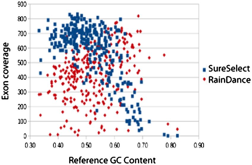
Page 4 of 10Wooderchak-Donahue et al. BMC Medical Genomics 2012, 5:50 Page 5 of 10 http://www.biomedcentral.com/1755-8794/5/50 (Figure 1A) and had substantially fewer exons with less the GC content of the surrounding sequence (Figure 2). than tenfold coverage (Figure 1B). SureSelect results For exons containing greater than 75% GC (for instance, were influenced by particularly low coverage for the exon 1 in the NOTCH1 and TGFβR1 genes) no coverage NOTCH1 gene (mean 189x), a feature not observed with was observed. the sequences generated by emulsion PCR. Low cover- In contrast to the strong correlation of GC content age may be related to the GC content of the NOTCH1 with read depth for the hybridization capture enrich- gene, in which 17 of 34 exons are > 65% GC. ment, exon coverage varied in an unpredictable manner Examining exons individuals, the SureSelect enrich- for the emulsion PCR enrichment method (Figure 3). To ment yielded fewer total exons with coverage below 10 assess the degree of coverage variability across samples, when compared to both RainDance and the whole we computed the standard deviation in coverage for exome capture (Figure 1B). Some 98.5% of all exons had each exon across samples for both custom-designed mean depth greater than 50 for SureSelect, as compared panel enrichment techniques. The mean of these devia- to 85.5% for RainDance and 70% for whole exome. tions across exons reflects the overall degree of coverage Despite relatively high read depths across most exons, variability across all samples, with zero indicating that several exons consistently had little or no coverage each exon was covered by the same number of reads in among the samples. Within the SureSelect-enchriched every sample. Between sample variability was signifi- sequences, much of the variation appears to be related to cantly higher for the emulsion PCR enrichment, with a Figure 1 Mean depth of coverage per aortopathies gene for the enrichment methods. In A, both custom-designed gene panels (SureSelect and RainDance) yielded significantly higher average coverage per gene compared to whole-exome sequencing (SureSelect 50 Mb All Exon Capture). Bars indicate one standard deviation. In B, the fraction of exons covered to the given depth for the enrichment methods is shown. The custom-designed SureSelect gene panel yielded greater average read depths across the 294 exons for all samples versus the RainDance or whole-exome enrichments.
Wooderchak-Donahue et al. BMC Medical Genomics 2012, 5:50                                                                         Page 6 of 10
http://www.biomedcentral.com/1755-8794/5/50

 Figure 2 Mean exon read depth as a function of reference sequence GC content for the custom-designed gene panels. High GC content
 resulted in decreased exon coverage in the SureSelect enriched samples. GC content had little to no effect on exon coverage of the RainDance
 enriched samples, which were more prone to sporadic amplicon failure.

mean of 330.4, compared to 215.8 for the hybridization                   (Additional file 1: Table S1). The sequences generated
enrichment panel (paired t-test, p < 2.2 × 10⁻¹⁶, Figure 4).             by the RainDance enrichment contained several more
                                                                         variants, 221 in total, including 26 not identified in
Variants analysis                                                        the SureSelect-enriched sequences. In contrast, the
In addition to the number of reads mapping to targeted                   SureSelect protocol yielded 214 total variants, of which
locations, we also assessed the quantity and character of                19 were not found using the emulsion PCR tech-
the variants identified by both enrichment methods. Be-                  nique. Among the variants common to both methods,
cause both methods targeted overlapping, but partially                   55 (28.2%) were in dbSNP (v. 132), while 2 of the 17 var-
distinct regions, we considered only those variants in the               iants (12%) unique to SureSelect were in dbSNP, and
intersection of the two capture regions. The intersection                3 of the 23 variants (13%) unique to emulsion PCR were
of the two capture regions was 60.58 Kb in size, com-                    in dbSNP. Assuming variants found in dbSNP are not
prising 64% of the bases targeted by SureSelect and 99%                  false positives in our samples, this suggests that even the
of bases in the RainDance capture.                                       relatively high coverage obtained by both enrichment
  In total, 252 unique variants were identified across all               methods did not guarantee accurate variant detection, as
samples, 195 of which were identified by both methods                    both the custom targeted enrichment methods identified

 Figure 3 Number of exons with mean read depth less than 20, across samples (x-axis) and genes (y-axis). The color of the block
 represents the number of exons with low coverage for a given sample and gene.
Wooderchak-Donahue et al. BMC Medical Genomics 2012, 5:50 Page 7 of 10
http://www.biomedcentral.com/1755-8794/5/50
mutation by Sanger sequencing and MLPA [20] were
evaluated using the aortopathy panel. Sample 5 was from
a 19-year-old female with borderline aortic dilatation,
mild tricuspid valve prolapse and mild mitral valve insuf-
ficiency, height of 178 cm (5’10”) (99th percentile), arm
span of 176 cm (5’9”) severe pectus carinatum, scoliosis,
arachnodactyly, joint mobility, positive wrist and thumb
signs, and skin striae. Analysis of the coding region
variants and splice site variants from both enrichment
methods revealed two nonsynonymous heterozygous
variants in SLC2A10 (c.616G > A, p.Ala206Thr and
c.1154C > G, p. Ala385Gly) and a NOTCH1 potential
splice site variant (IVS16-4C > T). These variants were
present in dbSNP and are likely not disease causing.
Sample 6 was from a 34-year-old female with mild aortic
dilatation in the setting of past aortic dissection and un-
known family history. Two heterozygous nonsynonymous
Figure 4 Variability in coverage: RainDance versus SureSelect.
variants, one in COL3A1 (c.2092G > A, p.Ala698Thr) and
Standard deviation in coverage for each exon across enrichment the other in FBN2 (c.2893G > A, p.Val965Ile) were both
samples was computed. The mean deviation across exons reflects present in dbSNP, rs1800255 and rs154001 at 19.8%
the overall degree of coverage variability across samples 1–6, with and 72.4% frequencies in 1000 genomes (November 2010
zero indicating that each exon was covered by the same number of release, 1000genomes.org), respectively. A novel heterozy-
reads in every sample. Between sample variability was significantly
higher for the RainDance enrichment.
gous COL3A1 variant (IVS9-7T > C) not found in dbSNP
or the COL3A1 locus specific mutation database was also
identified. Splice site prediction programs were run to
several variants not recognized by the other method. determine if the change altered splicing [24], but they pre-
Similarly discordant results have been found in other dicted that no change in splicing would occur. It is likely
recent studies [13]. that this variant is also not disease causing.
Examination of the discordant variants suggested sev- No synonymous splice site variants were identified in
eral sources for the disagreement. First, discordant var- either patient. It is possible that these two patients have
iants appeared in the intronic and untranslated areas at a large deletion or duplication of one of the nine aorto-
the periphery of individual capture regions. Ten SureSe- pathy genes, but this has yet to be evaluated. Currently,
lect and fifteen RainDance discordant variants were detection of structural variation cannot be reliably
located in non-exonic regions. Examination of read detected from NGS data alone. Patient samples that test
depth in these areas suggested that coverage was rela- negative from the NGS assay should be run on an exonic
tively low compared to the mean for the whole exon, im- level comparative genomic hybridization (CGH) array
plicating variation in read depth as a potential source of assay used to evaluate large copy number changes in the
discordant or false positive calls. same gene set. Until NGS data analysis programs im-
A second source of discordant variation was the prove in their ability and accuracy to detect structural
NOTCH1 gene. A relatively high fraction of the discord- variation, CGH array assays are the suggested approach
ant variants appeared in the exons of NOTCH1 (15 of for detecting such large deletions or duplications in multi-
45, or 33%), and these appeared with similar frequencies gene panels. Alternatively, another gene not included in
in both the RainDance and SureSelect samples (8 from the aortopathy panel may be responsible for the disorder
RainDance, 7 from SureSelect). NOTCH1 is likely to be in these patients.
problematic for two reasons. First, the high GC content
of NOTCH1 reduces the read depths obtained with the Discussion
SureSelect enrichment procedure. Second, NOTCH1 Enrichment performance
bears substantial sequence homology to other NOTCH All targeted enrichment methods evaluated yielded very
genes, potentially confounding alignment algorithms and high read depths in the aortopathies targeted regions,
generating spurious variant calls. with all genes except for NOTCH1 from the exome
capture obtaining a mean depth of coverage of over 50 ×
Clinical utility of the aortopathy panel (Figure 1). Overall, the custom-designed hybridization
Two clinically affected individuals who had previously capture enrichment yielded lower coverage in regions
tested negative for a FBN1, TGFBR1, and TGFBR2 of high GC content (Figure 2), but consistently highWooderchak-Donahue et al. BMC Medical Genomics 2012, 5:50 Page 8 of 10
http://www.biomedcentral.com/1755-8794/5/50
coverage in regions of low GC bias. The emulsion PCR high enough depth of coverage to accurately call variants
method, in contrast, yielded exons with very low cover- is much higher than for the targeted panel. Samples can
age that had no obvious correlate (Figure 2). Although also be indexed and pooled prior to hybridization capture
influenced by GC content, the hybridization capture [25], reducing the overall cost per sample even further.
nonetheless yielded fewer total exons with low coverage Despite the lower cost, the hybridization capture
than did the emulsion PCR technique (Figure 1B). Over- protocol is more time consuming, complex, and labor
all, emulsion PCR yielded significantly higher between- intensive when compared to the emulsion PCR tech-
sample variability in coverage. In a clinical setting, a high nique. However, the ability to automate this process in
degree of between sample consistency is desirable. Low the clinical laboratory alleviates these challenges and
coverage exons will likely require follow-up Sanger yields more consistent, reliable results [26]. Currently,
sequencing. Knowing which exons will require such the emulsion PCR instrument processes one sample/hour,
treatment beforehand may ameliorate the cost and add- limiting the total number of samples processed in a day
itional time resulting from designing new primer sets on to approximately twenty-four. With the increasing inter-
an as-needed basis. est in exome sequencing, automation of hybridization
Despite satisfactory coverage, the methods yielded only capture enrichment protocols have become readily avail-
partially overlapping sets of variants, with some 10% of able [26], and up to 96 different custom-designed NGS
variants identified by one method but not by the other panels and exome hybridizations can take place at the
(Additional file 1: Table S1). These inconsistencies may same time during the same run. Solution-based hybridi-
be related to variability in read depth as well as align- zation libraries can be ordered in larger volumes, ali-
ment ambiguity related to sequence homology. Similarly quoted for up to two-three freeze/thaw cycles, and stored
high discordancy rates were found in a recent study for up to one year. RainDance primer libraries have to be
comparing the RainDance and Fluidigm enrichment switched out after the eighth sample with the RDT1000
platforms [13], in which only 42% of variants identified instrument design, and if there are not eight samples to
were common to both methods, and 67% of the discord- run using the same primer library, the rest will be wasted.
ant variants were found in regions of less than 20-fold
coverage. Until procedures are devised that substantially Conclusions
decrease the number of regions with low coverage, The aortopathy panel offers a cost-effective, faster mo-
Sanger confirmation of suspected variants will likely be lecular diagnostic assay compared to the conventional
necessary prior to clinical action. gene-by-gene Sanger sequencing approach. Both Rain-
A high sample failure rate of 25% (2 of 8 samples) Dance and SureSelect enrichment methods accurately
was observed due to sample quality (sample 7, Rain- identified variants in the positive samples assayed, and
Dance and SureSelect) and a pipetting error (sample 8, overall coverage was high across most exons for all sam-
SureSelect) (Table 2). Based on our results, only DNA ples. Hybrid capture results demonstrated that genes with
samples of high quality (A260/280 is between 1.8 and 2.0) high homology to other genes and high GC rich regions
should be evaluated using NGS enrichment. If a sample can be problematic. However, we note that increases in
falls below this initial quality measurement, DNA should read length, currently available with the Ion Torrent or
be extracted again and/or the sample should be purified Illumina MiSeq instruments, are likely to alleviate
until an acceptable quality is obtained. For complex homology-related alignment issues. After comparing the
enrichment protocols such as the SureSelect method, cost, design flexibility, and versatility of the workflows of
pipetting errors can be avoided by incorporating auto- the two enrichment methods, the custom-designed
mation into sample preparation (discussed further hybridization capture design offers the best solution for
below). This is especially important as the number of enriching the aortopathy genes in a clinical laboratory set-
clinical samples increase. ting. Patients with Marfan syndrome and Marfan-like syn-
dromes featuring aortopathies will benefit from the timely
NGS enrichment in the clinical laboratory molecular diagnosis of this new testing approach which
When compared to emulsion PCR, the solution-based will lead to the appropriate surveillance and interventions
hybridization technique yielded higher coverage as well aimed at preventing the significant morbidity and mortal-
as greater predictability in performance. These attri- ity associated with these conditions.
butes, when combined with lower cost, strongly favor
the hybridization capture for enrichment of the aortopa- Additional file
thy panel in the clinical setting. Emulsion PCR enrich-
ment is significantly more expensive and requires the Additional file 1: Figure S1. NGS and Sanger sequencing confirm the
use of a costly instrument. Exome sequencing is also pathogenic FBN1 mutation in sample 1. A heterozygous nonsense
mutation (c.1585C>T, p.R529X) was detected using RainDance PCR
more costly because the cost to sequence the exome at aWooderchak-Donahue et al. BMC Medical Genomics 2012, 5:50 Page 9 of 10
http://www.biomedcentral.com/1755-8794/5/50
enrichment (panel A) and SureSelect capture enrichment (panel B). In C, 9. Guo DC, Pannu H, Tran-Fadulu V, Papke CL, Yu RK, Avidan N, Bourgeois S,
Sanger sequencing confirmed the mutation detected in both enrichment Estrera AL, Safi HJ, Sparks E, Amor D, Ades L, McConnell V, Willoughby CE,
strategies. The FBN1 gene is on the reverse strand, and appears in the 3’ Abuelo D, Willing M, Lewis RA, Kim DH, Scherer S, Tung PP, Ahn C, Buja LM,
to 5’ orientation in the NGS traces versus the Sanger sequencing trace Raman CS, Shete SS, Milewicz DM: Mutations in smooth muscle α-actin
which is 5’ to 3’. Table S1. Summary of variants detected from the (ACTA2) lead to thoracic aortic aneurysms and dissections. Nat Genet
RainDance and SureSelect enrichments. 2007, 39:1488–1493.
10. Coucke PJ, Willaert A, Wessels MW, Callewaert B, Zoppi N, De Backer J,
Fox JE, Mancini GM, Kambouris M, Gardella R, Facchetti F, Willems PJ,
Competing interests Forsyth R, Dietz HC, Barlati S, Colombi M, Loeys B, De Paepe A: Mutations
We have no financial, nonfinancial, or competing interests to declare. in the facilitative glucose transporter GLUT10 alter angiogenesis and
cause arterial tortuosity syndrome. Nat Genet 2006, 38:452–457.
Authors’ contributions 11. Garg V, Muth AN, Ransom JF, Schluterman MK, Barnes R, King IN, Grossfeld
WLWD participated in the design of the study, performed all experiments, PD, Srivastava D: Mutations in NOTCH1 cause aortic valve disease.
assisted in data analysis, and wrote the manuscript; BO analyzed the data Nature 2005, 437:270–274.
and wrote the manuscript; LVF participated in the design of the study and 12. El-Hamamsy I, Yacoub MH: Cellular and molecular mechanisms of
wrote the manuscript; JDD and PGR assisted in data analysis; PP assisted thoracic aortic aneurysms. Nat Rev Cardiol 2009, 6:771–786.
with experiments and data analysis; AFR and ATY were responsible for 13. Jones MA, Bhide S, Chin E, Ng BG, Rhodenizer D, Zhang VW, Sun JJ,
diagnosis and management of patients and participated in the design of the Tanner A, Freeze HH, Hegde MR: Targeted polymerase chain
study; PBT conceived the study, and participated in its design and reaction-based enrichment and next generation sequencing for
coordination. All authors read and approved the final manuscript. diagnostic testing of congenital disorders of glycosylation. Genet Med
2011, 13:921–932.
Acknowledgments 14. Berg JS, Evans JP, Leigh MW, Omran H, Bizon C, Mane K, Knowles MR, Weck
We thank Lin Pham, Ph.D. for her assistance in the design of the RainDance KE, Zariwala MA: Next generation massively parallel sequencing of
library. We thank Owen Hardy, Maria Celeste-Ramirez, and Christopher targeted exomes to identify genetic mutations in primary ciliary
Hopkins, Ph.D. for providing assistance in the design of the SureSelect library. dyskinesia: implications for application to clinical testing. Genet Med
This work was funded by the ARUP Institute for Clinical and Experimental 2011, 13:218–229.
Pathology. 15. Teer JK, Bonnycastle LL, Chines PS, Hansen NF, Aoyama N, Swift AJ,
Abaan HO, Albert TJ, Comparative Sequencing Program NISC, Margulies EH,
Author details Green ED, Collins FS, Mullikin JC, Biesecker LG: Systematic comparison of
1
ARUP Institute for Clinical and Experimental Pathology, Salt Lake City, USA. three genomic enrichment methods for massively parallel DNA
2
Department of Pathology, University of Utah, Salt Lake City, USA. sequencing. Genome Res 2010, 20:1420–1431.
3
Department of Pediatrics, Division of Medical Genetics, University of Utah, 16. Kiialainen A, Karlberg O, Ahlford A, Sigurdsson S, Lindblad-Toh K,
Salt Lake City, USA. 4Department of Pediatrics, Division of Cardiology, Syvänen AC: Performance of microarray and liquid based capture
University of Utah, Salt Lake City, USA. 5Molecular Genetics Department, methods for target enrichment for massively parallel sequencing and
ARUP Institute for Clinical and Experimental Pathology, 500 Chipeta Way, Salt SNP discovery. PLoS One 2011, 6:e16486.
Lake City, UT 84108, USA. 17. Hedges DJ, Guettouche T, Yang S, Bademci G, Diaz A, Andersen A,
Hulme WF, Linker S, Mehta A, Edwards YJ, Beecham GW, Martin ER,
Received: 18 July 2012 Accepted: 9 October 2012 Pericak-Vance MA, Zuchner S, Vance JM, Gilbert JR: Comparison of three
Published: 14 November 2012 targeted enrichment strategies on the SOLiD sequencing platform.
PLoS One 2011, 6:e18595.
References 18. Tewhey R, Warner JB, Nakano M, Libby B, Medkova M, David PH,
1. Klein DG: Thoracic aortic aneurysms. J Cardiovasc Nurs 2005, 4:245–250. Kotsopoulos SK, Samuels ML, Hutchison JB, Larson JW, Topol EJ, Weiner MP,
2. Verloes A, Sakalihasan N, Koulischer L, Limet R: Aneurysms of the Harismendy O, Olson J, Link DR, Frazer KA: Microdroplet-based PCR
abdominal aorta: familial and genetic aspects in three hundred thirteen enrichment for large-scale targeted sequencing. Nat Biotechnol 2009,
pedigrees. J Vasc Surg 1995, 21:646–655. 27:1025–1031.
3. Law C, Bunyan D, Castle B, Day L, Simpson I, Westwood G, Keeton B: 19. Gnirke A, Melnikov A, Maguire J, Rogov P, LeProust EM, Brockman W,
Heterozygous TGFΒR2 mutations in Marfan syndrome. Nat Genet 2004, Fennell T, Giannoukos G, Fisher S, Russ C, Gabriel S, Jaffe DB, Lander ES,
36:855–860. Nusbaum C: Solution hybrid selection with ultra-long oligonucleotides
4. Loeys BL, Chen J, Neptune ER, Judge DP, Podowski M, Holm T, Meyers J, for massively parallel targeted sequencing. Nat Biotechnol 2009,
Leitch CC, Katsanis N, Sharifi N, Xu FL, Myers LA, Spevak PJ, Cameron DE, 27:182–189.
De Backer J, Hellemans J, Chen Y, Davis EC, Webb CL, Kress W, Coucke P, 20. Furtado LV, Wooderchak-Donahue W, Rope AF, Yetman AT, Lewis T, Plant P,
Rifkin DB, De Paepe AM, Dietz HC: A syndrome of altered cardiovascular, Bayrak-Toydemir P: Characterization of large genomic deletions in the
craniofacial, neurocognitive and skeletal development caused by FBN1 gene using multiplex ligation-dependent probe amplification.
mutations in TGFΒR1 or TGFΒR2. Nat Genet 2005, 37:275–281. BMC Med Genet 2011, 12:119.
5. Pannu H, Fadulu VT, Chang J, Lafont A, Hasham SN, Sparks E, Giampietro PF, 21. Li H, Durbin R: Fast and accurate short read alignment with
Zaleski C, Estrera AL, Safi HJ, Shete S, Willing MC, Raman CS, Milewicz DM: Burrows-Wheeler transform. Bioinformatics 2009, 25:1754–1760.
Mutations in transforming growth factor-β receptor type II cause familial 22. Li H, Ruan J, Durbin R: Mapping short DNA sequencing reads and
thoracic aortic aneurysms and dissections. Circulation 2005, 112:513–520. calling variants using mapping quality scores. Genome Res 2008,
6. Gupta PA, Wallis DD, Chin TO, Northrup H, Tran-Fadulu VT, Towbin JA, 18:1851–1858.
Milewicz DM: FBN2 mutation associated with manifestations of Marfan 23. McKenna A, Hanna M, Banks E, Sivachenko A, Cibulskis K, Kernytsky A,
syndrome and congenital contractural arachnodactyly. J Med Genet 2004, Garimella K, Altshuler D, Gabriel S, Daly M, DePristo MA: The Genome
41:e56. Analysis Toolkit: a MapReduce framework for analyzing next-generation
7. Zhu L, Vranckx R, Khau Van Kien P, Lalande A, Boisset N, Mathieu F, DNA sequencing data. Genome Res 2010, 20:1297–1303.
Wegman M, Glancy L, Gasc JM, Brunotte F, Bruneval P, Wolf JE, Michel JB, 24. Cartegni L, Wang J, Zhu Z, Zhang MQ, Krainer AR: ESEfinder: a web
Jeunemaitre X: Mutations in myosin heavy chain 11 cause a syndrome resource to identify exonic splicing enhancers. Nucelic Acids Res 2003,
associating thoracic aortic aneurysm/aortic dissection and patent ductus 31:3568–3571.
arteriosus. Nat Genet 2006, 38:343–39. 25. Bell CJ, Dinwiddie DL, Miller NA, Hateley SL, Ganusova EE, Mudge J,
8. Khau Van Kien P, Mathieu F, Zhu L, Lalande A, Betard C, Lathrop M, Langley RJ, Zhang L, Lee CC, Schilkey FD, Sheth V, Woodward JE,
Brunotte F, Wolf JE, Jeunemaitre X: Mapping of familial thoracic aortic Peckham HE, Schroth GP, Kim RW, Kingsmore SF: Carrier testing for severe
aneurysm/dissection with patent ductus arteriosus to 16p12.2–p13.13. childhood recessive diseases by next generation sequencing. Sci Transl
Circulation 2005, 112:200–206. Med 2011, 3:65ra4.Wooderchak-Donahue et al. BMC Medical Genomics 2012, 5:50 Page 10 of 10
http://www.biomedcentral.com/1755-8794/5/50
26. Fisher S, Barry A, Abreu J, Minie B, Nolan J, Delorey TM, Young G, Fennell TJ,
Allen A, Ambrogio L, Berlin AM, Blumenstiel B, Cibulskis K, Friedrich D,
Johnson R, Juhn F, Reilly B, Shammas R, Stalker J, Sykes SM, Thompson J,
Walsh J, Zimmer A, Zwirko Z, Gabriel S, Nicol R, Nusbaum C: A scalable,
fully automated process for construction of sequence-ready human
exome targeted capture libraries. Genome Biol 2011, 12:R1.
doi:10.1186/1755-8794-5-50
Cite this article as: Wooderchak-Donahue et al.: A direct comparison of
next generation sequencing enrichment methods using an aortopathy
gene panel- clinical diagnostics perspective. BMC Medical Genomics 2012
5:50.
Submit your next manuscript to BioMed Central
and take full advantage of:
• Convenient online submission
• Thorough peer review
• No space constraints or color figure charges
• Immediate publication on acceptance
• Inclusion in PubMed, CAS, Scopus and Google Scholar
• Research which is freely available for redistribution
Submit your manuscript at
www.biomedcentral.com/submitYou can also read