Virological Characterization of the First 2 COVID-19 Patients Diagnosed in Italy: Phylogenetic Analysis, Virus Shedding Profile From Different ...
←
→
Page content transcription
If your browser does not render page correctly, please read the page content below
Open Forum Infectious Diseases
   MAJOR ARTICLE

Virological Characterization of the First 2 COVID-19
Patients Diagnosed in Italy: Phylogenetic Analysis, Virus
Shedding Profile From Different Body Sites, and Antibody
Response Kinetics
Francesca Colavita,a Daniele Lapa,a Fabrizio Carletti, Eleonora Lalle, Francesco Messina, Martina Rueca, Giulia Matusali, Silvia Meschi, Licia Bordi,
Patrizia Marsella, Emanuele Nicastri, Luisa Marchioni, Andrea Mariano, Laura Scorzolini, Tommaso Ascoli Bartoli, Antonino Di Caro, Giuseppe Ippolito,

                                                                                                                                                                            Downloaded from https://academic.oup.com/ofid/article/7/10/ofaa403/5900725 by guest on 08 December 2020
Maria Rosaria Capobianchi, and Concetta Castilletti; on behalf of the INMI COVID-19 Laboratory Team and INMI COVID-19 Study Group
National Institute for Infectious Diseases “L. Spallanzani” IRCCS, Rome, Italy

   Background. The pathogenesis of severe acute respiratory syndrome coronavirus 2 (SARS-CoV-2) infection remains unclear.
We report the detection of viral RNA from different anatomical districts and the antibody profile in the first 2 COVID-19 cases diag-
nosed in Italy.
   Methods. We tested for SARS-CoV-2 RNA clinical samples, either respiratory and nonrespiratory (ie, saliva, serum, urine,
vomit, rectal, ocular, cutaneous, and cervico-vaginal swabs), longitudinally collected from both patients throughout the hospitaliza-
tion. Serological analysis was carried out on serial serum samples to evaluate IgM, IgA, IgG, and neutralizing antibody levels.
   Results. SARS-CoV-2 RNA was detected since the early phase of illness, lasting over 2 weeks in both upper and lower respiratory
tract samples. Virus isolate was obtained from acute respiratory samples, while no infectious virus was rescued from late respiratory
samples with low viral RNA load, collected when serum antibodies had been developed. Several other specimens came back positive,
including saliva, vomit, rectal, cutaneous, cervico-vaginal, and ocular swabs. IgM, IgA, and IgG were detected within the first week
of diagnosis, with IgG appearing earlier and at higher titers. Neutralizing antibodies developed during the second week, reaching
high titers 32 days after diagnosis.
   Conclusions. Our longitudinal analysis showed that SARS-CoV-2 RNA can be detected in different body samples, which may
be associated with broad tropism and different spectra of clinical manifestations and modes of transmission. Profiling antibody re-
sponse and neutralizing activity can assist in laboratory diagnosis and surveillance actions.
   Keywords. antibody response; COVID-19; Italy; phylogenesis; SARS-CoV-2; viral culture; virus shedding.

In January 2020, a novel coronavirus was identified as the cause                                      illness caused by SARS-CoV-2), with 787 773 deaths have been
of pneumonia cases, with the first cases reported in December                                         reported worldwide [3].
2019 in Wuhan City, Hubei Province of China [1, 2]. The new                                              Transmission is mainly through respiratory droplets, but
pathogen belongs to betacoronavirus genus lineage B, and due                                          other routes cannot be excluded and are under investigation,
to its close phylogenetic relation to other bat severe acute res-                                     as SARS-CoV-2 was detected in several body fluids (ie, saliva,
piratory syndrome (SARS)–like coronaviruses, it was named                                             stool, ocular fluid) [1, 4–7]. Much still needs to be learned about
SARS coronavirus 2 (SARS-CoV-2). Initially linked to possible                                         this infection, and research is underway worldwide to better
exposure to infected wildlife, human-to-human transmission                                            understand the clinical features and extent of interhuman trans-
was identified, and the outbreak rapidly spread to other parts                                        mission. A better knowledge of viral RNA shedding kinetics
of China and outside the country [2]. As of August 20, 2020,                                          from different body districts could help us understand SARS-
22 431 929 coronavirus disease 2019 (COVID-19) cases (the                                             CoV-2 transmission and pathogenesis, supporting surveillance
                                                                                                      and clinical management. In addition, due to the current emer-
                                                                                                      gency context, very few data about the antibody response are
  Received 27 April 2020; editorial decision 26 August 2020; accepted 28 August 2020.
  a
   Equal contribution
                                                                                                      available in the literature.
  Correspondence: Maria Rosaria Capobianchi, PhD, Via Portuense 292, 00149 Rome (maria.                  Here, we report the kinetics of viral RNA shedding from dif-
capobianchi@inmi.it).
                                                                                                      ferent body sites and the concomitant antibody profile (IgM,
Open Forum Infectious Diseases®
© The Author(s) 2020. Published by Oxford University Press on behalf of Infectious Diseases Society   IgA, IgG, and neutralizing Ig) along the disease course in the
of America. This is an Open Access article distributed under the terms of the Creative Commons        first 2 COVID-19 confirmed cases reported in Italy and hospi-
Attribution License (http://creativecommons.org/licenses/by/4.0/), which permits unrestricted
reuse, distribution, and reproduction in any medium, provided the original work is properly cited.
                                                                                                      talized at the National Institute for Infectious Diseases “Lazzaro
DOI: 10.1093/ofid/ofaa403                                                                             Spallanzani” (INMI) in Rome.

                                                                                                                         SARS-CoV-2 Shedding and Antibodies • ofid • 1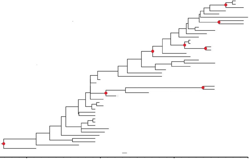
METHODS                                                            Capobianchi et al. [9]. Reads were de novo assembled, and con-
Clinical Samples
                                                                   sensus sequences were manually controlled and confirmed by
Clinical samples from the first 2 COVID-19 patients were lon-      Sanger sequencing.
gitudinally collected for diagnostic purposes starting from           The Bayesian phylogenetic tree was inferred using the
the first day of hospitalization (corresponding to day 1 from      Markov chain Monte Carlo (MCMC) approach in BEAST,
symptom onset [DSO], as declared by the patients at admis-         version 1.10.4, with BEAGLE, version 2.1.2. To infer time-
sion) up to DSO 32. These samples included upper (URT; ie,         measured phylogenic analysis, the mutation model Hasegawa
nasopharyngeal swab, nasal swab, throat swab) and lower (LRT;      Kishino Yano (HKY) was used, which assumes the nucleotides
ie, sputum and bronchoalveolar lavage [BAL]) respiratory tract     have different frequencies into genome and, transitions and
specimens and nonrespiratory specimens (ie, saliva, serum,         transversions occur at different rates [10.1007 / BF02101694].
urine, rectal swab, ocular swab, cervico-vaginal swab, cuta-       Moreover, constant population size and strict clock model
neous swab).                                                       over time were imposed as coalescent priors for independent
                                                                   Monte Carlo Marchov Chain (MCMC) runs, as reported in pre-

                                                                                                                                        Downloaded from https://academic.oup.com/ofid/article/7/10/ofaa403/5900725 by guest on 08 December 2020
Patient Consent Statement                                          vious study [10]. Chains were conducted for at least 100 × 106
The patients’ written consent was obtained. This study was ap-     generations with sampling every 10 000 steps and burn-in for
proved by the INMI Ethical Board.                                  10 × 106 generations. The convergence of the MCMC was as-
                                                                   sessed by calculating for each parameter the Effective Sample
Nucleic Acid Extraction and Molecular Tests                        Size (ESS) (accepted if the ESS > 250). A maximum clade cred-
Viral RNA was extracted by QIAsymphony (QIAgen,                    ibility tree was obtained from the trees’ posterior distributions
Hilden, Germany), and real-time reverse transcription poly-        using Tree-Annotator, version 1.10.4.
merase chain reaction (RT-PCR) targeting the E and RdRp viral
genes was used to assess the presence of SARS-CoV-2 RNA [8].       Serological Tests
Confirmation of diagnosis was performed by in-house RT-PCR         Indirect immunofluorescence assay (IFA) was used to detect
targeting the viral membrane protein (M) gene, followed by         specific IgM, IgA, and IgG on slides prepared in-house with
Sanger sequencing (327 bp). Follow-up of the infection course      Vero E6 cells infected with SARS-CoV-2 isolate, as described
was then performed using E gene real-time RT-PCR only. Other       elsewhere [11]. All sera were depleted of IgG using Eurosorb
respiratory tract infections were investigated using multiplex     reagent (Euroimmun, Lubecca, Germany) and tested using
nucleic acid testing (QIAstat-Dx Respiratory Panel, QIAgen,        1:20 screening dilution with titration by limiting dilution.
Hilden, Germany).                                                  FITC-conjugated antihuman IgM, IgA, and IgG antibodies
                                                                   (Euroimmun, Lubecca, Germany) were used as secondary an-
Virus Isolation                                                    tibody and Evans Blue as cell counterstain.
Viral culture was performed in the BSL-3 laboratory, and clin-        For neutralizing antibody evaluation, sera were heat-
ical samples (ie, nasopharyngeal swab, sputum, BAL, and oc-        inactivated, diluted 1:10, and titrated in duplicate in 2-fold di-
ular swab) were diluted in MEM (Corning, New York, USA)            lutions. Equal volumes of 100 TCID50/well SARS-CoV-2 and
plus viral inoculating broth (VIB) 1× containing antibiotics.      serum dilutions were mixed and incubated at 37°C for 30 minutes.
The mixture was kept at room temperature for 1 hour and then       Subsequently, 96-well plates with subconfluent Vero E6 cells were
inoculated on Vero E6 cells for 1 hour. Finally complete me-       incubated with 100 μL/well of virus-serum mixtures at 37°C, 5%
dium was replaced with MEM containing 2% FBS and 0.5×              CO2. Neutralizing antibody titers were calculated as the highest
VIB. Cytopathic effect (CPE) appearance was observed by            serum dilution not presenting CPE at day 6 postinfection.
light microscope and the Cytation 5 reader (Biotek, Winooski,
Vermont, USA). First, samples collected at diagnosis (nasopha-     RESULTS
ryngeal swabs on both patients and sputum of Pt1) were imme-
                                                                   On January 29, 2020, 2 spouses, a 66-year-old woman (Patient
diately inoculated into the cell culture for isolation purposes.
                                                                   1 [Pt1]) and a 67-year-old man (Patient 2 [Pt2]) visiting Rome
The follow-up samples (nasopharyngeal swabs, BAL, and oc-
                                                                   for vacation, were admitted at INMI as possible COVID-19
ular secretions) were stored at –80°C and never thawed before
                                                                   cases. Both patients arrived in Italy on January 23 from Wuhan,
inoculation for viral culture, which was performed 3 months
                                                                   Hubei Province, China, and beginning January 28 presented
after sample collection.
                                                                   relevant respiratory symptoms. Pt1 had a history of hyperten-
                                                                   sion, whereas Pt2 had no comorbidities and milder illness at
Next-Generation Sequencing and Bioinformatics Analysis             presentation, as recently reported [12].
NGS was performed using the Ion Torrent (Thermo Fisher,               Diagnosis of SARS-CoV-2 infection was confirmed by real-
Waltham, Massachusetts, USA) S5 platform as described in           time RT-PCR on nasopharyngeal swab and sputum for Pt1

2 • ofid • Colavita et al
Uninfected cells Cells with inoculum
40× 40×
Downloaded from https://academic.oup.com/ofid/article/7/10/ofaa403/5900725 by guest on 08 December 2020
100× 100×
Figure 1. Severe acute respiratory syndrome coronavirus 2 (SARS-CoV-2) isolation in cell culture. Mock-infected Vero E6 cells (left) and cells inoculated with sputum from
Pt1 (right) observed after 24 hours postseed. Magnification insets (100×) of selected regions are shown. Virus-induced cytopathic effect is evident in inoculated Vero E6 cells.
Real-time reverse transcription polymerase chain reaction test on spent cell growth medium confirmed SARS-CoV-2 replication (inoculum cycle threshold [Ct] value = 16.73
vs 24 hours postinoculum Ct value = 8.15). Images captured by Cytation 5, Biotek.
(cycle threshold [Ct]: 14.28 and 16.12, respectively) and on Bayesian phylogenetic analysis places Pt1’s sequence (re-
nasopharyngeal swab for Pt2 (Ct: 24.58), followed by viral M ferred as INMI1) in the V clade, characterized by G251V sub-
gene sequencing. Nasopharyngeal swabs at admission were stitution in the ORF3a gene, according to the NJ tree in the
negative for all other respiratory pathogens tested. Virus was GISAID EpiCov portal. In this analysis, the origin of the entire
isolated from Pt1 acute-phase sputum (named 2019-nCoV/ clade V appears to date back to January 14 (95% HPD: January
Italy-INMI1) (Figure 1). 5–23; node 2), which is highly consistent with the travel history
Both patients developed progressive respiratory failure on of the patients from China. This analysis inferred a mutation
DSO 4 and required mechanical ventilation support in the in- frequency of 1.824 * 10–3 (95% HPD: 1.01 * 10–3 – 2.73 * 10–3)
tensive care unit (ICU) on DSO 6 for Pt2 and DSO 7 for Pt1. (Figure 2).
During their stay in the ICU, both patients received 3 days of Several body fluids, including nonrespiratory specimens,
lopinavir/ritonavir therapy, followed by intravenous adminis- were tested daily for SARS-CoV-2 RNA for 32 DSO. For Pt1,
tration of remdesivir for 13 days. At the time of writing, both 148 samples, including 54 from the URT and LRT and 94 from
patients were discharged. other body sites (ie, saliva, vomit, serum, ocular swab, urine,
Full-genome sequences of Pt1 were obtained by NGS rectal swab, cervico-vaginal swab, cutaneous swab) were tested.
from both virus isolate and clinical sample (nasopharyngeal For Pt2, 119 samples were analyzed, including 48 from the URT
swab). As described in Capobianchi et al. [9], the analysis and LRT and 71 from other body sites (ie, saliva, serum, ocular
of consensus sequences from the clinical sample showed 2 swab, urine, rectal swab, cutaneous swab).
nonsynonymous changes with respect to the Wuhan-Hu-1 The dynamics of viral RNA levels in different specimens are
NCBI Reference Genome (accession number: MN908947.3), shown in Figure 3. Since DSO 1, for both patients, high viral
leading to change in Orf1a and in Orf3a. One additional syn- loads were detected in respiratory samples. Compared with
onymous substitution in Orf1a (A2269T) was detected in the Pt2, Pt1 presented higher viral RNA levels in the URT at ad-
isolate only [9]. The partial Pt2 sequence was very similar to mission (difference of ~10 Ct) and during their hospitalization.
the sequence of Pt1 and consistent with the full-genome se- During the progression of diseases, for both patients, speci-
quence of the strain isolated by the national reference center mens obtained from the LRT (ie, sputum and BAL) had higher
(GISAID accession ID: EPI_ISL_412974) from Pt2’s naso- SARS-CoV-2 RNA levels than those from the URT (Figure 3A
pharyngeal swab [13]. and D). The last positive-testing result from a respiratory
SARS-CoV-2 Shedding and Antibodies • ofid • 3hCoV-19/Singapore/9/2020|EPI_ISL_410715|2020-02-04
hCoV-19/Singapore/10/2020|EPI_ISL_410716|2020-02-04
hCoV-19/Singapore/5/2020|EPI_ISL_410536|2020-02-06
hCoV-19/Guangdong/GD2020087-P0008/2020|EPI_ISL_413863|2020-02-01
hCoV-19/Hong_Kong/VB20024950/2020|EPI_ISL_412029|2020-01-30
hCoV-19/Sweden/01/2020|EPI_ISL_411951|2020-02-07
hCoV-19/South_Korea/KUMC02/2020|EPI_ISL_413018|2020-02-06
hCoV-19/South_Korea/KUMC01/2020|EPI_ISL_413017|2020-02-06
hCoV-19/Sydney/3/2020|EPI_ISL_408977|2020-01-25
hCoV-19/Italy/INMI1-cs/2020|EPI_ISL_410546|2020-01-29
hCoV-19/Singapore/7/2020|EPI_ISL_410713|2020-01-27
hCoV-19/Australia/VIC01/2020|EPI_ISL_406844|2020-01-25
hCoV-19/France/IDF0372/2020|EPI_ISL_406596|2020-01-23
hCoV-19/France/IDF0373/2020|EPI_ISL_406597|2020-01-23
2 hCoV-19/France/IDF0386-is1P3/2020|EPI_ISL_411220|2020-01-28
hCoV-19/France/IDF0386-is1P1/2020|EPI_ISL_411219|2020-01-28
hCoV-19/USA/CA2/2020|EPI_ISL_406036|2020-01-22
hCoV-19/Taiwan/2/2020|EPI_ISL_406031|2020-01-23
hCoV-19/Singapore/2/2020|EPI_ISL_407987|2020-01-25
hCoV-19/France/IDF0626/2020|EPI_ISL_408431|2020-01-29
hCoV-19/Singapore/1/2020|EPI_ISL_406973|2020-01-23
hCoV-19/Zhejiang/WZ-02/2020|EPI_ISL_404228|2020-01-17
hCoV-19/Zhejiang/WZ-01/2020|EPI_ISL_404227|2020-01-16
hCoV-19/Shenzhen/SZTH-003/2020|EPI_ISL_406594|2020-01-16
hCoV-19/Wuhan/WH03/2020|EPI_ISL_406800|2020-01-01
hCoV-19/Wuhan/TVDC-HB-01/2019|EPI_ISL_402119|2019-12-30
hCoV-19/England/01/2020|EPI_ISL_407071|2020-01-29
hCoV-19/England/02/2020|EPI_ISL_407073|2020-01-29
hCoV-19/Shenzhen/SZTH-002/2020|EPI_ISL_406593|2020-01-13
hCoV-19/Wuhan/WHO04/2020|EPI_ISL_406801|2020-01-05
hCoV-19/China/WHU02/2020|EPI_ISL_406717|2020-01-02
hCoV-19/Wuhan-Hu-1/2019|EPI_ISL_402125|2019-12-31
hCoV-19/China/WHU01/2020|EPI_ISL_406716|2020-01-02
Downloaded from https://academic.oup.com/ofid/article/7/10/ofaa403/5900725 by guest on 08 December 2020
hCoV-19/Wuhan/IPBCAMS-WH-04/2019|EPI_ISL_403929|2019-12-30
hCoV-19/Wuhan/WTV07/2019|EPI_ISL_402130|2019-12-30
hCoV-19/Wuhan/IPBCAMS-WH-03/2019|EPI_ISL_403930|2019-12-30
hCoV-19/Wuhan/WTV04/2019|EPI_ISL_402124|2019-12-30
hCoV-19/Wuhan/WTV06/2019|EPI_ISL_402129|2019-12-30
hCoV-19/Wuhan/HBCDC-HB-01/2019|EPI_ISL_402132|2019-12-30
hCoV-19/Wuhan/TVDC-HB-04/2020|EPI_ISL_402120|2020-01-01
hCoV-19/Wuhan/WTV05/2019|EPI_ISL_402128|2019-12-30
hCoV-19/Wuhan/IPBCAMS-WH-05/2020|EPI_ISL_403928|2020-01-01
hCoV-19/Wuhan/TVDC-HB-05/2019|EPI_ISL_402121|2019-12-30
1 hCoV-19/Wuhan/WIV02/2019|EPI_ISL_402127|2019-12-30
hCoV-19/Wuhan/WH01/2019|EPI_ISL_406798|2019-12-26
hCoV-19/Wuhan/IPBCAMS-WH-01/2019|EPI_ISL_402123|2019-12-24
0.003
13 Dec 2019 01 Jan 2020 19 Jan 2020 07 Feb 2020
Figure 2. Estimated Bayesian maximum-clade-credibility tree of severe acute respiratory syndrome coronavirus 2 (SARS-CoV-2) whole-genome sequences. Red dots cor-
respond to nodes with >85% posterior probability. The INMI-1 Pt1 sequence is highlighted in red. The nodes leading to the INMI-1 sequence segregation are shown in red.
Chains were conducted for at least 100×106 generations with sampling every 10 000 steps and burn-in 10×106 generations. The convergence of the Markov chain Monte Carlo
was assessed by calculating for each parameter the ESS (accepted if ESS > 250). A maximum clade credibility tree was obtained from the trees’ posterior distributions with
the Tree-Annotator software, version 1.10.4.
sample was at DSO 26 for Pt1 (nasopharyngeal swab) and DSO specimen collected at DSO 5 from Pt1 tested positive with a
17 for Pt2 (BAL). Viral culture was attempted on late follow-up high RNA load (Ct: 19.49; not shown). SARS-CoV-2 RNA was
respiratory samples collected at DSO 14 (nasopharyngeal swab, detected also in cervico-vaginal swabs (Ct: 32.9 and 37.23) col-
Ct: 27.5; and BAL, Ct: 23.3) and DSO 25 (nasopharyngeal lected from Pt1 at DSO 7 and 20, respectively. Cutaneous swabs
swab, Ct: 34.1) from Pt1 and at DSO 14 (BAL, Ct: 30.3) from collected from the back of Pt2 were positive for SARS-CoV-2
Pt2. No replication-competent virus was recovered from any of at DSO 5 (Ct: 35,77) and negative at DSO 18; all cutaneous
these samples. None of the urine samples from either patient swabs available for Pt1 (at DSO 5 and 6) were negative (data
tested positive for SARS-CoV-2 RNA. Serial saliva specimens not shown).
came back negative for Pt2, but highly positive for Pt1 with a The kinetics of specific IgG, IgM, and IgA response was evalu-
discontinuous and fluctuant trend of viral loads (Figure 3B and ated on serial serum samples in a time frame between DSO 1
E). SARS-CoV-2 RNA was detected only in 1 out of 7 serum and 32 (Figure 3C and F). For both patients, IgG was detected
samples from Pt1 at DSO 5 (Ct: 35.5) and in none of the sera earlier and at higher titers than IgM and IgA, starting on DSO
from Pt2 (not shown). All ocular swabs from Pt2 came back 6 for Pt1 and DSO 3 for Pt2. The titer of all antibody classes
negative for SARS-CoV-2 (Figure 3E); on the contrary, viral steadily increased since the second week of illness, mirroring
RNA was detected in sequential ocular swabs collected from an inverse trend toward decreasing levels of viral RNA in respi-
Pt1, who presented persistent bilateral conjunctivitis that was ratory tract samples. Within the time frame considered for the
improved on 15 DSO and had resolved by DSO 20. In fact, as serological investigation, a 4-fold increase in IgM titers, a 6-fold
we have described elsewhere [14], SARS-CoV-2 RNA was de- increase in IgA, and a 8-fold increase in IgG were observed in
tected in Pt1 ocular swabs starting from DSO 3 up to DSO 21 Pt1; for Pt2, a 4-fold increase in IgM and IgA titers and a 9-fold
with declining viral RNA levels (Ct values from 21.66 to 36.56, increase in IgG titers were found. Both patients developed neu-
respectively); a relapse was observed after 5 days of negative tralizing antibodies, which were first detected at DSO 10 for Pt2
results, with a new positive result in the ocular swab sample and DSO 17 for Pt1. The neutralization titer steadily increased
collected at 27 DSO (Figure 3B). Notably, infectious virus was in both patients, reaching 1:320 in Pt1 and 1:80 in Pt2 at DSO
cultured from the first ocular sample, as detailed elsewere [13]. 32. As shown in Figure 3, an earlier and more robust serocon-
Rectal swabs were positive at DSO 5 (Ct: 30.10), 7 (Ct: 36.31), version occurred in Pt2 in comparison with Pt1; inversely, along
and 15 (Ct: 36.21) for Pt1 (Figure 3B) and at DSO 16 (Ct: the entire disease course, the virus RNA levels in virtually all
35.21) and 17 (Ct: 38.59) for Pt2 (Figure 3E). The unique vomit body sites were lower in Pt2 as compared with Pt1.
4 • ofid • Colavita et alA D
0 0 Nasopharyngeal swab
Nasopharyngeal swab
Throat swab Throat swab
SARS-CoV-2 RNA (Ct value)
SARS-CoV-2 RNA (Ct value)
10 Nasal swab 10 Nasal swab
Sputum BAL
20 BAL 20
30 30
40 40
Limit of Limit of
detection detection
2 4 6 8 10 12 14 16 18 20 22 24 26 28 30 32 2 4 6 8 10 12 14 16 18 20 22 24 26 28 30 32
Day from symptom onset Day from symptom onset
B E
0 0
Downloaded from https://academic.oup.com/ofid/article/7/10/ofaa403/5900725 by guest on 08 December 2020
Saliva Saliva
Ocular swab
SARS-CoV-2 RNA (Ct value)
SARS-CoV-2 RNA (Ct value)
Ocular swab
10 10
Rectal swab Rectal swab
Vaginal swab
20 20
30 30
40 40
Limit of Limit of
detection detection
2 4 6 8 10 12 14 16 18 20 22 24 26 28 30 32 2 4 6 8 10 12 14 16 18 20 22 24 26 28 30 32
Day from symptom onset Day from symptom onset
F
C 10240
10 240 IgM 5120 IgM
(reciprocal of dilution, log2 scale)
5120 IgA
(reciprocal of dilution, log2 scale)
2560 IgA
2560 IgG
1280 IgG
1280 NT Ab
640 NT Ab
Antibody titer
Antibody titer
640
320 320
160 160
80 80
40 40
20 Limit of 20 Limit of
10 detection 10 detection
2 4 6 8 10 12 14 16 18 20 22 24 26 28 30 32 2 4 6 8 10 12 14 16 18 20 22 24 26 28 30 32
Day from symptom onset Day from symptom onset
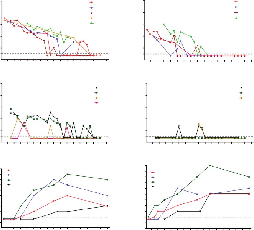
Figure 3. Kinetics of severe acute respiratory syndrome coronavirus 2 (SARS-CoV-2) RNA in different clinical samples and of antibody response in the first 2 coronavirus
disease 2019 patients diagnosed in Italy. Viral RNA levels detected in respiratory tract secretions (A) and in non–respiratory tract samples (B) and antibody titers (C). Pt1 is
shown on the left; pt2 is shown on the right. Antibody titers for IgM, IgG, IgA, and neutralizing antibodies (NT Ab) are expressed as the reciprocal of serum dilution and are
shown on a log2 scale; viral RNA levels are expressed as cycle threshold values (Ct) of E gene amplification. Dashed lines represent the limits of detection of immunofluores-
cence assay (1:20 in (C) and (F)) and of real-time reverse transcription polymerase chain reaction (Ct: 45 in (A), (B), (D), and (E)).
CONCLUSIONS Sequential sampling in a wide range of body fluids was per-
Here, we report virological and serological characterization of formed to monitor viral dissemination and shedding as well as
the first 2 COVID-19 cases diagnosed in Italy during the 2020 antibody kinetics throughout the illness.
pandemic. The 2 patients traveled from Whuan, China, to Italy SARS-CoV-2 RNA was detected both in URT and LRT sam-
on January 23, developed symptoms in Rome on January 28, ples since the initial phase of disease. Similarly to what was ob-
and were hospitalized the following day. A detailed description served in MERS and SARS patients, our analysis showed that
of the clinical presentation has been published [12]. LRT samples presented higher levels of SARS-CoV-2 RNA than
The patients harbored the same virus strain, clustering with those in paired samples from the URT [15–17]. The results are
clade V, characterized by a nonsynonymous mutation in the consistent with the expression of the candidate SARS-CoV-2
ORF3a gene (G251V), according to the GISAID EpiCov portal cell entry receptor, human angiotensin-converting enzyme 2
[9, 13]. Bayesian phylogenetic analysis is consistent with the (ACE2), which is found primarily in the LRT [18, 19].
plausible date of exposure that presumably occurred in China Nevertheless, the presence of high levels of viral RNA in the
before the start of their travel. URT samples during the early phase of illness, coupled with the
SARS-CoV-2 Shedding and Antibodies • ofid • 5isolation of infectious virus obtained by others groups [20] and virus was cultured from the urine of a COVID-19 case in China
by us on different patients (authors’ unpublished data), strongly [34]. The limited number of patients included in our study may ac-
suggests a high potential for SARS-CoV-2 transmission [20, count for the apparent discrepancy and does not allow us to estab-
21]. Duration of viral shedding in respiratory samples was lish a definitive role of urinary shedding in viral diagnosis. Attention
26 DSO for Pt1 and 17 for Pt2, in line with observations re- to the possible involvement of conjunctiva, either as the site of virus
porting 20 days as the median shedding duration for survivors entry or a source of contagion, has been suggested [35, 36]. Ocular
[22, 23]. Live virus was isolated from the respiratory samples samples collected from Pt1 (who presented conjunctivitis at admis-
collected at presentation from the 2 patients [13]. In line with sion and up to DSO 20) were positive for SARS-CoV-2 RNA from
previous reports [20, 23], despite numerous attempts in Vero the very early phase of infection up to DSO 27. Surprisingly, as we
E6 cell culture, no replication-competent virus was recovered have described elsewhere [14], infectious virus was cultured from
from later respiratory samples when antibodies were detected. the first acute ocular sample, supporting the evidence of persistent
Several factors, including suboptimal sensitivity of the virus sustained viral replication in conjunctiva and viral shedding from
culture system especially for low–viral load samples and storage this site [14]. No virus detection was observed for Pt2, who did not
Downloaded from https://academic.oup.com/ofid/article/7/10/ofaa403/5900725 by guest on 08 December 2020
at –80°C, as well as the presence of antibodies against SARS- present any ocular symptoms. These findings indicate that contact
CoV-2, may have contributed to unsuccessful virus culture at- with conjunctival secretion from COVID-19 patients with ocular
tempts. However, the difficulty isolating SARS-CoV-2 from late symptoms may represent a potential risk of infection; therefore,
samples supports the idea that despite the long duration of viral eye protection represents an important measure to prevent virus
RNA shedding, the transmission of the infection is likely lim- transmission especially in health care settings. SARS-CoV-2 RNA
ited to early infection, when the viral load is high and antibody was recovered from several additional nonrespiratory samples.
response has not yet been developed [23]. Shorter duration of Although at low levels, we found positive for viral RNA a cutaneous
viral shedding was observed for nonrespiratory samples, which swab from Pt2, and vomit samples and cervico-vaginal swabs from
showed lower viral loads since the early phase of illness and a Pt1. These findings are thus far unique [37] and need to be con-
more discontinuous trend. firmed in further studies in order to define the transmission poten-
Among the nonrespiratory samples, saliva was positive from tial linked to this wide RNA shedding.
the early to late phases of disease (up to DSO 26) for Pt1, sup- To date, knowledge on the antibody response during SARS-
porting the idea of transmission via saliva droplets [4]. Stool CoV-2 infection is limited. We monitored the kinetics of IgM,
represents another specimen of clinical and epidemiological in- IgA, IgG, and neutralizing antibodies in the 2 patients using IFA
terest, and stool sample testing for follow-up monitoring and based on whole virus in serial samples collected during hospi-
patient discharge has been suggested. In fact, the presence of talization. In line with other reports on COVID-19 cases, we
SARS-CoV-2 RNA in fecal samples has been reported even after observed seroconversion for all antibody classes within the first
viral clearance from the respiratory tract [5, 24], and our results week after diagnosis, which corresponds to the date of symptom
on rectal swabs partially support these data from the literature, onset based on the recorded anamnestic data provided by the
as fluctuant positivity was found during illness. However, to our patients [31, 38, 39]. Surprisingly, in both patients IgG was de-
knowledge there have been no reports of fecal–oral transmis- tected at 3 (Pt1) and 6 (Pt2) DSO at high titers, when IgM and
sion yet, and this issue is still under debate, although several au- IgA were still low or undetectable [40, 41]. We cannot exclude
thors have provided data suggesting that, at least in some cases, the occurrence of a pauci-symptomatic phase that may have
the gastrointestinal tract may harbor SARS-CoV-2 replication prolonged the effective time lapse from the initial infection and
[25, 26]. The presence of SARS-CoV-2 in blood is still contro- IgG appearance. The early appearance of high-titer IgG in con-
versial: Some reports on COVID-19 patients found no viral trast to IgM could be also due to an anamnestic response to
RNA, while in other studies occasional (10%) positivity was re- past infection with other endemic coronaviruses, as previously
ported, possibly associated with severe manifestations [27–31]. reported [20, 42].
In this study, only 1 serum sample from Pt1 tested positive for Increasing antibody levels were observed during the second
SARS-CoV-2 RNA with high real-time RT-PCR Ct values; this week, with high titers of IgG and IgA. In addition, in accordance
was collected at DSO 5 and corresponded to worsening of the with earlier findings, both patients developed neutralizing anti-
clinical picture and ICU admission. However, the detection of bodies during the second week of illness, reaching high levels at
low-level viral genome fragments in blood is not to be taken as DSO 32 [31]. IgA is predominantly present in mucosal tissues,
definitive evidence of bloodstream dissemination of the virus; including the URT, providing the first line of defense in mucosal
from our and other existing data, it seems that blood does not immunity. As shown in this study and others, detecting sero-
play a major role in virus transmission [23, 28, 32]. conversion of IgA as well as IgG and IgM can be useful to fully
We did not find viral RNA in urine samples from the patients. evaluate the humoral response in COVID-19 cases [38, 39].
However, urinary shedding of viral RNA has been occasionally re- Although our study examined 2 patients, which represents
ported with evidence of renal tropism [28, 31–33], and infectious a limitation of the present results, the description of virus
6 • ofid • Colavita et aldynamics based on daily monitoring of both virological and se- Micaela Maritti, Giulia Matusali, Silvia Meschi, Francesco Messina,
Chiara Montaldo, Silvia Murachelli, Emanuele Nicastri, Roberto
rological aspects during the course of disease can give impor-
Noto, Claudia Palazzolo, Emanuele Pallini, Virgilio Passeri,
tant insight into the pathogenesis and host response. Overall, Federico Pelliccioni, Antonella Petrecchia, Ada Petrone, Nicola Petrosillo,
the results show that, on one side, SARS-CoV-2 shedding and Elisa Pianura, Maria Pisciotta, Silvia Pittalis, Costanza Proietti,
its duration may involve several body sites and may be associ- Vincenzo Puro, Gabriele Rinonapoli, Martina Rueca, Alessandra
Sacchi, Francesco Sanasi, Carmen Santagata, Silvana Scarcia,
ated on different. spectrum of clinical manifestation (such as Vincenzo Schininà, Paola Scognamiglio, Laura Scorzolini, Giulia Stazi,
conjuntivitis). Further studies are needed for a better under- Francesco Vaia, Francesco Vairo, Maria Beatrice Valli.
standing of this aspect, which is important to inform clinical Financial support. This work was supported by the Ministry of Health
(Ricerca Corrente - Linea 1, Conto Capitale 2017, COVID-2020-12371817,
management and public health decision-making. On the other
COVID 2020 12371675) and the European Commission–Horizon 2020
side, the detection and profile of specific antibodies can assist (European Virus Archive GLOBAL—871029; EU project 101003544–
with diagnosis, provide valuable information for screening CoNVat; EU project 101003551- EXSCALATE4CoV).
Potential conflicts of interest. The authors declare that no conflicting fi-
of suspect cases (including in subclinical cases), and evaluate
nancial interests or other competing relationships exist. All authors have
the disease course. Furthermore, the evaluation of antibody
Downloaded from https://academic.oup.com/ofid/article/7/10/ofaa403/5900725 by guest on 08 December 2020
submitted the ICMJE Form for Disclosure of Potential Conflicts of Interest.
response will be crucial for surveillance and epidemiological Conflicts that the editors consider relevant to the content of the manuscript
studies of this novel disease and may be informative in vaccine have been disclosed.
development for SARS-CoV-2. Further investigation should
References
clarify the level and duration of protection following infection. 1. Lake MA. What we know so far: COVID-19 current clinical knowledge and re-
search. Clin Med (Lond) 2020; 20:124–7.
Acknowledgments 2. Velavan TP, Meyer CG. The COVID-19 epidemic. Trop Med Int Health 2020;
We acknowledge the contributors of genome sequences of the newly 25:278–80.
3. European Centre for Disease Control and Prevention. Geographical distribution
emerging coronavirus, that is, the originating and submitting laboratories,
of 2019-nCov cases. Available at: https://www.ecdc.europa.eu/en/geographical-
for sharing their sequences and other metadata used in this study through
distribution-2019-ncov-cases. Accessed 17 April 2020.
the GISAID Initiative. 4. To KKW, Tsang OTY, Yip CCY, et al. Consistent detection of 2019 novel corona-
INMI COVID-19 laboratory team. Isabella Abbate, virus in saliva. Clin Infect Dis. 2020; 71:841–3.
Chiara Agrati, Loredana Aleo, Tonino Alonzi, Alessandra Amendola, 5. Wang W, Xu Y, Gao R, et al. Detection of SARS-CoV-2 in different types of clinical
Claudia Apollonio, Nicolina Arduini, Barbara Bartolini, Giulia Berno, specimens. JAMA. 2020; 323:1843–4.
Silvia Biancone, Mirella Biava, Angela Bibbò, Licia Bordi, Carla Brega, 6. Xia J, Tong J, Liu M, Shen Y, Guo D. Evaluation of coronavirus in tears and con-
Marco Canali, Angela Cannas, Maria Rosaria Capobianchi, junctival secretions of patients with SARS‐CoV‐2 infection. J Med Virol. 2020;
Fabrizio Carletti, Stefania Carrara, Rita Casetti, Concetta Castilletti, 92:589–94.
7. Wu Y, Guo C, Tang L, et al. Prolonged presence of SARS-CoV-2 viral RNA in
Roberta Chiappini, Lucia Ciafrone, Eleonora Cimini, Sabrina Coen,
faecal samples. Lancet Gastroenterol Hepatol 2020; 5:434–5.
Francesca Colavita, Rossella Condello, Antonio Coppola, Silvia D’Arezzo,
8. Corman VM, Landt O, Kaiser M, et al. Detection of 2019 novel coronavirus
Antonino Di Caro, Stefania Di Filippo, Chiara Di Giuli, (2019-nCoV) by real-time RT-PCR. Eurosurveillance. 2020; 25:pii=2000045.
Lavinia Fabeni, Luisa Felici, Valeria Ferraioli, Federica Forbici, 9. Capobianchi MR, Rueca M, Messina F, et al. Molecular characterization of SARS-CoV-2
Anna Rosa Garbuglia, Emanuela Giombini, Caterina Gori, Silvia Graziano, from the first case of COVID-19 in Italy. Clin Microbiol Infect 2020; 26:954–6.
Cesare Ernesto Maria Gruber, Daniele Khouri, Eleonora Lalle, 10. Li X, Zai J, Zhao Q, et al. Evolutionary history, potential intermediate animal host,
Daniele Lapa, Barbara Leone, Patrizia Marsella, Chiara Massimino, and cross-species analyses of SARS-CoV-2. J Med Virol 2020; 92:602–11.
Giulia Matusali, Antonio Mazzarelli, Silvia Meschi, Francesco Messina, 11. Colavita F, Biava M, Castilletti C, et al. Inflammatory and humoral immune re-
Claudia Minosse, Claudia Montaldo, Stefania Neri, Carla Nisii, sponse during Ebola virus infection in survivor and fatal cases occurred in Sierra
Leone during the 2014–2016 outbreak in West Africa. Viruses 2019; 11:373.
Elisabetta Petrivelli, Fabrizio Petroni, Elisa Petruccioli, Marina Pisciotta,
12. Albarello F, Pianura E, Di Stefano F, et al; COVID 19 INMI Study Group. 2019-
Daniele Pizzi, Gianluca Prota, Fabrizio Raparelli, Gabriella Rozera,
novel coronavirus severe adult respiratory distress syndrome in two cases in Italy:
Martina Rueca, Rossella Sabatini, Silvia Sarti, Giuseppe Sberna, an uncommon radiological presentation. Int J Infect Dis 2020; 93:192–7.
Roberta Sciamanna, Marina Selleri, Carla Selvaggi, Catia Sias, 13. Stefanelli P, Faggioni G, Lo Presti A, et al. Whole genome and phylogenetic anal-
Chiara Stellitano, Antonietta Toffoletti, Silvia Truffa, Federica Turchi, ysis of two SARS-CoV-2 strains isolated in Italy in January and February 2020:
Maria Beatrice Valli, Carolina Venditti, Tiziana Vescovo, Donatella Vincenti, additional clues on multiple introductions and further circulation in Europe.
Antonella Vulcano, Emma Zambelli. Eurosurveillance. 2020; 25:pii=2000305.
COVID-19 INMI Study Group. Maria Alessandra Abbonizio, 14. Colavita F, Lapa D, Carletti F, et al. SARS-CoV-2 isolation from ocular secretions
Chiara Agrati, Fabrizio Albarello, Gioia Amadei, of a patient with COVID-19 in Italy with prolonged viral RNA detection. Ann
Intern Med 2020; 173:242–3.
Alessandra Amendola, Mario Antonini, Raffaella Barbaro,
15. Cheng PK, Wong DA, Tong LK, et al. Viral shedding patterns of coronavirus
Barbara Bartolini, Martina Benigni, Nazario Bevilacqua,
in patients with probable severe acute respiratory syndrome. Lancet 2004;
Licia Bordi, Veronica Bordoni, Marta Branca, 363:1699–700.
Paolo Campioni, Maria Rosaria Capobianchi, Cinzia Caporale, 16. Paules CI, Marston HD, Fauci AS. Coronavirus infections—more than just the
Ilaria Caravella, Fabrizio Carletti, Concetta Castilletti, Roberta common cold. JAMA 2020; 323:707.
Chiappini, Carmine Ciaralli, Francesca Colavita, Angela Corpolongo, 17. Kim JY, Ko J-H, Kim Y, et al. Viral load kinetics of SARS-CoV-2 infection in first
Massimo Cristofaro, Salvatore Curiale, Alessandra D’Abramo, two patients in Korea. J Korean Med Sci. 2020; 35:e86.
Cristina Dantimi, Alessia De Angelis, Giada De Angelis, 18. Zhou P, Yang XL, Wang XG, et al. A pneumonia outbreak associated with a new
Rachele Di Lorenzo, Federica Di Stefano, Federica Ferraro, coronavirus of probable bat origin. Nature 2020; 579:270–3.
19. Hoffmann M, Kleine-Weber H, Schroeder S, et al. SARS-CoV-2 cell entry de-
Lorena Fiorentini, Andrea Frustaci, Paola Gallì, Gabriele
pends on ACE2 and TMPRSS2 and is blocked by a clinically proven protease in-
Garotto, Maria Letizia Giancola, Filippo Giansante,
hibitor. Cell 2020; 181:271–80.e8.
Emanuela Giombini, Maria Cristina Greci, Giuseppe Ippolito, 20. Wölfel R, Corman VM, Guggemos W, et al. Virological assessment of hospitalized
Eleonora Lalle, Simone Lanini, Daniele Lapa, Luciana Lepore, patients with COVID-2019. Nature. 2020; 581:465–9.
Andrea Lucia, Franco Lufrani, Manuela Macchione, Alessandra 21. Zou L, Ruan F, Huang M, et al. SARS-CoV-2 viral load in upper respiratory spe-
Marani, Luisa Marchioni, Andrea Mariano, Maria Cristina Marini, cimens of infected patients. N Engl J Med 2020; 382:1177–9.
SARS-CoV-2 Shedding and Antibodies • ofid • 722. Zhou F, Yu T, Du R, et al. Clinical course and risk factors for mortality of adult 33. Puelles VgG, Lütgehetmann M, Lindenmeyer M, et al. Multiorgan and renal tro-
inpatients with COVID-19 in Wuhan, China: a retrospective cohort study. Lancet pism of SARS-CoV-2. N Engl J Med 2020; 383:590–2.
2020; 395:1054–62. 34. Sun J, Zhu A, Li H, et al. Isolation of infectious SARS-CoV-2 from urine of a
23. Walsh KA, Jordan K, Clyne B, et al. SARS-CoV-2 detection, viral load and infect- COVID-19 patient. Emerg Microbes Infect 2020; 9:991–3.
ivity over the course of an infection. J Infect 2020; 81:357–71. 35. Li JO, Lam DSC, Chen Y, Ting DSW. Novel coronavirus disease 2019 (COVID-
24. Wu Y, Guo C, Tang L, et al. Prolonged presence of SARS-CoV-2 viral RNA in 19): the importance of recognising possible early ocular manifestation and using
faecal samples. Lancet Gastroenterol Hepatol 2020; 5:434–5. protective eyewear. Br J Ophthalmol 2020; 104:297–8.
25. Lamers MM, Beumer J, van der Vaart J, et al. SARS-CoV-2 productively infects 36. Lu CW, Liu XF, Jia ZF. 2019-nCoV transmission through the ocular surface must
human gut enterocytes. Science 2020; 369:50–4. not be ignored. Lancet 2020; 395:e39.
26. Xiao F, Tang M, Zheng X, et al. Evidence for gastrointestinal infection of SARS- 37. Qiu L, Liu X, Xiao M, et al. SARS-CoV-2 is not detectable in the vaginal fluid of
CoV-2. Gastroenterology 2020; 158:1831–3.e3. women with severe COVID-19 infection. Clin Infect Dis 2020; 71:813–7.
27. Xie C, Jiang L, Huang G, et al. Comparison of different samples for 2019 novel corona- 38. Guo L, Ren L, Yang S, et al. Profiling early humoral response to diagnose novel
virus detection by nucleic acid amplification tests. Int J Infect Dis 2020; 93:264–7. coronavirus disease (COVID-19). Clin Infect Dis. 2020; 71:778–85.
28. Wang Y, Zhang L, Sang L, et al. Kinetics of viral load and antibody response in 39. Okba NMA, Müller MA, Li W, et al. Severe acute respiratory syndrome corona-
relation to COVID-19 severity. J Clin Invest. 2020; 138759. virus 2−specific antibody responses in coronavirus disease patients. Emerg Infect
29. Chen W, Lan Y, Yuan X, et al. Detectable 2019-nCoV viral RNA in blood is a strong Dis. 2020; 26:1478–88.
indicator for the further clinical severity. Emerg Microbes Infect 2020; 9:469–73. 40. Long Q, Liu B, Deng H, et al. Antibody responses to SARS-CoV-2 in patients with
30. Holshue ML, DeBolt C, Lindquist S, et al; Washington State 2019-nCoV Case COVID-19. Nat Med 2020; 26:845–8.
Downloaded from https://academic.oup.com/ofid/article/7/10/ofaa403/5900725 by guest on 08 December 2020
Investigation Team. First case of 2019 novel coronavirus in the United States. N 41. Shu H, Wang S, Ruan S, et al. Dynamic changes of antibodies to SARS-
Engl J Med 2020; 382:929–36. CoV-2 in COVID-19 patients at early stage of outbreak. Virol Sin. 2020;
31. Haveri A, Smura T, Kuivanen S, et al. Serological and molecular findings during 1–8.
SARS-CoV-2 infection: the first case study in Finland, January to February 2020. 42. Graham NR, Whitaker AN, Strother CA, et al. Kinetics and isotype assessment
Eurosurveillance 2020; 25. of antibodies targeting the spike protein receptor binding domain of SARS-
32. Peng L, Liu J, Xu W, et al. SARS‐CoV‐2 can be detected in urine, blood, anal CoV-2 in COVID-19 patients as a function of age and biological sex. medRxiv
swabs, and oropharyngeal swabs specimens. J Med Virol. 2020; doi:10.1002/ 2020.07.15.20154443 [Preprint]. 22 July 2020. Available at: https://doi.org/10.110
jmv.25936 1/2020.07.15.20154443. Accessed 25 August 2020.
8 • ofid • Colavita et alYou can also read