Gut Microbiome-Targeted Treatment for Diabetes: What's Your Gut Telling You?
←
→
Page content transcription
If your browser does not render page correctly, please read the page content below
Gut Microbiome-Targeted Treatment for Diabetes:
                   What’s Your Gut Telling You?

                                 Amanda K. Kitten, Pharm.D.
              Master of Science Graduate Student and Pharmacotherapy Resident
                                 Division of Pharmacotherapy
                    The University of Texas at Austin College of Pharmacy
                      Pharmacotherapy Education and Research Center
                                    UT Health San Antonio

                                      Friday, April 13, 2018

Learning Objectives
1. Identify potential mechanisms by which the microbiome affects human health
2. Explain how the microbiome influences the development of diabetes mellitus
3. Describe the differences seen in gut microbiome composition between patients with diabetes and
    healthy subjects
4. Evaluate microbiome-targeted therapies as potential interventions to prevent and treat type 2
    diabetes mellitus (T2DM)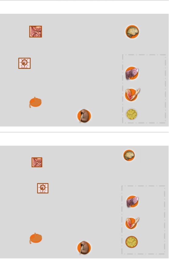
Role of the Microbiome in Human Health

I.      Overview of the human microbiome
           a. Definitions1
                i. Microbiota: microbes that collectively inhabit a given ecosystem
               ii. Microbiome: collection of all genomes of microbes in an ecosystem
              iii. Dysbiosis: disturbance or change in the composition and function of microbes
           b. Scope2
                i. Body’s bacteria would circle the Earth 2.5 times
               ii. Weighs up to 1 to 2 kg
              iii. Outnumber human cells by 10:1
              iv. 95% of bacteria located in gastrointestinal (GI) tract
           c. Studying the microbiome
                i. Transition from culture-based methods to culture-independent molecular assays
               ii. Methods are used to discern the structure (i.e., anatomy) and function (i.e., physiology)
                   of the microbiota

                                   Table 1. Tools for Analyzing Microbiome1
            Approach                     Data                            Platform
            16S rRNA gene sequencing     Community composition           Next-generation sequencing
            Metagenomics                 Whole genome sequencing         Next-generation sequencing
            Metatranscriptomics          Gene expression                 Next-generation sequencing
            Metaproteomics               Protein expression              Mass spectrometry
            Metabolomics                 Metabolic productivity          Mass spectrometry

              iii. Most common approach is 16S rRNA gene sequencing3
                            1. 16S gene encodes for the 16S rRNA molecule that is unique to bacteria and
                                 archaea, thus distinguishes these cells from human cells
                            2. 16S gene is amplified using polymerase chain reaction and sequenced using
                                 next-generation sequencing technology
                            3. Machine learning is used to cluster similar sequences and reference
                                 databases (e.g., Greengenes) assist with assigning taxonomy
            d. Composition
                i. Varies substantially by body site4
                            1. Outer body sites predominated by Gram-positive aerobic organisms from
                                 the Actinobacteria and Firmicutes phyla
                            2. Gut microbiome (represented by stool) predominated by anaerobic Gram-
                                 positive and Gram-negative bacteria
                                     a. Firmicutes (e.g., Lachnospiraceae, Ruminococcaceae)
                                     b. Bacteroidetes (e.g., Bacteroidaceae, provetellaceae)
                                     c. Actinobacteria (e.g., Bifidobacteriaceae)
               ii. Microbiota extensively conserved at high taxonomic levels; variation increases at
                   progressively lower taxonomic levels
              iii. Large inter-individual variability in microbiota composition, but not ecosystem function

                                                    Page 2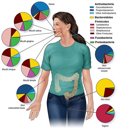
Figure 1. Dominant Bacterial Taxa by Body Site4

 II.   Global gut microbiota functions1
          a. Mature and train the immune system
          b. Inhibit invasion by pathogens
          c. Mediate host-cell proliferation and vascularization
          d. Regulate intestinal endocrine functions, neurologic signaling, and bone density
          e. Provide a source of energy biogenesis
          f. Biosynthesize vitamins, neurotransmitters, and related compounds
          g. Metabolize bile salts
          h. Xenobiotic metabolism and elimination

III.   Associations between gut dysbiosis and human disease1
           a. Endogenous and exogenous factors influence gut microbiota
               i. Neonatal mode of delivery                          vi. Environmental exposures
              ii. Host genetic features                             vii. Age
             iii. Host immune response                             viii. Physical activity
             iv. Diet                                                ix. Smoking
              v. Medications                                          x. Alcohol consumption
           b. Disruption of microbial communities associated with a host of chronic and acute diseases

                                                  Page 3
Table 2: Influence of Gut Microbiome Communities on Health5
              Health                       Microbial products or activities                 Disease
         Nutrient & energy      •   SCFA production & vitamin synthesis               Obesity & metabolic
               supply           •   Energy supply, gut hormones, & satiety                syndrome
                                •   Lipopolysaccharides, inflammation
         Cancer prevention      •   Butyrate production, phytochemical release         Cancer promotion
                                •   Toxin and carcinogen inflammation
                                •   Mediates inflammation
         Pathogen inhibition    •   SCFA production, intestinal pH, bacteriocins      Pathogen invasion
                                •   Competition for substrates and/or binding sites
                                •   Toxin production, tissue invasion, inflammation
            GI immune           •   Balance of pro- and anti-inflammatory signals             IBD
             function           •   Inflammation, immune disorders
            Gut motility        •   Metabolites (SCFAs, gases) from non-digestible    IBS (constipation,
                                    carbohydrates                                     diarrhea, bloating)
           Cardiovascular    •      Lipid & cholesterol metabolism                      Cardiovascular
               health                                                                       disease
            SCFA=short-chain fatty acid; IBD=inflammatory bowel disease; IBS=irritable bowel syndrome

     Gut Dysbiosis and Diabetes Mellitus

I.      Overview of diabetes mellitus
           a. Disease prevalence6,7
                     i. As of 2015, 30.3 million Americans (9.4% of the population) with diabetes
                            1. 23.1 million people diagnosed
                            2. 7.2 million people undiagnosed
                    ii. 29 million Americans (9% of the population) have T2DM
                   iii. Local prevalence8
                            1. San Antonio
                                    a. 14.2% of population diagnosed with type 1 diabetes mellitus
                                        (T1DM) or T2DM
                                    b. T2DM in San Antonio prevalence varies by race
                                              i. Whites: 8%
                                             ii. Blacks: 12%
                                            iii. Hispanics: 16%
                            2. Bexar county: prevalence 13%
                            3. Texas: prevalence 10%
                            4. United States: prevalence 9%
           b. Morbidity and mortality
                     i. Absolute number of deaths due to diabetes increased by 93% from 1990 to 20109
                    ii. In 2012 estimated annual cost of diabetes $245 billion8

                                                     Page 4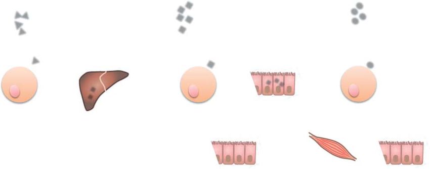
182    b-Cell–Centric Classification of Diabetes                                                                   Diabetes Care Volume 39, February 2016

 II.      Pathophysiology: Egregious Eleven10-13

                                                  Figure 2: b-cell-Centric Construct: Egregious Eleven7

               a. Describes pathways that contribute to development of diabetes
               b. Dysfunctional pathways
                        i. Pancreatic b-cells: decreased insulin production
                       ii. Muscle: disruptions in insulin signal transduction resulting in insulin resistance
                     iii. Liver: decreased inhibition of hepatic glucose production (HGP) by hyperinsulinemia
                      iv. Adipose: enlarged fat cells exhibit insulin resistance; fat “spill-over” can worsen
                           insulin resistance in muscle and liver
                       v. Decreased incretin effect
                               1. Glucagon-like peptide-1 (GLP-1) diminished in diabetes
                               2. GLP-1 aids in glucose disposal as well as inhibition of HGP
                      vi. a-cell: overproduction of glucagon in diabetes patients, contributing to increased
                           basal HGP
                     vii. Kidney: increased sodium-glucose cotransporter-2 (SGLT2) threshold
                    viii. Brain: delayed satiety in response to increases in insulin
                      ix. Stomach/small intestine: increased glucose absorption
                       x. Immune dysregulation/inflammation: macrophage and interleuin-1 (IL-1)
                           recruitment to pancreas results in b-cell apoptosis
                      xi. Colon/microbiome: influences host metabolism in three main ways that can affect
                           multiple other facets of Egregious Eleven

            Figure 3—b-Cell–centric construct: the egregious eleven. Dysfunction of the b-cells is the final common denominator in DM. A: Eleven currently
            known mediating pathways of hyperglycemia are shown. Many of these contribute to b-cell dysfunction (liver, muscle, adipose tissue [shown in red
            to depict additional association with IR], brain, colon/biome, and immune dysregulation/inflammation [shown in blue]), and others result from
            b-cell dysfunction through downstream effects (reduced insulin, decreased incretin effect, a-cell defect, stomach/small intestine via reduced
            amylin, and kidney [shown in green]). B: Current targeted therapies for each of the current mediating pathways of hyperglycemia. GLP-1,
            glucagon-like peptide 1; QR, quick release.

                                                                      Page 5
Review K H Allin and others Gut microbiota in T2DM 172:4 R170
A. Lipopolysaccharide B. Short-chain fatty acids C. Bile acids
Dietary fibres Butyrate Primary Secondary
Acetate bile acids bile acids
LPS
Propionate
TLR4 GPR41 Energy source
TGR5
GPR43
↑ Lipogenesis
↑ Gluconeogenesis
European Journal of Endocrinology
↑ Inflammation ↓ Inflammation
↑ GLP1 and PYY ↑ Energy expenditure ↑ GLP1
Figure 1 Figure 3: Microbiome and Host Metabolism9
Microbes and host metabolism. Microbes may influence host various effects depending on the cellular types affected. In
1. Increased production of lipopolysaccharides
metabolism through numerous mechanisms, of which three
(LPS)14,15 in decreased inflammation
immune cells, this signalling results
important mechanisms are depicted. (A) Lipopolysaccharide. and in the enteroendocrine L-cells it results in increased GLP1
a. from
Lipopolysaccharide (LPS) originates LPSstheshed
outer from
membraneGram-negative
and PYY levelsbacterial celltowalls
together leading (i.e.,
improved E. coli)
insulin sensitivity.
i. Bind
of Gram-negative bacteria and binds to Toll-like to4 toll-like
receptor receptor-4
(C) Bile acids. Primary(TLR4)/CD14 complex
bile acids are produced by the liver and
ii. TLR4
(TLR4), which activates pro-inflammatory signalling activatesrecirculated
pathways innate immune system,
to the liver from the gut. resulting
However, gutin pro-are
bacteria
resulting in low-grade inflammation and thus decreased insulin capable of deconjugating primary bile acids hindering their
inflammatoryrecirculation.
sensitivity. (B) Short-chain fatty acids. Bacteria in the colon
responseThe primary deconjugated bile acids are further
ferment dietary fibres to short-chain fattyiii.
acids Decrease
(mainly expression
metabolisedof bytight junction
gut bacteria proteins
to secondary andSecondary
bile acids. increase
mucosa are
butyrate, acetate and propionate). Acetate and propionate integrity
bile acids bind to the G protein-coupled receptor TGR5, which
b. Decreased integrity ofresults
used as substrates for gluconeogenesis and lipogenesis in the
intestinal mucosa increases release of LPS
in increased energy expenditure in muscles and GLP1
liver, whereas butyrate is an important energy substrate for 14 secretion in the enteroendocrine L-cells, both of which lead
into bloodstream
colonic mucosa cells. Moreover, short-chain fatty acids bind to to improved insulin sensitivity.
the G protein-coupled receptors GPR41 andi.GPR43 Higher plasma
resulting in LPS levels in DM patients than healthy
counterparts
15
high-fat diet (21) 2. Decreased
and mice short-chain
receiving antibiotics fatty acids
exhibited been (SCFAs)
proposed production
to be part of the signalling pathways
a. SCFAs (butyrate, acetate, propionate) produced by bacterial
lower levels of circulating LPSs and TNFa as well as affecting the development of metabolic syndrome as
decreased insulin resistance compared with pair-fed mice observed in studies of Tlr2- and Tlr5-deficient mice
(22). As a part of the immune system, fermentation of dietary
Toll-like receptors (23,fiber and resistant
24). Additional evidencestarches
of the importance of the
(TLRs) recognise microbial molecules i.andMain energy
activate the source
crosstalk for gut epithelium
among (mainlyinflammation
the immune system, butyrate)
innate immune system. LPSs bind toii.andBind G-protein
activate the andcoupled
metabolism receptors (GPCRs)
was observed in the41development
and 43 in of
TLR4/CD14 complex, which activates pro-inflammatory non-alcoholic fatty liver disease (NAFLD). Mice without
intestinal mucosa,
pathways. Other TLRs, such as TLR2 and TLR5, have also
immune cells, liver, and adipose tissues
the inflammasome complexes NLRP3 or NLRP5,
1. Intestinal mucosa: SCFAs bind to GPCRs on
www.eje-online.org
enterohepatic L-cells in colon à increase GLP-1
secretion
2. Immune cells: inhibit NF-KB activation; decrease
TNF-a and IL-6 suppression and decreased
inflammation
Page 6
3. Bile acids16
                                     a. Gut bacteria convert primary bile acids to secondary bile acids via
                                         bile salt hydrolases
                                     b. Secondary bile acids act as signaling molecules to induce GLP-1
                                         secretion from small intestine L-cells
              c. Gut microbes implicated in specific mechanisms of dysbiosis
                      i. LPS production by Gram-negative bacteria14
                            1. E. coli
                            2. Salmonella
                            3. Shigella
                            4. Pseudomonas
                            5. Neisseria
                            6. H. influenza
                            7. Bodetella pertussis
                            8. Vibrio cholerae
                     ii. Beneficial SCFA producers:17,18
                            1. Mainly species in the Firmicutes phyla
                                     a. Roseburia sp.
                                     b. Faecalibacterium prausnitzii
                                     c. Eubacterium hallii
                                     d. Eubacterium rectale
                    iii. Microbiota with beneficial bile salt hydrolases16
                            1. Lactobacillus
                            2. Bifidobacterium
                            3. Firmicutes
                            4. Enterococcus
                            5. Clostridum
                            6. Bacteroides

III.      American Diabetes Association (ADA) acknowledged importance of the relationship between
          microbiome and diabetes9
              a. 2014 ADA and JDRF Research Symposium: Diabetes and the Microbiome
                      i. First gathering of experts that focused on the link between the pathophysiology of
                         the microbiome of diabetes
                     ii. Symposium made several recommendations to guide future diabetes and
                         microbiome research

       The Gut Microbiome in Patients with Diabetes

  I.      Microbiome studies: associations with metabolic (dys)function
             a. Historically, studies have yielded diverse results19-21
             b. Several recent robust studies demonstrated differences between T2DM patients and
                 controls as well as complex relationships between bacterial taxa17,18

                                                      Page 7
RESEARCH ARTICLE
a Control-enriched MLGs T2D-enriched MLGs
Con-133
Bacteroides sp. 20_3 Desulfovibrio sp. 3_1_syn3
Unclassified
Actinobacteria
Con-131
Eggerthella R. intestinalis A. muciniphila
Con-122 F. prausnitzii C. bolteae
Bacteroidales
C. symbiosum E. coli
Alistipes Con-130
Bacteroides T2D-8
Con-120 E. rectale
Parabacteroides T2D-14
R. inulinivorans
Firmicutes Con-148 Con-152 Clostridium sp. HGF2
Lachnospiraceae Con-144 T2D-16
Con-155 T2D-12
Erysipelotrichaceae T2D-9
Clostridiales Clostridium ramosum T2D-2
Clostridium Con-104 Con-101 C. hathewayi
Con-109 E. lenta
Eubacterium
Roseburia T2D-62 T2D-170
T2D-30
Faecalibacterium T2D-73 T2D-165
Subdoligranulum Clostridiales sp. SS3/4
Proteobacteria B. intestinalis
H. parainfluenzae T2D-79 T2D-6
Desulfovibrio
Escherichia T2D-90
Haemophilus T2D-37
Con-142 Con-180 T2D-93
Verrucomicrobia
Akkermansia
b MLG: metagenomic linkage group T2D
17
Figure 4: Microbiota Trends in Metabolic (Dys)function
Gut microbiota Gut environment
Xenobiotics
Metabolism of cofactors and vitamins Cofactors
Butyrate-producing
c. Discordant findings due toCon-343
differences in:12,13,22bacteria
Con-3380 Con-1831 Con-1697 Vitamins
BCAA i. Diet v. Physical activity
ii. Age Cell motility vi. Smoking
Butyrate biosynthesis Butyrate
iii. Birth (Caesarian sectionbiodegradation and metabolism
Xenobiotics vii. Alcohol consumption
versus vaginal delivery)
BCAA transport CH4 metabolism
viii. MedicationsCH
4
iv. Host genotype ix. Geographic location
Oxidative stress Sulphate-reducing bacteria
H2S biosynthesis H2S
T2D-823
Oxidative stress resistance Drug resistance
Microbiome-Targeted Therapies for the Prevention and Treatment of Diabetes
Sugar related membrane transport
23
I. Personalized nutrition:
Akkermansia muciniphila
a. Mucin
Individualized
layer
dietary
T2D-317
plan based on an individual’s distinctive characteristics
Mucin degradation Mucin layer integrality
b. Host
Linktissues
between microbiome composition and post-prandial glucose response (PPRG)
gure 2 | Taxonomic and functionali.characterization
Identified microbiome as integral
of gut microbiota incomponent
(blue) or belowin formulating
20.4 (red).a b,
personalized
A schematic nutrition
diagram showing the main
plan to optimize
2D. a, A co-occurrence network was deduced from 47 MLGs that were PPGR of the gut microbes that had a predicted T2D association. Red text d
entified from 52,484 gene markers. Nodes depict MLGs with their ID enriched functions in T2D patients; blue text denotes depleted funct
splayed in the centre. Persize
A The person
of the profiling
nodes indicates gene number within the T2D patients; black Computational analysisfunctional role relative
text denotes an uncertain
LG. The colour of the nodes indicates their taxonomic Diary assignment.
(food, sleep, physical activity) The dashed line arrows point to the inference that was not detected di
Gut microbiome Using smartphone-adjusted website Main PPGR
onnecting lines represent Spearman
16S rRNAcorrelation coefficient values above 0.4 reported by previouscohort
5,435 days, 46,898 meals, 9.8M Calories, 2,532 exercises
studies. prediction
Metagenomics
Continuous glucose monitoring
Using a subcutaneous sensor (iPro2)
Blood tests
e previous findings in studies of inflammatory bowel disease
130K hours, 1.56M and This may indicate
glucose measurements 800that the gut environment of a T2D patient is
Participants
bese patients26. By contrast,
Questionnaires control-enriched markers were fre- stimulates
Standardized meals (50g available carbohydrates)
bacterial defence mechanisms against oxidativ
Validation Dietary
uently involved in cell motility Lifestyle and metabolism Day
Food frequency
of1 cofactors and (Supplementary Table
Day 2 Day 3 Day 4 Day 5 Day 6 Day 7 cohort
10). Similarly,
interventionwe found 14 KEGG orth
Medical
tamins (P , 0.002; Supplementary Fig. 9). G
markers
G F
related to drug resistance that were greatly enriched
At the module or pathway level, the gut microbiota of T2D patients patients, further supporting that T2D patients may have a mor
Anthropometrics Bread Bread Bread & Bread & Glucose Glucose Fructose
butter butter
100 Participants 26 Participants
as functionally characterized with our T2D-associated markers and gut environment, and the medical histories of these patients ma
owed enrichment in membrane transport of sugars, branched-chain this (Supplementary Table 10).
mino acid (BCAA) transport, methane Figure 5. Illustration
metabolism, xenobiotics of Experimental Design23Participant 141
B 45% 33% 22% C 76% 21% 3% D
gradation and metabolism, and sulphate reduction. By contrast, T2D-related Sleep
dysbiosis in gut microbiota
Glucose (mg/dl)
Frequency
Frequency
ere was a decrease in the level of bacterial chemotaxis, flagellar In light of the above MGWAS result and an ad
sembly, butyrate biosynthesis and metabolism of cofactorsPage and8 PERMANOVA27 (permutational multivariate analysis of v
tamins (Fig. 2b and Supplementary Table 10; see Supplementary analysis that clearly showed that T2D was a significant fa
g. 10 for the detailed information on butyrate-CoA transferase). explaining the variation in the examined gut microbialA Meal B Figure 4. Fac
c. 9Study design: (2)
carbohydrates three-part study of Postprandi
i. Part 1: Created PPG predictionSlope algorithm based on profiling of 800 Israeli 9
Partial dependence
7518 >0
PPGR (iAUC, mg/dl.h)
6 95.1%
(A) Partial dep
nt 4
3 participants (54% overweight, 22% obese), which included: rtic
ipa marginal contrib
Frequency
(a.u.) Pa
0 1. Continuous glucose monitoring (CGM) content to the
-3 8400 2. Real time diary: food, sleep, physical activity units) at each
-6 Participant 145
3. Gut microbiome analysis (16S rRNA and metagenomic analysis) (x axis). Red an
zero contributio
0 40 80 4.120Blood tests (HbA1c%, lipid levels)
5. Anthropometrics meals). Boxplo
Weight (g) Carbohydrate-PPGR slope Meal carbohydrates (g)
drates content
C Meal 6. Lifestyle,
D medical history
fat / carbohydrates (4) Participant 267 Participant 465 25, 50, 75, and
4 ii. Part 2: Algorithm validation in second cohort 40 of Israeli participants 40
across the coho
Partial dependence
R difference: R difference:
PPGR (iAUC, mg/dl.h)
2 iii.
8303 Part 3: Dietary intervention 0.21 0.11
30 (B) Histogram o
1. 26 new participants randomized to algorithm-produced diet plan
Meal fat (g)
Frequency
(a.u.)
0 pant) of a linear
-2
(intervention) or dietician-produced 20 diet plan (comparator) after week-long 20
drate content a
7611 profiling period 10 shown is an exa
-4
a. Two-week intervention included one week of “good” diet (e.g., slope and anoth
0 0
-2 -1 0 1 2 foods associated with lower PPGR) and one week of “bad” diet (e.g., (C) Meal fat/car
log2(fat/carbs)
food associated
R-difference with higher PPGR) Meal carbohydrates (g) (D) Histogram
E b. Time
PPRGfrom highly variable Meal between individuals 24-hour participant) betw
Meal sodium (5) last sleep (12) dietary fiber (14) Meal water (21) dietary fiber (25)
d. Analyzed partial dependence plots (PDPs) to better understand the role of various factors in
2 two linear regr
Partial dependence
the algorithm’s
1
7290 predictions 8697
PPGR and the
10947 7575
i. PDPs illustrate how individual variables contribute 5588 to model predictions another when
(a.u.)
0
ii. Values greater4971 than 0 indicate6968 positive contribution and content. Also s
10330values less than 8342
0 indicate
-1 8623
negative contribution hydrate and fat
pant with a rela
iii. Meal carbohydrate weight most significant contributor to PPGR
0 iv. 1000
correlates well
Relative abundance
2000 0 400 800 (RA)1200 of0 multiple
3 6 9bacterial
12 0 taxa 300contributed
600 0 to PPGR 20 as
40 well
Weight (mg) Time (min) Weight (g) Amount (ml) Weight (g) relatively high
content have lo
F M00514
TtrS-TtrR TCS (27)
M00496
NblS-NblR TCS (28)
M00256
Cell div. trans. sys. (30)
Bacteroides
dorei (45)
Alistipes
putredinis (48)
correspond to t
(E) Additional PD
Partial dependence
0.6 5
390 164 14 (F) Microbiome
0.3 79 334 323
(a.u.)
105 87 in which the mic
0 50 is indicated (left,
0
401 523 632 448 424 10–90 percentile
-0.3
(G) Heatmap of
n.d. 10-6 10-5 n.d. 10-6 10-5 n.d. 10-3 n.d. 10-4 10-3 10-2 n.d. 10-4 10-3 10-2 (Pearson) betw
Relative abundance Relative abundance Relative abundance Relative abundance Relative abundance beneficial (gree
Coprococcus catus Eubacterium rectale Parabacteroides Ratio mapped to Phylum several risk fact
PTR (53) PTR (59) distasonis (63) gene-set (93) Bacteroidetes (95)
0.3 See also Figure
Partial dependence
24
320 217
0.1 158 103 59 223 363
63
(a.u.)
77
101 144
-0.1 430
455 332 448 436
n.d. 1.1 1.2 n.d. 1.1 1.2 n.d. 10-4 10-3 10-2 0.75 0.8 0.85 n.d. 10-0.6 10-0.2
PTR PTR Relative abundance Ratio mapped Relative abundance
23
Figure 6: Microbiome PDP(ALT)
Alanine aminotransferase
G Age PDP Legend
BMI
Systolic blood pressure
Non-fasting total cholesterol
Feature name
Glucose fluctuations (noise, σ/μ) (Feature rank)
HbA1c%
Waist-to-hip ratio 0.6
85 - # undetected
514 TtrS-TtrR TCS (27)
S-LuxU-LuxO TCS (38)
2 NarQ-NarP TCS (41)
pherol biosynthesis (60)
00664 Nodulation (49)
ESCRT-III complex (70)
terium eligens PTR (79)
ceramide biosynth. (80)
ochrome c oxidase (98)
lum Euryarchaeota (99)
m Cyanobacteria (107)
Alistipes putredinis (48)
3 QseC-QseB TCS (50)
ubdoligranulumun (54)
ionine degradation (55)
terium rectale PTR (59)
cus salivarius PTR (65)
ia muciniphila PTR (82)
Alistipes finegoldii (83)
ides xylanisolvens (85)
ubacterium rectale (87)
mansia muciniphila (96)
0.3 372- # with
above-zero
contribution
Page 9 0
Negative trend:
Feature is beneficial
-0.3
497 - #with below-Glucose
Glucose
(w.r.t days 0-3)
(w.r.t days 0-3)
Fold change
Fold change
e. Post-intervention microbiota
1 2 3 4
alterations
5 6 1 2 3 4 5 6 1 2 3 4 5 6 1 2 3 4 5
i. Most significant microbiota
‘Bad’ diet week (day) changes ‘Good’ were dietinter-individual
week (day) pre- and‘Good’
post-diet week (day) ‘Bad’ diet week (day)
intervention Actinobacteria (C)
Alistipes putredinis (S)
Bifidobacterium (G)
Bifidobacterium pseudocatenulatum (S)
Parabacteroides merdae (S)
Streptococcus thermophilus (S)
Lactobacillus ruminis (S)
Bifidobacterium (G)
ii. Several bacterial taxa were significantly changed due to diet typeCorpobacter
Akkermansia muciniphila (S)
in all participants
fastidiosus (S) Bifidobacterium pseudocatenula
C Bacteria decreasing in ‘good’ diet week Bacteria increasing in ‘good’ diet week D Bifidobacterium adolescentis
P9
E14 ‘Good’ diet week
Paricipants - ‘good’ diet week
E6
P2
P8
E4
E12
Fold change (with respect to days 0-3)
P1
P10
E9
E2
E8
P6
E11
E5
E3
E7
P9
E14
E6
Paricipants - ‘bad’ diet week
P2
P8
E4
P4
E12
P1
P10
E9 ‘Bad’ diet week
E2
E8
P6
E11 Day
E5
E3
E1
E Roseburia inulinivorans
Actinobacteria (Phylum)
Actinobacteria (Class)
Bifidobacteriales (Order)
Coriobacteriales (Order)
Bifidobacteriaceae (Family)
Coriobacteriaceae (Family)
Bifidobacterium (Genus)
Collinsella (Genus)
Anaerostipes (Genus)
Dorea (Genus)
Gammaproteobacteria (Class)
Deltaproteobacteria (Class)
Betaproteobacteria (Class)
Bifidobacterium adolescentis (Species)
Collinsella aerofaciens (Species)
Anaerostipes hadrus (Species)
Eubacterium hallii (Species)
Dorea longicatena (Species)
Bacteroidetes (Phylum)
Viruses (Phylum)
Proteobacteria (Phylum)
Bacteroidia (Class)
Enterobacteriales (Order)
Bacteroidales (Order)
Burkholderiales (Order)
Viruses, noname (Order)
Desulfovibrionales (Order)
Prevotellaceae (Family)
Bacteroidaceae (Family)
Sutterellaceae (Family)
Prevotella (Genus)
Bacteroides (Genus)
Barnesiella (Genus)
Ruminococcus lactaris (Species)
Eubacterium eligens (Species)
Roseburia inulinivorans (Species)
Bacteroides vulgatus (Species)
Bacteroides stercoris (Species)
Alistipes putredinis (Species)
‘Good’ diet week
Fold change (days 4-7 vs. days 0-3) Fold change (with respect to days 0-3)
Statistically significant Statistically significant
decrease (PFold ch
‘Bad’ diet week
Day
E Roseburia inulinivorans
Coriobacteriales (Order)
Bifidobacteriaceae (Family)
Coriobacteriaceae (Family)
Bifidobacterium (Genus)
Collinsella (Genus)
Anaerostipes (Genus)
Dorea (Genus)
Bifidobacterium adolescentis (Species)
Collinsella aerofaciens (Species)
Anaerostipes hadrus (Species)
Eubacterium hallii (Species)
Dorea longicatena (Species)
Bacteroidetes (Phylum)
Viruses (Phylum)
Proteobacteria (Phylum)
Bacteroidia (Class)
Gammaproteobacteria (Class)
Deltaproteobacteria (Class)
Betaproteobacteria (Class)
Enterobacteriales (Order)
Bacteroidales (Order)
Burkholderiales (Order)
Viruses, noname (Order)
Desulfovibrionales (Order)
Prevotellaceae (Family)
Bacteroidaceae (Family)
Sutterellaceae (Family)
Prevotella (Genus)
Bacteroides (Genus)
Barnesiella (Genus)
Ruminococcus lactaris (Species)
Eubacterium eligens (Species)
Roseburia inulinivorans (Species)
Bacteroides vulgatus (Species)
Bacteroides stercoris (Species)
Alistipes putredinis (Species)
‘Good’ diet week
Fold change (with respect to days 0-3)
Fold change (days 4-7 vs. days 0-3)
cally significant Statistically significant
rease (PReview Current understanding of 228:3 R103
1. Decreased leakage of LPS from gut resulting in improved IS and decreased
H AN AND L HE
metformin effect
inflammation
AMPK NF-κB
AMPK ACC
LPS LPS
PTEN
Activation
Cell Host & Micr
Microbiota Permeability Insulin signaling
Inhibition
Preview
(intestine) (enterocyte) (hepatocyte)
Journal of Endocrinology
Figure 10: Metformin Mediation of LPS24
Figure 3
metformin also blocks LPS-mediated
Metformin improves insulin signaling in the liver. Metformin can alter the activation of the NF-kB signaling
microbiota in the intestine, resulting in a reduction in LPS production and pathway and PTEN induction. patients (Forslund et al., 2015).
translocation across the intestinal barrier. Activation of AMPK by
gether, the data lead Forslund
26,27 (2015) to suggest that changes in mi
h. Impact on gut microbiome
the development of insulin resistance, as evident in PTEN expression in pre-adipocyte 3T3 cells (Okamura et al.
2007, Lee et al. 2011). This metformin action is al taxonomy and function are ind
i. Metformin treatment results in microbiome
mice with NF-kB (p50) knockout (Gao et al. 2009). This
similar to non-diabetic subjects AMPK
dent of glycaemic
mouse model exhibited increased insulin sensitivity in the
ii. Increase
liver and in Escherichia
produced significantly and Intestinibacter
less glucose in a hyper-
dependent, as the metformin effect is lost in cells treated
withgenera
Compound C (an AMPK inhibitor) or with AMPK
Celllevels
Host and&due Micr to
T2D-associated disease phenotype
insulinemic–euglycemic clamp. Furthermore, inhibition
iii. Increase
of the NF-kB
iv. Increased
in Bifidobacterium
pathway improved insulin resistance in
RA of A. muciniphila
db/db mice (Kim et al. 2013). Of particular interest is the
depletion by shRNA. This report showed
downstream regulator of AMPK and that the AMPK–PTEN
that
pathway plays a critical role in regulating inflammatory
PTEN
not
is a
Preview
Importantly, Forslund et al. (2015)
retrieve signatures associated
finding that the activation of AMPK by AICAR inhibited response (Fig. 3). However, further studies will be needed
untreated T2D from the taxonomic
v. Directly
the NF-kB promotes
pathway (Cacicedo growth of Bifidobacterium
et al. 2004). Aligning with adolescentis
to demonstrate conclusively how metformin’s effect on
this, metformin-mediated AMPK activation attenuates PTEN occurs in the liver as well as muscle and determines mation. On the other hand, metf
the activation of the NF-kB pathway (Hattori et al. 2006, how activated AMPK suppresses PTEN expression. treatment status or drug treatm
Huang et al. 2009). Therefore, the inhibition of the NF-kB patients
blinded T2D (Forslund
samplesetcould al., be 2015).
sepa
pathway by metformin-mediated AMPK activation
Perspective gether, the data lead
implying that T2D metagenomic da Forslund
would lead to an improvement in hepatic insulin
signaling (Fig. 3).
(2015) to suggest
confounded that changes
by metformin in mi
treatme
Since the maximum metformin dose prescribed to
Phosphatase and tensin homolog (PTEN), a tumor patients with diabetes is w2.5 g/day, this high therapeutic al
order taxonomy and function
to investigate this are ind
further,
suppressor, can reverse PI3K (Phosphatidylinositol-4, 5- dose might affect multiple targets. As an oral dent agent, of glycaemic levels and due to
metformin-treated patients (n = 93)
bisphosphate 3-kinase) function by dephosphorylating metformin can change the composition of gut microbiota
T2D-associated disease phenotype
the PI(3,4,5)P3 to PI(4,5)P2, therefore, suppressing the (Shin et al. 2014) and activate mucosal AMPK compared (Duca to T2D-untreated patient
PI3K-PKB/AKT pathway (Myers et al. 1998, Stiles et al. et al. 2015) that will maintain intestinal barrier integrity Importantly,
106). UnivariateForslundtests showet al.a(2015)
statis
2004). Intriguingly, LPS can induce the expression of (Peng et al. 2009, Elamin et al. 2013). Together,not theseretrieve signatures associated
significant decrease in Intestinib
PTEN (Okamura et al. 2007), and metformin can suppress metformin effects will decrease LPS levels in the
untreated
spp. in allT2D from the
cohorts, andtaxonomic
an increa
http://joe.endocrinology-journals.org ! 2016 Society for Endocrinology Published by Bioscientifica Ltd.
mation. On spp.
Escherichia the other
in twohand,
out ofmetf the
DOI: 10.1530/JOE-15-0447 Printed in Great Britain
treatment status or the
cohorts (interestingly, drug
Chinesetreatmc
blinded
has elevated T2D samples couldspp.
Escherichia be sepa
in a
implying
tients when thatcompared
T2D metagenomic
to Swedendaa
confoundedcohorts).
Denmark by metforminThesetreatme differe
order
remain tosignificant
investigate whenthisnormalize
further,
metformin-treated
several parameterspatients (gender,(n body= 93)
compared
index, etc.)toand T2D-untreated
correlated patient
with fa
106). Univariate
serum concentrations tests show a statis
of metformin
significant
slund et al., decrease2015). While in changes
Intestinib
spp.
microbiotain all havecohorts,nowand been an observ
increa
Escherichia
many studies spp. in two et
(Karlsson outal.,of2013
the
cohorts (interestingly,
politano et al., 2014; the ShinChinese
et al., c2
has elevated Escherichia
the underlying causes remain spp. to in
bead
Figure 11. Gut
Figuremicrobiota
1. SchematicinIllustration patientsofwith T2DM with
the Interactions and the
between without
Anti-diabetic metforminDrug versusmined.healthy
tients when
Is metformin changing the a
compared to Sweden m
27 Denmark cohorts).
altering These
ratios ofdiffere
Metformin and the Microbiota of Type 2 Diabetic controls Patients in Cohorts from Denmark, Sweden, biota
remain
by: (1) sen
and China resistantsignificant
strains caused when by normalize
direct a
Type 2 diabetic patients display a dysbiotic and dysfunctional microbiota, which contributes to impaired several parameters (gender, body
glucose homeostasis. Metformin treatment of type 2 diabetic patients leads to positive taxonomic and of the drug on bacterial metabolism
functional changes in the microbiota. Microbial-associated changes possibly contribute to improved index,
reiro et al., 2013), (2) modifying thefa
etc.) and correlated with p
glycaemia but are also responsible for the side effects of the drug. serumofconcentrations
ology the host caused of by
metformin
impairin
slund
progressionet al., of 2015).
T2D,While
or (3) changes
a combin
microbiota have now been of observ
Page 12 of both? Testing the effects metf
T2D is associated with a decrease in the gut environment as a way to detoxify treatment many studies on the (Karlsson et al., 2013
gut microbiota of he
genera producing the short-chain fatty increased hydrogen peroxide that might humans politano could et al., untangle
2014; Shin theetspecifi
al., 2
acid butyrate (Roseburia spp., Subdoli- result from inflammation, a condition the fects underlying
of metformin causeson remainthe tomicro
be di. Are metformin’s effects on microbiome a result of influence on blood glucose or direct
effects on the microbiome?
Metformin (?)
Glycemic Healthy
control microbiome
Metformin (?)
Figure 12. Metformin’s Cyclical Mechanism
III. Fecal microbiota transplantation (FMT)
a. Wu et al. transferred fecal samples from T2DM patients before (M0) and 4 months after
(M4) initiation of metformin treatment to germ-free mice26
i. Mice who received M4 fecal transplants demonstrated better glucose tolerance
compared to M0 recipients
1. Glycemic control by metformin partly due to changes in microbiome
b. Vrieze et al. investigated short-term safety and efficacy of FMT from lean donors to treat
metabolic syndrome28
i. Prospective, blinded, randomized controlled study
Table 3: Study Subjects
Recipients Donors
2
Inclusion criteria: • Lean Caucasian males (BMITable 4: Insulin Sensitivity in Recipients
Basal state Clamp (step1) Clamp (step 2)
Characteristics Allogenic Autologous Allogenic Autologous Allogenic Autologous
a
Baseline EGP 10.0 10.0 3.8 4.6 * *
Day 60 EGP 9.8 10.3 3.8 4.8 * *
a
Baseline Rd ---- --- 11.6 10.5 26.2 18.9
a
Day 60 Rd ---- --- 13.6 10.3 45.3 19.5
EGP: endogenous glucose production, Rd: rate of glucose disposal, aunits: mcmol/kg/min,
*EGP completely suppressed
Table 5: Insulin Sensitivity in Donors
Basal state Clamp (step 1) Clamp (step 2)
a
EGP 13.0 2.2 *
a
Rd --- 22.5 65.0
EGP: endogenous glucose production, Rd: rate of glucose disposal, aunits:
mcmol/kg/min, *EGP completely suppressed
1. No significant changes in gut microbiota of autologous infusion subjects: 184 ±
71 species before transplantation compared to 211 ± 50 species after
2. Significant gut microbiota changes in allogenic infusion subjects
a. 178 ± 62 species before transplantation compared to 234 ± 40 species
after (P < 0.05)
b. Specific taxa closely related to Roseburia sp., indicating a role in
butyrate production
Table 6: Specific Bacteria Taxa Changes
Fold change
Phylum Bacterial taxa after/before q-value
transplantation
Firmicutes Dorea Formicigenerans 1.92 0.02
Firmicutes Clostridium sphenoides 1.95 0.02
Firmicutes Coprobacillus cateneformis 1.65 0.02
Firmicutes Ruminococcus lactaris 2.47 0.02
Firmicutes Clostridium nexile 2.09 0.03
Preoteobacteria Oxalobacter formigenes 1.70 0.02
v. Conclusions
1. Results indicate possible role for FMT in prevention and/or treatment of
metabolic disorders (e.g., diabetes)
2. Long-term follow-up needed to assess treatment longevity and effects on
weight, HbA1c, blood pressure, lipids, and other markers of metabolic health
3. Future studies examining FMT specifically in DM needed
Page 14IV. Potential microbiome targeted DM treatments
a. Probiotics29
i. Living bacteria or fungi that confer a health benefit for the host
ii. Modes of action: antimicrobial activity, improved intestinal integrity,
immunomodulation
iii. Example: Lactobacillus plantarum
b. Prebiotics29
i. Nondigestible compounds that lead to favorable changes in intestinal microbiota
which stimulate growth of selective and beneficial gut bacteria
ii. Often designed to increase abundance of lactobacilli and bifidobacteria
iii. Compounds in development: transgalactooligosaccharides, inulin, oligofructose,
xylooligosaccharide
c. Physical activity30-33
i. Animal studies show aerobic exercise contributes to improved intestinal integrity,
increased microbial diversity, and reduced inflammation
ii. Only observational human studies
V. Future directions & research gaps
a. Development of treatment modalities targeting the gut microbiome will depend on further
data collection in order to define the optimal microbiome composition
b. Need for RCTs assessing diet, prebiotics, physical activity, and microbiota replacement
therapies for diabetes treatment and prevention
c. ADA and JDFR recommend further research on the role of the gut microbiome in DM9
i. Need to define the distinction between the microbiomes of metabolically healthy
obese individuals and obese individuals who develop diabetes
Research Project: The
Page 15Research Project: The Microbiome as a Potential Mediator of Diabetes Health Disparities
Microbiome as a Potential mediator of Diabetes Health Disparities
Research Is Mexican American ethnicity a predictor of gut microbiome composition among patients with
Question diabetes independent of other factors commonly associated with health disparities in this population?
Methods
Design Prospective study of volunteers from San Antonio, TX from June 1, 2017 to May 31, 2018
Population Subjects to enroll: 50
Inclusion Criteria Exclusion criteria
• ≥ 18 years of age • Prior gastrointestinal surgery that has altered the anatomy of the
• Self-identify as esophagus, stomach, or small/large intestine
Mexican American • Chronic daily use of any medications that could alter gastrointestinal
secretory or motor function (e.g., prokinetic agents, narcotic
analgesics, laxatives, anticholinergics, anti-diarrheals)
• Use of antibiotics, gastric-acid suppressing medications, probiotics,
within two months of the stool sample collection
Protocol • Subjects to be divided into two groups: T2DM versus no T2DM
o T2DM defined as having been diagnosed and currently receiving treatment
• Subject recruitment and pre-screening (See Appendix 1)
• Data collection
o Survey items: country of birth, country of birth of parents and grandparents, age, sex,
socioeconomic status, height and weight, chronic comorbidities, medication use
o Food diary to be completed over first three study days
o Stool collection kit to be used on study day four
• Sample processing and sequencing
o DNA extraction with MoBio powerlyzer kit
o Amplify 16S rDNA V4 region
o 16S rRNA sequencing with Illumina MiSeq machine
• Microbiome analysis
o Process sequences with software
o Classify sequences into operational taxanomic units (OTUs) using Mothur’s Bayesian
Classifier and referenced to Greengenes database
of Diabetes
Summary
• Gut microbiome plays a major role in human health, especially with respect to metabolic health
• Have identified multiple mechanisms through which gut microbiome plays a role in diabetes
pathophysiology
• Various individual taxa implicated in gut dysbiosis contributing to diabetes, but conflicting study
results indicate complex relationship exists between gut microbiota, diet, age, physical activity,
genotype, age, and medication usage
• Novel treatment modalities targeting gut microbiota, including personalized dietary algorithm and
lean donor FMT, associated with beneficial effects on glycemic control and metabolic syndrome
• Randomized control trials of microbiome-targeted therapies needed as well as further microbiome
studies to distinguish healthy from dysbiotic gut microbiomes
Page 16References
1. Lynch SV, Pedersen O. The human intestinal microbiome in health and disease. New Engl J Med.
2016;375(24):2369-2379.
2. European Society for Neurogastroenterology & Motility. Gut microbiota for health. Available at:
http://www.gutmicrobiotaforhealth.com/en/resources/. Accessed April 12, 2018.
3. Ranjan R, Rani A, Metwally A, McGee HS, Perkins DL. Analysis of the microbiome: Advantages of
whole genome shotgun versus 16S amplicon sequencing. Biochem Biophysical Res Commun.
2016;469(4):967-977.
4. Cho I, Blaser MJ. The human microbiome: at the interface of health and disease. Nat Rev Genet.
2012;13(4):260-270.
5. Flint HJ, Scott KP, Louis P, Duncan SH. The role of the gut microbiota in nutrition and health. Nat
Rev Gastroenterol Hepatol. 2012;9(10):577-589.
6. American Diabetes Association. Statistics about diabetes. Available at:
http://www.diabetes.org/diabetes-basics/statistics/. Accessed April 11, 2018.
7. City of San Antonio Metropolitan Health District. Diabetes in Bexar County, 2014. Available at:
https://www.sanantonio.gov/Portals/0/Files/health/HealthyLiving/FactSheet-Diabetes-
English.pdf. Accessed April 11, 2018.
8. City of San Antonio Metropolitan Health District. Type 2 diabetes in Bexar County. Available at:
http://www.sanantonio.gov/Portals/0/Files/health/HealthyLiving/FactSheet-Diabetes.pdf.
Accessed April 11, 2018.
9. Semenkovich CF, Danska J, Darsow T, et al. American Diabetes Association and JDRF Research
Symposium: Diabetes and the Microbiome. Diabetes. 2015;64(12):3967-3977.
10. Schwartz SS, Epstein S, Corkey BE, Grant SF, Gavin JR, 3rd, Aguilar RB. The time is right for a new
classification system for diabetes: rationale and implications of the beta-cell-centric
classification schema. Diabetes Care. 2016;39(2):179-186.
11. Defronzo RA. Banting Lecture. From the triumvirate to the ominous octet: a new paradigm for
the treatment of type 2 diabetes mellitus. Diabetes. 2009;58(4):773-795.
12. Allin KH, Nielsen T, Pedersen O. Mechanisms in endocrinology: Gut microbiota in patients with
type 2 diabetes mellitus. Eur J Endocrinol. 2015;172(4):R167-177.
13. Tai N, Wong FS, Wen L. The role of gut microbiota in the development of type 1, type 2 diabetes
mellitus and obesity. Rev Endocr Metab Disord. 2015;16(1):55-65.
14. Liang H, Hussey SE, Sanchez-Avila A, Tantiwong P, Musi N. Effect of lipopolysaccharide on
inflammation and insulin action in human muscle. PloS One. 2013;8(5):e63983.
15. Carvalho BM, Saad MJ. Influence of gut microbiota on subclinical inflammation and insulin
resistance. Mediators Inflamm. 2013;2013:986734.
16. Chavez-Talavera O, Tailleux A, Lefebvre P, Staels B. Bile acid control of metabolism and
inflammation in obesity, type 2 diabetes, dyslipidemia, and nonalcoholic fatty liver disease.
Gastroenterology. 2017;152(7):1679-1694 e1673.
17. Qin J, Li Y, Cai Z, et al. A metagenome-wide association study of gut microbiota in type 2
diabetes. Nature. 2012;490(7418):55-60.
18. Karlsson FH, Tremaroli V, Nookaew I, et al. Gut metagenome in European women with normal,
impaired and diabetic glucose control. Nature. 2013;498(7452):99-103.
19. Turnbaugh PJ, Ley RE, Mahowald MA, Magrini V, Mardis ER, Gordon JI. An obesity-associated
gut microbiome with increased capacity for energy harvest. Nature. 2006;444(7122):1027-1031.
20. Ley RE. Obesity and the human microbiome. Curr Opin Gastroenterol. 2010;26(1):5-11.
21. Ridaura VK, Faith JJ, Rey FE, et al. Gut microbiota from twins discordant for obesity modulate
metabolism in mice. Science. 2013;341(6150):1241214.
Page 1722. Johnson EL, Heaver SL, Walters WA, Ley RE. Microbiome and metabolic disease: revisiting the
bacterial phylum Bacteroidetes. J Mol Med (Berl). 2017;95(1):1-8.
23. Zeevi D, Korem T, Zmora N, et al. Personalized nutrition by prediction of glycemic responses.
Cell. 2015;163(5):1079-1094.
24. An H, He L. Current understanding of metformin effect on the control of hyperglycemia in
diabetes. J Endocrinol. 2016;228(3):R97-106.
25. Forslund K, Hildebrand F, Nielsen T, et al. Disentangling type 2 diabetes and metformin
treatment signatures in the human gut microbiota. Nature. 2015;528(7581):262-266.
26. Wu H, Esteve E, Tremaroli V, et al. Metformin alters the gut microbiome of individuals with
treatment-naive type 2 diabetes, contributing to the therapeutic effects of the drug. Nat Med.
2017;23(7):850-858.
27. Cabreiro F. Metformin Joins Forces with Microbes. Cell Host Microbe. 2016;19(1):1-3.
28. Vrieze A, Van Nood E, Holleman F, et al. Transfer of intestinal microbiota from lean donors
increases insulin sensitivity in individuals with metabolic syndrome. Gastroenterology.
2012;143(4):913-916 e917.
29. Patel R, DuPont HL. New approaches for bacteriotherapy: prebiotics, new-generation probiotics,
and synbiotics. Clin Infect Dis. 2015;60 Suppl 2:S108-121.
30. Buford TW. (Dis)trust your gut: the gut microbiome in age-related inflammation, health, and
disease. Microbiome. 2017;5(1):80.
31. Mika A, Fleshner M. Early-life exercise may promote lasting brain and metabolic health through
gut bacterial metabolites. Immunol Cell Biol. 2016;94(2):151-157.
32. Campbell SC, Wisniewski PJ, Noji M, et al. The effect of diet and exercise on intestinal integrity
and microbial diversity in mice. PloS One. 2016;11(3):e0150502.
33. Clarke SF, Murphy EF, O'Sullivan O, et al. Exercise and associated dietary extremes impact on gut
microbial diversity. Gut. 2014;63(12):1913-1920.
Page 18Supplementary Figure 1. Overview of study scheme.
Appendix 1: Baseline Characteristics28
Supplementary Table 1. Characteristics of Study Subjects at Baseline and After 6 Weeks
Allogenic group (N ! 9) Autologic group (N ! 9)
Baseline 6 weeks Baseline 6 weeks
Age, y 47 " 4 53 " 3
Length, cm 185 " 2 178 " 2
Weight, kg 123 " 6 122 " 6 113 " 7 113 " 7
Body mass index, kg/m2 35.7 " 1.5 35.6 " 1.4 35.6 " 1.5 35.7 " 1.6
Body fat mass, % 40 " 1 40 " 1 39 " 2 39 " 1
Fasting plasma glucose, mmol/L 5.7 " 0.2 5.7 " 0.2 5.7 " 0.2 5.7 " 0.2
Glycated hemoglobin, mmol/mol 39 " 1.1 38 " 1.2 40 " 1.5 39 " 3
Cholesterol, mmol/L 4.5 " 0.4 4.6 " 0.4 4.8 " 0.3 4.8 " 0.2
HDLc 1.0 " 0.1 1.0 " 0.1 1.0 " 0.1 0.9 " 0.1
LDLc 3.1 " 0.4 3.0 " 0.3 2.9 " 0.2 2.9 " 0.2
TG 1.4 " 0.3 1.5 " 0.4 1.6 " 0.3 1.8 " 0.4
Plasma free fatty acid, mmol/L 0.5 " 0.1 0.5 " 0.1 0.7 " 0.2 0.5 " 0.1
Systolic blood pressure, mm Hg 138 " 3 132 " 6 140 " 2 142 " 8
Diastolic blood pressure, mm Hg 85 " 2 83 " 5 84 " 2 86 " 6
NOTE. Values are expressed as mean " standard error of the mean. The body mass index is the weight in kilograms divided by the square of
the height in meters. No significant differences in clinical variables were found between baseline and 6 weeks in both treatment groups.
HDLc, high-density lipoprotein cholesterol; LDLc, low-density lipoprotein cholesterol; TG, triglycerides.
Appendix 2. Prescreening Questions for Potential Subjects
Question NO YES
Are you at least 18 years of age? Exclude
Do you consider yourself Mexican American? Exclude
Do you have a history of major gastrointestinal surgery? Exclude
Do you have any use in the past two months of any of the following medications:
Acid reflux medications (e.g., Tums®, Zantac®, Prilosec®, Nexium®, Exclude
Prevacid®)?
Antibiotics (e.g., Keflex®, Bactrim®, minocycline, amoxicillin) Exclude
Probiotics (except dietary probiotics, like yogurt) Exclude
Anti-diarrhea medications (e.g., Imodium) Exclude
Laxatives (e.g., Ex-Lax) Exclude
Anti-depressants (e.g., Zoloft®, Celexa®, Effexor®) Exclude
Anti-anxiety medications (e.g., Xanax®, Ativan®, Klonopin®) Exclude
Narcotic pain medications (e.g., hydrocodone, codeine, morphine) Exclude
Have you been diagnosed with Type 2 Diabetes and are you currently using a Non-T2D T2D
diabetes medication? group group
Page 19You can also read


