In vivo human brain expression of histone deacetylases in bipolar disorder - Nature
←
→
Page content transcription
If your browser does not render page correctly, please read the page content below
Tseng et al. Translational Psychiatry (2020)10:224
                                    https://doi.org/10.1038/s41398-020-00911-5                                                                                                     Translational Psychiatry

                                     ARTICLE                                                                                                                                                    Open Access

                                    In vivo human brain expression of histone
                                    deacetylases in bipolar disorder
                                    Chieh-En J. Tseng 1, Tonya M. Gilbert 1, Mary C. Catanese1, Baileigh G. Hightower1, Amy T. Peters2, Anjali J. Parmar1,
                                    Minhae Kim1, Changning Wang1, Joshua L. Roffman1,2, Hannah E. Brown3, Roy H. Perlis2,4, Nicole R. Zürcher 1 and
                                    Jacob M. Hooker 1

                                      Abstract
                                      The etiology of bipolar disorder (BD) is unknown and the neurobiological underpinnings are not fully understood.
                                      Both genetic and environmental factors contribute to the risk of BD, which may be linked through epigenetic
                                      mechanisms, including those regulated by histone deacetylase (HDAC) enzymes. This study measures in vivo HDAC
                                      expression in individuals with BD for the first time using the HDAC-specific radiotracer [11C]Martinostat. Eleven
                                      participants with BD and 11 age- and sex-matched control participants (CON) completed a simultaneous magnetic
                                      resonance – positron emission tomography (MR-PET) scan with [11C]Martinostat. Lower [11C]Martinostat uptake was
                                      found in the right amygdala of BD compared to CON. We assessed uptake in the dorsolateral prefrontal cortex (DLPFC)
                                      to compare previous findings of lower uptake in the DLPFC in schizophrenia and found no group differences in BD.
                                      Exploratory whole-brain voxelwise analysis showed lower [11C]Martinostat uptake in the bilateral thalamus,
                                      orbitofrontal cortex, right hippocampus, and right amygdala in BD compared to CON. Furthermore, regional [11C]
                                      Martinostat uptake was associated with emotion regulation in BD in fronto-limbic areas, which aligns with findings
                  1234567890():,;
1234567890():,;
                  1234567890():,;
1234567890():,;

                                      from previous structural, functional, and molecular neuroimaging studies in BD. Regional [11C]Martinostat uptake was
                                      associated with attention in BD in fronto-parietal and temporal regions. These findings indicate a potential role of
                                      HDACs in BD pathophysiology. In particular, HDAC expression levels may modulate attention and emotion regulation,
                                      which represent two core clinical features of BD.

                                    Introduction                                                                                        contribute additional risk5. The connection between these
                                      The etiology and underlying pathology of bipolar dis-                                             two variables—genetic and environmental risk—has been
                                    order (BD) are poorly understood, and their elucidation is                                          difficult to assess but may be explained at a molecular
                                    complicated by misdiagnosis due to overlapping symp-                                                level by epigenetic mechanisms. Moreover, dysregulation
                                    toms with other neuropsychiatric disorders including                                                of gene transcription in animal models of neuropsychia-
                                    schizophrenia, unipolar depression, and impulse control1.                                           tric disorders is well-established and in part can be
                                    Polygenic risk has explained some of the heritability of                                            attributed to epigenetic enzymes, including histone dea-
                                    BD, which is estimated at ~60–80%2–4, and environ-                                                  cetylases (HDACs)6,7.
                                    mental factors including childhood trauma and life events                                             Genetic association and clinical pharmacology have
                                                                                                                                        been used to identify a potential role for HDACs in BD.
                                                                                                                                        For example, a recent systematic analysis of genome-wide
                                    Correspondence: Jacob M. Hooker (jhooker@mgh.harvard.edu)
                                    1                                                                                                   association study (GWAS) data incorporating gene path-
                                     Athinoula A. Martinos Center for Biomedical Imaging, Department of
                                    Radiology, Massachusetts General Hospital, Harvard Medical School,                                  way analysis, found that HDAC2 may be linked to
                                    Charlestown, MA 02129, USA
                                    2
                                                                                                                                        increased genetic risk for BD through its involvement in
                                     Department of Psychiatry, Massachusetts General Hospital, Harvard Medical
                                                                                                                                        the development of the amygdala, nucleus accumbens and
                                    School, Boston, MA 02114, USA
                                    Full list of author information is available at the end of the article                              hippocampus8. Moreover, pharmacological treatments for
                                    These authors contributed equally: Chieh-En J. Tseng, Tonya M. Gilbert

                                    © The Author(s) 2020
                                                        Open Access This article is licensed under a Creative Commons Attribution 4.0 International License, which permits use, sharing, adaptation, distribution and reproduction
                                                        in any medium or format, as long as you give appropriate credit to the original author(s) and the source, provide a link to the Creative Commons license, and indicate if
                                    changes were made. The images or other third party material in this article are included in the article’s Creative Commons license, unless indicated otherwise in a credit line to the material. If
                                    material is not included in the article’s Creative Commons license and your intended use is not permitted by statutory regulation or exceeds the permitted use, you will need to obtain
                                    permission directly from the copyright holder. To view a copy of this license, visit http://creativecommons.org/licenses/by/4.0/.
Tseng et al. Translational Psychiatry (2020)10:224 Page 2 of 9 BD act on HDACs. Specifically, valproic acid (VPA) is an healthy age- and sex-matched controls (CON) using HDAC inhibitor9, lithium downregulates HDAC110, and the HDAC-specific radiotracer [11C]Martinostat38–41 and lamotrigine increases histone acetylation levels in vitro11. positron emission tomography (PET). Our primary Additionally, hyper-activity was reduced by HDAC inhi- hypothesis was that HDACs would be differentially bition in preclinical models of mania12,13. Collectively, expressed in the amygdala of BD compared to CON with these observations beg the question of whether HDACs potential right lateralization, based on previous functional may represent a direct mechanistic link to BD. imaging findings of amygdalae activation42 and right Brain pathology has implicated a potential association hemisphere disturbances often reported in BD43. In a pre- between HDACs and BD in specific brain regions. For vious study, relative in vivo HDAC expression was shown to example, in the dorsolateral prefrontal cortex (DLPFC) be lower in the DLPFC in patients with schizophrenia and caudate, no differences were detected in HDAC1 or compared to unaffected controls36. This finding aligned HDAC2 mRNA transcript levels between BD donor with results from a separate large-scale postmortem study14, samples compared to controls14, while in the hippo- which also found no differences in this region in BD. Thus, campus SO-CA2/3 region, HDAC1 mRNA transcript we further hypothesized that relative in vivo HDAC levels were ~3-fold lower in BD donor samples compared expression in the DLPFC would not be different between to controls15. However, pathology studies are fraught with BD compared to CON. We also conducted voxelwise ana- classical challenges in interpreting results from post- lyses to assess HDAC expression across the entire brain in mortem tissue, including the analysis of a limited number an exploratory follow-up. In addition, to understand the of brain regions across studies with varying methodology. clinical implications of HDACs in BD, we investigated the Evidence from rodent studies also suggest involvement relationships between HDAC expression, emotion dysre- of HDACs in relation to BD-associated behavior or gulation, and attention disturbance, as measured by the treatment. For example, anti-manic effects of HDAC Measurement and Treatment Research to Improve Cogni- inhibition are suggested to be related to the amygdala, tion in Schizophrenia (MATRICS) consensus cognitive striatum, and prefrontal cortex, but not the hippocampus battery (MCCB)44–47. in rats12. Also, treatment of lithium or the HDAC inhi- bitor, sodium butyrate, increased the levels of acetylation Methods on histone H3, an indirect measure of altered HDAC Study design levels and/or activity in the amygdala in rats16. Addi- The main goal of this study was to measure in vivo tionally, HDAC inhibition in the basolateral amygdala HDAC expression using [11C]Martinostat and simulta- decreased fear extinction and increased memory con- neous MR-PET neuroimaging. This study was approved solidation in rodents17. However, to the best of our by the Partners HealthCare Institutional Review Board knowledge, HDAC expression has not been examined in (IRB) and the Massachusetts General Hospital (MGH) the amygdala in humans. Radioactive Drug Research Committee. All participants The amygdala represents an important neural substrate provided written informed consent according to the in BD due to its role in mood and emotion regulation18–21. Declaration of Helsinki. Participants underwent a physical Extant neuroimaging studies demonstrate structural and examination by a licensed physician or nurse practitioner functional abnormalities of the amygdala in BD22–24, as in order to determine study eligibility and to record well as within broader fronto-limbic neural circuitry18,19, medical history, medication use and smoking status. critical for top-down regulation of emotion and attentional Eleven participants with BD and 11 age- and sex-matched functions. Notably, emotion dysregulation and attention CON (Table 1) completed a [11C]Martinostat MR-PET disturbance are present in BD during both acute manic scan at the Athinoula A. Martinos Center for Biomedical and depressive episodes but also persist during periods of Imaging. Imaging studies were not blinded to diagnosis, relative euthymia25,26. Given that current medications do and no outliers were excluded (as assessed via the ROUT not adequately improve these processes for many patients method48 in GraphPad Prism version 8; the ROUT with BD27, molecular strategies to improve emotion reg- method combines robust nonlinear regression and outlier ulation and attention are acutely needed. The implication detection based on false discovery rate to determine of HDACs in emotion and attention has been shown in outliers). both rodents12,28–35 and humans36,37, however, the rela- tionship between HDAC expression and these clinical Study participants features has not been examined in patients with BD. BD and CON were group matched for age and sex. In To fill the translational gap that exists between genetic, order to obtain group matched CON, the CON were pharmacological and pathological studies demonstrating pooled from two studies. Participants were physically roles for HDACs in BD, we measured in vivo HDAC dis- healthy as determined by medical history and a physical tribution and relative expression levels in BD compared to examination. Patients with BD met DSM-IV criteria for
Tseng et al. Translational Psychiatry (2020)10:224                                                                                Page 3 of 9

Table 1 Demographic characteristics, medication,                                equivalent doses49 were calculated. Six of 11 BD were taking
administered radiotracer dose, and cognitive metrics of                         lithium or lamotrigine, which have demonstrated potential
study participants.                                                             inhibitory effects on HDACs in vitro10,11 (Table 1). BD
                                                                                medication usage is reported in Supplementary Table 1.
Demographic or cognitive metric Bipolar                Control       p-value
                                                                                Eligible participants were not using illicit drugs or recrea-
Age (year)                             38.2 ± 15.5     38.4 ± 15.3    0.91      tional marijuana, confirmed by a urine drug screen on the
                                                                                day of the scan (Discover Plus Drug Test Card DIS-DOA-
Sex (M/F)                              4/7             4/7           >0.999
                                                                                3124, American Screening Corp.). Furthermore, all partici-
Body mass index                        31.4 ± 8.9      26.0 ± 4.3     0.24      pants met requirements for both MR and PET scanning
Smoking status (%)                     0               0               –        safety regulations. Eligible female participants had a nega-
Parental socioeconomic index   a
                                       2.3 ± 0.8       3.1 ± 1.1      0.06      tive serum pregnancy test (Sure-Vue serum hCG-STAT,
                  b                                                             Fisher HealthCare) on the day of the scan.
Handedness (L/R)                       1/10            0/9           >0.999
Lithium/lamotrigine (%)                55              0               –        Radiosynthesis of [11C]Martinostat
Antipsychotics (%)                     73              0               –          [11C]Martinostat was synthesized through reductive
Chlorpromazine equivalent dose         193.9 ± 352.7   –               –        amination, followed by conversion into a hydroxamic acid
(mg/d)                                                                          in the presence of hydroxylamine and sodium hydroxide
                                                                                in accordance with cGMP guidelines as described in39.
Injected dose (mCi)                    5.3 ± 0.2       5.0 ± 0.3      0.0008
                                                                                [11C]Martinostat is a hydroxamic acid-based HDAC
Injected mass (μg)                     1.6 ± 0.9       1.1 ± 0.5      0.22      inhibitor containing an adamantyl group and radiolabeled
Molar activity (mCi/nmol)              1.8 ± 1.4       1.9 ± 0.9      0.37      with 11C.
MCCB speed of processing T-score       50.8 ± 16.9     52.4 ± 11.3    0.75
                                                                                MR-PET data acquisition
MCCB attention/vigilance T-score       46.8 ± 9.3      46.4 ± 11.8    0.93
                                                                                  [11C]Martinostat was injected through an intravenous
MCCB working memory T-score            45.5 ± 14.0     47.6 ± 14.1    0.75      catheter in the antecubital vein by a licensed nuclear
MCCB verbal learning T-score           51.2 ± 9.5      49.4 ± 6.7     0.40      medicine technologist. PET and MR images were
MCCB visual learning T-score           52.8 ± 14.8     58.3 ± 10.4    0.32      acquired simultaneously on a 3T Siemens TIM Trio with
                                                                                a BrainPET insert. PET data were collected for 90 min
MCCB reasoning and problem             49.4 ± 10.6     45.9 ± 8.3     0.47
                                                                                post-injection. The intrinsic spatial resolution of PET in
solving T-score
                                                                                the center field-of-view was
Tseng et al. Translational Psychiatry (2020)10:224 Page 4 of 9
period between cognitive and PET data collection for (GTM), a region-based partial volume correction (PVC)
this CON. method using PETSurfer tools available within Free-
Surfer62,63. The PVC SUVR values were extracted from
MR data processing and analysis the left and right amygdala in native space. The DLPFC
The MEMPRAGE images were reconstructed using was also selected as an a priori ROI based on previous
FreeSurfer’s automated segmentation and parcellation findings in postmortem brain tissue of differences in
(version 6.0; http://surfer.nmr.mgh.harvard.edu/). The HDAC2 mRNA expression in donors with schizophrenia
regions of interest (ROI) for the amygdala in native space but not donors with BD compared to controls14. Indivi-
were defined using these segmentations56 and visually dual SUVR maps were registered to MNI standard space
inspected. Volumes were corrected for estimated total and spatially smoothed at full width at half maximum
intracranial volume (eTIV) (corrected as a ratio of (FWHM) 8 mm. The DLPFC SUVR values were extracted
volume/eTIV). Because previous morphometry MR ima- from MNI standard space with the same DLPFC ROI used
ging studies detected reduced volume in multiple regions previously in Gilbert et al.36.
of the frontal cortex as well as the amygdala, hippo- Furthermore, to comprehensively interrogate in vivo
campus, and thalamus in BD compared to CON57,58, we HDAC expression in BD, we also conducted exploratory
investigated the relationship between volume and [11C] voxelwise analyses (described below in Statistical
Martinostat uptake. Volumes of anatomical regions analysis).
represented in posthoc regions showing differences in
[11C]Martinostat uptake between groups, or in correla- Statistical analysis
tions with [11C]Martinostat uptake, were extracted using Wilcoxon rank-sum test was used to assess between-
Freesurfer tools. group differences in demographic scores, MCCB T-
scores, [11C]Martinostat uptake (SUV/SUVR), and volu-
PET data processing and analysis metric data. Between-group differences in the ROI ana-
PET images were reconstructed using the Ordinary lysis were assessed with residuals of SUVR values (in the
Poisson Ordered Subset Expectation Maximization 3D amygdala and DLPFC) after controlling for age and sex
algorithm from prompt coincidences, corrected for nor- using Matlab’s fitlm linear regression function.
malization, dead time, isotope decay, photon attenuation, A whole-brain voxelwise group comparison for [11C]
and expected random and scatter coincidences. MR-based Martinostat uptake between BD and CON was conducted
attenuation correction was applied using Statistical using FSL’s FEAT (FMRIB software library, Oxford, UK;
Parametric Mapping (SPM)–based, pseudo–computed https://fsl.fmrib.ox.ac.uk/fsl/) with an unpaired t test,
tomography59. PET data were binned and reconstructed ordinary least squares (OLS) mixed-effects modeling, a
in units of SUV in 1.25 mm isotropic voxel size60. SUV significance threshold of Z > 2.3, and cluster correction of
maps normalized by the whole-brain mean37 (excluding pcluster < 0.0564. Age and sex were added to the model as
cerebrospinal fluid) (SUVR) were generated from 60 to regressors of no interest. Whole-brain voxelwise analyses
90 min post radiotracer injection. There was no difference correlating [11C]Martinostat uptake with MCCB emotion
in mean whole-brain SUV between the two groups (BD regulation and attention T-scores in BD was conducted
mean ± standard deviation: 3.44 ± 0.73; CON mean ± using FSL’s FEAT (Z > 2.3, pcluster < 0.05), with age and sex
standard deviation: 3.80 ± 0.73; U = 44, p = 0.30). Motion added to the model as regressors of no interest.
was assessed by calculating the absolute frame displace- Spearman’s rank-order correlation was used to corre-
ment between six 5-min frames of the PET window of late: (1) right amygdalar SUVR residuals with CPZ
interest (i.e. 60–90 min post radiotracer injection) and a equivalent dose in BD, (2) left and right amygdalar SUVR
reference frame. Motion estimates were not different residuals with MCCB emotion regulation and attention T-
between groups (BD mean ± standard deviation: 0.86 ± scores in BD and across the whole sample (BD and CON),
0.45 mm; CON mean ± standard deviation: 0.79 ± and (3) SUVR residuals, MCCB emotion regulation and
0.43 mm; U = 60, p > 0.999). The amount of motion is attention T-scores with volumes of anatomical regions
below the intrinsic spatial resolution of PET. represented in posthoc regions.
ROI analysis was used to quantify differences in [11C] All statistical tests other than whole-brain voxelwise
Martinostat uptake between BD and CON. The left and analyses were performed using GraphPad Prism version 8.
right amygdala were selected as a priori ROIs based on
evidence of structural and functional abnormalities in the Exclusion of controls in whole-brain voxelwise correlation
amygdala in BD23,24,61 and lateralized amygdala func- analyses
tions42. In order to account for the small size of the No correlations between [11C]Martinostat uptake and
amygdala and possible PET signal spillover from neigh- MCCB emotion regulation T-scores and MCCB attention
boring tissue, we applied geometric transfer matrix T-scores were found across the whole sample (BD andTseng et al. Translational Psychiatry (2020)10:224 Page 5 of 9
CON), or between [11C]Martinostat uptake and MCCB
attention T-scores in CON in whole-brain voxelwise
analyses using FSL’s FEAT (Z > 2.3, pcluster < 0.05), with
age and sex added to the model as regressors of no
interest. The association between [11C]Martinostat uptake
and MCCB emotion regulation in CON was previously
reported in37.
Results
Demographics and clinical characteristics
Demographic information and cognitive metrics of
participants are provided in Table 1 (n = 11 participants
per group). No differences in age, sex, body mass index Fig. 1 [11C]Martinostat uptake is lower in the right amygdala of
participants with bipolar disorder (BD) compared to matched
(BMI), smoking status, parental socioeconomic status65, or
healthy controls (CON). SUVR extracted from the left and right
cognitive performance as assessed by the MCCB were amygdala a priori ROIs in native space of BD compared to CON
detected between groups. Information on parental socio- (n = 11 subjects per group). Box plots display median, first quartile,
economic status and handedness was not available for 2 third quartile, and range of min-max. Geometric transfer matrix (GTM)
CON because they were from a study that did not inquire partial volume correction was applied.
this information. The injected [11C]Martinostat dose was
significantly different between BD and CON. As injected
dose is included in the calculation of SUV, and whole- amygdala of participants with BD was not related to CPZ
brain normalization which removes inter-individual dif- equivalent dose (Spearman’s r = −0.06, p = 0.85). SUVR in
ferences in global signal was used to calculate SUVR, a 6% the right amygdala did not differ between participants with
difference in injected dose across the two groups will not BD 1 or BD 2 (U = 13, p = 0.79, Supplementary Fig. 2b).
impact between-group differences. Medication usage of
participants with BD is detailed in Supplementary Table 1. Exploratory voxelwise analysis
In a whole-brain voxelwise comparison of SUVR
Region of interest analyses between groups (Z > 2.3, pcluster < 0.05), participants with
To characterize HDAC expression patterns between BD BD showed lower regional uptake in the bilateral thala-
and CON, simultaneous MR-PET was performed with mus, orbitofrontal cortex, right hippocampus, and right
[11C]Martinostat, a radiotracer selective for HDAC para- amygdala compared to CON (Fig. 2). No area showed
logs 1, 2, 3, and putatively 639,41. [11C]Martinostat uptake higher SUVR in BD compared to CON. The volumes of
was measured using SUVR from 60 to 90 min post these regions were not different between groups in native
radiotracer injection. SUVR was lower in the right amyg- space (U = 43, p = 0.27). No between-group differences
dala of BD compared to CON with a 7.2% mean percen- were found in the volumes of total gray matter (U = 53,
tage difference (U = 27, p = 0.03, Fig. 1). The difference in p = 0.65), white matter (U = 40, p = 0.19) or cere-
SUVR in the left amygdala was not statistically significant brospinal fluid (U = 48, p = 0.44).
between groups (U = 35, p = 0.10). SUVR in the left and
right amygdala did not correlate with motion estimates in Correlations between cognition and [11C]Martinostat
BD and CON (left amygdala: Spearman’s r = 0.25, p = uptake
0.26; right amygdala: Spearman’s r = 0.04, p = 0.86). There Emotion regulation
were no subjects identified as outliers using the ROUT There was no group difference in MCCB emotion reg-
method in GraphPad Prism. The volumes of the left and ulation T-scores between BD and CON (Table 1). There
right amygdala in native space were not significantly dif- was no association between SUVR in the left or right
ferent between groups (left: U = 47, p = 0.40; right: U = amygdala a priori ROIs and emotion regulation in BD
49, p = 0.48). SUVR in the DLPFC was not significantly (left: Spearman’s r = 0.03, p = 0.93, right: Spearman’s r =
different between groups (U = 41, p = 0.22, Supplemen- 0.25, p = 0.46). In an exploratory whole-brain voxelwise
tary Fig. 1) and did not correlate with motion estimates in analysis, higher SUVR in the right prefrontal white matter
BD and CON (Spearman’s r = −0.10, p = 0.67). and left perisylvian region were associated with higher
emotion regulation scores in BD (Z > 2.3, pcluster < 0.05;
Analyses of potential effects of medication Supplementary Fig. 3); the volumes of these regions were
SUVR in the right amygdala of participants with BD was not correlated to SUVR in native space. Additionally,
not related to lithium or lamotrigine prescription (U = 9, lower SUVR in the right middle frontal gyrus was asso-
p = 0.33, Supplementary Fig. 2a). SUVR in the right ciated with higher emotion regulation scores in BD,Tseng et al. Translational Psychiatry (2020)10:224 Page 6 of 9
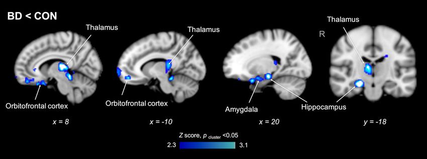
Fig. 2 [11C]Martinostat uptake is lower in the bilateral thalamus, orbitofrontal cortex, right hippocampus, and right amygdala of
participants with bipolar disorder (BD) compared to matched healthy controls (CON). Statistical maps from voxelwise comparison of SUVR
between groups, controlled for age and sex, overlaid onto the MNI 1 mm template in radiological orientation (Z > 2.3, pcluster < 0.05). Blue-light blue
represents regions significantly lower in BD compared to CON (n = 11 subjects per group).
however, smaller volume was associated with lower SUVR relative HDAC expression levels in BD and CON because
(Spearman’s r = 0.67, p = 0.03) and higher emotion reg- epigenetic mechanisms, such as those regulated by
ulation (Spearman’s r = −0.70, p = 0.02) in native space. HDACs, have the potential to reconcile contributions of
both genetic and environmental factors in neuropsychia-
Attention tric disorders including BD. Our primary results indicate
There was no group difference in MCCB attention T- lower relative HDAC expression in the right amygdala of
scores between BD and CON (Table 1). There was no BD, as well as within a broader fronto-limbic distribution
association between SUVR in the left or right amygdala a including the thalamus, orbitofrontal cortex, and hippo-
priori ROIs and attention in BD (left: Spearman’s r = campus. Moreover, relative HDAC expression was related
−0.46, p = 0.16, right: Spearman’s r = 0.16, p = 0.63). In to attention and emotion regulation selectively in BD.
an exploratory whole-brain voxelwise analysis, higher These results suggest a potential role for HDACs in the
SUVR in the bilateral hippocampus and pons, right fundamental pathophysiology of BD as well as in a subset
parahippocampal gyrus, left pallidum and inferior long- of its hallmark clinical features.
itudinal fasciculus (temporal regions) were associated Consistent with our hypothesis, [11C]Martinostat SUVR
with higher attention scores; and lower SUVR in the left was lower in the right amygdala of BD compared to CON.
middle frontal gyrus, pre- and postcentral gyrus, inferior Comparatively, relative HDAC expression was not found
parietal lobule, and lateral occipital cortex (fronto-parietal to differ in the amygdala of individuals with schizophrenia
regions) were associated with higher attention scores in (SCZ) or schizoaffective disorder (SAD) compared to
BD (Z > 2.3, pcluster < 0.05; Supplementary Fig. 4). Posthoc CON in a prior PET study36. Additionally, relative HDAC
assessments of volume showed no correlations between expression was lower in the DLPFC, a brain region rele-
attention and volume (temporal regions: Spearman’s vant to the pathophysiology of SCZ in a previous study
r = 0.35, p = 0.30; fronto-parietal regions: Spearman’s using [11C]Martinostat in SCZ/SAD36, but not in the
r = −0.54, p = 0.09), or between SUVR and volume current study of BD, results that further align with post-
(temporal regions: Spearman’s r = 0.50, p = 0.13; fronto- mortem data14. These observations suggest that lower
parietal regions: Spearman’s r = 0.38, p = 0.25). relative HDAC expression in the right amygdala may be a
specific etiological feature of BD. The group difference in
Discussion SUVR of the left amygdala approached but did not reach
BD is characterized by recurrent episodes of altered significance and may be consistent with the hemispheric
mood involving disruptions in emotion regulation and asymmetry hypothesis of BD, which implicates altered
cognitive processes which lead to overall functional right hemisphere brain function in bipolar depression72.
impairment. A number of structural, functional, and However, this result could reflect a limit in statistical
molecular neuroimaging studies implicate aberrant power to detect medium effect sizes in the current
fronto-limbic neural circuitry in BD24,66–71, however, the sample.
molecular mechanisms underlying structural and func- Exploratory whole-brain voxelwise analyses detected
tional alterations are not fully understood. In this study lower relative HDAC expression in BD compared to CON
we used [11C]Martinostat PET to measure and compare including the right amygdala and hippocampus, bilateralTseng et al. Translational Psychiatry (2020)10:224 Page 7 of 9
thalamus, and orbitofrontal cortex. The amygdala, hip- higher SUVR in temporal regions were correlated with
pocampus, thalamus, and orbitofrontal cortex are higher attention scores in BD. Sustained attention is
involved in mood regulation, sensory integration, and typically supported by engagement of fronto-parietal cir-
decision making, which are frequent clinical presentations cuitry and deactivation of temporo-limbic regions,
in patients with BD73. In particular, our finding in the including the parahippocampal gyrus87. Given that a
thalamus is primarily in the mediodorsal thalamus, which delicate balance of these two cognitive processes is likely
is involved in cognitive processes and attention74,75, likely needed for sustained attention, altered relative HDAC
due to its dense connections to the prefrontal cortex76. expression in these brain regions in BD may contribute to
The mediodorsal thalamus has been found to be under- differences in sustained attention. Overall, our results
connected to the prefrontal cortex in both patients with suggest that altered relative HDAC expression in BD may
BD and schizophrenia using resting state functional MR have impacts on emotion regulation and attention.
imaging77. Moreover, fronto-limbic regions were pre- We acknowledge several limitations of this work. Our
viously shown to be abnormal in BD via structural, study measures differences in [11C]Martinostat uptake
functional, and molecular neuroimaging studies as men- relative to the whole-brain mean (SUVR) and not absolute
tioned above, providing support to the hypothesis that uptake values. Therefore, future [11C]Martinostat PET
altered HDAC expression may contribute to the observed studies with arterial blood sampling in larger sample sizes
regional abnormalities. To date, concordance between will be necessary to validate HDAC expression differences
HDAC function in BD and the neural circuitry underlying in BD. Given substantial cognitive heterogeneity in BD,
behavior has only been indirectly extrapolated across the modest sample size of this study may explain why we
different postmortem and rodent studies15,78. Use of [11C] did not see differences in cognitive performance at the
Martinostat PET begins to fill this gap by identifying group level. Another possibility is that the participants
altered HDAC levels in participants with BD in vivo and with BD in this study are more high-functioning to be able
also offers the unique advantage of possible application to complete a 90-min PET scan, therefore these results
during simultaneous functional MR imaging, which may not be generalizable to all patients with BD. Fur-
should be undertaken in future studies to further dissect thermore, participants with BD were medicated and we do
the potential impact of altered HDAC levels on neural not have standardized rating scales of their mood symp-
circuitry in BD. Furthermore, it would be interesting to toms at the time of the scan. Therefore, future studies
consider anatomical subregions moving forward. For with more narrowly defined cohorts of participants with
instance, HDAC inhibition in the basolateral amygdala BD, including first-episode patients with limited medica-
enhances memory consolidation in rats17. The investiga- tion exposure are needed to assess the potential impact of
tion of amygdalar subnuclei may provide more insight medication status on HDAC expression levels. Despite
into the molecular mechanism of HDAC regulation in these limitations, we did test potential confounding fac-
relation to cognitive function in BD. tors such as anatomical volumes and potential effects of
HDACs regulate genes important for activity-dependent medication, and our findings were robust to these issues.
regulation of neuroplasticity79, as well as genes associated Nonetheless, a larger sample size will be needed to con-
with BD80–83. Therefore, it is possible that altered HDAC firm our findings.
expression may contribute to emotional or cognitive In conclusion, our study presents the first in vivo evi-
disturbances characteristic of BD through altered neuro- dence of altered relative HDAC expression in fronto-
plasticity. In this study, we explored whether relative limbic regions between participants with BD and age- and
HDAC expression levels correlate with emotion regula- sex-matched healthy CON. This work suggests a potential
tion and attention. Whole-brain analyses revealed that link between altered HDAC expression, attention, and
SUVR was associated with MCCB emotion regulation emotion dysregulation in BD.
performance, selectively in BD and within brain regions
that have relevance to emotion regulation. Specifically, the Acknowledgements
left perisylvian region is implicated in language compre- We thank A. Kendall and N. Nortelus for medical coverage; J. Sore and the
hension84, the superior temporal sulcus in multiple social radiopharmacy team for radiotracer synthesis; G. Arabasz, S. Hsu, and R.
Butterfield for assistance with MR-PET imaging; and A. Zhu and M. Hibert for
processes, including theory of mind85, and the middle imaging data collection. This research received funding from the National
frontal gyrus in social judgment86. These correlates of Institute of Mental Health (R21 MH11197101A1 to J.M.H.), Brain and Behavior
emotion regulation in BD differ from those identified in Foundation Independent Investigator Award (J.M.H.) and Young Investigator
Award (T.M.G.), MGH Research Scholar’s Program (J.M.H.), MGH ECOR Fund for
healthy participants (e.g. the inferior longitudinal and Medical Discovery (T.M.G.), and Athinoula A. Martinos Center Postdoctoral Pilot
fronto-occipital fasciculus and hippocampus37), raising Funding (T.M.G.). A.T.P was supported by NIMH T32 MH 112485. This research
the possibility that differential regional patterns of HDAC was carried out at the Athinoula A. Martinos Center for Biomedical Imaging at
MGH, using resources provided by the Center for Functional Neuroimaging
expression may underlie emotion dysregulation in BD. Technologies, P41EB015896, a P41 Biotechnology Resource Grant supported
Additionally, lower SUVR in fronto-parietal regions and by the National Institute of Biomedical Imaging and Bioengineering (NIBIB),Tseng et al. Translational Psychiatry (2020)10:224 Page 8 of 9
and the Neuroimaging Analysis Center, P41EB015902, a P41 supported by 6. Tsankova, N., Renthal, W., Kumar, A. & Nestler, E. J. Epigenetic regulation in
NIBIB. This work also involved the use of instrumentation supported by the psychiatric disorders. Nat. Rev. Neurosci. 8, 355–367 (2007).
National Institutes of Health (NIH) Shared Instrumentation Grant Program; 7. Nestler, E. J., Peña, C. J., Kundakovic, M., Mitchell, A. & Akbarian, S. Epigenetic
specifically, S10RR017208-01A1, S10RR026666, S10RR022976, S10RR019933, basis of mental illness. Neuroscientist 22, 447–463 (2016).
S10RR023043, and S10RR023401. 8. Xiang, B. et al. Systematic genetic analyses of genome-wide association study
data reveal an association between the key nucleosome remodeling and
Author details deacetylase complex and bipolar disorder development. Bipolar Disord. 20,
1 370–380 (2018).
Athinoula A. Martinos Center for Biomedical Imaging, Department of
Radiology, Massachusetts General Hospital, Harvard Medical School, 9. Phiel, C. J. et al. Histone deacetylase is a direct target of valproic acid, a potent
Charlestown, MA 02129, USA. 2Department of Psychiatry, Massachusetts anticonvulsant, mood stabilizer, and teratogen. J. Biol. Chem. 276,
General Hospital, Harvard Medical School, Boston, MA 02114, USA. 36734–36741 (2001).
3 10. Wu, S. et al. Lithium down-regulates histone deacetylase 1 (HDAC1) and
Department of Psychiatry, Boston University School of Medicine, Boston, MA
02118, USA. 4Center for Genomic Medicine, Massachusetts General Hospital, induces degradation of mutant huntingtin. J. Biol. Chem. 288, 35604–35616
Harvard Medical School, Boston, MA 02114, USA (2013).
11. Leng, Y., Fessler, E. B. & Chuang, D. M. Neuroprotective effects of the mood
Author contributions stabilizer lamotrigine against glutamate excitotoxicity: Roles of chromatin
T.M.G., C.-E.J.T., B.G.H., A.T.P., N.R.Z., M.C.C., and J.M.H. designed the study. T.M.G., remodelling and Bcl-2 induction. Int. J. Neuropsychopharmacol. 16, 607–620
B.G.H., M.C.C., A.J.P., M.K., C.W., J.L.R., H.E.B., and R.H.P. collected human imaging (2013).
data, demographic data, and/or cognitive data. C.-E.J.T., T.M.G., B.G.H., and N.R. 12. Arent, C. O. et al. Neuroanatomical profile of antimaniac effects of histone
Z. analyzed human imaging data. C.-E.J.T. and N.R.Z. performed statistical deacetylases inhibitors. Mol. Neurobiol. 43, 207–214 (2011).
analyses. T.M.G., C.-E.J.T., M.C.C., A.T.P, N.R.Z., and J.M.H. wrote the manuscript. 13. Moretti, M. et al. Behavioral and neurochemical effects of sodium butyrate in
All authors edited the manuscript. an animal model of mania. Behav. Pharmacol. 22, 766–772 (2011).
14. Schroeder, F. A. et al. Expression of HDAC2 but Not HDAC1 transcript is
reduced in dorsolateral prefrontal cortex of patients with schizophrenia. ACS
Data availability
Chem. Neurosci. 8, 662–668 (2017).
The data that support these findings are available from the corresponding
15. Benes, F. M. et al. Regulation of the GABA cell phenotype in hippo-
author, J.M.H., upon reasonable request. Human subject data will be
campus of schizophrenics and bipolars. Proc. Natl Acad. Sci. USA 104,
deidentified to protect confidentiality.
10164–10169 (2007).
16. Kwon, B. & Houpt, T. A. Phospho-acetylation of histone H3 in the amygdala
Code availability after acute lithium chloride. Brain Res. 1333, 36–47 (2010).
Custom codes for PET and MR data processing are available from the 17. Valiati, F. E. et al. Administration of a histone deacetylase inhibitor into the
corresponding author, J.M.H., upon reasonable request. basolateral amygdala enhances memory consolidation, delays extinction, and
increases hippocampal BDNF levels. Front. Pharmacol. 8, 1–8 (2017).
Conflict of interest 18. Phillips, M. & Swartz, H. A critical appraisal of neuroimaging studies of bipolar
The content is solely the responsibility of the authors and does not necessarily disorder: toward a new conceptualization of underlying neural circuitry and
represent the official views of Massachusetts General Hospital, Harvard roadmap for future research Mary. Am. J. Psychiatry 171, 829–884 (2014).
University, and its affiliated academic healthcare centers, or the NIH. 19. Strakowski, S. M. et al. The functional neuroanatomy of bipolar disorder: a
Intellectual property (IP) has been filed around [11C]Martinostat by J.M.H. and C. consensus model. Bipolar Disord. 14, 313–325 (2012).
W.; A portion of this IP has been licensed. J.M.H. is a co-founder and equity 20. Korgaonkar, M. S. et al. Amygdala activation and connectivity to emotional
holder of Eikonizo Therapeutics, Inc. and has received honoraria for speaking processing distinguishes asymptomatic patients with bipolar disorders and
or advisory service at non-profit academic institutions. In the past year, J.M.H. unipolar depression. Biol. Psychiatry Cogn. Neurosci. Neuroimaging 4, 361–370
has consulted for Psy Therapeutics, Inc., Amathus Therapeutics Inc., Evelo (2019).
Biosciences Inc., Rodin Therapeutics Inc., and the Alzheimer’s Drug Discovery 21. Davis, M. & Whalen, P. J. The amygdala: vigilance and emotion. Mol. Psychiatry
Foundation. T.M.G. is a current employee of Eikonizo Therapeutics, Inc. The 6, 13–34 (2001).
remaining authors declare no competing interests. 22. Costafreda, S. G., Brammer, M. J., David, A. S. & Fu, C. H. Y. Predictors of
amygdala activation during the processing of emotional stimuli: a meta-
analysis of 385 PET and fMRI studies. Brain Res. Rev. 58, 57–70 (2008).
Publisher’s note 23. Bora, E., Fornito, A., Yücel, M. & Pantelis, C. Voxelwise meta-analysis of gray
Springer Nature remains neutral with regard to jurisdictional claims in matter abnormalities in bipolar disorder. Biol. Psychiatry 67, 1097–1105 (2010).
published maps and institutional affiliations. 24. Chen, C. H., Suckling, J., Lennox, B. R., Ooi, C. & Bullmore, E. T. A quantitative
meta-analysis of fMRI studies in bipolar disorder. Bipolar Disord. 13, 1–15
Supplementary Information accompanies this paper at (https://doi.org/ (2011).
10.1038/s41398-020-00911-5). 25. Henry, C. et al. Emotional dysfunction as a marker of bipolar disorders. Front.
Biosci. 4, 2622–2630 (2012).
26. Quraishi, S. & Frangou, S. Neuropsychology of bipolar disorder: a review. J.
Received: 29 May 2020 Revised: 11 June 2020 Accepted: 18 June 2020
Affect. Disord. 72, 209–226 (2002).
27. Geddes, J. R. & Miklowitz, D. J. Treatment of bipolar disorder. Lancet 381,
1672–1682 (2013).
28. Fukada, M. et al. Loss of deacetylation activity of Hdac6 affects emotional
behavior in mice https://doi.org/10.1371/journal.pone.0030924 (2012).
References
29. Anshu, K. et al. Altered attentional processing in male and female rats in a
1. Angst, J. Bipolar disorders in DSM-5: strengths, problems and perspectives. Int.
prenatal valproic acid exposure model of autism spectrum disorder. Autism
J. Bipolar Disord. 1, 1–3 (2013).
Res. 10, 1929–1944 (2017).
2. McGuffin, P. et al. The heritability of bipolar affective disorder and the
30. Tremolizzo, L. et al. Valproate corrects the schizophrenia-like epigenetic
genetic relationship to unipolar depression. Arch. Gen. Psychiatry 60,
behavioral modifications induced by methionine in mice. Biol. Psychiatry 57,
497–502 (2003).
500–509 (2005).
3. Kerner, B. Genetics of bipolar disorder. Appl. Clin. Genet. 7, 33–42 (2014).
31. Schroeder, F. A. et al. A selective HDAC 1/2 inhibitor modulates chromatin and
4. Johansson, V., Kuja-Halkola, R., Cannon, T. D., Hultman, C. M. & Hedman, A. M. A
gene expression in brain and alters mouse behavior in two mood-related
population-based heritability estimate of bipolar disorder - In a Swedish twin
tests. PLoS ONE 8, e71323 (2013).
sample. Psychiatry Res. 278, 180–187 (2019).
32. Jakovcevski, M. et al. Prefrontal cortical dysfunction after overexpression of
5. Aldinger, F. & Schulze, T. G. Environmental factors, life events, and trauma in
histone deacetylase 1. Biol. Psychiatry 74, 696–705 (2013).
the course of bipolar disorder. Psychiatry Clin. Neurosci. 71, 6–17 (2017).Tseng et al. Translational Psychiatry (2020)10:224 Page 9 of 9
33. Gräff, J. et al. An epigenetic blockade of cognitive functions in the neurode- 61. Keener, M. T. & Phillips, M. L. Neuroimaging in bipolar disorder: a critical review
generating brain. Nature 483, 222–226 (2012). of current findings. Curr. Psychiatry Rep. 9, 512–520 (2007).
34. Guan, J. S. et al. HDAC2 negatively regulates memory formation and synaptic 62. Greve, D. N. et al. Cortical surface-based analysis reduces bias and variance in
plasticity. Nature 459, 55–60 (2009). kinetic modeling of brain PET data. Neuroimage 92, 225–236 (2014).
35. Fischer, A., Sananbenesi, F., Wang, X., Dobbin, M. & Tsai, L. H. Recovery of 63. Greve, D. N. et al. Different partial volume correction methods lead to different
learning and memory is associated with chromatin remodelling. Nature 447, conclusions: an 18 F-FDG PET study of aging douglas. Neuroimage 132,
178–182 (2007). 334–343 (2016).
36. Gilbert, T. M. et al. PET neuroimaging reveals histone deacetylase dysregulation 64. Smith, S. M. et al. Advances in functional and structural MR image analysis and
in schizophrenia. J. Clin. Invest 129, 364–372 (2018). implementation as FSL. Neuroimage 23, S208–S219 (2004).
37. Gilbert, T. M. et al. Neuroepigenetic signatures of age and sex in the living 65. Hollingshead, A. Four Factor Index of Social Status. Unpublished manuscript
human brain. Nat. Commun. 10, 2945 (2019). (Yale University, New Haven, CT, USA, 1975).
38. Schroeder, F. A. et al. PET imaging demonstrates histone deacetylase target 66. Townsend, J. & Altshuler, L. L. Emotion processing and regulation in bipolar
engagement and clarifies brain penetrance of known and novel small disorder: a review. Bipolar Disord. 14, 326–339 (2012).
molecule inhibitors in rat. ACS Chem. Neurosci. 5, 1055–1062 (2014). 67. Perry, A., Roberts, G., Mitchell, P. B. & Breakspear, M. Connectomics of bipolar
39. Wang, C. et al. In vivo imaging of histone deacetylases (HDACs) in the central disorder: a critical review, and evidence for dynamic instabilities within inter-
nervous system and major peripheral organs. J. Med. Chem. 57, 7999–8009 oceptive networks. Mol. Psychiatry 24, 1296–1318 (2019).
(2014). 68. Drevets, W. C. et al. Glucose metabolism in the amygdala in depression:
40. Wey, H.-Y. et al. Kinetic analysis and quantification of [11C]Martinostat for relationship to diagnostic subtype and plasma cortisol levels. Pharmacol.
in vivo HDAC imaging of the brain. ACS Chem. Neurosci. 6, 708–715 (2015). Biochem. Behav. 71, 431–447 (2002).
41. Wey, H.-Y. et al. Insights into neuroepigenetics through human histone dea- 69. Ketter, T. A. et al. Effects of mood and subtype on cerebral glucose meta-
cetylase PET imaging. Sci. Transl. Med. 8, 351ra106 (2016). bolism in treatment-resistant bipolar disorder. Biol. Psychiatry 49, 97–109
42. Baas, D., Aleman, A. & Kahn, R. S. Lateralization of amygdala activation: a (2001).
systematic review of functional neuroimaging studies. Brain Res. Rev. 45, 70. Suhara, T. et al. D1 dopamine receptor binding in mood disorders
96–103 (2004). measured by positron emission tomography. Psychopharmacology 106,
43. Caligiuri, M. P. et al. A functional magnetic resonance imaging study of cortical 14–18 (1992).
asymmetry in bipolar disorder. Bipolar Disord. 6, 183–196 (2004). 71. Oquendo, M. A. et al. Brain serotonin transporter binding in depressed
44. Nuechterlein, K. H. et al. The MATRICS Consensus Cognitive Battery, part 1: test patients with bipolar disorder using positron emission tomography. Arch. Gen.
selection, reliability, and validity. Am. J. Psychiatry 165, 203–213 (2008). Psychiatry 64, 201–208 (2007).
45. Kern, R. et al. The MATRICS Consensus Cognitive Battery, part 2: co-norming 72. Harmon-Jones, E. & Gable, P. A. On the role of asymmetric frontal cortical
and standardization. Am. J. Psychiatry 165, 214–220 (2008). activity in approach and withdrawal motivation: an updated review of the
46. Zhu, Y. et al. The relationship between cognitive dysfunction and symptom evidence. Psychophysiology https://doi.org/10.1111/psyp.12879 (2018).
dimensions across schizophrenia, bipolar disorder, and major depressive dis- 73. Price, J. L. & Drevets, W. C. Neurocircuitry of mood disorders. Neuropsycho-
order. Front. Psychiatry 10, 1–8 (2019). pharmacology 35, 192–216 (2010).
47. Bo, Q. et al. Use of the MATRICS consensus cognitive battery (MCCB) to 74. Mitchell, A. & Chakraborty, S. What does the mediodorsal thalamus do? Front.
evaluate cognitive deficits in bipolar disorder: a systematic review and meta- Syst. Neurosci. 7, 37 (2013).
analysis. PLoS ONE 12, 176212 (2017). 75. Salgado-Pineda, P. et al. Decreased cerebral activation during CPT perfor-
48. Motulsky, H. J. & Brown, R. E. Detecting outliers when fitting data with non- mance: structural and functional deficits in schizophrenic patients. Neuroimage
linear regression – a new method based on robust nonlinear regression and 21, 840–847 (2004).
the false discovery rate. BMC Bioinform. 7, 123 (2006). 76. Ouhaz, Z., Fleming, H. & Mitchell, A. S. Cognitive functions and neurodeve-
49. Woods, S. W. Chlorpromazine equivalent doses for the newer atypical anti- lopmental disorders involving the prefrontal cortex and mediodorsal thala-
psychotics. J. Clin. Psychiatry 64, 663–667 (2003). mus. Front. Neurosci. 12, 33 (2018).
50. Kolb, A. et al. Technical performance evaluation of a human brain PET/MRI 77. Anticevic, A. et al. Characterizing thalamo-cortical disturbances in schizo-
system. Eur. Radiol. 22, 1776–1788 (2012). phrenia and bipolar illness. Cereb. Cortex 24, 3116–3130 (2013).
51. Tisdall, M. D. et al. Volumetric navigators for prospective motion correction 78. Bahari-Javan, S. et al. HDAC1 regulates fear extinction in mice. J. Neurosci. 32,
and selective reacquisition in neuroanatomical MRI. Magn. Reson. Med. 68, 5062–5073 (2012).
389–399 (2012). 79. Haggarty, S. J. & Tsai, L.-H. Probing the role of HDACs and mechanisms
52. Mayer, J. D., Salovey, P., Caruso, D. R. & Sitarenios, G. Measuring emotional of chromatin-mediated neuroplasticity. Neurobiol. Learn. Mem. 96, 41–52
intelligence with the MSCEIT V2.0. Emotion 3, 97–105 (2003). (2011).
53. Cornblatt, B. A., Risch, N. J., Faris, G., Friedman, D. & Erlenmeyer-Kimling, L. The 80. Simonini, M. V. et al. The benzamide MS-275 is a potent, long-lasting brain
continuous performance test, identical pairs version (CPT-IP): I. new findings region-selective inhibitor of histone deacetylases. Proc. Natl Acad. Sci. USA 103,
about sustained attention in normal families. Psychiatry Res. 26, 223–238 1587–1592 (2006).
(1988). 81. Dong, E. et al. Reelin and glutamic acid decarboxylase67 promoter remo-
54. Clark, L. & Goodwin, G. M. State- and trait-related deficits in sustained attention deling in an epigenetic methionine-induced mouse model of schizophrenia.
in bipolar disorder. Eur. Arch. Psychiatry Clin. Neurosci. 254, 61–68 (2004). Proc. Natl Acad. Sci. USA 102, 12578–12583 (2005).
55. McCleery, A. et al. Longitudinal stability of social cognition in schizophrenia: a 82. Tremolizzo, L. et al. An epigenetic mouse model for molecular and behavioral
5-year follow-up of social perception and emotion processing. Schizophr. Res. neuropathologies related to schizophrenia vulnerability. Proc. Natl Acad. Sci.
176, 467–472 (2016). USA 99, 17095–17100 (2002).
56. Fischl, B. et al. Whole brain segmentation: automated labeling of neuroana- 83. Gavin, D. P., Kartan, S., Chase, K., Jayaraman, S. & Sharma, R. P. Histone dea-
tomical structures in the human brain. Neuron 33, 341–355 (2002). cetylase inhibitors and candidate gene expression: an in vivo and in vitro
57. Hibar, D. P. et al. Cortical abnormalities in bipolar disorder: an MRI analysis of approach to studying chromatin remodeling in a clinical population. J. Psy-
6503 individuals from the ENIGMA Bipolar Disorder Working Group. Mol. chiatr. Res. 43, 870–876 (2009).
Psychiatry 23, 932–942 (2018). 84. Koenigs, M. et al. Areas of left perisylvian cortex mediate auditory-verbal short-
58. Hibar, D. P. et al. Subcortical volumetric abnormalities in bipolar disorder. Mol. term memory. Neuropsychologia 49, 3612–3619 (2011).
Psychiatry 21, 1710–1716 (2016). 85. Beauchamp, M. S. The social mysteries of the superior temporal sulcus. Trends
59. Izquierdo-Garcia, D. et al. An SPM8-based approach for attenuation correction Cogn. Sci. 19, 489–490 (2015).
combining segmentation and nonrigid template formation: application to 86. Mukherjee, P. et al. Altered amygdala connectivity within the social brain in
simultaneous PET/MR brain imaging. J. Nucl. Med. 55, 1825–1830 (2014). schizophrenia. Schizophr. Bull. 40, 152–160 (2013).
60. Chonde, D. B., Izquierdo-Garcia, D., Chen, K., Bowen, S. L. & Catana, C. Masa- 87. Lawrence, N. S., Ross, T. J., Hoffmann, R., Garavan, H. & Stein, E. A. Multiple
mune: a tool for automatic dynamic PET data processing, image recon- neuronal networks mediate sustained attention. J. Cogn. Neurosci. 15,
struction and integrated PET/MRI data analysis. EJNMMI Phys. 1, A57 (2014). 1028–1038 (2003).You can also read