Reversal of brain aging by targeting telomerase: A nutraceutical approach - Spandidos ...
←
→
Page content transcription
If your browser does not render page correctly, please read the page content below
INTERNATIONAL JOURNAL OF MOLECULAR MEDICINE 48: 199, 2021

                               Reversal of brain aging by targeting
                              telomerase: A nutraceutical approach
          DIMITRIS TSOUKALAS1‑3*, ANA MARIA BUGA4*, ANCA OANA DOCEA5*, EVANGELIA SARANDI2,6,
              RADU MITRUT7, ELISAVET RENIERI6,8, DEMETRIOS A. SPANDIDOS9, ION ROGOVEANU10,
          LILIANA CERCELARU11, MIHAELA NICULESCU11, ARISTIDIS TSATSAKIS6 and DANIELA CALINA1

          1
            Department of Clinical Pharmacy, University of Medicine and Pharmacy of Craiova, 200349 Craiova, Romania;
                 2
                   Metabolomic Medicine, Health Clinic for Autoimmune and Chronic Diseases, 10674 Athens, Greece;
          3
           European Institute of Nutritional Medicine (E.I.Nu.M.), I‑00198 Rome, Italy; Departments of 4Biochemistry and
     5
       Toxicology, University of Medicine and Pharmacy of Craiova, 200349 Craiova, Romania; 6Laboratory of Toxicology and
            Forensic Sciences, Medical School, University of Crete, 71003 Heraklion, Greece; 7Department of Cardiology,
     University and Emergency Hospital, 050098 Bucharest, Romania; 8Environmental Engineering Laboratory, Department of
         Chemical Engineering, Aristotle University of Thessaloniki, 54124 Thessaloniki; 9Laboratory of Clinical Virology,
               Medical School, University of Crete, 71409 Heraklion, Greece; Departments of 10Internal Medicine and
              11
                 Anatomy and Embryology, University of Medicine and Pharmacy of Craiova, 200349 Craiova, Romania

                                         Received 22 July 2021; Accepted September 7, 2021

                                                       DOI: 10.3892/ijmm.2021.5032

Abstract. Telomeres, the protective caps of chromosomes,              by ELISA and immunohistochemistry. In addition, the find‑
shorten with age, as telomerase, the enzyme responsible               ings from PCR‑ELISA demonstrated an enhanced telomerase
for the compensation of telomere erosion, is inactive in the          activity in the cerebellum and cortex cells in the brains of rats
majority of cells. Telomere shortening and subsequent cell            treated with the ‘Reverse™’ supplement. The histopathological
senescence lead to tissue aging and age‑related diseases.             findings confirmed a structural reversibility effect close to the
Neurodegenerative disorders, characterized by the progressive         differentiation observed in the young control group of rats
loss of neurons among other hallmarks of aged tissue, and             treated with two capsules/kg body weight of the ‘Reverse™’
poor cognitive function, have been associated with a short            supplement. On the whole, the findings of the present study
telomere length. Thus, telomerase activity has emerged as a           provide a strong indication that an increased telomerase
therapeutic target, with novel agents being under investigation.      activity and TERT expression may be achieved not only in
The present study aimed to examine the effects of a novel             the postnatal or embryonic period, but also in the brains of
natural telomerase activator, ‘Reverse™’, containing Centella         middle‑aged rats through nutraceutical supplementation. The
asiatica extract, vitamin C, zinc and vitamin D3 on the brains        use of the ‘Reverse™’ supplement may thus contribute to the
of 18‑month‑old rats. The administration of the ‘Reverse™’            potential alleviation of a number of central nervous system
supplement for 3 months restored telomerase reverse tran‑             diseases.
scriptase (TERT) expression in the brains of rats, as revealed
                                                                      Introduction

                                                                      Telomerase is a ribonucleoprotein enzyme which functions as
                                                                      a reverse transcriptase to maintain the length of telomeres, by
Correspondence to: Professor Anca Oana Docea, Department
of Toxicology, University of Medicine and Pharmacy of Craiova,        adding tandem short‑sequence repeats at the end of chromo‑
2 Petru Rareș Street, 200349 Craiova, Romania                         somes, thus compensating for the loss of deoxyribonucleic acid
E‑mail: ancadocea@gmail.com                                           (DNA) during genome replication. It consists of the protein
                                                                      telomerase reverse transcriptase (TERT) and the telomerase
Professor Daniela Calina, Department of Clinical Pharmacy,
                                                                      RNA component (TERC), which facilitate telomere synthesis
University of Medicine and Pharmacy of Craiova, 2 Petru Rareș
Street, 200349 Craiova, Romania                                       and is regulated by several proteins that are bound to TERT
E‑mail: calinadaniela@gmail.com                                       and TERC, forming the enzymatically active telomerase (1).
                                                                      In addition, TERT has been reported to perform additional
*
    Contributed equally                                               to telomere elongation functions, including its translocation
                                                                      to the mitochondria and its contribution to the decrease in
Key words: telomerase activators, telomerase reverse transcriptase,   oxidative stress‑related complications upon the accumulation
aging, Centella asiatica, vitamin C, zinc, vitamin D                  of reactive oxygen species (ROS) (2). It has been demonstrated
                                                                      that upon increased levels of oxidative stress or other less
                                                                      common stimuli, TERT is located in the mitochondria where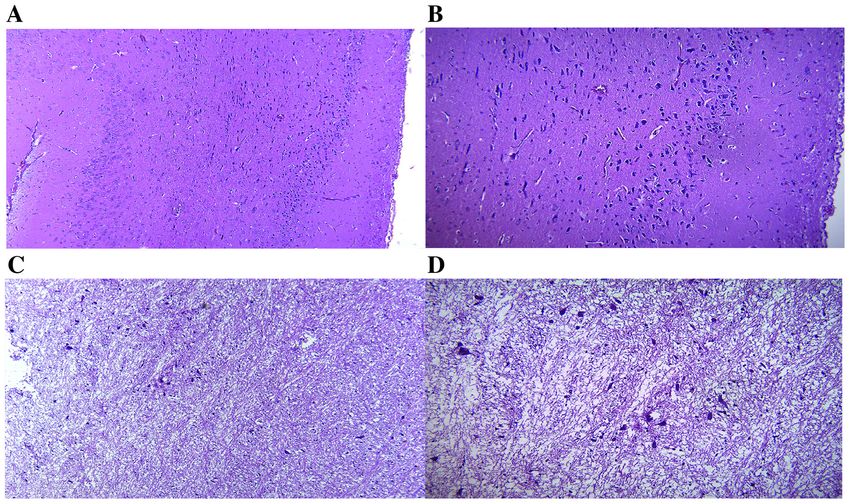
2                           TSOUKALAS et al: REVERSAL OF BRAIN AGING BY TARGETING TELOMERASE

it reduces mitochondrial, as well as nuclear DNA damage (3),         parameters (20). Further analysis has demonstrated that cyclo‑
enhances the natural antioxidant and detoxification cellular         astragenol is the active compound responsible for the increased
mechanisms (4) and improves respiratory chain function,              telomerase activity. Other known telomerase activators include
thereby reducing ROS production (5,6). Telomerase is inactive        product B, a combination of antioxidants, and the synthetic
or expressed at low levels in the majority of somatic cells;         GRN510, which has a similar structure to cycloastragenol (21).
however, an increased activity is observed in highly prolifer‑       A previous comparative in vitro analysis of natural compounds
ating cells, such as adult stem cells, male sperm cells, activated   identified a novel telomerase activator 08AGTL formula‑
lymphocytes and some epidermal cells (7,8). Consequently,            tion containing Centella asiatica extract; this formulation
somatic cells have a definite number of divisions, upon              was found to induce telomerase activation by up to 8.8‑fold
which they become senescent or undergo apoptosis through             compared with 2.2‑fold in cells treated with TA‑65® (22). The
a mechanism that involves telomeres and the DNA damage               present study aimed to investigate the potency of the novel
response (9). Briefly, telomeres are DNA sequences at the very       natural formulation ‘Reverse™’ containing Centella asiatica
end of chromosomal DNA consisting of the TTAGGG pattern,             extract, vitamin C, zinc and vitamin D3 on telomerase expres‑
bound to the shelterin proteins, forming a DNA‑protective            sion and telomerase activity in the brains of rats.
structure known as the T‑loop (10). This structure protects
the coding DNA sequences lying before the telomeres from             Materials and methods
exonuclease activity or DNA damaging agents (11). However,
telomere length decreases with each cell replication by several      Animal study design. Male Sprague‑Dawley rats (n=6;
hundred bases due to the inability of DNA polymerase to              3 months old, with a body weight between 320 and 380 g)
replicate the 3' telomeres and maintain their length to the          and 18 male Sprague‑Dawley rats (18 months old, with a
daughter DNA strands. Telomeres shorten gradually up to a            body weight between 500 and 580 g) obtained from the
critical point at which they signal cell proliferation arrest to     Animal Facility of the University of Medicine and Pharmacy
prevent cellular and DNA damage (12). Senescent or dying             of Craiova (Craiova, Romania) were included in the present
cells are replaced by somatic stem cells of the respective tissue    study. The animal experiments were performed according
to maintain tissue function. The dominant hypothesis of aging        to the EU Directive 2010/63/E.U. as amended by Regulation
and age‑related diseases suggests that the shortening of the         E.U.2019/1010 regarding animal experiments and approved
telomeres of somatic stem cells hampers their tissue renewal         by the Ethics Committee of the University of Medicine and
capacity, which leads to the accumulation of senescent cells         Pharmacy of Craiova, Craiova Romania with the license
in the tissue and, respectively, to the aged phenotype of the        no. 102/23.09.2019. Prior to the start of the experiment, the
tissue (13).                                                         animals were allowed to acclimatize to their environment for
     Neurodegenerative disorders, such as Alzheimer's disease        2 weeks. The animals were housed 2‑3 animals per cage, with
(AD) and Parkinson's disease (PD), are characterized by the          a 12‑h dark/light cycle, and a constant humidity (50±5%) and
progressive loss of neurons that ultimately leads to the decline     temperature (21±2˚C). During the study, the animals had free
of brain function, and the deterioration of cognition and/or         access to standard animal diet and tap water.
locomotor activity (14,15). Among the main risk factors of               The animals were divided into four groups, with 6 animals
such disorders is the accumulation of protein aggregates,            per group, and received the treatment once per day via gavage
such as α‑synuclein, amyloid‑β and pathological tau proteins,        for a period of 3 months, as presented in Table I. Based on
as well as oxidative stress and mitochondrial dysfunc‑               power analysis, the minimum number of animals per group
tion (16‑18). Epigenetic modifications have been suggested to        was estimated at 5; however, due to the fact that the experi‑
regulate neuroinflammation and subsequent neurodegenera‑             ments included middle‑aged animals, 6 animals per group
tion. Thus, several epigenetic biomarkers have emerged in brain      were used to prevent the unexpected loss of animals. The
diseases, such as patterns of methylation and the expression of      workflow of the study is presented in Fig. 1.
specific microRNAs (miRNAs/miRs), as well as the levels of
serum extracellular vesicle‑derived circular RNAs (19,20). The       Treatment and dose selection. The treatment consisted of
epigenome is dynamically regulated through diet and lifestyle,       the administration of the ‘Reverse™’ supplement (Natural
thus opening a novel avenue for neurodegenerative disorder           Doctor S.A), notified as a food supplement (Notification
therapeutics through the combination of pharmaceutical and           no. 6704/21‑1‑2020) at the Greek National Organization for
nutritional strategies (21,22).                                      Medicines under current legislation for food supplements.
     Of note, a short telomere length has been shown to be               The details regarding the dose selection have been previ‑
associated with poor cognitive function and neurodegenerative        ously described (10). Briefly, the animals from the treatment
diseases. Although the etiopathogenesis of neurodegenerative         groups received 1 or 2 capsules/kg body weight/day of the
diseases has yet to be fully elucidated, telomere attrition and      ‘Reverse™’ supplement suspended in corn oil at a final volume
telomerase expression have emerged as novel mechanisms and           of 1.5 ml. Each capsule contains 9 mg Centella asiatica (L.)
potential therapeutic targets (19). Several telomerase activators    extract [consisting of a >90% high purity single chemical
exist, including both natural and synthetic compounds, acting        entity, as assessed by high‑performance liquid chromatog‑
through genetic manipulation mediated by adeno‑associated            raphy and gas chromatography (data not shown)], vitamin C
viruses. A widely known natural compound is TA‑65 ®                  (200 mg as magnesium ascorbate), zinc (5 mg as zinc citrate)
containing Astragalus membranaceus extract that has been             and vitamin D3 (50 µg as cholecalciferol) per capsule.
shown to activate telomerase and to be associated with longer            The administered doses and treatment duration were
telomeres of peripheral blood monocytes and improved health          in accordance with clinical recommendations relative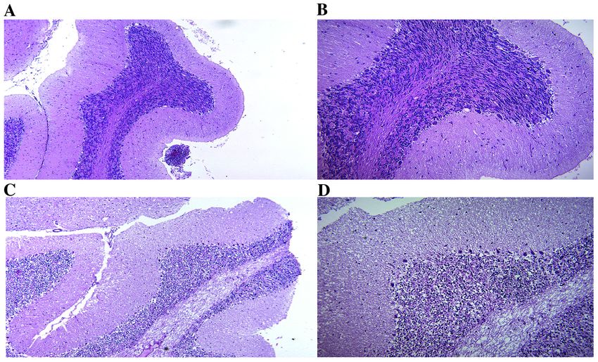
INTERNATIONAL JOURNAL OF MOLECULAR MEDICINE 48: 199, 2021 3
Table I. Groups and corresponding treatments in the present study.
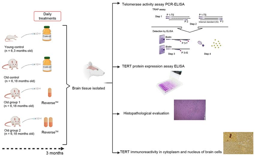
Animal groups Young control, (n=6) Old‑aged control, (n=6) Old‑aged group 1, (n=6) Old‑aged group 2, (n=6)
Age of the animals 3 18 18 18
at the beginning
of the study (months)
Treatments Corn oil Corn oil 1 capsule/kg 2 capsules/kg
body weight of body weight of
‘Reverse™’ ‘Reverse™’
(Natural Doctor S.A) (Natural Doctor S.A)
Figure 1. Workflow of the study design. TRAP, telomeric repeat amplification protocol; TERT, telomerase reverse transcriptase.
to body weight and with respect to previous studies for For treatment group 2, the rat dose = 2/60x6.2x10=2.06 capsule/
vitamin C (23,24), vitamin D3 (25,26) and zinc (27). For kg/body weight, approximate to 2 capsules kg/body weight per
Centella asiatica, no established clinical recommendations rat.
exist and the administered dose in the present study was mark‑ The animals from the young and old‑aged control group
edly lower than that used in previous research (28). received only corn oil used as a solvent in a volume of 1.5 ml
The doses used for the rats were converted from those used once per day. The duration of the administration was set as
for humans using the factor method. The allometric scaling 3 months that correspond to subchronic administration for
approach, following the Food and Drug Administration guide‑ rats. This was set based on a previous study by the authors
lines, was applied for calculations and more specifically, the using healthy human volunteers and using other vitamin
correction factor (km) for rats used was equal to 6.2 and the supplements, where it was demonstrated that the modulation of
safety factor value for converting rat doses to humans was telomerase and telomere length is usually observed following
equal to 10 (29). The dose values, as previously described (10) 6‑12 months of treatment (7).
were calculated as follows: Rat dose = human dose x6.2x10,
where human dose = 1/60 capsule/kg/body weight (reference Brain collection. Following 3 months of treatment, the animals
human body weight = 60 kg (29). were sacrificed by exsanguination from the abdominal aorta
For treatment group 1 corresponding to the human dose under 5% sevoflurane anesthesia. The brains were collected,
of 1 capsule/day, the rat dose = 1/60x6.2x10=1.03 capsule/ and half were formalin‑fixed for evaluation using immuno‑
kg/body weight, approximate to 1 capsule/kg/body weight per rat. histochemistry and histopathology, and the other half of the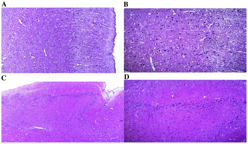
4                         TSOUKALAS et al: REVERSAL OF BRAIN AGING BY TARGETING TELOMERASE

cortex and cerebellum were separated and shock‑frozen in         with 3,3'‑diaminobenzidine (DAB; Vector Laboratories, Inc.),
small sections in liquid nitrogen and kept ‑80˚C until further   the sections were dried overnight at 37˚C and covered using
analysis.                                                        mounting medium (Vector Laboratories, Inc.). The visualiza‑
                                                                 tion and image collection of the slides was performed using
Telomerase activity assay. Telomerase activity in the cortex     a Nikon 90i motorized microscope equipped with a DS‑Ri2
and cerebellum was measured using a commercial rat telom‑        16 MP complementary metal oxide semiconductor (CMOS)
erase PCR‑ELISA (TeloTAGGG Telomerase PCR ELISA                  camera and the Nikon NIS Elements AR software package
PLUS; cat. no. 12 013 789 001, Roche Diagnostics), based on      Ver5.11.03 (Nikon Corporation).
a photometric enzyme immunoassay for quantitative deter‑
mination utilizing a telomeric repeat amplification protocol     Histopathological evaluation. Brain tissue was collected
(TRAP) (30). TRAP reaction was performed according to            and kept for 48 h in a 4% paraformaldehyde solution. The
the manufacturer's protocol using the Applied Biosystems         samples were successively dehydrated in solution with
PCR system (30 cycles at 50˚C; cat. no. 4485701; Applied         increasing concentrations of ethanol (1 h in 70% solution, 1 h
Biosystems; Thermo Fisher Scientifiic, Inc.); the reactions      in 90% solution and 5 h in 100% solution) and then cleared in
were performed in triplicate. The detection of amplicons         xylene for 2 h. The tissue was then embedded in paraffin. From
was further quantified using ELISA (StatFax 4700 ELISA           the paraffin block, sections of 25 µm thickness were cut and
reader system, Awareness Technology, Inc.). The absorbance       stained with hematoxylin and eosin (Sigma‑Aldrich) according
values are reported as the A450nm reading against the blank.     to the standard protocol (31). The histopathological examina‑
The mean of the absorbance reading of the negative control       tion was performed under the Panthera L research microscope
from those of the sample was extracted. Samples are regarded     (Motic Europe, S.L.U.) with the licensed Panthera L photo‑
as telomerase‑positive if the difference in the absorbance was   micrographic image acquisition software Motic Images
>0.2 units.                                                      Plus 2.0ML for Microsoft Windows Legacy Version w/o MI
                                                                 Devices (Motic Europe, S.L.U.).
TERT protein expression assay. The TERT levels in the cortex
and cerebellum were measured using a commercial rat TERT         Statistical analysis. Statistical analysis was performed using
sandwich ELISA kit (cat. no. SEC241Ra; Cloud‑Clone Corp.)        GraphPad Prism version 5.0 software for Windows (GraphPad
according to the manufacturer's instructions. Brain tissue       Software, Inc.). One‑way analysis of variance (ANOVA; para‑
homogenate was prepared as follows: After microdissection,       metric) for independent samples was used for the comparisons
the brain tissues (cortex and cerebellum) were weighted and      of the mean values for more than two independent groups.
homogenized in 0.5 ml of 0.01 M PBS (pH 7.0) using a tissue      One‑way ANOVA was followed by post hoc analysis using the
homogenizer on ice. The suspensions were subjected to two        Bonferroni adjusted t‑test. A P‑value ≤0.05 was considered to
freeze/thaw cycles to further break the cell membranes and       indicate a statistically significant difference.
were centrifuged for 5 min at 500 x g at 4˚C. The supernatant
was stored at ‑20˚C until analysis (no more than 1 month).       Results
    TERT assay was performed using 100 µl supernatant,
according to the manufacturer's instructions and measured        Telomerase activity. The telomerase activity levels were
spectrophotometrically at a wavelength of 450 nm using an        significantly lower in the brain cortex of the old‑aged control
ELISA reader system (StatFax 4700, Awareness Technology,         group (0.17±0.01; P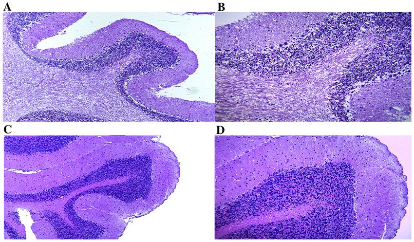
INTERNATIONAL JOURNAL OF MOLECULAR MEDICINE 48: 199, 2021                                                           5

Figure 2. Telomerase activity following 3 months of treatment expressed as absorbance units (A450 nm). (A) In the cerebellum and (B) in the cortex. *P
6 TSOUKALAS et al: REVERSAL OF BRAIN AGING BY TARGETING TELOMERASE
Figure 5. Microscopic structure of the cerebral cortex. (A) Young control group. Fragment with the microscopic structure of the cerebral cortex with normal
differentiation. H&E staining; magnification, x100. (B) Young control group. More detailed view of the image in panel A. H&E staining; magnification, x200.
(C) Old‑aged control group. Fragment with the microscopic structure of the cerebral cortex with cytoarchitectonic structure modified by edema and a poor
neuronal cell distribution is shown. H&E staining; magnification, x100. (D) Old‑aged control group. More detailed view of the image in panel C, with rare large
and medium pyramidal neurons dissociated by marked edema. H&E staining; magnification, x200. H&E, hematoxylin and eosin.
Figure 6. Microscopic structure of the cerebral cortex. (A) Old‑aged group 1. Fragment of the microscopic structure of cerebral cortex with cytoarchitectonic
structure modified by edema and poor neuronal cellular distribution is shown. H&E staining; magnification, x100. (B) Old‑aged group 1. More detailed view of
the image in panel A, with the identification of cerebral cortex layers. H&E staining; magnification, x200. (C) Old‑aged group 2. Fragment of the microscopic
structure of the cerebral cortex with relative normal cytoarchitectonic structure is shown. H&E staining; magnification, x100; (D) More detailed view of the
image in panel C, with the identification of the deep layers of the cerebral cortex. H&E staining; magnification, x200. H&E, hematoxylin and eosin.
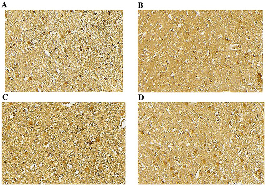
differences in TERT protein levels were observed between distribution with cytoarchitectonics modified by poor neuronal
the old‑aged control group and old‑aged group 1 in both the cellularity. Several different areas of each cerebral cortex were
cortex and cerebellum tissue (Fig. 3). Staining with anti‑TERT observed and the results were similar; thus, the most represen‑
antibody revealed that TERT immunoreactivity was present in tative images are presented (Fig. 5C and D). By contrast, the
the cytoplasm and nucleus of the brain cells in the rat cortex young control group exhibited a normal microscopic structure
stained with DAB (Fig. 4). (Fig. 5A and B). Treatment with 1 capsule/kg body weight
of the ‘Reverse™’ supplement for 3 months led to a slight
Histopathological evaluation decrease in edema; however, the cytoarchitectonic structure
Histopathological aspect of the cerebral cortex. The micro‑ remained altered and a poor neuronal cellular distribution was
scopic evaluation of the rat cerebral cortex of the old‑aged observed (Fig. 6A and B). In the old‑aged group 2, following
control group, revealed an increase in edema that dissociated treatment with 2 capsules/kg body weight of the ‘Reverse™’
the layers of the cerebral cortex and appeared as an unequal supplement for 3 months, the cytoarchitecture of the cerebral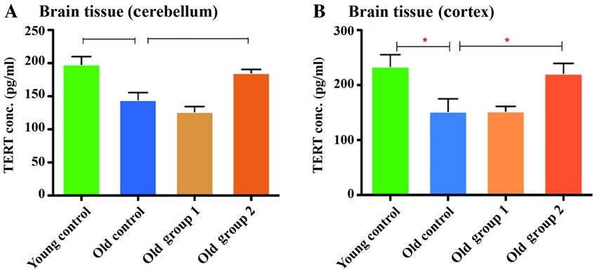
INTERNATIONAL JOURNAL OF MOLECULAR MEDICINE 48: 199, 2021                                                               7

Figure 7. Microscopic structure of the cerebellar cortex. (A) Young control group. Fragment of the microscopic structure of cerebellar cortex with normal
differentiation is shown. H&E staining; magnification, x100. (B) Young control group. More detailed view of the image in panel A, in which the layers of the
cerebellar cortex are observed . H&E staining; magnification, x200. (C) Old‑aged control group. Fragment of the microscopic structure of the cerebellar cortex
with the modified architecture of the layers of the cerebellar cortex is shown. H&E staining; magnification, x100; (D) Old‑aged control group. More detailed
view of the image in panel C, in which the modification of the architecture of the layers of the cerebellar cortex is observed, with increased interneuronal
edema. H&E staining; magnification, x200. H&E, hematoxylin and eosin.

Figure 8. Microscopic structure of the cerebellar cortex. (A) Old‑aged group 1. Fragment of the microscopic structure of the cerebellar cortex with the modified
architecture of the layers of the cerebellar cortex with discrete interneuronal edema is shown. H&E staining; magnification, x100. (B) Old‑age group 1. More
detailed view of the image in panel A. H&E staining; magnification, x200. (C) Old‑aged group 2. Fragment of the microscopic structure of cerebellar cortex
with a relatively normal cytoarchitectonic structure is shown. H&E staining; magnification, x100. (D) More detailed view of the image in panel C revealing
the layers of the cerebellar cortex. H&E staining; magnification, x200. H&E, hematoxylin and eosin.

cortex appeared relatively normal, similar to the differentia‑                    of each cerebellar cortex were observed and the results were
tion observed in the young control group and the edema was                        similar; thus, the most representative images are presented
discrete in the intermediate layers of the cerebral cortex                        (Fig. 7C and D). By contrast, the young control group exhibited
(Fig. 6C and D).                                                                  a normal microscopic structure (Fig. 7A and B). In the group
    Microscopic aspect of the cerebellar cortex. The micro‑                       treated with 1 capsule/kg body weight of the ‘Reverse™’
scopic evaluation of the rat cerebellar cortex of the old‑aged                    supplement for 3 months (old‑aged group 1) the modified cyto‑
control group revealed an increase in edema that dissociated                      architecture at the level of the cerebellar layers, through edema,
the layers of the cerebellar cortex; a modified cytoarchitec‑                     remained and the effects were slightly milder compared with
ture was also observed that was accentuated at the level of                       the those in the old‑aged control group (Fig. 8A and B). In
the cerebellar layers, although not in the same measure as the                    old‑aged group 2 that received 2 capsules/kg body weight of
one identified in the cerebral cortex. Several different areas                    the ‘Reverse™’ supplement for 3 months, the cytoarchitecture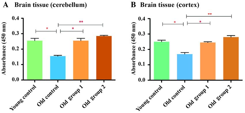
8                          TSOUKALAS et al: REVERSAL OF BRAIN AGING BY TARGETING TELOMERASE

of the cerebellar cortex was relatively normal, similar to the      2 capsules/kg body weight per rat of the ‘Reverse™’ dietary
differentiation observed in the young control group, and the        supplement is more effective for telomerase activation and
edema was discrete in the intermediate layers of the cerebellar     TERT level restoration in the brain than the 1 capsule dose.
cortex (Fig. 8C and D).                                                 The same microscopic changes produced by aging were
                                                                    noted following the comparative analysis performed on
Discussion                                                          the cerebral and cerebellar cortex. The edema was constant
                                                                    and accentuated in both types of cortex with a modi‑
The present study aimed to investigate the activity of telom‑       fied cytoarchitecture and poor neuronal cellularity in the
erase and the expression of TERT in the brains of rats treated      21‑month‑old rats. Previous research supports these findings.
with a novel telomerase activator containing Centella asiatica      Desbordes and Cohadon (35) demonstrated that in rat brains,
extract, vitamin C, zinc and vitamin D3 for 3 months.               cellular swelling increased with aging, while extracellular
    Brain ageing is a key risk factor and mechanism respon‑         edema was less evident. These findings can be explained by
sible for a number of neurodegenerative diseases (32,33), and       the decreased activity of Na+‑K+‑ATPase activity. It has been
it has been demonstrated that telomerase activity and TERT          demonstrated that at 3 months of age, which corresponds to the
abundance may play a protective role in neurons and brain cell      end of adolescence, rats reach the peak number of neurons both
functions (34).                                                     in the cerebellum and cortex. From this age on, the progressive
    To the best of our knowledge, the present study demon‑          decline in the number of neurons commences (36). Morterá and
strates for the first time that the administration of the dietary   Herculano‑Houzel (37) demonstrated that by 22 months of age,
supplement ‘Reverse™’ containing Centella asiatica extract,         the decrease in neurons in the cerebellum and cortex of rats is
vitamin C (as magnesium ascorbate), zinc (as zinc citrate)          almost 30% less than the value observed at the age of 3 months.
and vitamin D3 (as cholecalciferol), to 18‑month‑old rats           In the present study, the administration of 1 capsule/kg body
for 3 months, restores the decline in TERT expression and           weight of the ‘Reverse™’ supplement for 3 months exerted
enhances telomerase activity. The findings presented herein         only a slight beneficial effect on the decline in the number of
demonstrated that the administration of the dietary supple‑         neurons. The administration of 2 capsules/kg body weight of
ment ‘Reverse™’ resulted in a moderate increase in TERT             the ‘Reverse™’ supplement for 3 months to the 18‑month‑old
expression, and the telomerase activity was highly increased        rats (old‑aged group 2) exerted a structural reversibility effect
between old‑aged groups 1 and 2, and the young control              close to the differentiation of the gray matter from the young
group. The findings also demonstrated that the activation           control group that is explained by telomerase activation and the
of telomerase activity could be achieved despite the limited        restoration of TERT levels in the brain observed in this group.
increase in TERT expression. However, further studies using         It has been demonstrated that TERT inhibits the death of cells
larger‑sized groups are warranted to establish whether there        and promote the survival of neurons (38). Even if neurons are
is a dose‑response association. These findings are in line with     non‑dividing cells, telomerase expression in neurons plays an
those in a previous study by the authors which demonstrated         additional role in regulating apoptosis beyond its function in
that ‘Reverse™’ improved motor performance and decreased            telomere maintenance (39).
the stress levels of rats (10).                                         Previous studies on telomerase expression enhancement
    Specifically, in the present study, telomerase activity and     in the brain have been conducted either via genetic interven‑
TERT expression were shown to be significantly lower in             tion or by the use of synthetic or natural enzyme activators,
21‑month‑old rats compared with 6‑month‑old rats in both the        as reviewed by Saretzki and Wan (34). As regards natural
brain cortex and cerebellum, suggesting the age‑dependent           activators, TA‑65® and GRN510 have yielded encouraging
expression of TERT in rats, in line with other findings (10).       results on the beneficial effects of telomerase activation on
The TERT levels regulate telomerase activity, and it has been       brain function. Specifically, as previously demonstrated, the
described that telomerase activity decreases during embryo‑         administration of TA‑65® to aged mice ameliorated several
genesis. By contrast, the TERT levels are maintained, at least      physiological parameters, including neuromuscular coordina‑
in humans, possibly to protect neurons from oxidative stress        tion and cognition accompanied by the increased expression
and neurodegeneration (34).                                         of the mouse TERT (mTERT). Furthermore, treatment with
    The dose of 1 capsule of ‘Reverse™’ supplement per kg           TA‑65® and GRN510 yielded similar results in brain‑specific
body weight per rat (old‑aged group 1) resulted in a statisti‑      TERT expression, but only TA‑65® decreased mitochondrial
cally significant increase in telomerase activity in the cortex     ROS accumulation and α‑synuclein aggregates (40). However,
and cerebellum of 21‑month‑old rats. Comparable results             telomerase activation with TA‑65®, which contains Astragalus
were obtained for TERT expression, although these did not           membranaceus extract, has been shown to be significantly
reach statistical significance. The effect was more evident         lower than with formulations including Centella asiatica
with the 2‑capsule dose (old‑aged group 2) where the levels of      extract (22). ‘Reverse™’ is a dietary supplement containing
telomerase activity were significantly higher than those in the     Centella asiatica extract, vitamin C, vitamin D3 and zinc,
old‑aged control group, reaching the levels of the young group      constituents with known antioxidant properties previously
both in the cortex and the cerebellum of the rats. Accordingly,     described in terms of their relevance to the present study (10).
the TERT expression levels were higher in the old‑aged group 2      In particular, Centella asiatica has been shown to exert neuro‑
than in the old‑aged group 1 and the old‑aged control group.        protective effects, while enhancing cognitive function (41).
Anti‑TERT immunohistochemistry staining in the cortex               Vitamin C and zinc have been reported to play a neuropro‑
and cerebellum tissue slides confirmed the aforementioned           tective role and function synergistically to improve synaptic
results. These findings suggest that the oral administration of     activity and detoxification, and to maintain the physiological
INTERNATIONAL JOURNAL OF MOLECULAR MEDICINE 48: 199, 2021 9
function of protein degradation mediated by ubiquitin (42). functional outcomes following brain lesions, such as stroke or
Vitamin D deficiency is a known and well‑described risk factor neurodegenerative diseases, and may prove to play a crucial
for several neurodegenerative diseases (43,44). Vitamin D has role in the management of a number of CNS diseases.
been described as a key neuroprotector through its role as an AD and PD are slowly progressive neurodegenerative
immunomodulator, a regulator of calcium levels in neurons disorders that occur upon irreversible brain damage, extensive
and a mediator of the detoxification mechanism (45,46). inflammation and oxidative stress, leading to dementia and
Telomerase activity has been reported to be enhanced in motor/non‑motor traits, respectively. In AD, telomere attri‑
previous in vivo studies on vitamin C, vitamin D, zinc and tion emerges as a critical mechanism possibly related to the
Centella asiatica (47,48). However, additional studies are increased oxidative stress that precedes the accumulation of
required to determine whether the combination of these amyloid and neurofibrillary tangles (58). TERT expression
compounds acts synergistically in terms of telomerase activa‑ inversely correlates with tau expression in neurons, indicating
tion and to investigate the underlying molecular mechanisms. an oxidative stress‑mediated mechanism of AD. AD can occur
Overall, the beneficial effects of natural products on oxida‑ during the later or early stages of life (known as early‑onset
tive stress and inflammation is a widely studied field; thus, AD) and occurs prior to the age of 60. There is evidence to
they have emerged as potential therapeutic targets in several indicate that midlife interventions focusing on modifiable risk
neurodegenerative disorders. Indeed, there is evidence to factors can prevent dementia (59). Accordingly, PD etiopatho‑
indicate that phytochemicals, such as polyphenols, found in genesis includes the accumulation of α‑synuclein caused by
pomegranate (47), aloe vera, red fruits and other natural prod‑ folding impairment and its defective clearance through the
ucts are important for normal brain function (15,17,49‑51). autophagic pathway. It has been demonstrated that telomerase
Detailed analyses of the different modes of action of natural activators can successfully facilitate the normal function of
phytochemicals in AD have suggested that certain compounds autophagy and the reduction of protein aggregates, possibly
may serve as potent therapeutic agents as they are effective, mediated by the increase in TERT levels. In addition, TERT is
easy‑to access, safe and lower‑cost options compared to other known for its non‑canonical function as an antioxidant defense
drugs (14,32,52,53). mediator, which further supports the physiological protein
Although not aiming to extrapolate the present findings, post‑translational folding and mitochondrial function (60).
it may be noted that the beneficial effects of ‘Reverse™’ on In conclusion, to the best of our knowledge, the present
telomerase/TERT levels may be mediated by the reduction of study provides the first evidence of the potency of a novel
oxidative stress in the brain microenvironment through the telomerase activator based on the formulation ‘Reverse™’
synergistic effects of the formulation constituents. However, containing Centella asiatica extract, vitamin C, vitamin D3
a limitation of the present study is the lack of oxidative and zinc as a treatment against brain aging and related disor‑
stress damage measurements and the comparative analysis ders using an in vivo animal model. These results may have
of separate compounds contained in ‘Reverse™’ to validate a positive impact on the management of a number of CNS
this hypothesis. As regards the synergy of nutrients, there is diseases. However, they need to be further validated in clinical
significant evidence to indicate that combined treatment with trials to establish the dosing and duration in order to achieve
nutraceuticals may prove to be extremely useful in the manage‑ the optimal therapeutic effects.
ment of age‑related diseases (49,54,55). Specifically for brain
health, it has been demonstrated that the administration of a Acknowledgements
multi‑nutrient supplement can prevent brain ischemia more
potently than single nutrients in vitro (56). Future studies are Not applicable.
required however to validate the additive effects of ‘Reverse™’
constituents on TERT expression and telomerase activity. Funding
Further studies are also required to investigate the association
between telomere length, telomerase activity and motor func‑ No funding was received.
tion post‑treatment with ‘Reverse™’, to provide mechanistic
insight into aging prevention and management strategies for Availability of data and materials
common neurodegenerative diseases.
The clinical relevance of the present study refers to the The datasets used and/or analyzed during the current study are
potency of ‘Reverse™’ to increase TERT expression and telom‑ available from the corresponding author on reasonable request.
erase activity in the brains of middle‑aged rats. Middle‑aged
rats at 18 months old correspond to 45‑year‑old humans; Authors' contributions
thus, rats at this age represent a good experimental model
to examine the effects of the intervention on the early‑aged DC and DT designed the study and wrote the manuscript as a
brain that could be used as a tool to delay neurodegenerative special part of a PhD thesis from the University of Medicine
diseases (57). The results of the present study demonstrate that and Pharmacy of Craiova, Romania. AMB, AOD, ES, RM, ER,
the increase inh TERT expression in the brains of middle‑aged LC, DAS, IR and MN performed the analyses, statistical anal‑
rats and the increase in telomerase activity can be achieved ysis and presentation of the results and wrote the manuscript.
through nutraceutical supplementation, and not only in the DT, AT, DAS, IR, AMB, MN and DC critically reviewed the
postnatal or embryonic period [when central nervous system text and prepared the figures. AT, AOD, AMB and DC criti‑
(CNS) development occurs]. The reactivation of such path‑ cally assessed the design of the study and the interpretation
ways that are active during CNS development can improve of the findings. DT, AMB, AOD confirm the authenticity of10 TSOUKALAS et al: REVERSAL OF BRAIN AGING BY TARGETING TELOMERASE
all the raw data. All authors have read and approved the final 12. Victorelli S and Passos JF: Telomeres and Cell Senescence ‑ Size
Matters Not. EBioMedicine 21: 14‑20, 2017.
version of the manuscript. 13. Carneiro MC, de Castro IP and Ferreira MG: Telomeres in
aging and disease: Lessons from zebrafish. Dis Model Mech 9:
Ethics approval and consent to participate 737‑748, 2016.
14. Calina D, Buga AM, Mitroi M, Buha A, Caruntu C, Scheau C,
Bouyahya A, El Omari N, El Menyiy N and Docea AO: The
The present study was conducted according to the guidelines Treatment of Cognitive, Behavioural and Motor Impairments
of the Declaration of Helsinki, and approved by the Ethical from Brain Injury and Neurodegenerative Diseases through
Cannabinoid System Modulation‑Evidence from In Vivo Studies.
Committee of the University of Medicine and Pharmacy J Clin Med 9: 28, 2020.
of Craiova, Craiova, Romania (no. 102/23.09.2019). All 15. Salehi B, Sestito S, Rapposelli S, Peron G, Calina D,
the procedures used in this experiment were according Sharifi‑Rad M, Sharopov F, Martins N and Sharifi‑Rad J:
Epibatidine: A Promising Natural Alkaloid in Health.
to the European directives for the animal experiments Biomolecules 9: 10, 2018.
(EU Directive 2010/63/EU as amended by Regulation 16. Salehi B, Quispe C, Chamkhi I, El Omari N, Balahbib A,
EU 2019/1010). Sharifi‑Rad J, Bouyahya A, Akram M, Iqbal M, Docea AO, et al:
Pharmacological Properties of Chalcones: A Review of
Preclinical Including Molecular Mechanisms and Clinical
Patient consent for publication Evidence. Front Pharmacol 11: 592654, 2021.
17. Salehi B, Sharifi‑Rad J, Cappellini F, Reiner Ž, Zorzan D,
Imran M, Sener B, Kilic M, El‑Shazly M, Fahmy NM, et al: The
Not applicable. Therapeutic Potential of Anthocyanins: Current Approaches
Based on Their Molecular Mechanism of Action. Front
Competing interests Pharmacol 11: 1300, 2020.
18. Radu G, Bordejevic AD, Buda V, Tomescu MC, Dragan I,
Dehelean L, Cocos IL, Cheveresan A and Andor M:
DAS is the Editor‑in‑Chief for the journal, but had no personal Cardiovascular risk factors for different types of psychiatric
involvement in the reviewing process, or any influence in terms pathologies. A correlative study. Farmacia 68: 835‑842, 2020.
19. Lex K, Maia Gil M, Lopes‑Bastos B, Figueira M, Marzullo M,
of adjudicating on the final decision, for this article. DT is a Giannetti K, Carvalho T and Ferreira MG: Telomere shortening
scientific advisor for Natural Doctor S.A. The other authors produces an inflammatory environment that increases tumor
declare that they have no competing interests. incidence in zebrafish. Proc Natl Acad Sci USA 117: 15066‑15074,
2020.
20. Liu P, Zhao H and Luo Y: Anti‑Aging Implications of Astragalus
References membranaceus (Huangqi): A Well‑Known Chinese Tonic. Aging
Dis 8: 868‑886, 2017.
1. de Lange T: T‑loops and the origin of telomeres. Nat Rev Mol 21. Harley CB, Liu W, Flom PL and Raffaele JM: A natural product
Cell Biol 5: 323‑329, 2004. telomerase activator as part of a health maintenance program:
2. Miwa S, Czapiewski R, Wan T, Bell A, Hill KN, von Zglinicki T Metabolic and cardiovascular response. Rejuvenation Res 16:
and Saretzki G: Decreased mTOR signalling reduces mito‑ 386‑395, 2013.
chondrial ROS in brain via accumulation of the telomerase 22. Tsoukalas D, Fragkiadaki P, Docea AO, Alegakis AK, Sarandi E,
protein TERT within mitochondria. Aging (Albany NY) 8: Thanasoula M, Spandidos DA, Tsatsakis A, Razgonova MP and
2551‑2567, 2016. Calina D: Discovery of potent telomerase activators: Unfolding
3. Singhapol C, Pal D, Czapiewski R, Porika M, Nelson G and new therapeutic and anti‑aging perspectives. Mol Med Rep 20:
Saretzki GC: Mitochondrial telomerase protects cancer cells 3701‑3708, 2019.
from nuclear DNA damage and apoptosis. PLoS One 8: e52989, 23. Institute of Medicine (US) Panel on Dietary Antioxidants and
2013. Related Compounds: Dietary Reference Intakes for Vitamin C,
4. Martens A, Schmid B, Akintola O and Saretzki G: Telomerase Vitamin E, Selenium, and Carotenoids. National Academies
Does Not Improve DNA Repair in Mitochondria upon Stress but Press (US), Washington, DC, 2000.
Increases MnSOD Protein under Serum‑Free Conditions. Int J 24. Sil S, Ghosh T, Gupta P, Ghosh R, Kabir SN and Roy A:
Mol Sci 21: 21, 2019. Dual Role of Vitamin C on the Neuroinflammation Mediated
5. Rosen J, Ja kobs P, A le‑Agha N, A ltsch m ied J and Neurodegeneration and Memory Impairments in Colchicine
Haendeler J: Non‑canonical functions of Telomerase Reverse Induced Rat Model of Alzheimer Disease. J Mol Neurosci 60:
Transcriptase ‑ Impact on redox homeostasis. Redox Biol 34: 421‑435, 2016.
101543‑101543, 2020. 25. Holick MF, Binkley NC, Bischoff‑Ferrari HA, Gordon CM,
6. Padureanu R, Albu CV, Mititelu RR, Bacanoiu MV, Docea AO, Hanley DA, Heaney RP, Murad MH and Weaver CM: Guidelines
Calina D, Padureanu V, Olaru G, Sandu RE, Malin RD, et al: for preventing and treating vitamin D deficiency and insuf‑
Oxidative Stress and Inflammation Interdependence in Multiple ficiency revisited. J Clin Endocrinol Metab 97: 1153‑1158, 2012.
Sclerosis. J Clin Med 8: 11, 2019. 26. Williamson L, Hayes A, Hanson ED, Pivonka P, Sims NA and
7. Tsoukalas D, Fragkiadaki P, Docea AO, Alegakis AK, Sarandi E, Gooi JH: High dose dietary vitamin D3 increases bone mass and
Vakonaki E, Salataj E, Kouvidi E, Nikitovic D, Kovatsi L, et al: strength in mice. Bone Rep 6: 44‑50, 2017.
Association of nutraceutical supplements with longer telomere 27. Institute of Medicine (US) Panel on Micronutrients: Dietary
length. Int J Mol Med 44: 218‑226, 2019. Reference Intakes for Vitamin A, Vitamin K, Arsenic, Boron,
8. Tsoukalas D, Fragoula k is V, Sa randi E, Docea AO, Chromium, Copper, Iodine, Iron, Manganese, Molybdenum,
Papakonstaninou E, Tsilimidos G, Anamaterou C, Fragkiadaki P, Nickel, Silicon, Vanadium, and Zinc. National Academies Press
Aschner M, Tsatsakis A, et al: Targeted Metabolomic Analysis of (US), Washington, DC, 2001.
Serum Fatty Acids for the Prediction of Autoimmune Diseases. 28. Rao SB, Chetana M and Uma Devi P: Centella asiatica treatment
Front Mol Biosci 6: 120, 2019. during postnatal period enhances learning and memory in mice.
9. Tsoukalas D, Sarandi E, Thanasoula M, Docea AO, Tsilimidos G, Physiol Behav 86: 449‑457, 2005.
Calina D and Tsatsakis A: Metabolic Fingerprint of Chronic 29. Nair AB and Jacob S: A simple practice guide for dose conversion
Obstructive Lung Diseases: A New Diagnostic Perspective. between animals and human. J Basic Clin Pharm 7: 27‑31, 2016.
Metabolites 9: 18, 2019. 30. Grin Y, Admoni T and Priel E: Telomerase activity in the various
10. Tsoukalas D, Zlatian O, Mitroi M, Renieri E, Tsatsakis A, regions of mouse brain: Non‑radioactive telomerase repeat
Izotov BN, Burada F, Sosoi S, Burada E, Buga AM, et al: A amplification protocol (TRAP) assay. J Vis Exp 91: e51865, 2014.
Novel Nutraceutical Formulation Can Improve Motor Activity 31. Iordache AM, Buga AM, Albulescu D, Vasile RC, Mitrut R,
and Decrease the Stress Level in a Murine Model of Middle‑Age Georgiadis G, Zisis IE, Mamoulakis C, Tsatsakis A,
Animals. J Clin Med 10: 624, 2021. Docea AO, et al: Phosphodiesterase‑5 inhibitors ameliorate
11. Palm W and de Lange T: How shelterin protects mammalian structural kidney damage in a rat model of contrast‑induced
telomeres. Annu Rev Genet 42: 301‑334, 2008. nephropathy. Food Chem Toxicol 143: 111535, 2020.INTERNATIONAL JOURNAL OF MOLECULAR MEDICINE 48: 199, 2021 11
32. Salehi B, Prakash Mishra A, Nigam M, Karazhan N, 48. Wei F, Qu C, Song T, Ding G, Fan Z, Liu D, Liu Y, Zhang C,
Shukla I, Kiełtyka‑Dadasiewicz A, Sawicka B, Głowacka A, Shi S and Wang S: Vitamin C treatment promotes mesenchymal
Abu‑Darwish MS, Hussein Tarawneh A, et al: Ficus plants: State stem cell sheet formation and tissue regeneration by elevating
of the art from a phytochemical, pharmacological, and toxico‑ telomerase activity. J Cell Physiol 227: 3216‑3224, 2012.
logical perspective. Phytother Res 35: 1187‑1217, 2021. 49. Islam MS, Quispe C, Hossain R, Islam MT, Al‑Harrasi A,
33. Aloizou AM, Siokas V, Pateraki G, Liampas I, Bakirtzis C, Al‑Rawahi A, Martorell M, Mamurova A, Seilkhan A,
Tsouris Z, Lazopoulos G, Calina D, Docea AO, Tsatsakis A, et al: Altybaeva N, et al: Neuropharmacological Effects of Quercetin:
Thinking outside the Ischemia Box: Advancements in the Use of A Literature‑Based Review. Front Pharmacol 12: 665031, 2021.
Multiple Sclerosis Drugs in Ischemic Stroke. J Clin Med 10: 19, 50. Sharifi‑Rad J, Dey A, Koirala N, Shaheen S, El Omari N, Salehi B,
2021. Goloshvili T, Cirone Silva NC, Bouyahya A, Vitalini S, et al:
34. Saretzki G and Wan T: Telomerase in Brain: The New Kid Cinnamomum Species: Bridging Phytochemistry Knowledge,
on the Block and Its Role in Neurodegenerative Diseases. Pharmacological Properties and Toxicological Safety for Health
Biomedicines 9: 490, 2021. Benefits. Front Pharmacol 12: 600139‑600139, 2021.
35. Desbordes P and Cohadon F: Brain water and aging. J Gerontol 42: 51. Zarini GG, McLean M, Vaccaro J, Exebio J, Ajabshir S
655‑659, 1987. and Huffman FG: Effect of Vitamin D3 Supplementation
36. Spear LP: The adolescent brain and age‑related behavioral mani‑ on Telomerase Activity in Hispanics with Type 2 Diabetes.
festations. Neurosci Biobehav Rev 24: 417‑463, 2000. FASEB J 30: 1156.1‑1156.1, 2016.
37. Morterá P and Herculano‑Houzel S: Age‑related neuronal loss in 52. Salehi B, Calina D, Docea AO, Koirala N, Aryal S,
the rat brain starts at the end of adolescence. Front Neuroanat 6: Lombardo D, Pasqua L, Taheri Y, Marina Salgado Castillo C,
45, 2012. Martorell M, et al: Curcumin's Nanomedicine Formulations for
38. Saretzki G: Telomerase, mitochondria and oxidative stress. Exp Therapeutic Application in Neurological Diseases. J Clin Med 9:
Gerontol 44: 485‑492, 2009. 35, 2020.
39. Ahmed S, Passos JF, Birket MJ, Beckmann T, Brings S, 53. Deshpande P, Gogia N and Singh A: Exploring the efficacy of
Peters H, Birch‑Machin MA, von Zglinicki T and Saretzki G: natural products in alleviating Alzheimer's disease. Neural
Telomerase does not counteract telomere shortening but protects Regen Res 14: 1321‑1329, 2019.
mitochondrial function under oxidative stress. J Cell Sci 121: 54. Sharifi‑Rad M, Anil Kumar NV, Zucca P, Varoni EM, Dini L,
1046‑1053, 2008. Panzarini E, Rajkovic J, Tsouh Fokou PV, Azzini E, Peluso I, et al:
40. Wan T, Weir EJ, Johnson M, Korolchuk VI and Saretzki GC: Lifestyle, Oxidative Stress, and Antioxidants: Back and Forth in
I ncreased telomerase i mproves motor f unction a nd the Pathophysiology of Chronic Diseases. Front Physiol 11: 694,
alpha‑synuclein pathology in a transgenic mouse model of 2020.
Parkinson's disease associated with enhanced autophagy. Prog 55. Gupta C and Prakash D: Nutraceuticals for geriatrics. J Tradit
Neurobiol 199: 101953‑101953, 2021. Complement Med 5: 5‑14, 2014.
41. Puttarak P, Dilokthornsakul P, Saokaew S, Dhippayom T, 56. Sharifi‑Rad J, Quispe C, Herrera‑Bravo J, Martorell M,
Kongkaew C, Sruamsiri R, Chuthaputti A and Chaiyakunapruk N: Sharopov F, Tumer TB, Kurt B, Lankatillake C, Docea AO,
Effects of Centella asiatica (L.) Urb. on cognitive function and Moreira AC, et al: A Pharmacological Perspective on
mood related outcomes: A Systematic Review and Meta‑analysis. Plant‑derived Bioactive Molecules for Epilepsy. Neurochem
Sci Rep 7: 10646‑10646, 2017. Res 46: 2205‑2225, 2021.
42. Han QQ, Shen TT, Wang F, Wu PF and Chen JG: Preventive and 57. Wyss‑Coray T: Ageing, neurodegeneration and brain rejuve‑
Therapeutic Potential of Vitamin C in Mental Disorders. Curr nation. Nature 539: 180‑186, 2016.
Med Sci 38: 1‑10, 2018. 58. Guan JZ, Guan WP, Maeda T, Guoqing X, GuangZhi W and
43. Islam MT, Salehi B, Karampelas O, Sharifi-Rad J, Docea AO, Makino N: Patients with multiple sclerosis show increased
Martorell M and Calina D: High skin melanin content, vitamin oxidative stress markers and somatic telomere length shortening.
D deficiency and immunity: potential interference for severity of Mol Cell Biochem 400: 183‑187, 2015.
COVID‑19. Farmacia 68: 970‑983, 2020. 59. Guo T, Zhang D, Zeng Y, Huang TY, Xu H and Zhao Y:
44. Sidiropoulou P, Docea AO, Nikolaou V, Katsarou MS, Molecular and cellular mechanisms underlying the pathogenesis
Spandidos DA, Tsatsakis A, Calina D and Drakoulis N: of Alzheimer's disease. Mol Neurodegener 15: 40‑40, 2020.
Unraveling the roles of vitamin D status and melanin during 60. Apostolova N and Victor VM: Molecular strategies for targeting
Covid‑19 (Review). Int J Mol Med 47: 92‑100, 2021. antioxidants to mitochondria: Therapeutic implications. Antioxid
45. Islam MT, Quispe C, Martorell M, Docea AO, Salehi B, Calina D, Redox Signal 22: 686‑729, 2015.
Reiner Ž and Sharifi‑Rad J: Dietary supplements, vitamins and
minerals as potential interventions against viruses: Perspectives
for COVID‑19. Int J Vitam Nutr Res: Jan 13, 2021 (Epub ahead
of print).
46. Ragab D, Soliman D, Samaha D and Yassin A: Vitamin D status This work is licensed under a Creative Commons
and its modulatory effect on interferon gamma and interleukin‑10 Attribution-NonCommercial-NoDerivatives 4.0
production by peripheral blood mononuclear cells in culture. International (CC BY-NC-ND 4.0) License.
Cytokine 85: 5‑10, 2016.
47. Farahzadi R, Fathi E, Mesbah‑Namin SA and Zarghami N:
Zinc sulfate contributes to promote telomere length extension
via increasing telomerase gene expression, telomerase activity
and change in the TERT gene promoter CpG island methylation
status of human adipose‑derived mesenchymal stem cells. PLoS
One 12: e0188052, 2017.You can also read