Micro-computed tomography imaging reveals the development of a unique tooth mineralization pattern in mackerel sharks Chondrichthyes; ...
←
→
Page content transcription
If your browser does not render page correctly, please read the page content below
www.nature.com/scientificreports

                         OPEN      Micro-computed tomography
                                   imaging reveals the development of a
                                   unique tooth mineralization pattern
Received: 20 February 2019
Accepted: 18 June 2019             in mackerel sharks (Chondrichthyes;
                                   Lamniformes) in deep time
Published: xx xx xxxx

                                   Patrick L. Jambura 1, René Kindlimann2, Faviel López-Romero1, Giuseppe Marramà                            1
                                                                                                                                                 ,
                                   Cathrin Pfaff 1, Sebastian Stumpf 1, Julia Türtscher1, Charlie J. Underwood 3,
                                   David J. Ward 4 & Jürgen Kriwet 1
                                   The cartilaginous fishes (Chondrichthyes) have a rich fossil record which consists mostly of isolated
                                   teeth and, therefore, phylogenetic relationships of extinct taxa are mainly resolved based on dental
                                   characters. One character, the tooth histology, has been examined since the 19th century, but its
                                   implications on the phylogeny of Chondrichthyes is still in debate. We used high resolution micro-CT
                                   images and tooth sections of 11 recent and seven extinct lamniform sharks to examine the tooth
                                   mineralization processes in this group. Our data showed similarities between lamniform sharks and
                                   other taxa (a dentinal core of osteodentine instead of a hollow pulp cavity), but also one feature that
                                   has not been known from any other elasmobranch fish: the absence of orthodentine. Our results
                                   suggest that this character resembles a synapomorphic condition for lamniform sharks, with the
                                   basking shark, Cetorhinus maximus, representing the only exception and reverted to the plesiomorphic
                                   tooth histotype. Additionally, †Palaeocarcharias stromeri, whose affiliation still is debated, shares the
                                   same tooth histology only known from lamniform sharks. This suggests that †Palaeocarcharias stromeri
                                   is member of the order Lamniformes, contradicting recent interpretations and thus, dating the origin of
                                   this group back at least into the Middle Jurassic.

                                   Lamniform sharks include some of the most iconic shark species, like the great white shark (Carcharodon car-
                                   charias) and the biggest macropredatory shark that has ever roamed the world’s oceans, †Otodus megalodon1–3.
                                   Both, molecular and morphological data support the monophyly of this group, which today comprises seven
                                   families with 15 species. Together with the orders Carcharhiniformes, Orectolobiformes and Heterodontiformes
                                   they form the superorder Galeomorphii, which is the sister clade to the Squalomorphii (Hexanchiformes,
                                   Pristiophoriformes, Squatiniformes and Squaliformes)4–8. The oldest confirmed lamniform sharks are from the
                                   Valanginian (Early Cretaceous)9, but the origin of this group remains ambiguous, because the systematic position
                                   of †Palaeocarcharias stromeri from the early Tithonian (Late Jurassic) remains unclear as being either a stem
                                   lamniform10,11, an extinct sister group to lamniforms12,13, or sister to a clade comprising Carcharhiniformes and
                                   Lamniformes14.
                                       A unique pattern of sharks is the tooth renewal with constantly forming series of teeth resulting in that func-
                                   tional teeth are shed and replaced in a constant and controlled succession (polyphyodont dentition)15–17. Teeth are
                                   initially formed within the dental lamina and during their development move from a lingual into a labial position
                                   in a conveyer belt like fashion18,19. The continuous shedding of teeth and the lack of a bony skeleton led to the
                                   preservation of a rich fossil record based predominantly on taxa known from isolated teeth only. Consequently,

                                   1
                                    University of Vienna, Department of Palaeontology, Vienna, Austria. 2Haimuseum und Sammlung R. Kindlimann,
                                   Aathal-Seegräben, Switzerland. 3Birkbeck, University of London, Department of Earth and Planetary Sciences,
                                   London, UK. 4Natural History Museum, Department of Earth Sciences, London, UK. Correspondence and requests
                                   for materials should be addressed to P.L.J. (email: patrick.jambura@gmail.com)

        Scientific Reports |   (2019) 9:9652 | https://doi.org/10.1038/s41598-019-46081-3                                                            1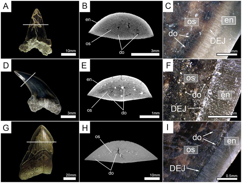
www.nature.com/scientificreports/                                                              www.nature.com/scientificreports

                           tooth characters such as crown and root morphologies or root vascularization patterns mostly are the only fea-
                           tures that can be used to infer the systematic position of extinct sharks16,17.
                               Glickman20,21 attempted to resolve the systematic positions of fossil chondrichthyans based solely on tooth
                           histologies of the crown and introduced the concept of histotype inferring. He distinguished between two dif-
                           ferent tooth histologies - the orthodont and the osteodont tooth histotypes. Accordingly, the orthodont type is
                           characterized by the presence of a hollow pulp cavity which is encased by dentine that has tightly packed parallel
                           tubules giving it a compact appearance (orthodentine)22–24. In contrast, teeth displaying the osteodont histotype
                           have the pulp cavity filled by dentine that is composed of numerous vascular canals surrounded by concentric
                           layers of dentine, similar to osteons in spongy bone (osteodentine)22–24, which intrudes from the root into the
                           crown and fills the pulp cavity25,26.
                               Although the phylogeny of chondrichthyans has been drastically improved by adding more dental and mor-
                           phological characters in recent years5,27–29, the tooth histotype concept still is used to distinguish elasmobranch
                           groups, as in rajiform and myliobatiform batomorphs17,30 or in galeomorph sharks between lamniform and car-
                           charhiniform sharks, with lamniforms displaying the osteodont tooth histotype and carcharhiniforms the ortho-
                           dont histotype26,31,32, with one exception: the carcharhiniform shark Hemipristis elongata that was assumed to
                           have the osteodont tooth histology16,17,25,33.
                               Recent examinations of the alleged osteodont carcharhiniform shark Hemipristis, however, revealed the pres-
                           ence of a third histotype - the pseudoosteodont tooth histotype26. Teeth of Hemipristis have an osteodentine core
                           that fills the hollow pulp cavity and that is encased by a layer of orthodentine. This is in contrast to the tooth
                           histology of recently examined lamniform sharks (Lamnidae and Alopiidae) which lack an orthodentine layer
                           and only have osteodentine26,31,34. Recognition of the pseudoosteodont tooth histotype that is based on misin-
                           terpreted osteodont histologies makes a re-evaluation of previously interpreted osteodont histotypes in various
                           elasmobranchs necessary to infer the taxonomic and systematic importance of tooth histotypes in sharks, rays,
                           and skates.
                               Here we re-evaluate the tooth histotype of lamniform sharks based on teeth of eight fossil taxa, including the
                           enigmatic galeomorph shark †Palaeocarcharias stromeri and 11 extant species using micro-computed tomogra-
                           phy (micro-CT) and traditional tooth sections.
                               Previously published information about the tooth histology of other lamniform sharks that were not exam-
                           ined here was added to this study. This results in the description of tooth mineralization patterns of a wide
                           range of species, from the basalmost lamniform sharks assigned to Eoptolamnidae (sensu Kriwet et al.35), or
                           Pseudoscapanorhynchidae (sensu Herman36) to 14 of the 15 extant species (Supplementary Table S1). Therefore,
                           this study represents the most comprehensive synopsis of tooth mineralization patterns in lamniform sharks to
                           date and discusses the phylogenetic relevance of the tooth histotype for systematic interpretations and the origin
                           of lamniform sharks in deep time.

                           Results
                           Tooth mineralization patterns in lamniform sharks. Micro-CT images of tooth files from the upper
                           jaw (palatoquadrate cartilage, PC) of the basking shark (Cetorhinus maximus) (7-692/RZ) and the left upper jaw
                           (LPC) of the crocodile shark (Pseudocarcharias kamoharai) (7-693/RZ) were 3D reconstructed and virtually sec-
                           tioned to examine the tooth mineralization sequence of both species. The tooth mineralization sequences were
                           consistent through all tooth files of each investigated species, with little variations due to different numbers of
                           teeth per tooth file. The code specification of the abbreviations used here is depicted in the Material and Methods
                           section.
                               Pseudocarcharias kamoharai has five to eight teeth in tooth files of the upper jaw, with zero to two functional
                           teeth per tooth file. In the youngest developmental stages, the only mineralized structure of the tooth is the super-
                           ficial enameloid. The enameloid first starts to mineralize at the apex (LPC1R6) and mineralization continues to
                           the tooth crown base (LPC1R5). Mineralization of the enameloid is completed in LPC1R4. Until this position, the
                           osteodentine formation has neither started in the root, nor in the crown and enameloid is the only mineralized
                           structure. Osteodentine starts forming in the root and in the center of the crown simultaneously after completion
                           of enameloid (LPC1R3), until it has fully filled the pulp cavity (LPC1R1 & LPC2R2). LPC1R1 is already fully
                           mineralized, but not in a functional position and, therefore, regarded as the first replacement tooth. LPC1F1
                           and LPC1F2 are fully mineralized and are in an erect position on the outer edge of the jaw cartilage, allowing
                           them to be utilized. During tooth development, no orthodentine can be identified at any stage, resulting in fully
                           mineralized teeth consisting of only one sort of dentine – osteodentine (Fig. 1C,D). The virtual section of an
                           isolated functional tooth also demonstrates the presence of only one layer of dentine, which is traversed by small
                           canals and surrounded only by the hypermineralized enameloid (Fig. 2E). A manual tooth section confirms this,
                           showing that dentinal osteons are spreading throughout the whole crown and are also present in close proximity
                           or next to the enameloid (Fig. 2F). The presence of osteodentine and the absence of orthodentine implies the
                           osteodont tooth mineralization pattern for teeth of P. kamoharai (Figs 1 and 2; Supplementary Tables S2 and S3).
                               The basking shark, Cetorhinus maximus has five to seven teeth within tooth files of the fractional part of the
                           upper jaw of which three to four can be regarded as functional teeth because of their position along the margin
                           of the jaw cartilage. Teeth have already an enameloid cap covering the whole of the crown in the earliest mineral-
                           ization stages. A thin layer of orthodentine is present and osteodentine in the root already has started to form at
                           this stage (PCaR3). In the adjacent file, only enameloid is present at the corresponding position, but no dentine
                           (PCbR3) (Supplementary Fig. S1). Orthodentine can be distinguished from enameloid due to different densities.
                           Dense tissues are represented by lighter shades (the hypermineralized enameloid is typically white in our recon-
                           structions) and less dense tissues by darker shades (dentine is typically grey in our reconstructions). Osteodentine
                           starts forming in the root and basally in the crown along the walls underneath the orthodentine. A hollow pulp
                           cavity remains in the center of the tooth (PCaR3). During the next stages, the orthodentine layer becomes thicker

Scientific Reports |   (2019) 9:9652 | https://doi.org/10.1038/s41598-019-46081-3                                                             2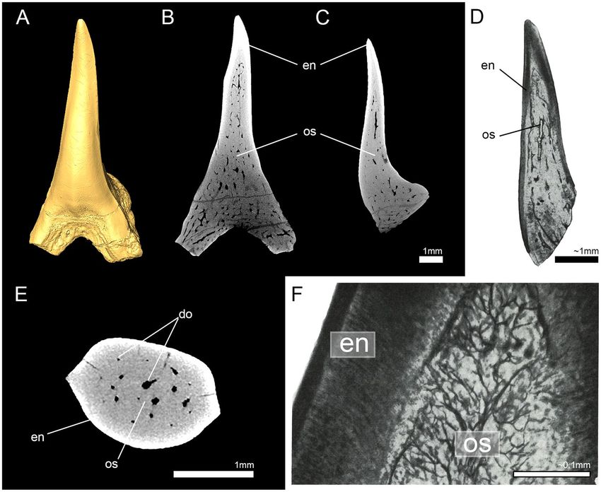
www.nature.com/scientificreports/                                                                www.nature.com/scientificreports

                           Figure 1. 3D micro-CT isosurfaces of the upper jaws and 2-D images of the virtually sectioned tooth row
                           reveal two different patterns of tooth mineralization in lamniform sharks. The basking shark Cetorhinus
                           maximus (7-692/RZ) (A,B) develops two layers of dentine (orthodentine and osteodentine representing the
                           pseudoosteodont histotype), while the crocodile shark Pseudocarcharias kamoharai (7-693/RZ) (C,D) lacks
                           orthodentine and only develops osteodentine (osteodont histotype). en, enameloid; or, orthodentine; os,
                           osteodentine; pc, pulp cavity.

                           and is fully mineralized in the first replacement tooth PCaR1. After the formation of osteodentine in the earliest
                           stages, it intrudes basally into the pulp cavity until it fully fills the center of the crown. The osteodentine in the
                           root is fully mineralized at the same developmental stage as orthodentine (PCaR1), but not in the crown until the
                           next stage (PCaF3). The first replacement tooth (PCaR1) is already in a functional position, but the osteodentine
                           in the crown has not fully filled the pulp cavity at this point. Therefore, completion of the mineralization process
                           in the crown determines the earliest functional tooth (PCaF3) (Fig. 1A,B; Supplementary Tables S2 and S4).
                           Manual and virtual tooth sections of an isolated functional tooth confirm the presence of both types of dentine - a
                           dentine core which is made up of both, dentinal osteons and interosteonal tissue, giving it a spongiose appearance
                           (osteodentine), which is surrounded by a prominent layer of dentine, lacking any vertical canals or pores but hori-
                           zontal tubules, which are arranged parallel to each other and give the dentine layer a very compact appearance
                           (orthodentine). The development of both, orthodentine and osteodentine in the crown shows that C. maximus
                           has a pseudoosteodont mineralization pattern (Figs 1 and 2; Supplementary Tables S2 and S4).
                               To clarify which of the two mineralization patterns is present among the remaining members of the order
                           Lamniformes, teeth of nine additional extant species were micro-CT scanned and virtually sectioned to deter-
                           mine their tooth histology. Additionally, teeth of all extant specimens (except the megamouth shark, Megachasma
                           pelagios) also were manually sectioned horizontally and compared to the high-resolution images of the virtually
                           sectioned teeth. Both methods demonstrate the presence of only one layer of dentine - osteodentine. Therefore, all
                           extant species (except for Cetorhinus maximus) display the osteodont tooth histotype (Figs 2–4, Supplementary
                           Fig. S2).

                           Tooth histology patterns in fossil lamniform sharks. To clarify the plesiomorphic condition for lamni-
                           form sharks, fossil teeth of seven selected taxa (including supposedly basal (i.e. Eoptolamnidae (sensu Kriwet et al.35)
                           and derived taxa) of extinct lamniform sharks were examined using micro-computed tomography images.
                           Additionally, an isolated tooth of the extant species Megachasma pelagios from the Pliocene (2.5–5 mya) is
                           included here as well.

Scientific Reports |   (2019) 9:9652 | https://doi.org/10.1038/s41598-019-46081-3                                                                3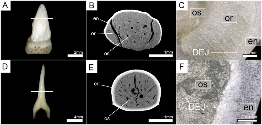
www.nature.com/scientificreports/                                                              www.nature.com/scientificreports

                           Figure 2. Horizontal virtual micro-CT sections and manual tooth sections of the basking shark (Cetorhinus
                           maximus, Cetorhinidae) (EMRG-Chond-T-24) and the crocodile shark (Pseudocarcharias kamoharai,
                           Pseudocarchariidae) (EMRG-Chond-T-28). (A) picture of a functional tooth prior to the tooth section, (B)
                           virtual section, and (C) tooth section of C. maximus under normal light illustrating the presence of two layers of
                           dentine - compact orthodentine surrounding a core of osteodentine. (D) picture of a functional tooth prior to
                           the tooth section, (E) virtual section, and (F) tooth section of P. kamoharai under polarized light illustrating the
                           presence of one layer of dentine - osteodentine. White lines indicate the approximate plane of the sections. do,
                           dentinal osteons; en, enameloid; or, orthodentine; os, osteodentine.

                               Rendered high resolution micro-CT images of the virtual sections (in axial and sagittal view) reveal the
                           absence of orthodentine in teeth of †Leptostyrax sp. (†Eoptolamnidae (sensu Kriwet et al.35)), †Palaeocarcharodon
                           orientalis (Lamniformes incertae fam.), and Megachasma pelagios (Megachasmidae). The hypermineralized enam-
                           eloid (white) is clearly distinguishable from the dentine (grey) as a result of density differences. From the core of
                           the tooth to the enameloid-dentine border, the entire dentine layer is traversed by dentinal osteons (Fig. 4). The
                           same histology pattern can be identified in teeth of †Scapanorhynchus rapax (Mitsukurinidae) and †Squalicorax
                           pristodontus (†Anacoracidae), which thus also exhibit the osteodont tooth histotype (Supplementary Fig. S3).
                               In three cases (teeth of †Dwardius woodwardi (Lamniformes incertae fam.), †Squalicorax sp. (†Anacoracidae),
                           and †Otodus megalodon (†Otodontidae)) the presence or absence of orthodentine could not be properly identi-
                           fied based solely on micro-CT images. The enameloid-dentine border was less well defined in micro-CT images
                           than in other examined teeth and some areas of dentine were indistinct, probably due to taphonomic processes,
                           not allowing to identify the presence or absence of dentinal osteons (Fig. 5B,E,H). This was especially evident in
                           †Otodus megalodon, where tooth enameloid could not be distinguished from the dentinal core and only a few
                           coarse canals were visible (Fig. 5H). Therefore, the tooth crowns were manually sectioned horizontally and exam-
                           ined under a light microscope. These tooth sections demonstrate the presence of dentinal osteons, traversing the
                           entire dentine core from the center to the enameloid-dentine border (Fig. 5C,F,I). In †Dwardius woodwardi small
                           dentinal tubuli are visible that originate in the osteons and penetrate the adjacent enameloid. Since they originate
                           in the osteons they should be regarded as extensions of the osteon rather than representing an additional dentine
                           layer. Orthodentine was not identified in any fossil tooth and all examined fossil taxa therefore have the osteodont
                           tooth histotype.

                           Tooth histology of †Palaeocarcharias stromeri.                 The systematic position of †Palaeocarcharias
                           stromeri still has to be considered as ambiguous despite all recent advances (see supplementary information
                           (Supplementary Discussion 1) for a discussion of the most recent phylogenetic analysis14). Here, tooth histol-
                           ogy can provide additional and important information. For this, a tooth of the holotype (JME-SOS-2294) was
                           scanned using micro-computed tomography. Virtual sections through three planes (frontal, sagittal, and axial)
                           show a dentine core that is entirely traversed by a network of dentinal osteons (osteodentine) and covered by
                           enameloid. The dentinal osteons are distributed within the entire dentine layer, from the center of the crown to
                           close to the enameloid-dentine border. A compact layer of dentine (orthodentine) between the osteodont core
                           and the hypermineralized enameloid is not present in the type specimen of †Palaeocarcharias stromeri, which
                           therefore represents the osteodont histotype (Fig. 6B,C,E).

                           Discussion
                           Tooth mineralization patterns in sharks of the order Lamniformes.                    We identified two different
                           tooth mineralization patterns within lamniform sharks resulting in the osteodont and pseudoosteodont histo-
                           types. According to our results, the basking shark (Cetorhinus maximus) is the only lamniform shark having the
                           pseudoosteodont tooth histotype, consisting of an osteodentine core that is covered by orthodentine. All other
                           lamniform sharks displayed the osteodont tooth histotype, developing only one type of dentine (osteodentine).

Scientific Reports |   (2019) 9:9652 | https://doi.org/10.1038/s41598-019-46081-3                                                             4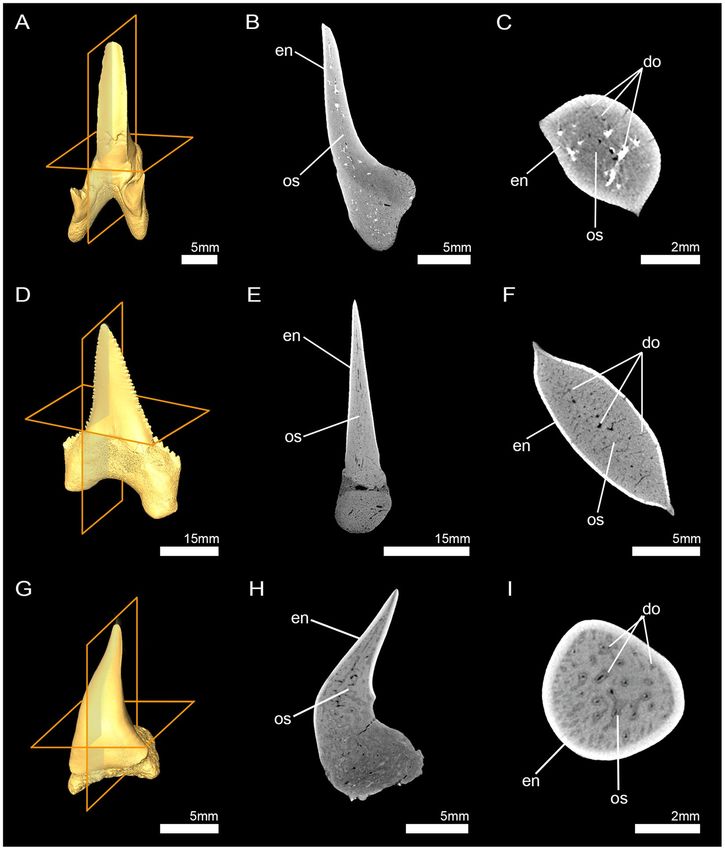
www.nature.com/scientificreports/ www.nature.com/scientificreports
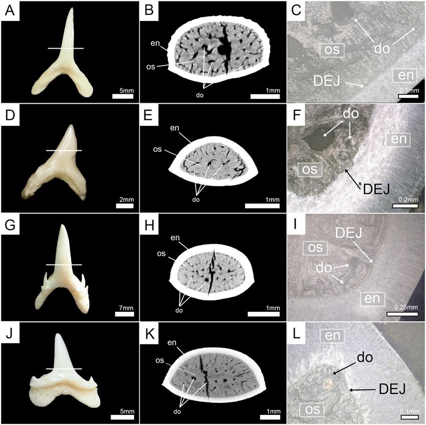
Figure 3. Horizontal virtual micro-CT sections and manual tooth sections of extant lamniform sharks under
normal light. (A–C) goblin shark (Mitsukurina owstoni, Mitsukurinidae) (EMRG-Chond-T-1); (D–F) common
thresher (Alopias vulpinus, Alopiidae) (EMRG-Chond-T-27); (G–I) smalltooth sand tiger (Odontaspis ferox,
Odontaspididae) (EMRG-Chond-T-2); (J–L) porbeagle shark (Lamna nasus, Lamnidae) (EMRG-Chond-T-4).
White lines indicate the approximate plane of the sections. do, dentinal osteons; en, enameloid; os, osteodentine.
There was no lamniform species that showed the orthodont tooth histotype (a hollow pulp cavity surrounded
by orthodentine) that is known from carcharhiniform sharks25,26,31–33,37–41. Teeth of the great white shark
(Carcharodon carcharias)31,32,42,43, shortfin mako (Isurus oxyrinchus)26,34, and the salmon shark (Lamna ditropis)44
are also lacking orthodentine and thus 13 out of 14 examined extant lamniform shark species follow an osteo-
dont tooth mineralization pattern. Unfortunately, there is no data for the longfin mako (Isurus paucus), but as all
lamnids as well as its closest relative, Isurus oxyrinchus, have osteodont teeth26,34, it seems eligible to assume that
its teeth also follow an osteodont mineralization pattern. Additionally, teeth of extinct species, from the assumed
basal-most family †Eoptolamnidae (sensu Kriwet et al.35) (†Leptostyrax sp.) from the Albian/Cenomanian (Early
Cretaceous, 94–113 mya) to †Otodus megalodon from the Miocene (Neogene, 5–23 mya) all showed the osteo-
dont histotype. The latter species was previously described as being osteodont45, but also pseudoosteodont46. In
both studies appropriate tooth sections were not prepared but fractured parts of the tooth were only superficially
inspected. The tooth section prepared for this study unambiguously demonstrates that the whole interior of the
crown is filled with osteodentine, while orthodentine is absent supporting †O. megalodon to be osteodont. The
osteodont tooth histology for lamniforms is also confirmed by a number of publications: for a more detailed list,
see Supplementary Table S1.
Phylogenetic implications of different tooth histotypes and the origin of osteodonty. Many
sharks, rays and skates (Elasmobranchii) previously have been considered to have the osteodont histotype accord-
ing to the traditional definition of histotypes (pulp cavity filled with osteodentine)20,21,25: Myliobatiformes22,
Ptychodus22,47,48, Hexanchiformes41, Squatiniformes49, Heterodontiformes22, Orectolobiformes49, Hemipristis
Scientific Reports | (2019) 9:9652 | https://doi.org/10.1038/s41598-019-46081-3 5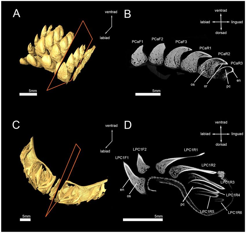
www.nature.com/scientificreports/                                                              www.nature.com/scientificreports

                           Figure 4. 3D reconstructions and virtual micro-CT sections (sagittal and axial plane) of fossil lamniform
                           shark teeth. (A–C) †Leptostyrax sp. (†Eoptolamnidae (sensu Kriwet et al.35)) (Inv.nr. 7–690); (D–F)
                           †Palaeocarcharodon orientalis (Lamniformes incertae fam.) (EMRG-Chond-T-50); (G–I) Megachasma pelagios
                           (Megachasmidae) (EMRG-Chond-T-44). do, dentinal osteons; en, enameloid; os, osteodentine.

                           spp. (Carcharhiniformes)25,26,40, Lamniformes26,31,34,43. However, tooth crowns of these groups consist of both,
                           ortho- and osteodentine and, therefore, should be referred to as being pseudoosteodont sensu Jambura et al.26.
                           Consequently, Lamniformes (except for Cetorhinus) is the only group in which osteodentine alone constitutes the
                           interior of the tooth crown and, therefore, should be the only group to be referred to as being osteodont.
                               Many Palaeozoic and hybodontiform sharks (the putative sister group to modern sharks50,51) are also referred
                           to as being osteodont22,52–55 according to the traditional definition of histotypes. However, as in modern groups
                           (except for the lamniform sharks) both, orthodentine and osteodentine form the interior of the crown and, there-
                           fore, they have the pseudoosteodont histotype. There seems to be only one exception: apparently the teeth of
                           †Aztecodus harmsenae, a Devonian chondrichthyan, also lack orthodentine53. The presence of pseudoosteodonty
                           in Palaeozoic sharks, †Hybodontiformes and in many extant elasmobranchs (sharks, rays and skates) strongly
                           indicates that this is the plesiomorphic condition for the modern sharks, and not a modification of the orthodont
                           tooth histotype as previously suggested26,40. The osteodont tooth histotype exclusively found in sharks of the order
                           Lamniformes represents a highly derived synapomorphic condition for this group within the elasmobranch fishes
                           (Fig. 7).

Scientific Reports |   (2019) 9:9652 | https://doi.org/10.1038/s41598-019-46081-3                                                            6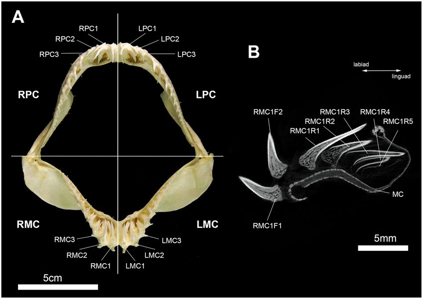
www.nature.com/scientificreports/                                                              www.nature.com/scientificreports

                           Figure 5. Horizontal virtual micro-CT sections and manual tooth sections of extinct lamniform sharks.
                           (A–C) †Dwardius woodwardi (Lamniformes incertae fam.) (EMRG-Chond-T-53); (D–F) †Squalicorax sp.
                           (†Anacoracidae) (EMRG-Chond-T-54); (G–I) †Otodus megalodon (†Otodontidae) (EMRG-Chond-T-57).
                           White lines indicate the plane of the sections. do, dentinal osteons; en, enameloid; os, osteodentine.

                               The basking shark Cetorhinus maximus however, represents a deviation from the general lamniform pattern
                           in that it displays the pseudoosteodont tooth histology. In phylogenetic analyses based solely on tooth morphol-
                           ogy, C. maximus and Megachasma pelagios formed a clade at the base of the lamniform sharks, representing a
                           primitive sister group to other lamnid sharks56. Although this would explain a plesiomorphic state of the tooth
                           histology in Cetorhinus to some extent, it conflicts the derived state in its putative sister group Megachasma.
                           Furthermore, other morphological characters and molecular data suggest C. maximus to be a highly derived
                           lamniform shark4–6,8, indicating that teeth of both planktivores became vestigial and, therefore, dental characters
                           were reduced or lost completely56,57. This might also be the case for the tooth histology of C. maximus which
                           reverted to a plesiomorphic state. However, M. pelagios, the second planktivorous species of this order still shares
                           its derived tooth histology with all other lamniform sharks and, therefore, does not support an ecophenotypic
                           link between tooth histology and feeding behaviour. What is also curious about the tooth histology in Cetorhinus
                           maximus is that it was not reverted to the most probable ancestral state of lamniform and carcharhiniform sharks
                           (orthodont), but to the ancestral state of all galeomorph sharks (pseudoosteodont) (Fig. 7). The reason for this
                           reversal of the tooth mineralization pattern in C. maximus remains ambiguous, but seemingly is neither linked to
                           its phylogenetic position, nor to its planktivorous feeding behaviour.

                           †Palaeocarcharias stromeri and the origin of lamniform sharks. High resolution micro-CT images
                           of a tooth of the holotype specimen of †Palaeocarcharias stromeri (JME-SOS-2294) display two types of tis-
                           sues - a prominent outer layer of hypermineralized tissue (enameloid) and a core of less dense tissue (dentine).
                           Dentinal osteons traversed the whole dentine core and were also present very close to the enameloid layer, which
                           is characteristic for osteodentine. There were no signs of a compact orthodentine layer which can be found in
                           pseudoosteodont teeth, thus teeth of †P. stromeri consist of only one layer of dentine (osteodentine) and display
                           the osteodont histotype, which is characteristic for lamniform sharks26,31,34,41.
                               Our findings contradict the original description of the tooth histology of †P. stromeri by Beaumont10, who
                           indicated the presence of three layers of dentine - a core of “trabecular dentine” (osteodentine) that was sur-
                           rounded by a mantle of orthodentine, which again was covered by vitrodentine10 (vitrodentine is one of many
                           synonyms that were used for enameloid before its true nature was resolved58). The presence of both, orthoden-
                           tine and osteodentine, within the crown would suggest that †P. stromeri displays the pseudoosteodont histotype.
                           According to the most recent study on †P. stromeri, vitrodentine and orthodentine were misinterpreted in the
                           original work and instead are components of the multi layered enameloid14. Vitrodentine resembles the SCE
                           (‘Single Crystallite Enameloid’) unit, while the tissue described as orthodentine in fact was the BCE (‘Bundled

Scientific Reports |   (2019) 9:9652 | https://doi.org/10.1038/s41598-019-46081-3                                                            7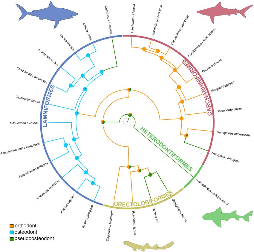
www.nature.com/scientificreports/                                                               www.nature.com/scientificreports

                           Figure 6. High resolution micro-CT images of virtual tooth sections and manual tooth sections of the original
                           description of †Palaeocarcharias stromeri. (A) 3D reconstruction, (B,C,E) virtual tooth sections in (B) frontal,
                           (C) sagittal and (E) axial view. (D,F) are modified pictures of the manual tooth sections from the original
                           description of †P. stromeri by Beaumont10. Do, dentinal osteons; en, enameloid; os, osteodentine.

                           Crystallite Enameloid’) unit of the enameloid. Misinterpretations of the BCE unit (or parts of it) to be orthoden-
                           tine occurred also in other studies, i.e. for Lamna sp.59 and Lamna nasus60. Berkovitz and Shellis61 reported the
                           presence of orthodentine in Carcharias sp. However, dentinal tubules in the tissue they identified as orthodentine
                           in their figure (figure 11.30)61 do not originate in this tissue but come from the adjacent osteodentine which is
                           known to occur at the dentine-enameloid junction31. Furthermore, the presence of a sharp junction between oste-
                           odentine and “orthodentine”, which is known from the enameloid-dentine border, but not from dentine-dentine
                           borders31,61 is another indicator for this misinterpretation. Therefore, it is apparent that Carcharias sp. has the
                           osteodont tooth histotype reported here for Carcharias taurus and other lamniform sharks.
                                Another indicator for the multi-layered enameloid being misinterpreted as orthodentine in the original work
                           of †P. stromeri is given by our micro-CT images. Computed tomography discriminates between tissues of differ-
                           ent densities - in our case, the very dense hypermineralized enameloid appears white, while the less dense dentine
                           is grey. If we compare the virtual section in sagittal view (Fig. 6C) with the original tooth section (also in sagittal
                           view) (Fig. 6D), we can see that the hypermineralized tissue in the virtual section (white) resembles the combined
                           layers of “vitrodentine” and “orthodentine” in the original tooth section (dark) in thickness. If only the thin layer
                           of “vitrodentine” in the original tooth section was enameloid, the hypermineralized structure in the micro-CT
                           scan would be much thinner and less prominent, since the enameloid has a higher mineral content and, thus, den-
                           sity than dentine58,62,63, which is also visible on the micro-CT images26,34. Therefore, the micro-CT images prove
                           the misinterpretation of orthodentine in †P. stromeri, which means that it does not display the pseudoosteodont
                           tooth histotype, but the osteodont histotype, which only occurs in lamniform sharks.
                                Although †P. stromeri is known from well-preserved articulated material, only a few studies were conducted
                           to resolve the systematic position of this species, which was in debate for decades12,13. In the original description,
                           †P. stromeri was placed at the base of the Lamniformes10. Other authors agreed that the tooth morphology is char-
                           acteristic for lamniform sharks11,16,17 but the body form was similar to that of orectolobiform sharks11, making it a
                           transitional taxon between both clades or the basal sister group of lamniforms12,13. In the most recent work on the
                           phylogenetic position of †P. stromeri, it was suggested to be the sister to the clade Carcharhiniformes + Lamnifor
                           mes14. Unfortunately, this phylogenetic hypothesis is inconclusive (Supplementary Discussion S1). Consequently,
                           it is most parsimonious to assume that the osteodont tooth histotype is a unique feature for Lamniformes and
                           thus we conclude that the osteodont tooth histology of †P. stromeri adds very strong support of this shark being

Scientific Reports |   (2019) 9:9652 | https://doi.org/10.1038/s41598-019-46081-3                                                               8
www.nature.com/scientificreports/ www.nature.com/scientificreports
Figure 7. Stochastic character mapping of tooth histotypes and ancestral state reconstructions in sharks of the
superorder Galeomorphii. The maximum credibility tree of the galeomorph sharks is based on whole mtDNA
sequences. Ancestral states at the nodes are coded as pie charts proportions of the probability distribution,
calculated from 100 stochastic mappings for the three histotypes present (orthodonty, osteodonty and
pseudoosteodonty).
the oldest known lamniform shark. Therefore, the origin of this group dates back at least into the Middle Jurassic
(Bathonian)64, a period when major diversifications of elasmobranch fishes took place65,66.
Strengths and weaknesses of micro-CT imaging. Similar to previous studies, micro-CT imaging
turned out to be a powerful tool for non-invasive investigations of internal structures26,31,43,67. This is especially
evident in examining tooth mineralization processes within tooth files in extant lamniform sharks. However, our
study also points out weaknesses of micro-CT imaging, especially for fossil specimens related either to tapho-
nomic alterations or to insufficient resolution of the CT scan. This is especially apparent in the tooth of †Otodus
megalodon, in which the enameloid was not distinguishable from the dentine and the peripheral vascular system
with its tiny canaliculi was not detectable in the micro-CT images, but was visible in manual tooth section. The
latter effect is easily explained by the low resolution (around 30 µm), which was caused by the size of the tooth.
In extant sharks, enameloid has a much higher degree of crystallinity and a very low organic content compared
to dentine62,63, which makes it appear denser in micro-CT images26,34. The poor results for fossil teeth can be dis-
torted by diagenetic processes leading to changes in the chemical constitution of enameloid63,68 resulting in less
differences of densities between enameloid and dentine. Nonetheless, in most cases micro-CT scanning generated
images that sufficiently resolved the internal structures of both, extant and fossil shark teeth without damaging or
destroying the material and therefore, should be regarded as a reliable non-invasive alternative to conventional
thin sectioning.
Material and Methods
Material. Teeth and jaws of 11 extant and seven extinct taxa of sharks of the order Lamniformes were exam-
ined. Additionally, a tooth of the holotype specimen of †Palaeocarcharias stromeri (JME-SOS-2294) housed in the
Jura Museum Eichstätt, Germany, the putative basal most lamniform shark from the Upper Jurassic (Tithonian),
is included in this study.
Scientific Reports | (2019) 9:9652 | https://doi.org/10.1038/s41598-019-46081-3 9www.nature.com/scientificreports/ www.nature.com/scientificreports
Figure 8. Terminology used to describe the topology of teeth within the jaws and tooth files. (A) Jaws of
Pseudocarcharias kamoharai (Inv.nr. 7-693/RZ) in frontal view, (B) virtual section through the tooth file RMC1.
F, functional tooth; F1, first (oldest) functional tooth; F2, second functional tooth; LMC, left Meckel’s cartilage;
LPC, left palatoquadrate cartilage; MC, Meckel’s cartilage (lower jaw); PQ, palatoquadrate cartilage (upper jaw);
R, replacement tooth; R1, first (oldest) replacement tooth; R2, second replacement tooth; R3, third replacement
tooth; R4, fourth replacement tooth; R5, fifth replacement tooth; RMC, right Meckel’s cartilage; RPC, right
palatoquadrate cartilage.
Extant material consisted of two jaws (basking shark Cetorhinus maximus (Inv.nr. 7-692/RZ), crocodile shark
Pseudocarcharias kamoharai (Inv.nr. 7-693/RZ)) and isolated teeth of all 11 species. The fossil material consisted
exclusively of isolated teeth and includes species from the Mesozoic and Cenozoic Era (five and three species
respectively). Daggers preceding taxon names denote extinct species (Supplementary Table S5).
Tooth terminology. To specify the tooth position within the jaw, we employed a previously published
code26,34. The first four letters define the position of the tooth file (the developmental sequence of replacement
and functional teeth sensu Moyer et al.31), if it is right (R) or left (L) of the symphysis or coming from the upper
(palatoquadrate PC) or lower jaw (Meckel’s cartilage MC). The number following the first three letters determines
the position of the file distally to the symphysis. For example, the tooth file illustrated in Fig. 1B is the first file
right to the symphysis in the lower jaw and, therefore, coded as RMC1. We distinguished between functional (F)
and replacement teeth (R), with functional teeth being fully mineralized and in an erect or semi-erect position,
allowing them to be utilized for food gathering (e.g. cutting, grasping, etc.). Replacement teeth are located lin-
gually to the functional teeth and are not fully mineralized at this point of development. The tooth position within
the tooth file is numbered, thus the first functional tooth of the RMC1 file is RMC1F1, the first replacement tooth
of the same file is RMC1R1 (Fig. 8).
Micro-CT scanning and imaging. Tooth mineralization and histology patterns for each species were
investigated using a SkyScan1173 micro-CT device (Bruker/Skyscan, Kontich, Belgium) at the Department
of Palaeontology (University of Vienna, Austria). Settings for each specimen are provided in the Supporting
Information section (Supplementary Table S6). The generated slice file stacks were loaded into the software pack-
age DataViewer (version 1.5.1.2 (64 bit), SkyScan (Bruker micro-CT, Kontich, Belgium)) and Amira software
package (version 5.4.5, FEI Visualization Sciences Group, Oregon, USA) to visualize and investigate 2D and 3D
images of the studied material. This allowed us to set clipping planes through the jaws and teeth with different
angles and digitally dissect through the material to examine the internal anatomy. Editing colour balance, contrast
and labeling of the resulting 2D images was conducted in Adobe Photoshop CS6 (version 13.0, Adobe Systems,
San José, USA).
Tooth sectioning. The quality of the results was tested by comparing the digital sections with actual tooth
sections. For this, tooth sections were prepared for all extant specimens and three fossil specimens (†Dwardius
woodwardi, †Otodus megalodon, †Squalicorax sp.). For better handling of the small teeth, they were embedded
in an adhesive medium using the two-component adhesive Araldite 2020/A and Araldite 2020/B, which were
merged with a ratio of 100:30. The embedded teeth were cut horizontally through the crown and the exposed
Scientific Reports | (2019) 9:9652 | https://doi.org/10.1038/s41598-019-46081-3 10www.nature.com/scientificreports/ www.nature.com/scientificreports
surface was polished using grinding powder (grain size 600 and 1000). Afterwards, the surface was treated with
a 2 molar HCl solution for 10–60 sec and examined under the digital microscope Keyence VHX-6000 (Keyence
International, Belgium). Pictures of the teeth were taken prior the sectioning process with an Olympus-OMD E5
mirrorless camera or the digital microscope.
Phylogenetic tree and ancestral state reconstruction. A phylogeny for 26 galeomorph sharks with
known tooth histology was built from whole mitochondrial DNA sequences retrieved from the nucleotide data-
base in GeneBank (accession number of the sequences can be found in the supplementary file (Supplementary
Table S7)). A complete sequenced genome of Heterodontus portusjacksoni was not available in the database, there-
fore, two other species of Heterodontus (H. francisci and H. zebra) were used as an outgroup of the clade consist-
ing of [Orectolobiformes + [Carcharhiniformes + Lamniformes]. The sequences were aligned in MAFFT69 and a
matrix of approximately 16.5kbp resulted after trimming the edges. To construct the phylogeny the GTR + G + I
substitution model was employed. The alignment was used in BEAST 2 software70, two parallel MCMC runs were
performed over 10,000,000 generations sampling every 1000 generations.10% of the generations were set as a
burn-in on TreeAnnotator70 to obtain a maximum credibility tree on which the topology was used for stochastic
character mapping with the make.simmap function in phytools71 to perform an ancestral state reconstruction
on the tooth histotype. Data for the tooth histology of the 26 species was retrieved from this study and the litera-
ture22,26,31,34,43,44,49. The final tree was edited in FigTree (v. 1.4.4).
Data Availability
All specimens are deposited in either of the following collections and are publicly accessible: 1. collection of the
Department of Palaeontology, University of Vienna, Vienna, Austria; 2. Haimuseum und Sammlung R. Kind-
limann, Aathal-Seegräben, Switzerland; 3. Jura Museum Eichstätt, Germany. Micro-CT scans are stored at the
Department of Palaeontology, University of Vienna, Vienna, Austria. All data generated and analyzed during this
study are included in this published article (and its Supplementary Information files). Detailed deposition infor-
mation can be found in Supplementary Table S5.
References
1. Pimiento, C. & Balk, M. A. Body-size trends of the extinct giant shark Otodus megalodon: a deep-time perspective on marine apex
predators. Paleobiology. 41, 479–490 (2015).
2. Razak, H. & Kocsis, L. Late Miocene Otodus (Megaselachus) megalodon from Brunei Darussalam: Body length estimation and
habitat reconstruction. Neues Jahrb. Geol. Palaontol. Abh. 288, 299–306 (2018).
3. Pimiento, C., Cantalapiedra, J. L., Shimada, K., Field, D. J. & Smaers, J. B. Evolutionary pathways toward gigantism in sharks and
rays. Evolution. 13680, https://doi.org/10.1111/evo.13680 (2019).
4. Compagno, L. J. V. Relationships of the megamouth shark, Megachasma pelagios (Lamniformes: Megachasmidae), with comments
on its feeding habits. NOAA Tech. Rep. NMFS. 90, 357–379 (1990).
5. Shimada, K. Phylogeny of lamniform sharks (Chondrichthyes: Elasmobranchii) and the contribution of dental characters to
lamniform systematics. Paleontol. Res. 9, 55–72 (2005).
6. Naylor, G. J. et al. Elasmobranch phylogeny: a mitochondrial estimate based on 595 Species in Biology of Sharks and their Relatives,
Edition 2 (eds Carrier, J. C., Musick, J. A., Heithaus, M. R.) 31–56 (CRC Press, 2012).
7. Ebert, D. A., Fowler, S. & Compagno, L. J. V. Sharks of the World: A Fully Illustrated Guide. Plymouth. 1–528 (Wild Nature Press,
2013).
8. Amaral, C. R. L., Pereira, F., Silva, D. A., Amorim, A. & de Carvalho, E. F. The mitogenomic phylogeny of the Elasmobranchii
(Chondrichthyes). Mitochondrial DNA A DNA Mapp. Seq. Anal. 29, 867–878 (2018).
9. Rees, J. Neoselachian shark and ray teeth from the Valanginian, Lower Cretaceous of Wawal, Central Poland. Palaeontology. 48,
209–221 (2005).
10. de Beaumont, G. Observations préliminaires sur trois Sélaciens nouveaux du calcaire lithographique d’Eichstätt (Bavière). Eclogae
Geol. Helv. 53, 315–328 (1960).
11. Duffin, C. J. The upper jurassic selachian Palaeocarcharias de Beaumont (1960). Zool. J. Linn. Soc. 94, 271–286 (1988).
12. Kriwet, J. & Klug, S. Late Jurassic selachians (Chondrichthyes, Elasmobranchii) from southern Germany: Re-evaluation on
taxonomy and diversity. Zitteliana. A 44, 67–95 (2004).
13. Kriwet, J. & Klug, S. Knorpelfische (Chondrichthyes) in Solnhofen – Ein Fenster in die Jurazeit (eds Arratia, G., Schultze, H. P.,
Tischlinger, H. & Viohl, G.) 334–359 (Verlag Dr. Friedrich Pfeil, 2015).
14. Landemaine, O., Thies, D. & Waschkewitz, J. The Late Jurassic shark Palaeocarcharias (Elasmobranchii, Selachimorpha) – functional
morphology of teeth, dermal cephalic lobes and phylogenetic position. Palaeontogr. Abt. A Palaeozool-Stratigr. 312, 103–165 (2018).
15. Owen, R. Lectures on the Comparative Anatomy and Physiology of the Vertebrate Animals. Part I. Fishes. London. 1–307 (Longman,
Brown, Green, and Longmans, 1846).
16. Cappetta, H. Handbook of Paleoichthyology, Vol 3B: Chondrichthyes II. Stuttgart. 1–192 (Gustav Fischer Verlag, 1987).
17. Cappetta, H. Handbook of Paleoichthyology, Vol 3E: Chondrichthyes - Mesozoic and Cenozoic Elasmobranchii: Teeth. München. 1–512
(Verlag Dr. Friedrich Pfeil, 2012).
18. Reif, W. E. Pattern regulation in shark dentitions in Pattern formation: a primer in developmental biology (ed. Malacinski, G. M.)
603–621 (Macmillan, 1984).
19. Smith, M. M., Johanson, Z., Underwood, C. & Diekwisch, T. G. H. Pattern formation in development of chondrichthyan dentitions:
a review of an evolutionary model. Hist. Biol. 25, 127–142 (2013).
20. Glickman, L. S. Akuly paleogena i ikh stratigraficheskoe znachenie. Moscow. 1–228 (Nauka Press, 1964).
21. Glickman, L. S. Subclass Elasmobranchii in Fundamentals of Palaeontology (eds Orlov, Y. A., Obruchev, D. V.) 292–352 (Israel
Program for Scientific Translations, 1967).
22. Radinsky, L. Tooth histology as a taxonomic criterion for cartilaginous fishes. J. Morphol. 109, 73–92 (1961).
23. Ørvig, T. Phylogeny of tooth tissues: evolution of some calcified tissues in early vertebrates in Structural and Chemical Organization
of Teeth (ed. Miles, A. E. W.) 45–110 (Academic Press, 1967).
24. Smith, M. M. & Sanson, I. J. Evolutionary origins of dentine in the fossil record of early vertebrates: diversity, development and
function in Development, Function and Evolution of Teeth (eds Teaford, M. F., Smith, M. M. & Ferguson, M. W. J.) 65–81 (Cambridge
University Press, 2000).
25. Compagno, L. J. V. Sharks of the Order Carcharhiniformes. New Jersey. 1–486 (Princeton University Press, 1988).
Scientific Reports | (2019) 9:9652 | https://doi.org/10.1038/s41598-019-46081-3 11www.nature.com/scientificreports/ www.nature.com/scientificreports
26. Jambura, P. L., Pfaff, C., Underwood, C. J., Ward, D. J. & Kriwet, J. Tooth mineralization and histology patterns in extinct and extant
snaggletooth sharks, Hemipristis (Carcharhiniformes, Hemigaleidae)-Evolutionary significance or ecological adaptation? PloS
ONE. 13, e0200951, https://doi.org/10.1371/journal.pone.0200951 (2018).
27. Shirai, S. Phylogenetic interrelationships of neoselachians (Chondrichthyes: Euselachii) in Interrelationships of fishes (eds Stiassny,
M. L. J., Parenti, L. R. & Johnson, G. D.) 9–34 (Academic Press, 1996).
28. Aschliman, N. C. The batoid tree of life: recovering the patterns and timing of the evolution of skates, rays and allies (Chondrichthyes:
Batoidea). PhD dissertation, Florida State University, June (2011).
29. Aschliman, N. C. Interrelationships of the durophagous stingrays (Batoidea: Myliobatidae). Environ. Biol. Fish. 97, 967–979 (2014).
30. Cappetta, H. N. R. Nouveaux Rhinobatoidei (Neoselachii, Rajiformes) à denture spécialisée du Maastrichtien du Maroc. Remarques
sur l’évolution dentaire des Rajiformes et des Myliobatiformes. Neues Jahrb. Geol. Palaontol. Abh. 187, 31–52 (1992).
31. Moyer, J. K., Riccio, M. L. & Bemis, W. E. Development and microstructure of tooth histotypes in the blue shark, Prionace glauca
(Carcharhiniformes: Carcharhinidae) and the great white shark, Carcharodon carcharias (Lamniformes: Lamnidae). J. Morphol. 276,
797–817 (2015).
32. Moyer, J. K. & Bemis, W. E. Shark teeth as edged weapons: serrated teeth of three species of selachians. Zoology. 120, 101–109 (2017).
33. Compagno, L. J. V. Interrelationship of living elasmobranchs. Zool. J. Linn. Soc. 53, 15–61 (1973).
34. Schnetz, L., Pfaff, C. & Kriwet, J. Tooth development and histology patterns in lamniform sharks (Elasmobranchii, Lamniformes)
revisited. J. Morphol. 277, 1584–1598 (2016).
35. Kriwet, J., Klug, S., Canudo, J. I. & Cuenca-Bescos, G. A new Early Cretaceous lamniform shark (Chondrichthyes, Neoselachii). Zool.
J. Linn. Soc. 154, 278–290 (2008).
36. Herman, J. Réflexions sur la systématique des Galeoidei et sur les affinités du genre Cetorhinus à l’occasion de la découverte
d'éléments de la denture d’un exemplaire fossile dans les sables du Kattendijk à Kallo (Pliocène inférieur, Belgique). Ann. Soc. géol.
Belg. 102, 357–377 (1979).
37. Jacobshagen, E. Grundlinien einer vergleichenden Anatomie des Zahnbeines und der Zähne niederer Wirbeltiere. Z. Mikrosk. Anat.
Forsch. 49, 225–272 (1940).
38. Salomon, C. D. Dentin of Carcharhinus milberti (Shark): a comparative histological and histochemical study. J. Dent. Res. 48,
196–205 (1969).
39. Herman, J., Hovestadt-Euler, M. & Hovestadt, D. C. Contributions to the study of the comparative morphology of teeth and other
relevant ichthyodorulites in living supraspecific taxa of chondrichthyan fishes. Part A: Selachii. No. 2c: Order: Carcharhiniformes,
Families: Proscylliidae, Hemigaleidae, Pseudotriakidae, Leptochariidae and Carcharhinidae. Bull. Inst. R. Sci. Nat. Belg. Biol. 61,
73–120 (1991).
40. Hovestadt, D. C. & Hovestadt-Euler, M. The vascularization system in teeth of Selachii in Élasmobranches et stratigraphie (eds
Herman, J. & van Waes, H.) 241–258 (Ministère des affaires économiques, Service géologique de Belgique, 1993).
41. Herman, J., Hovestadt-Euler, M. & Hovestadt, D. C. Contributions to the study of the comparative morphology of teeth and other
relevant ichthyodorulites in living supraspecific taxa of chondrichthyan fishes. Part A: Selachii. Addendum to 1: Order
Hexanchiformes-Family Hexachidae, 2: Order Carcharhiniformes, 2a: Family Triakidae, 2b: Family Scyliorhinidae, 2c: Family
Carcharhinidae, Hemigaleidae, Leptochariidae, Sphyrnidae, Proscylliidae and Pseudotriakidae, 3: Order Squaliformes: Family
Echinorhinidae, Oxynotidae and Squalidae. Tooth vascularization and phylogenetic interpretation. Bull. Inst. R. Sci. Nat. Belg. Biol.
73, 5–26 (2003).
42. Goto, M. Evolutionary trends of the tooth structure in Chondrichthyes in Mechanisms and Phylogeny of Mineralization in Biological
Systems (eds Suga, S., Nakahara, H.) 447–451 (Springer Japan, 1991).
43. Moyer, J. K., Hamilton, N. D., Hadlock Seeley, R., Riccio, M. L. & Bemis, W. E. Identification of shark teeth (Elasmobranchii:
Lamnidae) from a historic fishing station on Smuttynose Island, Maine, using computed tomography imaging. Northeast. Nat. 22,
585–597 (2015).
44. Kakizawa, Y. On the teeth of salmon shark, Lamna ditropis Hubbs & Follet. Nihon Univ. dent. J. 58, 59–69 (1984).
45. Bendix-Almgreen, S. E. Carcharodon megalodon from the Upper Miocene of Denmark, with comments on elasmobranch tooth
enameloid: coronoin. Bull. geol. Soc. Denmark. 32, 1–32 (1983).
46. Medina-Gavilán, J. L., Toscano, A., Muñiz, F. & Delgado, F. J. First description of a tooth of the extinct giant shark Otodus megalodon
(Aggassiz, 1835) found in the province of Seville (SW Iberian Peninsula) (Otodontidae). BVnPC. 4, 107–114 (2015).
47. Mutter, R. J., Iturralde-Vinent, M. & Fernández Carmona, J. The first mesozoic caribbean shark is from the Turonian of Cuba:
Ptychodus cyclodontis sp. nov. (Neoselachii). J. Vertebr. Paleontol. 25, 976–978 (2005).
48. Hoffman, B. L., Hageman, S. A. & Claycomb, G. D. Scanning electron microscope examination of the dental enameloid of the
Cretaceous durophagous shark Ptychodus supports neoselachian classification. J. Paleontol. 90, 741–762 (2016).
49. Herman, J., Hovestadt-Euler, M. & Hovestadt, D. C. Contributions to the study of the comparative morphology of teeth and other
relevant ichthyodorulites in living supraspecific taxa of chondrichthyan fishes. Part A: Selachii. No.4: Order: Orectolobiformes,
Families Brachaeluridae, Ginglymostomidae, Hemiscyllidae, Orectolobidae, Parascylliidae, Rhiniodontidae, Stegostomatidae,
Order: Pristiophoriphormes Family: Pristiophoridae, Order Squatiniformes Family: Squatinidae. Bull. Inst. R. Sci. Nat. Belg. Biol. 62,
193–254 (1992).
50. Hay, O. P. Bibliography and Catalogue of the Fossil Vertebrata of North America. Washington. 1–882 (US Government Printing Office
179, 1902).
51. Maisey, J. G. The braincase of the Middle Triassic shark Acronemus tuberculatus (Bassani 1886). Palaeontology. 54, 417–428 (2011).
52. Zangerl, R. Handbook of Paleoichthyology, Vol. 3A: Paleozoic Elasmobranchii. Stuttgart-New York. 1–115 (Gustav Fischer Verlag,
1981).
53. Hampe, O. & Long, J. A. The histology of Middle Devonian chondrichthyan teeth from southern Victoria Land, Antarctica. Rec.
Aust. Mus. 57, 23–36 (1999).
54. Rees, J. & Underwood, C. J. The status of the shark genus Lissodus Brough, 1935, and the position of nominal Lissodus species within
the Hybodontoidea (Selachii). J. Vertebr. Paleontol. 22, 471–479 (2002).
55. Ivanov, A. & Märss, T. New data on Karksiodus (Chondrichthyes) from the Main Devonian Field (East European Platform). Est. J.
Earth Sci. 63, 156–165 (2014).
56. Long, D. J. & Waggoner, B. M. Evolutionary relationships of the white shark: a phylogeny of lamniform sharks based on dental
morphology in Great white sharks: the biology of Carcharodon carcharias (eds Klimley, A. P. & Ainley, D. G.) 37–47 (Academic Press,
1996).
57. Shimada, K. Dental homologies in lamniform sharks (Chondrichthyes: Elasmobranchii). J. Morphol. 251, 38–72 (2002).
58. Reif, W. E. Morphologie und Ultrastruktur des Hai-“Schmelzes”. Zool. Scr. 2, 231–250 (1973).
59. Yabe, H. & Goto, M. Terminology of the elasmobranchian teeth. J. Fos. Res. 32, 14–20 (1999).
60. Marquard, E. Beiträge zur Kenntnis des Selachiergebisses. Rev. suisse Zool. 53, 73–132 (1946).
61. Berkovitz, B. & Shellis, P. The Teeth of Non-Mammalian Vertebrates. London. 1–342 (Academic Press, 2017).
62. Goto, M. Histological and biochemical studies on recent and fossil shark teeth. Tsurumi U. Dent. J. 4, 85–104 (1978).
63. Enax, J., Prymak, O., Raabe, D. & Epple, M. Structure, composition, and mechanical properties of shark teeth. J. Struct. Biol. 178,
290–299 (2012).
Scientific Reports | (2019) 9:9652 | https://doi.org/10.1038/s41598-019-46081-3 12www.nature.com/scientificreports/ www.nature.com/scientificreports
64. Wills, S., Bernard, E. L., Brewer, P., Underwood, C. J. & Ward, D. J. Palaeontology, stratigraphy and sedimentology of Woodeaton
Quarry (Oxfordshire) and a new microvertebrate site from the White Limestone Formation (Bathonian, Jurassic). Proc. Geol. Assoc.
130, 170–186 (2019).
65. Kriwet, J., Kiessling, W. & Klug, S. Diversification trajectories and evolutionary life-history traits in early sharks and batoids. Proc. R.
Soc. Lond., B, Biol. Sci. 276, 945–951 (2009).
66. Guinot, G., Adnet, S. & Cappetta, H. An analytical approach for estimating fossil record and diversification events in sharks, skates
and rays. PLoS ONE. 7, e44632, https://doi.org/10.1371/journal.pone.0044632 (2012).
67. Iurino, D. A., Danti, M., Della Sala, S. W. & Sardella, R. Modern techniques for ancient bones: vertebrate palaeontology and medical
CT analysis. Boll. Soc. Paleontol. Ital. 52, 145–155 (2013).
68. de Renzi, M., Manzanares, E., Marin-Monfort, M. D. & Botella, H. Comments on “Dental lessons from past to present: ultrastructure
and composition of teeth from plesiosaurs, dinosaurs, extinct and recent sharks” by A. Lübke, J. Enax, K. Loza, O. Prymak, P.
Gaengler, H.-O. Fabritius, D. Raabe and M. Epple, RSC Adv., 2015, 5, 61612. RSC Adv. 6, 74384–74388 (2016).
69. Katoh, K., Misawa, K., Kuma, K. & Miyata, T. MAFFT: a novel method for rapid multiple sequence alignment based on fast Fourier
transform. Nucleic Acids Res. 30, 3059–3066 (2002).
70. Bouckaert, R. et al. BEAST 2: A Software Platform for Bayesian Evolutionary Analysis. PLoS Comput. Biol. 10, e1003537 (2014).
71. Revell, L. J. phytools: an R package for phylogenetic comparative biology (and other things). Methods Ecol. Evol. 3, 217–223 (2012).
Acknowledgements
We want to thank Pierre Morinière from the Aquarium La Rochelle (France) and the staff from the Oceanário de
Lisboa (Portugal) for providing us with teeth of Carcharias taurus. Valentin Perlinger assisted during the tooth
section preparation. We also want to thank the Jura Museum Eichstätt for lending us a tooth of the holotype
specimen of †Palaeocarcharias stromeri, and Neil Mancktelow, president of the Swiss Geological Society, who
gave permission to use the pictures of the tooth sections of †P. stromeri from its original description. Open access
funding provided by University of Vienna.
Author Contributions
P.L.J. and J.K. conceived the project. C.P. and S.S. conducted the micro-CT scans. P.L.J. wrote most of the
main manuscript, produced all 3D reconstructions, virtual sections, and figures. P.L.J. and J.T. prepared the
tooth sections. The phylogenetic analyses in Supplementary material were performed by G.M. and J.K. The
reconstruction of the phylogenetic tree and the subsequent ancestral state analysis was conducted by F.L.-R. and
P.L.J. Material was provided by J.K., C.J.U., D.J.W. and R.K. All authors contributed equally to the interpretation
of results. P.L.J. and J.K. drafted the manuscript. All authors revised the manuscript.
Additional Information
Supplementary information accompanies this paper at https://doi.org/10.1038/s41598-019-46081-3.
Competing Interests: The authors declare no competing interests.
Publisher’s note: Springer Nature remains neutral with regard to jurisdictional claims in published maps and
institutional affiliations.
Open Access This article is licensed under a Creative Commons Attribution 4.0 International
License, which permits use, sharing, adaptation, distribution and reproduction in any medium or
format, as long as you give appropriate credit to the original author(s) and the source, provide a link to the Cre-
ative Commons license, and indicate if changes were made. The images or other third party material in this
article are included in the article’s Creative Commons license, unless indicated otherwise in a credit line to the
material. If material is not included in the article’s Creative Commons license and your intended use is not per-
mitted by statutory regulation or exceeds the permitted use, you will need to obtain permission directly from the
copyright holder. To view a copy of this license, visit http://creativecommons.org/licenses/by/4.0/.
© The Author(s) 2019
Scientific Reports | (2019) 9:9652 | https://doi.org/10.1038/s41598-019-46081-3 13You can also read