Disulfide disruption reverses mucus dysfunction in allergic airway disease
←
→
Page content transcription
If your browser does not render page correctly, please read the page content below
ARTICLE
                  https://doi.org/10.1038/s41467-020-20499-0                OPEN

                  Disulfide disruption reverses mucus dysfunction
                  in allergic airway disease
                  Leslie E. Morgan1,13, Ana M. Jaramillo1,13, Siddharth K. Shenoy 2,3, Dorota Raclawska1,
                  Nkechinyere A. Emezienna1,4, Vanessa L. Richardson1, Naoko Hara1, Anna Q. Harder1, James C. NeeDell1,
                  Corinne E. Hennessy1, Hassan M. El-Batal5, Chelsea M. Magin1,5, Diane E. Grove Villalon6, Gregg Duncan 2,7,
                  Justin S. Hanes2,3,8,9, Jung Soo Suk 2,3,9, David J. Thornton 10, Fernando Holguin1, William J. Janssen1,11,12,
                  William R. Thelin6 & Christopher M. Evans 1,12 ✉
1234567890():,;

                  Airway mucus is essential for lung defense, but excessive mucus in asthma obstructs airflow,
                  leading to severe and potentially fatal outcomes. Current asthma treatments have minimal
                  effects on mucus, and the lack of therapeutic options stems from a poor understanding of
                  mucus function and dysfunction at a molecular level and in vivo. Biophysical properties of
                  mucus are controlled by mucin glycoproteins that polymerize covalently via disulfide bonds.
                  Once secreted, mucin glycopolymers can aggregate, form plugs, and block airflow. Here we
                  show that reducing mucin disulfide bonds disrupts mucus in human asthmatics and reverses
                  pathological effects of mucus hypersecretion in a mouse allergic asthma model. In mice,
                  inhaled mucolytic treatment loosens mucus mesh, enhances mucociliary clearance, and
                  abolishes airway hyperreactivity (AHR) to the bronchoprovocative agent methacholine. AHR
                  reversal is directly related to reduced mucus plugging. These findings establish grounds for
                  developing treatments to inhibit effects of mucus hypersecretion in asthma.

                  1 Department of Medicine, School of Medicine, University of Colorado, Aurora, CO, USA. 2 Center for Nanomedicine at the Wilmer Eye Institute, Johns

                  Hopkins University School of Medicine, Baltimore, MD, USA. 3 Department of Ophthalmology, Johns Hopkins University School of Medicine, Baltimore, MD,
                  USA. 4 Department of Obstetrics and Gynecology, Howard University College of Medicine, Washington, DC, USA. 5 Department of Bioengineering, College of
                  Engineering, Design, and Computing, University of Colorado, Denver | Anschutz Medial Campus, Denver, CO, USA. 6 Parion Sciences, Inc., Durham, NC, USA.
                  7 Fischell Department of Bioengineering, School of Engineering University of Maryland, College Park, MD, USA. 8 Department of Pharmacology & Molecular

                  Sciences, Johns Hopkins University School of Medicine, Baltimore, MD, USA. 9 Department of Chemical & Biomolecular Engineering, Johns Hopkins
                  University, Baltimore, MD, USA. 10 Wellcome Trust Centre for Cell-Matrix Research and the Lydia Becker Institute of Immunology and Inflammation, School
                  of Biological Sciences, The University of Manchester, Manchester, UK. 11 Department of Medicine National Jewish Health, Denver, CO, USA. 12 Department of
                  Immunology and Microbiology, School of Medicine, University of Colorado, Aurora, CO, USA. 13These authors contributed equally: Leslie E. Morgan, Ana M.
                  Jaramillo. ✉email: Christopher.Evans@cuanschutz.edu

                  NATURE COMMUNICATIONS | (2021)12:249 | https://doi.org/10.1038/s41467-020-20499-0 | www.nature.com/naturecommunications                                1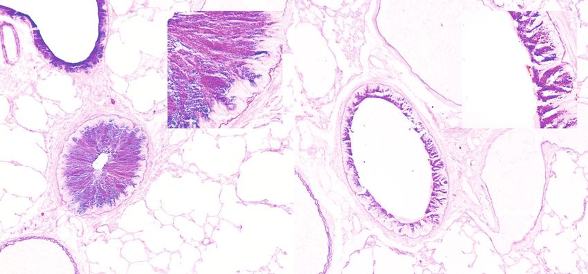
ARTICLE                                                               NATURE COMMUNICATIONS | https://doi.org/10.1038/s41467-020-20499-0

W
            ith daily exposures to >8000 liters of air containing            airway obstruction in asthma can be improved by disrupting
            billions of particles and potential pathogens, respira-          mucin disulfides with the reducing agent tris(2-carboxyethyl)
            tory tissues embody the need for robust host defense.            phosphine (TCEP) (Fig. 1b).
Airway mucus is critical for protection, but poor control of mucus              We chose TCEP due to its stability in aqueous solutions
function is central to numerous lung diseases1–5. In patients who            compared to reducing agents such as dithiothreitol, and also due
die during asthma exacerbations, mucus obstruction is a feature              to the ability of TCEP to rapidly reduce disulfides, including
long-recognized by pathologists6, with plugging observed in                  mucins19,20. In a recent study using a mouse model of chronic
>90% of cases and thus considered a major cause of fatal                     mucus dehydration similar to CF, TCEP was able to disrupt
obstruction5. Mucus obstruction is also prominent in non-fatal               mucus plugs in chronically obstructed airways21. Accordingly, we
cases of severe asthma2, but effective mucolytic therapies are               hypothesized that TCEP could be applied to test the ability of
lacking. Accordingly, asthma treatments could be significantly                disulfide reduction to reverse the effects of acute mucus hyper-
improved by determining molecular mechanisms of mucus                        secretion in a setting of allergic asthma.
dysfunction7.                                                                   We show that TCEP disrupts human asthmatic mucus and
   The predominant macromolecules in mucus are polymeric                     reverses pathological effects of mucus hypersecretion in a mouse
mucin glycoproteins7,8. In health, effective host defense requires           allergic asthma model. These effects of TCEP were mediated by
homeostatic mucin synthesis and secretion1. By contrast, exces-              loosening of mucus gel structure, enhancement of mucociliary
sive mucin production and secretion are demonstrated by alcian               clearance, and reduction of airway obstruction. These data sup-
blue-periodic acid Schiff’s (AB-PAS) staining within airway sur-             port the therapeutic potential of mucolytic treatments to inhibit
face and submucosal gland epithelial cells, and in plugs filling              the effects of mucus hypersecretion in muco-obstructive diseases,
small airways in fatal asthma (Fig. 1a). Mucin glycoprotein                  including asthma.
overproduction is also prominent in mild-to-moderate disease9,
suggesting a potentially broad etiological role for mucus hyper-
                                                                             Results
secretion in asthma.
                                                                             Polymeric mucins are targets for disulfide disruption of mucus
   Two polymeric mucin glycoproteins are abundant in airway
                                                                             in human asthma. Using mucus from human asthma patients,
mucus. The chief airway mucin in healthy mucus is MUC5B,
                                                                             we verified the presence of targets that could be reduced with
whose absence in mice results in impaired mucociliary clearance,
                                                                             TCEP under physiologic conditions (pH 7.4, 37 °C). In histologic
pathogen accumulation, and spontaneous lethal infections1. In
                                                                             samples from fatal asthma, AB-PAS positive plugs were sensitive
humans with asthma, MUC5B decreases in many patients9,10,
                                                                             to TCEP as demonstrated by alkylation of reduced thiols with
and lower MUC5B levels are associated with worsened disease10.
                                                                             biotinylated maleimide (Fig. 1c). Furthermore, in mucus aspi-
On the other hand, excessive MUC5B is also a risk factor for
                                                                             rated from bronchial airways of the same patient, TCEP reduced
developing pulmonary fibrosis11,12, and Muc5b has a gene dosage
                                                                             the sizes of massive MUC5AC and MUC5B polymers in a con-
effect on lung fibrosis in mice13. The other airway polymeric
                                                                             centration dependent manner (Fig. 1d). Based on these findings,
mucin, MUC5AC, is expressed at low levels at baseline, but it is
                                                                             we tested whether mucin polymer disruption could improve the
dramatically up-regulated in human asthma9,10 and in mouse
                                                                             physical properties of asthmatic mucus.
models14 where it is required for asthma-like mucus obstruction
                                                                                We used multiple particle tracking to assess mucus biophysical
and airway hyperreactivity (AHR)15.
                                                                             properties by quantifying mean square displacement (MSD) of 2 μm
   Taken together, MUC5AC and MUC5B play significant func-
                                                                             diameter carboxylated micro-particles. In fatal asthma mucus, MSD
tions in the lungs, but their precise roles in health and disease are
                                                                             was significantly increased in samples treated with TCEP (Fig. 1e, f).
complex7. The genetic, developmental, and environmental factors
                                                                             When converted to rheologic parameters22,23, these results demon-
that cause aberrant MUC5AC and MUC5B gene expression are
                                                                             strated that TCEP treatment rendered mucus less viscoelastic
active areas of investigation. However, given the inherent het-
                                                                             (Supplementary Fig. 1a), an effect that was driven by a significant
erogeneity among these pathways, finding selective gene expres-
                                                                             reduction in its elastic modulus (Supplementary Fig. 1b). The rapid
sion or signal transduction targets that can prevent mucus
                                                                             depolymerization of mucins and improvement of rheologic proper-
dysfunction while still preserving (or improving) mucus defense
                                                                             ties in asthmatic mucus in vitro suggested that TCEP could improve
remains an on-going challenge7,8,16. As an alternative, we propose
                                                                             mucus functions in an animal model of asthma in vivo.
that pathological effects of mucus hypersecretion can be reversed
while healthy functions are enhanced by directly targeting mucin
glycoprotein polymers.                                                       Mucolytic treatment reverses mucus dysfunction in a mouse
   Polymeric mucins are evolutionary precursors of the hemos-                model of allergic asthma. To produce allergic asthma-like inflam-
tasis protein von Willebrand factor (vWF). Accordingly, they                 matory, mucous, and AHR phenotypes, BALB/c mice were exposed
possess vWF-like amino (N-) and carboxyl (C-) terminal                       to a fungal allergen, Aspergillus oryzae extract (AOE), by aerosol
cysteine-rich domains that form covalent disulfide intermolecular             weekly for a total of four challenges15. Endpoints were studied 48 h
linkages to form large glycopolymers17. MUC5AC and MUC5B                     after the last AOE challenge, a time point of robust inflammation
first assemble in the endoplasmic reticulum as C-terminal dis-                and mucin overproduction (Supplementary Fig. 2a)15. Nebulized
ulfide dimers. Then in the Golgi, along with becoming heavily                 TCEP was used to determine the effects of mucolytic treatment on
glycosylated, they further multimerize via N-terminal disulfide               mucus properties and functions in vivo.
linkages18. Upon secretion, MUC5AC and MUC5B become                             To evaluate mucus directly on mouse airway surfaces, we
hydrated, extend into strands, and form a porous mesh-like gel               employed multiple particle tracking to quantify MSD of muco-
that traps particles and mediates mucociliary clearance.                     inert nanoparticles (MIPs) aerosolized onto mouse tracheas
   Due to the nature of mucus as a gel polymer, it is exquisitely            ex vivo24–26. Tracheas were removed from saline or AOE
sensitive to changes in the concentrations of solid materials                challenged mice, opened, placed on glass coverslips, and treated
within its matrix. Accordingly, when mucins are overproduced                 with nebulized 100 nm diameter MIPs suspended in saline vehicle
and hypersecreted in asthma, mucociliary clearance dysfunction               or TCEP (Fig. 2a). Compared to uninflamed controls, MIP
reflects aberrant gel behavior that can be corrected by disrupting            diffusion was heterogeneous and impaired in the tracheal mucus
its polymeric mucin components8. Therefore, here we test whe-                of AOE-challenged mice (Fig. 2b), resulting in a 2.7-fold decrease
ther aberrant mucus gel structure, mucociliary clearance, and                (p = 0.02) in median MSD values (Fig. 2c). Upon mucolytic

2               NATURE COMMUNICATIONS | (2021)12:249 | https://doi.org/10.1038/s41467-020-20499-0 | www.nature.com/naturecommunications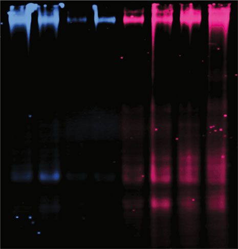
NATURE COMMUNICATIONS | https://doi.org/10.1038/s41467-020-20499-0 ARTICLE
a.
airway
surface
gland duct
Trachea Bronchus Bronchiole
b. c.
MUC5AC MUC5B AB-PAS Biotin-mal. (Veh) Biotin-mal. (TCEP)
N- -C N- -C
disulfide assembly
MUC5AC
MUC5B
polymerized
reduction
depolymerized
d. TCEP (mM)
e. f.
35 25
Veh
Frequency (%)
=1s
0 .1 .3 1 3 10 0 .1 .3 1 3 10 15 20
2.5
TCEP -4
* p < 10
MSD (μm2)
pol.
15
2.0
1.5 10
1.0
5
depol.
0.5
0 0
−4 −2 0 2
MUC5AC MUC5B Veh TCEP Log MSD (μm2) =1s
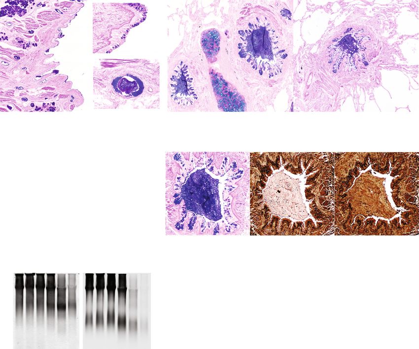
Fig. 1 Polymeric mucins in asthmatic airways are targets for disulfide disruption. a Alcian blue/periodic acid-Schiff (AB-PAS) stained tissues from the lungs
of patients (n = 2) who died during asthma exacerbations demonstrate mucin glycoproteins in large and small airways. Scale bars, 500 μm and 100 μm in insets
(trachea), 200 μm (bronchus and bronchiole). b MUC5AC and MUC5B assemble via amino (N-) and carboxyl (C-) terminal disulfide bonds that are sensitive to
reducing agents. c A mucus plug in a fatal asthma airway was examined in consecutive sections stained with AB-PAS (purple), or labeled with biotinylated-
maleimide (brown) after incubation at 37 °C for 10 min with saline vehicle (Veh) or tris(2-carboxyethyl)phosphine (TCEP, 10 mM). Scale bars, 250 μm; image
representative of airways from n = 2 patients. d–f. Expectorated sputum (n = 3 patients) was treated with TCEP (0.1–10 mM, 37 °C, 30 min), separated by
electrophoresis (1% SDS/agarose, non-reducing), and detected by immunoblot for MUC5AC and MUC5B (d). Note that full reduction causes epitope loss for both
anti-mucin antibodies resulting in decreased signal intensities at higher TCEP concentrations in d. Diffusion of 2-µm carboxylated micro-particles was evaluated in
mucus samples aspirated from fatal asthma bronchi (e, f). Compared to vehicle controls (Veh, cyan, n = 1303), particle mean square displacement (MSD)
increased significantly after TCEP (magenta, n = 1425). Bars in e are means ± sem of summarized linear-scale values, with individual points (open circles). Data in
f are log distributions of particles with curves showing Gaussian non-linear fits (r2 = 0.99 for Veh and TCEP). Significance was determined by two-tailed
Mann–Whitney U-tests with p-value and “*” denoting significance from Veh in f. Inverted arrows in f indicate locations of median values. Source data are provided
as a Source Data file.
treatment, MSD normalized to levels and homogeneity that were (Fig. 2d and Supplementary Fig. 4). Effects of TCEP on mucolysis
indistinguishable from non-allergic controls (Fig. 2b, c), reflecting an and mucociliary clearance were dose dependent, with mucins
increase in computed mucus mesh spacing (Supplementary Fig. 3). demonstrating significant depolymerization in mice treated with
Taken together, these findings suggested that mucolytic treatment aerosols of 50 and 500 mM TCEP solutions (Fig. 2e, f and
normalized mucus gel microstructure uniformity, which we Supplementary Fig. 5). Concordant with mucolysis, there were acute
postulated would also improve mucociliary function. decreases in inflammatory cell numbers in lung lavage fluid (Fig. 2f
We thus investigated the efficacy of inhaled mucolytic and Supplementary Table 1). With increasing doses of TCEP,
treatment on mucociliary clearance in allergically inflamed mice eosinophils were cleared resulting in a >5-fold decrease in total
in vivo. Animals were allergen challenged with AOE, and 48 h numbers recovered from lung lavage from mice treated with the
after the challenge, they received nose-only aerosol treatments highest concentration of TCEP delivered (500 mM). The rapid
with TCEP (5–500 mM) for 40 min (see Supplementary Fig. 2b). elimination of leukocytes from airway surfaces upon mucolytic
Immediately after mucolytic treatment, lungs were lavaged, and treatment provided direct functional evidence of improved
the disruption of mucin polymers and the elimination of clearance13.
inflammatory cells from airspaces were assessed13. In TCEP- Collectively, our in vitro, ex vivo, and in vivo results showed
treated mice, mucins demonstrated faster electrophoretic migra- that mucolytic treatment reversed mucus dysfunction in a setting
tion relative to controls thereby validating effective depolymerization of allergic inflammation and excessive mucin production. These
NATURE COMMUNICATIONS | (2021)12:249 | https://doi.org/10.1038/s41467-020-20499-0 | www.nature.com/naturecommunications 3ARTICLE NATURE COMMUNICATIONS | https://doi.org/10.1038/s41467-020-20499-0
a. b. c. p = 0.02
Tracheal mucus MPT q < 0.05
100 1 p = 0.04
Cumul. frequency (%)
AOE + Veh *
nebulizer AOE + Tx
Healthy
0 * *
*
Log MSD
*
MIPs 50 * –1
*
*
* –2
0 –3
O eh
−4 −3 −2 −1 0 1
H Tx
y
lth
V
+
Log MSD
+
ea
E
E
O
A
A
d. Veh Tx e. p = 0.007
f. p = 0.01
100 p = 0.002 15
Eosinophils (×104)
Percent unreduced
unred.
75 10
50
* * 5
red.
25 *
0 5 50 500 0 5 50 500
TCEP (mM) TCEP (mM)
Fig. 2 Mucolytic treatment improves mucus function in allergic mouse airways. a Mucus was probed in tracheal preparations ex vivo using 100 nm muco-
inert particles (MIPs). b, c In Aspergillus oryzae extract (AOE) challenged mice receiving 500 mM aerosol TCEP treatment (Tx, magenta, n = 13 biological
replicates), MIP diffusion measured as mean square displacement (MSD) increased significantly compared to vehicle (Veh, cyan, n = 14 biological replicates) and
non-AOE exposed animals (Healthy, gray, n = 7 biological replicates). Cumulative distribution data in b show the distributions of MSD values (means ± sem).
Scatter plot data in c show individual median MSD values per animal. d–f Mucociliary clearance was tested in AOE challenged mice treated by nose-only aerosol
with TCEP in a concentration dependent manner or Veh, followed by immediate lung lavage. Lectin blot analysis using Ulex europaeus agglutinin I (UEA-1) (α1,2-
fucose) in d shows disruption of mucin polymers in TCEP-treated animals (Tx, 500 mM, magenta) compared to vehicle (cyan). Image shows four samples per
group. Intensities of high molecular weight (polymerized) and low molecular weight (depolymerized) mucins were evaluated using Image Studio software (e).
Numbers of total eosinophils (f) recovered in lung lavage decreased significantly in Tx (magenta, n = 8 biological replicates) vs. Veh (cyan circles, n = 6 biological
replicates) exposed animals. Lines and error bars in c–f are means ± sem. ‘*’ denotes significance using a cut-off of 0.05 determined by unpaired two-tailed t-tests
using a two-stage step-up method at a 5% false discovery rate from Veh in b, by two-tailed Mann–Whitney U-test in c, and by non-parametric one-way ANOVA in
e, f. Source data are provided as a Source Data file.
findings led us to hypothesize that mucolytic treatment could also improvements in RL and RAW (Fig. 3a–d). There was also
protect against asthma-like airflow obstruction. To test this, we significant protection of resistance (GTI) and elastance (HTI) in
developed a two-route challenge and treatment analysis, with peripheral lung tissues (Fig. 3e–h). Thus, inhaled TCEP was not
methacholine administered intravenously (i.v.) to cause obstruction, acutely detrimental to pulmonary surfactant function, and it
and with TCEP administered by inhalation to cause mucolysis. appeared to confer protection from AHR by preserving airway
Methacholine and TCEP treatments were given during pulmonary patency. Indeed, the protective effects of TCEP treatment in
function tests (Supplementary Fig. 2c). allergic wild type mice were indistinguishable from benchmarks
Since we previously found that Muc5ac absence prevents AHR to in allergic Muc5ac−/− mice (p = 0.98), strongly supporting the
inhaled methacholine, which results in both airway smooth muscle hypothesis that mucolytic protection from AHR was linked to
contraction and mucin secretion15, we first tested whether Muc5ac reversal of mucus plugging.
was similarly required for AHR to methacholine administered To validate the role of mucolysis in protection from AHR in
intravenously (without mucolytic treatment). Compared to non- TCEP-treated mice, we confirmed mucin polymer reduction by
allergic animals, AOE challenged mice demonstrated significantly examining lung lavage fluid via immunoblot (Supplementary Fig. 6).
exaggerated increases in total lung resistance (RL) and airway We also quantified mucin accumulation in airspaces histologically15.
resistance (RAW) in response to i.v. methacholine (Fig. 3a–d). During methacholine-induced bronchoconstriction, secreted mucin
Importantly, these AHR responses were abolished in Muc5ac gene volume, airway obstruction, and heterogeneous plugging were
deficient mice (Fig. 3a–d), thereby validating a role for mucus significantly reversed throughout the airways of TCEP-treated
hypersecretion in this methacholine challenge model. The degrees of animals (Fig. 4). Thus, protection from AHR was directly related
protection observed in this chronic mucus prevention setting also to the ability of mucolytic treatment to reduce airway plugging.
established benchmarks for comparing effects of acute mucolytic
rescue. Accordingly, we next tested whether inhaled TCEP protected
mice from AHR by reversing mucus plugging. Discussion
In AOE exposed wild type mice treated with inhaled TCEP The studies reported here show that disrupting mucin polymers
during i.v. methacholine challenge, we observed significant improves mucus microstructure, enhances mucus transport, and
4 NATURE COMMUNICATIONS | (2021)12:249 | https://doi.org/10.1038/s41467-020-20499-0 | www.nature.com/naturecommunicationsNATURE COMMUNICATIONS | https://doi.org/10.1038/s41467-020-20499-0 ARTICLE
a. Total lung resistance c. Airway resistance e. Tissue resistance g. Tissue elastance
Veh
TCEP
GTI (cm H2O·s(1-α)·ml-1)
+/+ Saline, Veh
HTI (cm H2O·s(1-α)·ml-1)
18 6 75 120
RAW (cm H2O·s·ml-1)
+/+ AOE, Veh
RL (cm H2O·s·ml-1) +/+ AOE, Tx
−/− AOE, Veh
12 4 50 80
6 2 25 40
0 0 0 0
4 8 16 32 64 4 8 16 32 64 4 8 16 32 64 4 8 16 32 64
MCh (μg∙kg-1∙min-1, i.v.) MCh (μg∙kg-1∙min-1, i.v.) MCh (μg∙kg-1∙min-1, i.v.) MCh (μg∙kg-1∙min-1, i.v.)
b. 0.0009 0.006 0.02 d. 0.01 0.04 0.03 f. 0.0009 0.004 0.005 h. 0.08
n.s.
0.1 0.1
30 * 0.3 * 3 * 6
RAW slope
GTI slope
RL slope
HTI slope
20 0.2 2 4
10 0.1 1 2
0 0 0 0
AOE − + + + AOE − + + + AOE − + + + AOE − + + +
Tx − − + − Tx − − + − Tx − − + − Tx − − + −
Muc5ac +/+ +/+ +/+ −/− Muc5ac +/+ +/+ +/+ −/− Muc5ac +/+ +/+ +/+ −/− Muc5ac +/+ +/+ +/+ −/−
Fig. 3 Mucolytic treatment reverses allergic airway hyperreactivity. Dose response curves to methacholine (MCh, 4–64 μg kg−1 min−1, i.v.) were
generated in AOE challenged allergic wild type (WT) mice (magenta, n = 7 biological replicates) treated with TCEP between MCh doses (100 mM,
inverted yellow triangles). For comparison, saline challenged non-allergic WT mice (gray, n = 6 biological replicates), AOE challenged allergic WT mice
(cyan, n = 12 biological replicates), and AOE challenged Muc5ac−/− mice (black, n = 4 biological replicates) were treated with nebulized vehicle (Veh)
during i.v. MCh dose response tests. Values are means ± sem. For each mouse, dose response curves were fitted by log-linear best-fit regression analysis,
and slopes of regression lines were analyzed by one-way ANOVA. “*”, p < 0.05 using Dunnett’s test for multiple comparisons relative to AOE-challenged
Veh-treated WT mice (p-values are shown). Total lung resistance (RL in a, b), conducting airway resistance (RAW in c, d), tissue resistance (GTI in e, f) and
tissue elastance (HTI in g, h) are shown. Source data are provided as a Source Data file.
protects airflow in allergic asthma settings. In patients, mucin and it was conducted using large (500–600 µm diameter) metal
overproduction and pathologic changes in mucus biophysical particles. Given the loads imparted by these materials and the
properties are correlated with asthma exacerbations and forces required to displace them, inhibitory consequences of
fatalities2,5,7,9,27. These features are observed frequently by TCEP treatment on exogenous transport in that report may not
pathologists, but they are usually ignored during clinical assess- be directly comparable to findings investigating endogenous
ments since diagnostic tests and interventions have lagged. On clearance here. In our studies, TCEP treatment normalized
the diagnostic side, mucus obstruction is becoming more recog- mucus, improved clearance, and reversed acute airway plugging
nizable through high resolution imaging2, but treatment options (see Figs. 2–4). Thus, even under diseased conditions, mucolytic
are still constrained by a lack of efficacious mucolytics. effects were examined within physiologic constraints.
Indeed, while potentially beneficial in laboratory settings, Our findings suggest that an inhaled mucolytic treatment could
mucolytics and expectorants are not widely used ther- confer acute protection from obstruction in asthma. Airway
apeutically. The only FDA-approved reducing agent available narrowing initiated by smooth muscle contraction is clearly
as an inhaled mucolytic is N-acetylcysteine (NAC), but its important, and obstruction is amplified by mucus15. In bronch-
efficacy is low due to NAC’s weak activity at airway pH and oalveolar lavage (BAL) fluid from patients with mild-to-moderate
high mucin concentrations28–30. We propose that inhaled asthma, TCEP rapidly depolymerizes MUC5AC and MUC5B
mucolytic agents can reduce plugging and thus improve airway (Supplementary Fig. 7), demonstrating the presence of targets in
function in asthma. Nonetheless, it will be critical for any asthma patients even under stable disease conditions. To translate
strategy that reverses the detrimental effects of mucus hyper- our findings in mice to a clinically relevant setting, it will be
secretion to do so while also protecting mucus-mediated crucial to achieve pharmacokinetic and pharmacodynamic indi-
defense. Since disulfide assembly is a critical process in the ces that are effective, tolerable, and safe.
formation of viscoelastic mucus, mucin reduction remains an Full reduction of disulfide bonds could result in disruption of
attractive target for disrupting mucus plugs in human airways mucins to monomer fragments that form a poorly transported
(see Fig. 1). In addition, it is also plausible that the use of a viscous liquid. In our studies, partial depolymerization of mucins
reducing agent could affect other targets. For example, oxi- (Fig. 2d, e and Supplementary Fig. 5) was sufficient to facilitate
dants released from cells as a result of injury and inflammation mucociliary clearance such that eosinophil numbers decreased by
could be directly neutralized, but the effects of transient anti- 81% in vivo (Fig. 2f). Accordingly, partial reduction could be
oxidant treatment need to be tested13. effective for destabilizing aggregated mucus in patients with
In contrast to the findings reported here, a recent study heterogenous muco-obstruction in large and small diameter air-
investigating mucociliary transport showed inhibitory effects of ways. Like other inhalation therapies, the ability to achieve distal
TCEP on mucus transport in non-diseased pigs31. However, that lung deposition will also depend on aerosol droplet sizes and
investigation focused on tracheobronchial glandular secretions, chemical composition.
NATURE COMMUNICATIONS | (2021)12:249 | https://doi.org/10.1038/s41467-020-20499-0 | www.nature.com/naturecommunications 5ARTICLE NATURE COMMUNICATIONS | https://doi.org/10.1038/s41467-020-20499-0
a. Veh Tx
b. c. d.
All airways
p = 0.02
Mucin occlusion (%) p = 0.03 q ≤ 0.02
18 50 100
* *
Mucus volume (μl)
*
Frequency (%)
40 80
12
30 60
20 40 * Veh
6 * Tx
10 * 20
0 0 0
Veh Tx Veh Tx 0 10 20 30 40 50 60 70 80 90 100
Mucin occlusion (%)
e. f. g.
Bronchi
p = 0.006 p = 0.05 q < 0.001
Mucin occlusion (%)
18 50 100
Mucus volume (μl)
*
Frequency (%)
40 80 *
12
30 60
20 40 * Veh
Tx
6
*
* 10 20
0 0 0
Veh Tx Veh Tx 0 10 20 30 40 50 60 70 80 90 100
Mucin occlusion (%)
h. i. j.
Bronchioles
n.s. p = 0.01 q ≤ 0.01
Mucin occlusion (%)
18 50 100
* *
Mucus volume (μl)
Frequency (%)
40 80
12
30 60
20
* Veh
40 Tx
6
10 * 20
0 0 0
Veh Tx Veh Tx 0 10 20 30 40 50 60 70 80 90 100
Mucin occlusion (%)
Fig. 4 Mucolytic treatment disrupts mucus plugging. a AB-PAS stained lungs obtained after methacholine dose response tests show mucin glycoproteins
obstructing airspaces in vehicle (Veh, n = 8 biological replicates) treated mice that are disrupted by mucolytic treatment (Tx, n = 7 biological replicates).
Scale bars, 500 μm (low power) and 25 μm (high power). b–j Calculated mucus volumes (b, e, h), mean fractional occlusion (c, f, i), and heterogeneous
plugging (d, g, j) were significantly decreased in AOE-challenged mice receiving mucolytic (Tx, magenta, n = 7 biological replicates) compared to controls
(Veh, cyan, n = 8 biological replicates). Mucolytic treatment significantly reduced obstruction across all airways (b–d), and this effect was most prevalent
in bronchi (e–g). Although total mucus volume in bronchioles was low (h), occasional mucus aggregates were obstructive and are sensitive to TCEP
treatment (i, j). Data in b, c, e, f, h, and i are means ± sem on scatter plots, with p-values shown and significance (*) indicating p < 0.05 by two-tailed
Mann–Whitney U-test. Cumulative frequency distributions in d, g, and h show percentages of airways demonstrating occlusion, with circles and error bars
identifying means ± sem, and “*” demonstrating significance by t-test using a two-stage step-up method at a 5% false discovery rate. Source data are
provided as a Source Data file.
6 NATURE COMMUNICATIONS | (2021)12:249 | https://doi.org/10.1038/s41467-020-20499-0 | www.nature.com/naturecommunicationsNATURE COMMUNICATIONS | https://doi.org/10.1038/s41467-020-20499-0 ARTICLE
Recent reports have shown that mucin concentration and Spontaneously expectorated CF sputum samples were collected from patients
polymerization are critical factors that determine mucus (ages 31–52, n = 3 male/2 female, non-smokers, no CFTR modulator therapies) at
the adult CF clinic at Johns Hopkins University. Studies were approved by the
viscoelasticity8,32. A prolonged disulfide reduction strategy simi- Johns Hopkins IRB, and participants gave informed consent as above.
lar to what was applied here was shown to improve chronic
inflammation, airway injury, and abnormal tissue repair in a Mucin protein detection. Histochemical staining with alcian blue-periodic acid
mouse model of pulmonary fibrosis13. Furthermore, TCEP was Schiff’s (AB-PAS) stain was performed using standard techniques38. For immuno-
able to disrupt plugs in chronically mucus obstructed airways in a detection, human, and mouse mucins were detected using rabbit-anti-human
mouse model of CF21. MUC5AC (MAN5AC, Dr. Thornton’s laboratory, diluted 1:1000)39, mouse-anti-
The findings reported here build upon these prior works by MUC5AC (clone 45M1, ThermoFisher, diluted 1:1000), rabbit-anti-human
MUC5B (H300, Santa Cruz, diluted 1:5000), and rabbit-anti-mouse Muc5b (Dr.
showing mucolytic efficacy in a common disease, by demonstrating Evans’s laboratory, diluted 1:5000)1. For biochemical labeling, mucins were also
protective mucolytic effects during acute hypersecretion, and by detected using biotinylated Ulex europaeus agglutinin I (UEA-I) lectin to detect
validating that reversing mucus plugging has direct effects on pul- fucose residues (Vector, Burlingame, CA) or with biotinylated maleimide to
monary function (see Figs. 1 and 4). Taken together, these results alkylate and detect reduced sulfhydryls (ThermoFisher).
support the concept that protection could be conferred in both
chronic and acute respiratory diseases by preventing or reversing Immuno-/ lectin blotting. Lung lavage and mucus specimens were separated on
SDS/agarose gels to determine mucin profiles and changes in mucin polymer sizes
mucus dysfunction by disrupting mucin disulfide bonds. upon reduction40. A dot-blot ELISA was performed to assess MUC5B levels in each
Nonetheless, in addition to disulfide targets formed during human lavage sample, and lanes were equilibrated to contents of MUC5B, the
mucin biosynthesis, there are other potential targets for mucolytic predominant mucin in these patients with non-exacerbated asthma. In allergic
intervention. These include non-disulfide covalent linkages such mice, although lung lavage fluid contains mixtures of both Muc5ac and Muc5b,
they are found at higher concentrations and can be more homogeneously sampled
as sugars added during glycosylation, as well as non-covalent (see below). Therefore, mouse lung lavage samples were loaded into gels using
mucin polymer interactions formed during mucin packaging into equal volumes. After electrophoresis and transfer to PVDF membranes by vacuum
secretory granules. In addition, in the post-secretory environment blot40, mucins were probed with antimucin antibodies (1:1000–5000 dilutions as
oxidant-mediated mucin cross-linking is observed in cystic indicated), and then detected with goat antimouse IRDye 680RD or goat antirabbit
IRDye 800CW antibodies (LI-COR, Lincoln, NE, diluted 1:20,000). Image analysis
fibrosis (CF)33 and asthma2, and it is reported to occur on free was performed using Image Studio software (LI-COR).
thiols in these in static mucus aggregates in these settings.
Additional non-covalent mucus interactions associated with Particle tracking in human mucus. For human airway mucus samples, 2.0 µm
macromolecules such as DNA34, or effects mediated by ionic and fluorescent carboxylated microspheres (ThermoFisher) were used to estimate
pH environments may have additional effects on interstitial fluid mucus microrheology. Frozen mucus samples from the patients above were thawed
surrounding mucus mesh networks and could be also targeted35–37. on ice, distributed into 100 µl aliquots. A 10 µl solution of TCEP (100 mM) or
saline (vehicle) containing 100,000 microspheres was added to each mucus sample.
Although these are not directly affected by reduction per se, agents Samples were incubated at 37 °C for 10 min.
that disassemble disulfides could be applied in combination with Immediately after incubation, samples were loaded onto chambered slides and
other therapies to improve mucus hydration and transport. Thus, video imaged at five randomly chosen sites on a BX63 microscope for 30 s (7.5
treatments that reverse mucus dysfunction could then be con- frames per sec) using a DP80 camera (Olympus, Center Valley, PA). Particle
sidered as possible adjunct therapies. tracking was performed using Olympus cellSens software, and analyses were made
to derive MSD values using a Matlab program (Mathworks, Natick, MA) that was
Having identified the ability to deliver a mucolytic agent, published previously25,26,41. Using the frequency-dependent Stokes–Einstein
promote mucociliary clearance, and reverse AHR in a mouse equation, we translated MSD values to obtain approximations of micro-rheological
model of allergic asthma, findings reported here support the properties of airway mucus22,23.
concept that mucin disulfide disruption could be applied to
human lung pathologies where mucus dysfunction is Mice. Studies were conducted with approval of the University of Colorado Denver
significant7,8. Inflammatory or injurious causes of mucus dys- and Johns Hopkins University Institutional Animal Care and Use Committees.
Housing rooms are maintained at 22 °C, 30–40% humidity, and a 14/10 (h/h) light/
function vary within and across diseases, yet they still result in a dark cycle and at least 12 fresh-air changes per h. Male and female BALB/cJ wild
similar outcome—excessive polymeric mucin production. Indeed, type mice were purchased from the Jackson Labs (Bar Harbor, ME). Muc5ac−/−
mucus from fatal asthma exhibited comparable microstructure to mice were previously crossed onto a congenic BALB/cJ strain background15.
CF sputum, and both samples had almost identical concentra- Animals were housed under specific pathogen-free conditions and used in allergic
asthma studies beginning at ages 6–8 weeks.
tions of mucus solids (Supplementary Fig. 8) despite having To induce allergic inflammation and mucin overproduction, mice were
unrelated causes for abnormal hydration. challenged using aerosolized Aspergillus oryzae extract15 (AOE; Sigma), as shown
To this end, even in diseases with diverse primary causes, there in Supplementary Fig. 2. Mice received four weekly challenges, and endpoint
could be benefits that derive from optimizing airway mucus. analyses were studied 48 h after the last AOE challenge. To induce mucolysis, mice
and mucus samples were exposed to tris(2-carboxyethyl)phosphine (TCEP, neutral
Furthermore, while reducing obstruction, mucin depolymeriza- pH solution, Thermo Scientific, Cat. no. 77720). Mice exposed to saline challenges
tion could enhance the deposition and absorption of inhaled were used as controls.
bronchodilator, anti-inflammatory, or antibiotic agents. Addi-
tional studies focused on developing effective mucolytic therapies Particle tracking in sputum samples and freshly excised mouse tracheas.
and potentially mucin-selective treatments are needed. MIPs were prepared using 5-kDa methoxy-polyethylene glycol (PEG)-amine
(Creative PEGWorks, Chapel Hill, NC) molecules that were densely conjugated to
the surface of 100 nm carboxyl-functionalized polystyrene beads (PS-COOH;
Methods Product #F8801, 580/605, Molecular Probes, Eugene, OR)25. Covalent attachment
Human specimens. BAL fluid was obtained from volunteers with asthma recruited to the carboxyl end groups on the PS-COOH particles was achieved using a
for a University of Colorado Institutional Review Board (IRB) approved study (ages crosslinker, 1-ethyl-3-(3-dimethylaminopropyl) carbodiimide hydrochloride
18–53, n = 2 male, 2 female, non-smokers). Volunteers gave informed consent in (Sigma-Aldrich, No. E7750) and sulfo-N-hydroxysulfosuccinimide sodium salt
accordance with the Declaration of Helsinki. BAL was collected from the left upper (Sigma-Aldrich, No. 56485) in borate buffer (200 mM, pH 8.0, Growcells, No.
lobe (lingula projection), and a 2 ml sample was separated and stored “neat” MRGL-1100). Physicochemical properties of MIPs were then measured by a
(without centrifugation) to preserve high molecular weight mucins that sediment at Zetasizer Nano ZS90 (Malvern Analytical, Malvern, UK). Resulting 100 nm MIPs
low centrifugation speeds. exhibited hydrodynamic diameters of 98.0 ± 6.9 nm, polydispersity indices of 0.1 ±
For fatal asthma samples, samples were obtained from lungs donated to Oregon 0.01 and ζ-potentials (i.e., indicative of particle surface charges) of −4.1 ± 0.2 mV
Health and Sciences University (kindly provided by Dr. David Jacoby) and measured in 10 mM NaCl pH 7.4 at 25 °C.
National Jewish Health. Samples from both subjects were de-identified prior to use CF and asthmatic mucus samples were stored at 4°C immediately after
and were thus human subject exempt. Tissue sections were made from formalin- collection for up to 24 h for particle tracking experiments to ensure that the
fixed paraffin embedded samples from both donors. Mucus aspirated from the inherent mucus microstructure is preserved42. Aliquots of 30 µl of sputum were
trachea and lobar bronchi of samples at National Jewish health. added to custom microscopy chambers. Next, 0.5 µl of MIP (0.00001% v/v) were
NATURE COMMUNICATIONS | (2021)12:249 | https://doi.org/10.1038/s41467-020-20499-0 | www.nature.com/naturecommunications 7ARTICLE NATURE COMMUNICATIONS | https://doi.org/10.1038/s41467-020-20499-0
mixed gently with sputum sample to evenly distribute particles within the sample. Reporting summary. Further information on research design is available in the Nature
The chamber was sealed with a circular coverslip and incubated at room Research Reporting Summary linked to this article.
temperature for 30 min prior to imaging.
Fluorescent MIPs were administered to the mucosal surfaces of tracheas freshly
dissected from mice. Briefly, tracheal tissues harvested from animals were cut along Data availability
the dorsal cranial-caudal axis and laid flat to expose mucosal surfaces. MIPs were All data supporting the findings of this study will be available from the corresponding
diluted in saline (i.e., vehicle control) or TCEP (500 mM) at 0.02% w/v and authors upon reasonable request. Source data are provided with this paper.
administered inNATURE COMMUNICATIONS | https://doi.org/10.1038/s41467-020-20499-0 ARTICLE
27. Lachowicz-Scroggins, M. E. et al. Abnormalities in MUC5AC and MUC5B Author contributions
protein in airway mucus in asthma. Am. J. Respir. Crit. Care Med. 194, L.M., S.S., C.M., J.H., J.S., D.T., F.H., W.J., W.T., and C.E. designed the study; L.M., A. J.,
1296–1299 (2016). S.S., D.R., N.E., V.R., N.H., A.H., J.N., C.H., H.E., D.V., and G.D. planned, performed,
28. Tam, J., Nash, E. F., Ratjen, F., Tullis, E. & Stephenson, A. Nebulized and oral and analyzed experiments. L.M. and C.E. wrote the manuscript with help from all
thiol derivatives for pulmonary disease in cystic fibrosis. Cochrane Datab. Syst. coauthors. C.E. supervised the manuscript preparation.
Rev. 7, CD007168 (2013).
29. Bylin, G., Hedenstierna, G., Lagerstrand, L. & Wagner, P. D. No influence of
acetylcysteine on gas exchange and spirometry in chronic asthma. Eur. J.
Competing interests
The muco-inert particle technology described in this article is being developed by Kala
Respir. Dis. 71, 102–107 (1987).
Pharmaceuticals. J.H. declares a financial, a management/advisor, and a paid consulting
30. Gillissen, A. et al. Nacystelyn, a novel lysine salt of N-acetylcysteine, to
relationship with Kala Pharmaceuticals. J.H. is a cofounder of Kala Pharmaceuticals and
augment cellular antioxidant defence in vitro. Respir. Med. 91, 159–168
owns company stock, which is subject to certain restrictions under Johns Hopkins
(1997).
University policy. C.E. is a paid consultant with Eleven P15, a company focused on early
31. Fischer, A. J. et al. Mucus strands from submucosal glands initiate mucociliary
detection and treatment of pulmonary fibrosis. C.M. is a paid consultant with Sharklet
transport of large particles. JCI Insight https://doi.org/10.1172/jci.
Technologies, a company that uses surface texture to reduce biological adhesion to
insight.124863 (2019).
medical devices. The terms of these arrangements are being managed by Johns Hopkins
32. Hill, D. B. et al. Pathological mucus and impaired mucus clearance in cystic
University and the University of Colorado in accordance with respective institutional
fibrosis patients result from increased concentration, not altered pH. Eur.
conflict-of-interest policies. W.T. and D.V. are employees of Parion Sciences, Inc., a
Respir. J. https://doi.org/10.1183/13993003.01297-2018 (2018).
company that designs and tests novel mucolytic agents. No proprietary mucolytic agents
33. Yuan, S. et al. Oxidation increases mucin polymer cross-links to stiffen airway
were used in this study. All other authors declare no conflicts of interest.
mucus gels. Sci. Transl. Med. 7, 276ra227 (2015).
34. Lieberman, J. Dornase aerosol effect on sputum viscosity in cases of cystic
fibrosis. JAMA 205, 312–313 (1968). Additional information
35. Trillo-Muyo, S. et al. Granule-stored MUC5B mucins are packed by the non- Supplementary information is available for this paper at https://doi.org/10.1038/s41467-
covalent formation of N-terminal head-to-head tetramers. J. Biol. Chem. 020-20499-0.
https://doi.org/10.1074/jbc.RA117.001014 (2018).
36. Button, B. et al. A periciliary brush promotes the lung health by separating the Correspondence and requests for materials should be addressed to C.M.E.
mucus layer from airway epithelia. Science 337, 937–941 (2012).
37. Muraglia, K. A. et al. Small-molecule ion channels increase host defences in Peer review information Nature Communications thanks Franz-Georg Hanisch, Niclas
cystic fibrosis airway epithelia. Nature 567, 405–408 (2019). Karlsson and Bruce Rubin for their contribution to the peer review of this work. Peer
38. Evans, C. M. et al. Mucin is produced by clara cells in the proximal airways of reviewer reports are available.
antigen-challenged mice. Am. J. Respir. Cell Mol. Biol. 31, 382–394 (2004).
39. Thornton, D. J., Carlstedt, I. & Sheehan, J. K. Identification of glycoproteins Reprints and permission information is available at http://www.nature.com/reprints
on nitrocellulose membranes and gels. Mol. Biotechnol. 5, 171–176 (1996).
40. Piccotti, L., Dickey, B. F. & Evans, C. M. Assessment of intracellular mucin Publisher’s note Springer Nature remains neutral with regard to jurisdictional claims in
content in vivo. Methods Mol. Biol. 842, 279–295 (2012). published maps and institutional affiliations.
41. Xu, Q. et al. Nanoparticle diffusion in, and microrheology of, the bovine
vitreous ex vivo. J. Control Release 167, 76–84 (2013).
42. Duncan, G. A., Jung, J., Hanes, J. & Suk, J. S. The mucus barrier to inhaled
Open Access This article is licensed under a Creative Commons
gene therapy. Mol. Ther. 24, 2043–2053 (2016).
Attribution 4.0 International License, which permits use, sharing,
43. Sears, P. R., Yin, W. N. & Ostrowski, L. E. Continuous mucociliary transport
adaptation, distribution and reproduction in any medium or format, as long as you give
by primary human airway epithelial cells in vitro. Am. J. Physiol. Lung Cell.
appropriate credit to the original author(s) and the source, provide a link to the Creative
Mol. Physiol. 309, L99–L108 (2015).
Commons license, and indicate if changes were made. The images or other third party
material in this article are included in the article’s Creative Commons license, unless
indicated otherwise in a credit line to the material. If material is not included in the
Acknowledgements article’s Creative Commons license and your intended use is not permitted by statutory
This study was funded by NIH grants HL080396 (C.E. and C.M.) and ES023384 (C.E.), regulation or exceeds the permitted use, you will need to obtain permission directly from
HL130938 (C.E. and W.J.), HL125169 (J.H.); by U.S. Department of Defense grants the copyright holder. To view a copy of this license, visit http://creativecommons.org/
W81XWH-17-1-0597 (C.E.), W81XWH-19-1-0172 (C.E.), and PR192068 (C.M.); Cystic licenses/by/4.0/.
Fibrosis Foundation grants EVANS18IO (C.E.), JARAMI20F0 (A.J.), and HANES16XX0
(J.H.); the National Science Foundation NSF CAREER 1941401 (C.M.), and by the
Medical Research Council grant MR/R002800/1 (D.T). © The Author(s) 2021
NATURE COMMUNICATIONS | (2021)12:249 | https://doi.org/10.1038/s41467-020-20499-0 | www.nature.com/naturecommunications 9You can also read