Who Bites Me? A Tentative Discriminative Key to Diagnose Hematophagous Ectoparasites Biting Using Clinical Manifestations - horizon ird
←
→
Page content transcription
If your browser does not render page correctly, please read the page content below
diagnostics
Review
Who Bites Me? A Tentative Discriminative Key to
Diagnose Hematophagous Ectoparasites Biting Using
Clinical Manifestations
Mohammad Akhoundi 1, *, Denis Sereno 2,3 , Anthony Marteau 1 , Christiane Bruel 4 and
Arezki Izri 1
 1    Parasitology-Mycology Department, Avicenne Hospital, AP-HP, 93000 Bobigny, France;
      anthony.marteau@aphp.fr (A.M.); arezki.izri@aphp.fr (A.I.)
 2    MIVEGEC, IRD, Montpellier University, 34032 Montpellier, France; denis.sereno@ird.fr
 3    InterTryp, IRD, Montpellier University, 34032 Montpellier, France
 4    Agence Régionale de Santé (ARS) Île-de-France, 35, rue de la Gare, 75935 Paris CEDEX 19, France;
      christiane.bruel@ars.sante.fr
 *    Correspondence: m.akhoundi@yahoo.com

 Received: 12 April 2020; Accepted: 8 May 2020; Published: 15 May 2020

 Abstract: Arthropod blood feeders are vectors of several human pathogenic agents, including viruses
 (e.g., yellow fever, chikungunya, dengue fever), parasites (e.g., malaria, leishmaniasis, lymphatic
 filariasis), or bacteria (e.g., plague). Besides their role as a vector of pathogens, their biting activities
 cause a nuisance to humans. Herein, we document clinical symptoms associated with the biting
 of ten clusters of hematophagous arthropods, including mosquitoes, biting midges and sandflies,
 lice, ticks, tsetse flies, blackflies, horse flies, fleas, triatomine and bed bugs. Within the framework of
 clinical history and entomo-epidemiological information, we propose a tentative discriminative key
 that can be helpful for practicing physicians in identifying hematophagous arthropods biting humans
 and delivering treatment for the associated clinical disorders.

 Keywords: hematophagous arthropods; blood feeding; bite spot; clinical manifestation; diagnosis

1. Introduction
     Arthropods are a large group of invertebrates, having a significant impact on human health.
They emerged in the late Precambrian period, approximately 550 million years ago (MYA) [1].
Bloodsucking arthropods are involved in the transmission of a wide diversity of pathogens such as
bacteria, viruses, protozoa, and microfilariae, and are also a significant cause of nuisances for humans,
worldwide (Table 1).

Diagnostics 2020, 10, 308; doi:10.3390/diagnostics10050308                     www.mdpi.com/journal/diagnostics
Diagnostics 2020, 10, 308 2 of 15
Table 1. Entomo-epidemiological criteria of hematophagous arthropods activity.
Hematophagous Vectorial Role Sex/ Stage/ Telmophagy/ Time/ Seasonal Geographical
Arthropod Disease Pathogenic Agent Bloodfeeding Bloodfeeding Solenophagy Bloodfeeding Activity Dispersion
Malaria, yellow fever,
Plasmodium sp., Flaviviridae,
Culicidae chikungunya, dengue fever, Zika, Sunset and Commonly
Togaviridae, Wuchereria sp., Brugia sp., Female Adult S1 Global
(Mosquito) lymphatic filariasis, night * warm seasons
Japanese encephalitis virus
and japanese encephalitis
Leishmaniasis, bartonellosis,
Ceratopogonidae Leishmania sp., Bartonella sp.,
bluetongue disease, African horse
(Biting midges) Bluetongue virus, Phlebovirus, Sunset and Commonly
sickness and epizootic Female Adult T2 Global
Phlebotominae Toscana virus, African horse virus, night warm seasons
hemorrhagic disease,
(Sandflies) Epizootic hemorrhagic disease virus
pappataci fever
Pediculidae, Epidemic typhus, trench fever and Rickettsia prowazekii, Bartonella quintana, Nymph and Throughout
Both sexes S Every time Global
Pthiridae (Lice) louse-borne relapsing fever Borrelia recurrentis adult the year
Lyme disease, Rocky Mountain Borrelia burgdorferi, Rickettsia rickettsii,
spotted fever, tularemia, Colorado Francisella tularensis, Colorado tick Larva,
Ixodidae, Throughout
tick fever, human tick-borne fever (CTF) virus, Ehrlichia chaffeensis, Both sexes Nymph and T Every time Global
Argasidae (Tick) the year
ehrlichiosis, babesiosis, tick E. ewingii, Babesia microti, Borrelia sp., adult
paralysis, relapsing fever, Q fever Coxiella burnetii
Throughout
the year but
Plague, murine typhus, Tungiansis, Yersinia pestis, Rickettsia typhi,
Pulicidae (Flea) Both sexes Adult T Every time mainly Global
Tularemia Francisella tularensis
inwarm
seasons
Glossinidae Trypanosoma brucei gambiense, Throughout Sub-Sahara
Sleep sickness Both sexes Adult T Diurnal
(Tsetse fly) Trypanosoma brucei rhodesiense the year countries
Reduviidae Latin and
Nymph and Throughout
(Kissing bug, Chagas Trypanosoma cruzi Both sexes S Nocturnal South
adult the year
Triatomine bug) America
Africa, Latin
Simuliidae Throughout
Onchocerciasis (River blindness) Onchocerca volvulus Female Adult T Diurnal and South
(Black fly) the year
America
Tabanidae Throughout
Loiasis Loa loa Female Adult T Diurnal Global
(Horse fly) the year
Cimicidae Nymph and Throughout
- - ** Both sexes S Nocturnal Global
(Bed bug) adult the year
*: Depends mainly on the species; **: Suspected to be involved in transmission of over 40 pathogens; 1 : Solenophagy; 2 : Telmophagy.
Diagnostics 2020, 10, 308 3 of 15
Arthropods biting occur for diverse purposes of defense mechanism, paralyzing, parasitizing their
hosts, or for feeding [2]. Hematophagy has emerged in many orders and families, among arthropods
like Anoplura (lice), Siphonaptera (fleas), Ixodida (ticks), Hemiptera (bed bugs and triatomines) and
Diptera (mosquitoes) [3]. Blood feeder arthropods took the high nutritional value of blood for their
own advantages. The feeding behavior is classified into two categories, explaining in part differences
in clinical presentation and dermatological reactions: (i) telmatophagy/telmophagy (pool feeding),
in which the feeders cut the epidermis and create a pool of blood that they suck (e.g., sandfly), or
(ii) solenophagy (vessel feeding), in which the feeders insert their specialized mouthparts into the
blood vessel for taking blood (e.g., mosquitoes and bed bugs) [4]. This feeding habit signifies them
as temporary (e.g., mosquitoes, sandflies, tsetse flies or tabanids), permanent (e.g., lice) or periodic
(e.g., fleas and ticks) ectoparasites [3]. It can be restricted to one sex (e.g., only female mosquitoes are
blood feeder) or to a peculiar developmental stage (e.g., nymphal stage in bed bugs), or to both sexes
and at all developmental life stages (e.g., lice, bed bugs).
Several protein-based factors present in the saliva facilitate blood-feeding. These include
vasodilator and inhibitor agents of blood coagulation and platelet aggregation, as well as the proteins
with the anesthetic role [2,5]. In general, reactions following arthropod bites are not specific and vary
widely from one individual to another. The immunoglobulin E (IgE) and G (IgG) responses directed
against these proteins of insect saliva are in part responsible for the individualized manifestation of
arthropod bite [6]. Besides immunological reactions, several factors play a role in clinical manifestations.
They include feeding behavior (solenophagy/telmatophagy), quietness, volume, and number (one or
multiple bites by the same insect) of arthropod blood-feeding activity, but also the environmental
temperature or host-associated cues (CO2 and heat), etc. [5]. The impact of vector-borne pathogens on
the bite spot clinical manifestation is excluded in the present review.
2. Clinical Manifestations of Arthropod Bites
2.1. Culicidae (Mosquitoes)
Culicidae belongs to the Diptera order and Nematocera sub-order. To date, 3546 species
from 111 genera are described worldwide, which small numbers of them feed on humans [7].
They are the largest vector group of pathogens, transmissible to humans (i.e., Plasmodium sp., filariae,
and arboviruses). These vessel feeders (solenophagy) possess long mouthparts for piercing and
blood-sucking with a feeding habit restricted to females (Figure 1). In general, mosquito bites occur
during sunset or at night, but some species may bite daytime (e.g., Aedes sp.) [8].
Clinical manifestations: Reactions to mosquitos’ initial bite varies in severity between individuals,
and delayed local skin reactions could appear after a second exposition. After repeated bites, pruritic
papules develop quickly on the skin. People experiencing continuous exposition of the same
mosquito species could encounter a loss of the immediate reaction towards biting. Some people may
express more adverse serious reactions, like blistering or large skin rash, accompanied by fever [9].
The hypersensitivity to mosquito bites (HMB) is characterized by an intense local skin reaction with
fever and regional lymphadenopathy [10]. This affection is mainly reported in south-eastern Asian
countries. In the absence of immediate care, anaphylactic shock may occur, which can be fatal.
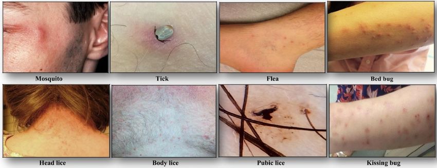
Bite spot diagnosis: Bites appear most commonly in the exposed body area, they are individualized
and scattered on the skin (see Figure 2). The common cutaneous manifestations to mosquito bites
consist typically of red itchy papules that resolve within few hours to several days [11]. The biting
activity is strongly season-dependent, occurring commonly during sunset and night in summer of
temperate regions, and all the year in the tropical region.
Diagnostics 2020, 10, 308                                                                                    4 of 15
  Diagnostics
  Diagnostics2020,
              2020,10,
                    10,xxFOR
                          FORPEER
                             PEERREVIEW
                                  REVIEW                                                                5 5ofof15
                                                                                                                15

                             Figure
                            Figure  1.1.Hematophagous
                              Figure1.  Hematophagous
                                        Hematophagousarthropods
                                                      arthropodsfeeding
                                                      arthropods  feedingon
                                                                 feeding   onhuman
                                                                          on  human
                                                                             human blood.
                                                                                     blood.
                                                                                    blood.

                    Figure
                     Figure2.2.2.Clinical
                   Figure        Clinical manifestations
                                          manifestationsfollowing
                                  Clinicalmanifestations followinghematophagous
                                                         following hematophagous
                                                                   hematophagousarthropods
                                                                                 arthropodsbiting.
                                                                                  arthropodsbiting.
                                                                                              biting.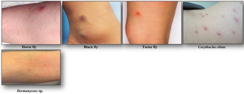
Diagnostics 2020, 10, 308                                                                         5 of 15

2.2. Ceratopogonidae (Biting Midges) and Phlebotominae (Sandflies)
     Ceratopogonidae or biting midges include more than 5000 species with a worldwide
distribution [12]. They are small insects with a length of 1–3 mm. They are the vector of infectious
diseases of veterinary importance, like bluetongue disease, mansonellosis, African horse sickness,
and epizootic hemorrhagic disease [13]. Members of the Phlebotominae family (sand flies) are insects
of about 2–4 mm in length, holding their wings in vertical V-shape during resting time. They are
proven vectors of viral and non-viral diseases, including bartonellosis, arboviruses and leishmaniasis
caused by Bartonella sp., Phlebovirus or Vesiculovirus and Leishmania sp., respectively [14]. The female
members of both families are pool feeders (telmophagy) (Table 1).
     Clinical manifestation: Biting midges can cause acute discomfort, irritation, and severe local
reactions. The latter is characterized by an acute pruritus, eczema or hypersensitivity. In the case of
repeated biting, people may become desensitized, expressing a mild or no reaction [15].
     Bite spot diagnosis: Bites caused by “biting midges” are painful and itchy with clinical symptoms
that range from small reddish bump and a burning sensation at the bite spot to local irritations that
cause significant itching [16]. Sandfly bites are also painful and cause small red bumps and blisters,
but often remain unnoticed. These bumps and blisters can become itchy, infected or cause dermatitis
or skin inflammation, and can persist for days or weeks [17] (Table 2).
Diagnostics 2020, 10, 308 6 of 15
Table 2. Discriminative clinical characters associated with hematophagous arthropods biting.
Hematophagous Arthropod
Criteria Biting Midge, Triatomine
Mosquito Louse Tsetse Fly Horse Fly Black Fly Tick Flea Bed Bug
Sandfly Bug
Macule,
Small painful red Small itchy papule, nodule,
Reddish sore Pruritic
spot or skin rash, Volcanic bumps vesicle, bullae,
(chancre), papules, Red, swollen
inflammation papule with surrounded by erythematous
Red itchy Small reddish itchy skin Reddish vesicules, and itchy skin,
Clinical manifestations and irritation central hole, reddish and pruritic
papules swollen bump rash, raised rash pruritus and anaphylactic
with blue spots reddish wheal inflamed skin, symptoms,
boil-like erythematus shock
or small spots of or plaque macule, papule, allergy,
swelling weals
blood on the skin nodule systematic
reaction
Bite feeling Painless Painful Painful Painful Painful Painful Painless Painless Painful Painless
Cluster of
Cluster of
Bite spot Sporadic and Sporadic and Sporadic and Sporadic and Sporadic and separated separated and Cluster of 2, 3 or Zigzag or
separated small
pattern separated separated separated separated separated small red sporadic more bites straight line
red bumps
bumps
Bite spot
diagnosis Evening and Evening and
Biting time Any time Diurnal Diurnal Diurnal Night Any time Any time Night
night night
Throughout
Lower
body
extremities,
Location Exposed area Exposed area particularly on Exposed area Exposed area Exposed area Exposed area Exposed area Exposed area
rarely upper
the scalp, neck
body
and shoulders
Washing with
Washing with Washing with Washing Washing with
Washing Removing the Washing with soap and water,
soap and water, soap and water, with soap soap and water,
Combing, Cold with soap tick by soap and water, corticosteroids
ice compress, ice compress, and water, ice compress,
pediculicidal compress, and water, tweezers, ice compress, (triamcinolone),
Treatment antihistamine antihistamine antihistamine epinephrine (in
shampoo or calamine, ice compress, topical steroids Antihistamine antipruritic
such as such as such as case of
lotion hydrocortisone Benadryl, and oral such as Benadryl, medications
Benadryl, Benadryl, Benadryl, systemic
Calamine antihistamines Calamine (paroxime,
Calamine Calamine Calamine reaction)
doxepin)
Diagnostics 2020, 10, 308                                                                            7 of 15

2.3. Pediculidae, Pthiridae (Lice)
      Lice are blood-sucking insects, obligate ectoparasites, widely distributed around the world.
They belong to the order of Phthiraptera Haeckel, 1896, which has about 5000 species, parasites of
warm-blooded animals [18]. The Anoplura sub-order has more than 550 species; most of them express
strict host specificity [19]. Only three taxa are obligate ectoparasites of humans and are of concern for
public health, including head lice, body lice, and pubic lice (Figure 1).
      Clinical manifestation and bite spot diagnosis: Biting occurs all year long with localization on the
human body that depends on the species.
      Head lice (Pediculus humanus capitis de Geer, 1778) are obligate ectoparasites of 2 to 3.5 mm
long, widespread throughout the world particularly among school-aged children [20] (Figure 1).
The infestation occurs in all socio-economical levels and all ethnic groups. Males, females and larvae
are strict hematophagous and feed exclusively on human blood, several times a day and reside close to
the scalp, in order to maintain its body temperature and humidity. The eggs (nits) are laid by females
and cemented at the base of the hair using an “adhesive” secretion produced by the female called
cementum [21]. They take advantage of the slightest direct contact, head to head, to grab the hair
of the new host. This passage from one host to another during direct contact is the main mode of
transmission. Common symptoms of head lice bites are intense pruritus and papules on the scalp,
neck, ears, and shoulders [21]. The bites often appear as small reddish or pink bumps sometimes with
coagulated blood (Figure 2). The rash caused by pruritic reaction and sleeping disturbance due to
irritability, are among additional symptoms. The role of Pediculus h. capitis in disease transmission is
debatable but in spite of detection of several pathogens in this insect, no formal evidence certifying its
vectorial role is available [22].
      Body lice (Pediculus humanus humanus Linnaeus, 1758) are morphologically similar to head lice
(3 to 4 mm length) (Figure 1). They are common in populations, suffering from a lack of standard
hygienic conditions, such as homeless, prisoners, and war refugees [23]. Up to now, only body lice are
reported as the recognized vectors of at least three pathogens namely Rickettsia prowazekii (epidemic
typhus), Bartonella quintana (trench fever), and Borrelia recurrentis (louse-borne relapsing fever) [24]
(Table 1). Body lice can be found alive in the belongings of an infected person, and be transferred
to another one by close contact or by the exchange of infested clothing. The symptoms of biting are
pruritus and small red lesions at the biting spots or skin rash, which develops to papule. In the case of
massive infestations, hematomas or melanodermic lesions are sometimes observed (Figure 2, Table 2).
In the case of continuous scratching, the bite spots can be itchy and can lead to secondary bacterial or
fungal infection. Bite spots are observed in various body parts, in particular, chest, armpits, and other
hairy parts. In persistent infestation cases, the skin may become thickened and discolored (vagabond’s
disease) [25]. In the case of an allergic reaction, pruritus and rash are common visible symptoms,
observed throughout the body. The discriminating symptom caused by body lice with those caused by
pubic lice is the severe itch and pruritus, observed in the case of pubic lice infestation [26].
      Pubic lice (Pthirus pubis Linnaeus, 1758) are yellow-gray ectoparasites, smaller (1–2 mm in length)
than the body and head lice (Figure 1). They are found in the pubic region, and the transmission
occurs by close and/or sexual contact [27]. Nevertheless, they are also found in other body parts, like
eyelashes, armpit, chest or facial hairs [27]. Itching in the genital region that becomes more intense at
nights, because of lice activity, is a common sign of pubic lice infestation [28]. The itching is due to an
allergic reaction to lice saliva. Scratching can cause additional inflammation and irritation with blue
spots or small spots of blood on the skin [27] (Figure 2). Other symptoms include occasional pruritus,
fever, irritability, and bluish spots near the bite spots.

2.4. Ixodidae, Argasidae (Ticks)
      Ticks are blood-sucking ectoparasites of mammals, birds, and reptiles (Figure 1). About 850 species
of tick are described [29]. Although they are active throughout the year, most of the tick bites occur
in spring and summer. Life cycle of the ticks includes a six-legged larval stage and one or more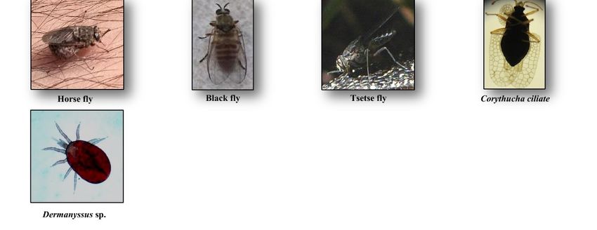
Diagnostics 2020, 10, 308                                                                             8 of 15

eight-legged nymphal stages. These immature stages need a blood-meal before proceeding to the
next stage. Two morphological and biological distinct families of ticks are described, Ixodidae
(hard ticks) comprising about 700 species and Argasidae (soft ticks) comprising about 200 species [30].
The mouthparts in hard ticks are visible at the front of the body, differentiating them from the soft
ticks [31]. Hard ticks take one day to one week to resume feeding, while soft ticks feed quickly and
leave their host. They are vectors of a wide range of pathogens, including bacteria, rickettsia, protozoa,
and viruses, causing Lyme disease, tick-borne encephalitis, Crimean–Congo hemorrhagic fever, rocky
mountain spotted fever, tularemia, Colorado tick fever, human tick-borne ehrlichiosis, babesiosis, tick
paralysis, relapsing fever, and the Mediterranean spotted fever [31] (Table 1).
     Clinical manifestation: Ticks cause acute or chronic skin diseases via physical trauma, salivary
secretions, feces or body part deposition [32]. When a tick attaches to his host, the initial lesion
development is due to the toxicity and the anti-coagulating activity of tick saliva, but also caused
by the physical injuries caused by mouthparts of the tick. Tick bites can cause a flu-like symptom,
vomiting, and even an anaphylactic shock [33]. They can also give symptoms by themselves with their
delivered saliva toxin (paralysis tick). They prefer warm and moist areas of the body; therefore once
they attach to their host, they migrate to other body parts, where they find adequate conditions to
survive, like armpits or groins [34].
     Bite spot diagnosis: Tick bites are most of the time painless. Bite spots are well separated and
sporadic, occurring generally in uncovered body parts, most commonly lower limbs [35]. In the case
of an attached tick, it can be removed and identified at the species level. The bite spot presentation is
similar to a volcanic papule with a central hole (“black dot”), and reddish wheal or plaque (Figure 2,
Table 2). The lesion can be hard, itchy or surrounded by redness. Skin lesions can persist and evolve
to papules, nodules or necrotic black spots [35]. The latter is a scar with a crust. The acute pruritic
papular dermatitis is a symptom, occurring following tick bites. Secondary infection of bite spots by
bacteria can also be observed.

2.5. Glossinidae (Tsetse Flies)
      Tsetse flies or Glossina spp. are hematophagous flies with about 6 to 15 mm in length (Figure 1),
transmitting African trypanosomiasis (sleeping sickness), caused by Trypanosoma brucei (Kinetoplastida:
Trypanosomatidae) in sub-Saharan Africa [36] (Table 1). About 30 species and subspecies of tsetse
flies are known divided into three distinct groups or subgenera: Austenia (G. fusca group), Nemorhina
(G. palpalis group) and Glossina (G. morsitans group) [37]. Both female and male tsetse flies feed
exclusively on blood. The female is larviparous. Most tsetse flies are diurnal and active all the year.
They are differentiated from other biting flies by their forward-pointing mouthparts (proboscis) and
characteristic wing venation [38]. They are prevalent mainly in Central Africa. Besides their vectorial
role, they cause African animal trypanosomiasis (nagana disease) resulting in gradual health decline in
infected livestock, reduced milk and meat production, and increased abortion rates [39].
      Clinical manifestation: The most common clinical signs of tsetse bites are reddish bumps or small
red ulcers at the bite spot.
      Bite spot diagnosis: The bite of tsetse fly is often painful and can develop a reddish sore (chancre)
or itchy skin rash that may evolve after several days, to a boil-like swelling (Figure 2, Table 2).

2.6. Pulicidae (Fleas)
      Fleas are small (1.5 to 4 mm in length), wingless and flat insects with three pairs of legs adapted
for jumping (Figure 1). They feed exclusively on the blood of mammals and birds. About 2574 species
belonging to 16 families and 238 genera have been described, but only a minority is synanthropic, that
is they live in close association with humans [40,41]. They are vectors of bacterial (e.g., Yersinia pestis,
Bartonella henselae, Rickettsia typhi and R. felis) and viral (e.g., Myxoma virus and feline leukemia virus)
diseases [42] (Table 1). Among flea species, Pulex irritans favors blood-feeding on humans. The oriental
rat flea, Xenopsylla cheopis, is the primary vector of Yersinia pestis, a Gram-negative coccobacillus,
Diagnostics 2020, 10, 308 9 of 15
which causes plague [43]. People can be bitten by fleas via contact with an infected animal. Both sexes
at the adult stage are hematophagous and bite at any time while active throughout the year [44].
The larvae feed mainly on organic debris left on their host’s skin or on the feces (dried blood) expelled
by the adults which accumulates, along with the eggs.
Clinical manifestation: Flea bites are very itchy, with surrounding skin that may become sore or
painful. They also cause skin irritation, flea bite allergy, hair loss, and reddish skin. Bites in humans
are usually located in lower extremities like legs or ankles [45]. They can also cause anemia in extreme
cases [46]. Persistent scratching can further damage the skin and cause a secondary bacterial infection.
Bite spot diagnosis: The most commonly observed symptom is small itchy red bumps, surrounded
by reddish inflamed skin. The lesion initially exhibits punctuate hemorrhagic area representing the
site of probing by the insect. Bites occur sporadically or in clusters and sometimes form a scatter or
line pattern on the skin [45] (Figure 2, Table 2).
2.7. Cimicidae (Bed Bugs)
Cimex lectularius and C. hemipterus, commonly named “bed bugs”, are parasitic insects of 5 to
7 mm in length (Figure 1). They have a major impact on public health and are probably one of
the most common ectoparasites with a worldwide distribution. Both sexes at all stages feed on a
human. Infestation occurs in all ethnic groups and at all socioeconomic levels. During the two last
decades, the infestation of human habitats has drastically increased, leading to rising concerns about
bed bugs. They are responsible for several clinical and psychological disorders [47]. They are also
suspected of being involved in the transmission of over 40 infectious agents. These include bacteria
(e.g., Borrelia recurrentis, B. duttoni, Coxiella burnetii and Rickettsia rickettsii), fungi (e.g., Aspergillus spp.),
viruses (e.g., Hepatitis B and HIV), filariae and parasites (e.g., Trypanosoma cruzi) [47] (Table 1).
The competence of C. lectularius to act as a vector of T. cruzi (responsible agent of Chagas disease) and
Bartonella quintana (causative agent of trench fever), has been recently probed in the laboratory [48,49],
but no evidence on the vectorial role of Cimex spp. in the endemic areas is available [47].
Clinical manifestation: At least 10% to 30% of the people who are bitten do not develop
any reactions [15]. Nevertheless, the bed bugs bites can cause a wide spectrum of dermatological
manifestations that at least in part, depend on the individual’s immune response. These reactions
include macule, papule, nodule, vesicle, bullae, erythematous lesion, pruritic reaction, secondary
infection, and systemic sign [47]. The most commonly encountered clinical signs, following bed bug
bites are pruritic maculopapular and erythematous lesions [47]. In the case of repeated bites, enlarged
pruritus can occur. Symptoms often occur immediately, but in some cases, bullous rashes can emerge
in the following few days. Without further irritation, symptoms typically resolve spontaneously after
one to two weeks. After persistent scratching, a secondary bacterial or fungal infection may also occur.
In rare cases, some other clinical disorders such as asthmatic reaction, urticaria or anaphylactic shock
are observed [50].
Bite spot diagnosis: Bed bug bites are painless and occur on any exposed part of the body.
Bed bugs feed mostly at night, but in case of hyper-infestation, their activity can take place all day
long. No seasonality is noticed. Lesions are typically distributed in a linear or zigzag pattern, so-called
“breakfast, lunch, dinner” bite pattern [47] (Figure 2, Table 2).
2.8. Reduviidae (Kissing Bugs)
Triatomine bugs are hematophagous insects with a long, cone-shaped head and a dark brown
or black body (3/4 to 11/4 inches) that are mostly active at night. They comprise 15 genera and 148
species and subspecies that are mainly found in Latin and South America, with a few species present
in Asia, Africa, and Australia [51,52]. Triatomine bugs are a proven vector of T. cruzi, causative agent
of American trypanosomiasis, called chagas disease (Figure 1). The bugs ingest the parasites when
they feed on an infected animal or person. Infected bugs then deposit the parasites with their feces on
the skin of another person during or shortly after feeding. Scratching or rubbing the parasites leads toDiagnostics 2020, 10, 308 10 of 15
enter the body through the bite wound or broken skin [53]. The disease is associated with poverty in
rural areas or in favelas. Both sexes and all the five instars feed on blood (Table 1).
Clinical manifestation: The symptoms can be quite variable and range from no symptoms to
severe and distressing signs. The common symptoms include skin rash, swollen lymph nodes,
fever, headaches, body aches, fatigue, and nausea [54]. Proteins released via their bites can provoke
anaphylaxis in sensitized individuals [55].
Bite spot diagnosis: Triatomine bugs’ bite occurs in any exposed body region, including the face,
head, legs, and arms. They are generally painless and asymptomatic, but in some occasions can cause
papules with hemorrhagic puncta, vesiculobullous lesions, eczematic reaction (the surrounding skin of
bite spot might become red, swollen and itchy) or anaphylactic shock [56]. Swelling, redness of skin
(called chagomas), and rash are common observable signs of kissing bugs’ bite (Figure 2, Table 2).
2.9. Simuliidae (Blackflies)
Simuliidae is a medically important family with over 2141 species, formally described [57].
They are usually small (1.5–4 mm), black, gray or brown, with short legs and antennae (Figure 1).
Simulids are cosmopolitan insects, which are found in running water, near rivers, waterfalls, etc.,
where the larvae are fixed under stones or vegetable matter using the abdomen apex [58]. They are
active in the morning and in the evening. Adult males feed on nectar, while females feed on blood
before laying eggs. They are vectors of human diseases, notably human onchocerciasis or river
blindness, the second ranking cause of infectious blindness [59]. The latter is caused by the nematode
Onchocerca volvulus, and transmitted by Simulium damnosum (S. damnosum) and S. neavei in Africa and
S. callidum, S. metallicum and S. ochraceum in Central and South America [60] (Table 1).
Clinical manifestation: “Black fly fever” is caused by the intense feeding of these flies. It is
accompanied by headache, nausea, fever, and swollen lymph nodes. A severe anaphylactic reaction is
a less common manifestation that may require hospitalization [61].
Bite spot diagnosis: Since females cut a small hole in the skin for blood-feeding (telmophagous),
their biting spot becomes painful and itchy. Feeding is facilitated by anticoagulant proteins in
the flies’ saliva that reduces the host’s awareness of being bitten, extending the feeding time.
Various clinical presentations are described (edematous, erythematous-edematous, erysipeloid,
inflammatory-indurative, phlegmonoid, and hemorrhagic), but the itching, localized swelling,
inflammation eruptions of pruritic papules, vesicles, intense pruritus, and erythematous wheals
are common symptoms, resulting from a hypersensitive reaction to black fly bites [62] (Figure 2,
Table 2). The Simulium dermatitis is a concern with a high risk of anaphylaxis and acute cardiotoxicity
in hypersensitive individuals [61].
2.10. Tabanidae (Horse Flies)
Horse-flies are a large family of the order Diptera with 4455 species belonging to 137 genera
that mostly have a diurnal activity [63]. They are found all over the world, except in polar regions.
Tabanid species have a body length between 5 and 25 mm mostly occur in warm areas with suitable
moist locations for breeding [64]. Because of weak mouthparts, males do not feed on blood, but in
contrast, the females bite animals to get enough protein for egg production. The mouthparts of females
cut the skin to form a pool of blood, so they then aspirate (Figure 1). They are known to act as biological
or mechanical vectors of various pathogens such as Loa loa worm, equine infectious anemia virus,
Trypanosoma spp., cattle, and sheep anthrax and tularemia [65] (Table 1).
Clinical manifestation: The biting spot is painful due to the physical action of the fly’s mouthparts.
Clinical manifestation includes large red and raised rash or temporary swollen skin. In exceptional
cases, reactions to saliva may result in general toxicity and immune response that lead to stress and
immunosuppression [66].
Bite spot diagnosis: The bite spots are usually red surrounded by a raised skin (Figure 2, Table 2).
The pain, redness, and weal help to identify horsefly bites. Other symptoms may include urticaria,Diagnostics 2020, 10, 308 11 of 15
dizziness, weakness, wheezing, and angioedema (a temporary itchy, pink or red swelling occurring
around the eyes or lips) [67].
Diagnostics 2020, 10, x FOR PEER REVIEW 12 of 15
3. Other Non-Common Hematophagous Arthropods
3. Other Non-Common Hematophagous Arthropods
Identification of clinical manifestations due to these arthropods’ bites can be difficult for
Identification of clinical manifestations due to these arthropods’ bites can be difficult for
physicians who are not familiar with these arthropods, and the emerging associated clinical disorders.
physicians who are not familiar with these arthropods, and the emerging associated clinical
Dermanyssus sp. and Corythucha ciliate (Figure 1) are such examples of arthropods that occasionally bite
disorders. Dermanyssus sp. and Corythucha ciliate (Figure 1) are such examples of arthropods that
humans (Figure 2) [68,69].
occasionally bite humans (Figure 2) [68,69].
4. Treatment
4. Treatment
Cutaneous symptoms due to arthropods’ bite typically resolve within one or two weeks without
Cutaneous symptoms due to arthropods’ bite typically resolve within one or two weeks without
intervention. The first-line action in the treatment of arthropods’ bite is cleaning bite spots with soap
intervention. The first-line action in the treatment of arthropods’ bite is cleaning bite spots with soap
and water, which prevents secondary skin infection and reduces itchiness. Ice or cold water relieves
and water, which prevents secondary skin infection and reduces itchiness. Ice or cold water relieves
itching and reduces swelling and inflammation. In the case of persistent itching, the application of
itching and reduces swelling and inflammation. In the case of persistent itching, the application of
topical steroids or oral antihistamines could be beneficial [47]. In the case of a secondary bacterial
topical steroids or oral antihistamines could be beneficial [47]. In the case of a secondary bacterial
skin infection, antibiotic ointment or oral antibiotics can be prescribed by physicians. For severe
skin infection, antibiotic ointment or oral antibiotics can be prescribed by physicians. For severe
systemic allergic reaction, oral antihistaminic compounds or injection of epinephrine, antihistamine,
systemic allergic reaction, oral antihistaminic compounds or injection of epinephrine, antihistamine,
and corticosteroids are the mainstays of pre-hospital treatment [70,71].
and corticosteroids are the mainstays of pre-hospital treatment [70,71].
For ticks that remain attached to the skin, tweezers or tick removers will help the removal [72].
For ticks that remain attached to the skin, tweezers or tick removers will help the removal [72].
Sometimes
Sometimesthe the rostrum
rostrum breaks
breaks andand remains
remains inin the
the skin,
skin, without
without consequence
consequencefor forthe
thebitten
bittenperson.
person.
The
Therostrum
rostrumwillwillbe
berejected
rejected later
later as
as an
an external
external object. In the
object. In the case
case of
of head
head lice,
lice,the
thelouse
lousecomb
combwill
will
be helpful for physical removal of the lice and their nits. Medications like ivermectin
be helpful for physical removal of the lice and their nits. Medications like ivermectin or chemicals, or chemicals,
such
suchasasisopropyl
isopropylmyristate,
myristate, benzyl
benzyl alcohol,
alcohol, spinosad, dimeticon and
spinosad, dimeticon and malathion
malathionare arerecommended
recommended
for
forthe
thetreatment
treatmentof oflice
liceinfestation
infestation [73].
[73]. Malathion
Malathion isis also
also available,
available, formulated
formulatedas asaalotion,
lotion,shampoo
shampoo
or cream. Laundering (60 ◦ C), dry cleaning, ironing, and replacement of clothing and linens help to
or cream. Laundering (60 °C), dry cleaning, ironing, and replacement of clothing and linens help to
remove
removethethebody
bodyandandpubic
pubiclice.
lice.
5.5.Discussion
Discussionand
and Conclusions
Conclusions
Physicians
Physicians and
and other
other healthcare
healthcare personnel
personnel areare frequently
frequently confronted
confronted with
with patients
patientswith
withskin
skin
lesions,
lesions,attributed
attributedtoto
thethe
bite of an
bite of unidentified
an unidentifiedarthropod. The The
arthropod. reactions include
reactions a wide
include a range of clinical
wide range of
manifestations from simple red bumps to allergic or systemic reactions. Without a formal
clinical manifestations from simple red bumps to allergic or systemic reactions. Without a formal identification
ofidentification
arthropods, the clinical history
of arthropods, of the biting
the clinical together
history with knowledge
of the biting of the
together with entomo-epidemiological
knowledge of the entomo-
and clinical signs makes it plausible to infer the putative arthropod species.
epidemiological and clinical signs makes it plausible to infer the putative arthropod species. A flowchart for the
A
discrimination of arthropods’ bites is resumed in the Figure 3. These species-specific
flowchart for the discrimination of arthropods’ bites is resumed in the Figure 3. These species-specific criteria help
clinical
criteriapractitioners
help clinical for precise diagnosis
practitioners for preciseanddiagnosis
proposing the
and proper treatment.
proposing the proper treatment.
Figure3.3.Clinical
Figure Clinical manifestations
manifestations of
of arthropods
arthropods bite
bite spots.
spots. Due
Due toto similarities
similaritiesin
inclinical
clinicalmanifestation
manifestation
and blood feeding activity, the mosquitoes, sandflies and biting midges grouped together.
and blood feeding activity, the mosquitoes, sandflies and biting midges grouped together.
Funding: This research received no external funding.
Conflicts of Interest: The authors declare no conflict of interest.
Reference
1. Jensen, S. The Proterozoic and Earliest Cambrian Trace Fossil Record; Patterns, Problems and Perspectives.
Integr. Comp. Biol. 2003, 43, 219–228.Diagnostics 2020, 10, 308 12 of 15
Funding: This research received no external funding.
Conflicts of Interest: The authors declare no conflict of interest.
References
1. Jensen, S. The Proterozoic and Earliest Cambrian Trace Fossil Record; Patterns, Problems and Perspectives.
Integr. Comp. Biol. 2003, 43, 219–228. [CrossRef]
2. Ribeiro, J.M.; Francischetti, I.M. Role of arthropod saliva in blood feeding: Sialome and post-sialome
perspectives. Annu. Rev. Entomol. 2003, 48, 73–88. [CrossRef] [PubMed]
3. Choe, J.C. Encyclopedia of Animal Behavior; Elsevier: San Diego, CA, USA, 2019; Volume 2.
4. Bouchet, F.; Lavaud, F. Solenophagy and telmophagy: Biting mechanisms among various hematophagous
insects. Allerg. Immunol. 1999, 31, 346–350.
5. Andrade, B.B.; Teixeira, C.R.; Barra, A.; Barral-Netto, M. Haematophagous arthropod saliva and host defense
system: A tale of tear and blood. An. Acad. Bras. Ciênc. 2005, 77, 15. [CrossRef]
6. Rizzo, M.C.; Arruda, L.K.; Chapman, M.D.; Fernandez-Caldas, E.; Baggio, D.; Platts-Mills, T.A.; Naspitz, C.K.
IgG and IgE antibody responses to dust mite allergens among children with asthma in Brazil. Ann. Allergy
1993, 71, 152–158. [PubMed]
7. Harbach, R.E. Mosquito Taxonomic Inventory. Available online: http://mosquito-taxonomic-inventory.info
(accessed on 6 December 2017).
8. Rozendaal, J.A. Chapter 1-Mosquitos and other biting Diptera. In Vector Control-Methods for Use by Individuals
and Communities; World Health Organization: Geneva, Switzerland, 1997.
9. Engler, R.J. Mosquito bite pathogenesis in necrotic skin reactors. Curr. Opin. Allergy. Clin. Immunol. 2001, 1,
349–354. [CrossRef]
10. Chiu, T.M.; Lin, Y.M.; Wang, S.C.; Tsai, Y.G. Hypersensitivity to mosquito bites as the primary clinical
manifestation of an Epstein-Barr virus infection. J. Microbiol. Immunol. Infect. 2016, 49, 613–616. [CrossRef]
11. Manuyakorn, W.; Itsaradisaikul, S.; Benjaponpitak, S.; Kamchaisatian, W.; Sasisakulporn, C.; Jotikasthira, W.;
Matangkasombut, P. Mosquito allergy in children: Clinical features and limitation of commercially-available
diagnostic tests. Asian Pac. J. Allergy Immunol. 2017, 35, 186–190.
12. Boorman, J. Biting midges (Ceratopogonidae). In Medical Insects and Arachnids; Lane, R.P., Crosskey, R.W., Eds.;
Springer: Dordrecht, The Netherlands, 1993; Volume 1, pp. 288–309.
13. Linley, J.R. Biting Midges (Diptera: Ceratopogonidae) as Vectors of Nonviral Animal Pathogens.
J. Med. Entomol. 1985, 22, 589–599. [CrossRef]
14. Akhoundi, M.; Kuhls, K.; Cannet, A.; Votýpka, J.; Marty, P.; Delaunay, P.; Sereno, D. A Historical Overview of
the Classification, Evolution, and Dispersion of Leishmania Parasites and Sandflies. PLoS Negl. Trop. Dis.
2016, 10, e0004349. [CrossRef]
15. Pali-Schöll, I.; Blank, S.; Verhoeckx, K.; Mueller, R.; Janda, J.; Marti, E.; Seida, A.A.; Rhyner, C.; DeBoer, D.J.;
Jensen-Jarolim, E. EAACI position paper: Comparing insect hypersensitivity induced by bite, sting, inhalation
or ingestion in human beings and animals. Allergy 2019, 74, 874–887. [CrossRef] [PubMed]
16. Borkent, A. The biting midges, the Ceratopogonidae (Diptera). In Biology of Disease Vectors, 2nd ed.;
Marquardt, W.C., Ed.; Elsevier: Burlington, MA, USA, 2004; Volume 2, pp. 113–126.
17. Linley, J.R.; Hoch, A.L.; Pinheiro, F.P. Biting midges (Diptera: Ceratopogonidae) and human health.
J. Med. Entomol. 1983, 20, 347–364. [CrossRef] [PubMed]
18. Price, R.D.; Hellenthal, R.A.; Palma, R.L.; Johnson, K.P.; Clayton, D.H. The Chewing Lice: World Checklist and
Biological Overview; Illinois Natural History Survey: Urbana, IL, USA, 2003; Volume 24, 501p.
19. Durden, L.A.; Musser, G.G. The sucking lice (Insecta: Anoplura) of the world: A taxonomic checklist with
records of mammalian hosts and geographical distributions. Bull. Am. Mus. Nat. Hist. 1994, 218, 1–90.
20. Mazurek, C.M.; Lee, C.P. How to manage head lice. West J. Med. 2000, 172, 342–345. [CrossRef]
21. Burkhart, C.N.; Burkhart, C.G. Head lice: Scientific assessment of the nit sheath with clinical ramifications
and therapeutic options. J. Am. Acad. Dermatol. 2005, 53, 129–133. [CrossRef]
22. Badiaga, S.; Brouqui, P. Human louse-transmitted infectious diseases. Clin. Microbiol. Infect. 2012, 18,
332–337. [CrossRef]
23. Bonilla, D.L.; Durden, L.A.; Eremeeva, M.E.; Dasch, G.A. The Biology and Taxonomy of Head and Body
Lice—Implications for Louse-Borne Disease Prevention. PLoS Pathog. 2013, 9, e1003724. [CrossRef]Diagnostics 2020, 10, 308 13 of 15
24. Raoult, D.; Roux, V. The body louse as a vector of reemerging human diseases. Clin. Infect. Dis. 1999, 29,
888–911. [CrossRef]
25. Brouqui, P. Arthropod-Borne Diseases Associated with Political and Social Disorder. Annu. Rev. Entomol.
2011, 56, 357–374. [CrossRef]
26. Anderson, A.L.; Chaney, E. Pubic Lice (Pthirus pubis): History, Biology and Treatment vs. Knowledge and
Beliefs of US College Students. Int. J. Environ. Res. Public Health 2009, 6, 592–600. [CrossRef]
27. Akhoundi, M.; Cannet, A.; Arab, M.K.; Marty, P.; Delaunay, P. An old lady with Pediculosis pubis on the
head hair. J. Eur. Acad. Dermatol. Venereol. 2016, 30, 885–887. [CrossRef] [PubMed]
28. Veraldi, S.; Schianchi, R.; Ramoni, S.; Nazzaro, G. Pubic hair removal and Phthirus pubis infestation. Int. J.
STD AIDS 2018, 29, 103–104. [CrossRef] [PubMed]
29. Furman, D.P.; Loomis, E.C. The ticks of California (Acari: Ixodida). Bull. California Insect Survey 1984, 25,
1–239.
30. Guglielmone, A.A.; Robbing, R.G.; Dmitry, A.; Trevor, N.P.; Estrada-Peña, A.; Horak, I.G.; Shao, R.; Barker, S.C.
The Argasidae, Ixodidae and Nuttalliellidae (Acari: Ixodida) of the world: A list of valid species names.
Zootaxa 2010, 2528, 1–28. [CrossRef]
31. Brites-Neto, J.; Roncato Duarte, K.M.; Fernandes Martins, T. Tick-borne infections in human and animal
population worldwide. Vet. World 2015, 8, 301–315. [CrossRef]
32. Parola, P.; Paddock, C.D.; Raoult, D. Tick-Borne Rickettsioses around the World: Emerging Diseases
Challenging Old Concepts. Clin. Microbiol. Rev. 2005, 18, 719–756. [CrossRef]
33. Van Wye, J.E.; Hsu, Y.P.; Terr, A.I.; Moss, R.B.; Lane, R.S. Anaphylaxis from a tickbite. NEJM 1991, 324,
777–778.
34. Rahlenbeck, S.; Fingerle, V.; Doggett, S. Prevention of tick-borne diseases: An overview. Br. J. Gen. Pract.
2016, 66, 492–494. [CrossRef]
35. Haddad, V., Jr.; Raineri Haddad, M.; Santos, M.; Luiz Costa Cardoso, J. Skin manifestations of tick bites in
humans. An. Bras. Dermatol. 2018, 93, 251–255. [CrossRef]
36. Krafsur, E.S. Tsetse flies: Genetics, evolution, and role as vectors. Infect. Genet. Evol. 2009, 9, 124–141.
[CrossRef]
37. Gooding, R.H.; Krafsur, E.S. Tsetse Genetics: Contributions to Biology, Systematics, and Control of Tsetse
Flies. Annu. Rev. Entomol. 2005, 50, 101–123. [CrossRef] [PubMed]
38. Service, M.W. A Guide to Medical Entomology; Palgrave Macmillan: London, UK, 1980.
39. Muhanguzi, D.; Mugenyi, A.; Bigirwa, G.; Kamusiime, M.; Kitibwa, A.; Gloria Akurut, G.; Ochwo, S.;
Amanyire, W.; George Okech, S.; Hattendorf, J.; et al. African animal trypanosomiasis as a constraint to
livestock health and production in Karamoja region: A detailed qualitative and quantitative assessment.
BMC Vet. Res. 2017, 13, 355.
40. Lewis, R.E. Resume of the Siphonaptera (Insecta) of the World. J. Med. Entomol. 1999, 35, 377–389.
41. Lewis, R.E. Notes on the geographical distribution and host preferences in the order Siphonaptera. Part 8.
New taxa described between 1984 and 1990, with a current classification of the order. J. Med. Entomol. 1993,
30, 239–256. [CrossRef]
42. Dobler, G.; Pfeffer, M. Fleas as parasites of the family Canidae. Parasit. Vectors 2011, 4, 139. [CrossRef]
43. Leulmi, H.; Socolovschi, C.; Laudisoit, A.; Houemenou, G.; Davoust, B.; Bitam, I.; Raoult, D.; Parola, P.
Detection of Rickettsia felis, Rickettsia typhi, Bartonella species and Yersinia pestis in fleas (Siphonaptera)
from Africa. PLoS Negl. Trop. Dis. 2014, 8, e3152. [CrossRef]
44. Mathison, B.A.; Prittb, B.S. Laboratory Identification of Arthropod Ectoparasites. Clin. Microbiol. Rev. 2014,
27, 48–67. [CrossRef]
45. Galy, A.; Loubet, P.; Peiffer-Smadja, N.; Yazdanpanah, Y. The plague: An overview and hot topics.
Rev. Med. Interne. 2018, 39, 863–868. [CrossRef]
46. Mullen, G.R.; Durden, L.A. Medical and Veterinary Entomology, 2nd ed.; Academic Press: Boston, MA, USA,
2009; 637p.
47. Goddard, J.; deShazo, R. Bed bugs (Cimex lectularius) and clinical consequences of their bites. JAMA 2009,
301, 1358–1366. [CrossRef]
48. Salazar, R.; Castillo-Neyra, R.; Tustin, A.W.; Borrini-Mayorí, K.; Náquira, C.; Levy, M.Z. Bed bugs (Cimex
lectularius) as vectors of Trypanosoma cruzi. Am. J. Trop. Med. Hyg. 2015, 92, 331–335. [CrossRef]Diagnostics 2020, 10, 308 14 of 15
49. Leulmi, H.; Bitam, I.; Berenger, J.M.; Lepidi, H.; Rolain, J.M.; Almeras, L.; Raoult, D.; Parola, P. Competence of
Cimex lectularius Bed Bugs for the Transmission of Bartonella quintana, the Agent of Trench Fever. PLoS Negl.
Trop. Dis. 2015, 9, e0003789.
50. Bircher, A.J. Systemic immediate allergic reactions to arthropod stings and bites. Dermatology 2005, 210,
119–127. [CrossRef] [PubMed]
51. Galvão, C.; Carcavallo, R.; Rocha, D.S.; Jurberg, J. A checklist of the current valid species of the subfamily
Triatominae Jeannel, 1919 (Hemiptera, Reduviidae) and their geographical distribution, with nomenclatural
and taxonomic notes. Zootaxa 2003, 202, 1–36. [CrossRef]
52. Schofield, C.J.; Galvão, C. Classification, evolution, and species groups within the Triatominae. Acta Trop.
2009, 110, 88–100. [PubMed]
53. Clayton, J. Chagas disease 101. Nature 2010, 465, S4–S5. [CrossRef]
54. Klotz, J.H.; Dorn, P.L.; Logan, J.L.; Stevens, L.; Pinnas, J.L.; Schmidt, J.O.; Klotz, S.A. “Kissing Bugs”: Potential
Disease Vectors and Cause of Anaphylaxis. Clin. Infect. Dis. 2010, 50, 1629–1634. [CrossRef]
55. Anderson, C.; Belnap, C. The Kiss of Death: A Rare Case of Anaphylaxis to the Bite of the “Red Margined
Kissing Bug”. Hawaii J. Med. Public Health 2015, 74, 33–35.
56. Steen, C.J.; Carbonaro, P.A.; Schwartz, R.A. Arthropods in dermatology. J. Am. Acad. Dermatol. 2004, 50,
819–842. [CrossRef]
57. Adler, P.H. World Blackflies (Diptera: Simuliidae): A Comprehensive Revision of the Taxonomic and
Geographical Inventory. Ph.D. Thesis, Department of Plant and Environmental Sciences, Clemson University,
Clemson, SC, USA, 2017. Available online: https://biomia.sites.clemson.edu/pdfs/blackflyinventory.pdf
(accessed on 29 May 2018).
58. Lock, K.; Adriaen, T.; Goethal, P. Effect of water quality on blackflies (Diptera: Simuliidae) in Flanders
(Belgium). Limnologica 2014, 44, 58–65. [CrossRef]
59. Adler, P.H.; Cheke, R.A.; Post, R.J. Evolution, Epidemiology, and population genetics of black flies (Diptera:
Simuliidae). Infect. Gen. Evol. 2010, 10, 846–865. [CrossRef]
60. Procunier, W.S. Cytological approaches to simuliid biosystematics in relation to the epidemiology and control
of human onchocerciasis. Genome 1989, 32, 559–569. [CrossRef] [PubMed]
61. Chattopadhyay, P.; Goyary, D.; Dhiman, S.; Rabha, B.; Hazarika, S.; Veer, V. Immunomodulating effects and
hypersensitivity reactions caused by Northeast Indian black fly salivary gland extract. J. Immunotoxicol. 2014,
11, 126–132. [CrossRef]
62. Farkas, J. Simuliosis. Analysis of dermatological manifestations following blackfly (Simuliidae) bites as
observed in the years 1981–1983 in Bratislava (Czechoslovakia). Derm. Beruf. Umwelt. 1984, 32, 171–173.
[PubMed]
63. Morita, S.I.; Bayless, K.M.; Yeates, D.K.; Wiegmann, B.M. Molecular phylogeny of the horse flies: A framework
for renewing tabanid taxonomy. Systematic Entomol. 2015, 41, 56–72. [CrossRef]
64. Croof, H.; Nour, M.; Ali, N. Morphological Identification of Horse Flies (Diptera: Tabanidae) and Estimation
of their Seasonal Abundance in Al-Showak District, Gedaref State, Eastern Sudan. IRA-Int. J. Appl. Sci. 2017,
6, 41–54. [CrossRef]
65. Cheng, T.C. General Parasitology, 2nd ed.; Elsevier Science: Amsterdam, The Netherlands, 2012; 660p.
66. Hemmer, W.; Focke, M.; Vieluf, D.; Berg-Drewniok, B.; Götz, M.; Jarisch, R. Anaphylaxis induced by horsefly
bites: Identification of a 69 kd IgE-binding salivary gland protein from Chrysops spp. (Diptera Tabanidae)
by western blot analysis. J. Allergy Clin. Immunol. 1998, 101, 134–136. [CrossRef]
67. Veraldi, S.; Esposito, L. Skin lesions caused by Tabanus bovinus bites. J. Travel Med. 2017, 24, 1–5. [CrossRef]
68. Sparagano, O.A.E.; George, D.R.; Harrington, D.W.J.; Giangaspero, A. Significance and Control of the Poultry
Red Mite, Dermanyssus gallinae. Ann. Rev. Entomol. 2014, 59, 447–466. [CrossRef]
69. Izri, A.; Andriantsoanirina, V.; Chosidow, O.; Durand, R. Dermatosis Caused by Blood-Sucking Corythucha
Ciliata. JAMA Dermatol. 2015, 151, 909–910. [CrossRef]
70. Foex, B.A. Oral antihistamines for insect bites. Emerg. Med. J. 2006, 23, 721–727. [CrossRef]
71. Kemp, S.F.; Lockey, R.F.; Simons, F.E.R.; World Allergy Organization ad hoc Committee on Epinephrine
in Anaphylaxis. Epinephrine: The Drug of Choice for Anaphylaxis-A Statement of the World Allergy
Organization. World Allergy Organ J. 2008, 1, S18–S26. [CrossRef] [PubMed]Diagnostics 2020, 10, 308 15 of 15
72. Aberer, E. What should one do in case of a tick bite? Curr. Probl. Dermatol. 2009, 37, 155–166. [PubMed]
73. Pariser, D.M.; Meinking, T.L.; Bell, M.; Ryan, W.G. Topical 0.5% Ivermectin Lotion for Treatment of Head
Lice. NEJM 2012, 367, 1687–1693. [CrossRef] [PubMed]
© 2020 by the authors. Licensee MDPI, Basel, Switzerland. This article is an open access
article distributed under the terms and conditions of the Creative Commons Attribution
(CC BY) license (http://creativecommons.org/licenses/by/4.0/).You can also read